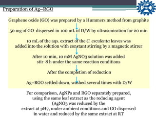
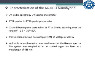
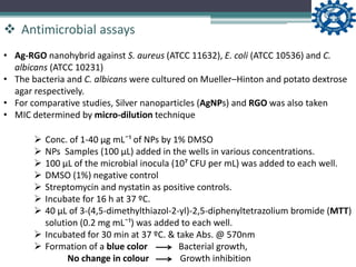
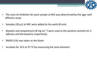
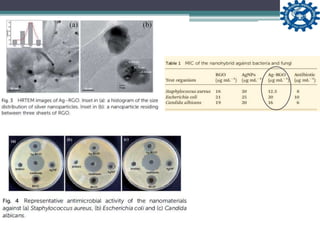
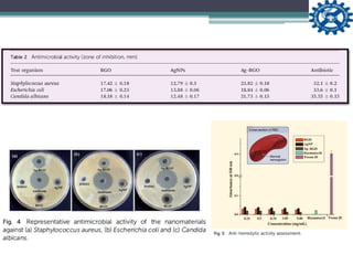
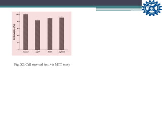
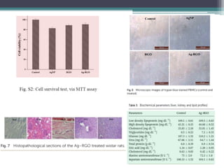
By- Govinda R. Navale
One step preparation of a biocompatible,
antimicrobial reduced graphene oxide–silver
nanohybrid as a topical antimicrobial agent
S. Baruaª; S. Thakurª; L. Aidewᵇ; A. K. Buragohainᵇ; P. Chattopadhyayͨ and N. Karakª*
RSC Adv., 2014
ªAdvanced Polymer and Nanomaterial Laboratory, Department of Chemical Sciences,
Tezpur University, Napaam-784028, Assam, India. E-mail: karakniranjan@yahoo.com
ᵇDepartment of Molecular Biology and Biotechnology, Tezpur University, Napaam-
784028, Assam, India
ͨ Defence Research Laboratory, Tezpur, Assam, India
Received : 19th November 2013
Accepted : 12th December 2013
Contents
• Introduction
• Experimental
 Preparation of Silver-reduced Graphene Oxide (Ag–RGO)
 Characterization of the nanohybrid
 Antimicrobial assays
 Hemolytic assay
 In vitro cytotoxicity assessment with Peripheral blood
mononuclear cells (PBMCs)
 Acute dermal toxicity study
• Results and discussion
• Conclusion
Introduction
 Graphene has recently been added to the class of carbon based
nanomaterials. Compatible with biological systems & has
antimicrobial activity
 Preparation of RGO by other methods like direct reduction of
graphene oxide (GO), using chemical reducing agents. like sodium
borohydride, hydrazine, hydroquinone and sodium hydroxide etc.
 Presence of trace amounts of reducing agents may be toxic to
biological systems
 Replaced by leaf extract such as Colocasia esculenta and Mesua
ferrea Linn. and an aqueous peel extract of orange (Citrus sinensis)
 Silver nanoparticles widely used in biomedical applications such as in
bandages, ointments.
 Graphen based nano material has applications such as nanomedicine,
sensors, catalysis etc.
 AG-RGO nanohybrid acts as a activity enhancer
 Compatibility of the nanohybrid with PBMCs as well as with mammalian red
blood cells (RBCs).
 Peripheral blood mononuclear cells (PBMCs) are responsible for the
contraction and healing of wounds
 A recent report focused on the skin irritation study of a AG-RGO nanohybrid
on rat models.
 Here a complete dermal toxicity of Ag–RGO studied
 The overall work reports a facile green method for the preparation of
antimicrobial Ag–RGO and its compatibility with mammalian RBCs and
PBMCs. In addition, acute dermal toxicity tested on wistar rat models.
…Continued
• Preparation of aqs. extract of Colocasia esculenta
Experimental
• Preparation of aqs. extract of Colocasia esculenta
2 g of Colocasia esculenta leaves
washed thoroughly with distilled water
ground using a domestic blender
stirred for 20 min in 100 mL of water at 45 ⁰C
aqueous extract obtained was filtered using a muslin cloth
Experimental
Preparation of Ag–RGO
Graphene oxide (GO) was prepared by a Hummers method from graphite
50 mg of GO dispersed in 100 mL of D/W by ultrasonication for 20 min
10 mL of the aqs. extract of the C. esculenta leaves was
added into the solution with constant stirring by a magnetic stirrer
After 10 min, 10 mM AgNO3 solution was added
stir 8 h under the same reaction conditions
After the completion of reduction
Ag–RGO settled down, washed several times with D/W
For comparison, AgNPs and RGO separately prepared,
using the same leaf extract as the reducing agent
(AgNO3 was reduced by the
extract at pH7, under ambient conditions and GO dispersed
in water and reduced by the same extract at RT
 UV-visible spectra by UV spectrophotometer
 FTIR spectra by FTIR spectrophotometer
 X-ray diffractograms were taken at RT at 5 min, scanning over the
range of 2 θ = 30º–80º.
 Transmission electron microscopy (TEM) at voltage of 200 kV
 A double monochromator was used to record the Raman spectra.
The system was coupled to an air cooled argon ion laser at a
wavelength of 488 nm
 Characterization of the AG-RGO Nanohybrid
 Antimicrobial assays
• Ag-RGO nanohybrid against S. aureus (ATCC 11632), E. coli (ATCC 10536) and C.
albicans (ATCC 10231)
• The bacteria and C. albicans were cultured on Mueller–Hinton and potato dextrose
agar respectively.
• For comparative studies, Silver nanoparticles (AgNPs) and RGO was also taken
• MIC determined by micro-dilution technique
 Conc. of 1-40 µg mLˉ¹ of NPs by 1% DMSO
 NPs Samples (100 µL) added in the wells in various concentrations.
 100 µL of the microbial inocula (10⁷ CFU per mL) was added to each well.
 DMSO (1%) negative control
 Streptomycin and nystatin as positive controls.
 Incubate for 16 h at 37 ºC.
 40 µL of 3-(4,5-dimethylthiazol-2-yl)-2,5-diphenyltetrazolium bromide (MTT)
solution (0.2 mg mLˉ¹) was added to each well.
 Incubated for 30 min at 37 ºC. & take Abs. @ 570nm
 Formation of a blue color Bacterial growth,
No change in colour Growth inhibition
• The zone of inhibition for each sample at MIC was determined by the agar well
diffusion assay
• Samples (50 µL) at MIC were added to the wells (8 mm)
• Nystatin and streptomycin (8 mg mLˉ¹) were used as the positive controls for C.
albicans and the bacteria respectively.
• DMSO (1%) was taken as the blank
• Incubate for 16 h at 37 ºC by measuring the zone-diameter
Goat blood collected in a heparinized tube (containing 4% sodium citrate)
3000 rpm (503 g) for 20 min
Erythrocytes washed with PBS
To obtain a 5% haematocrit, erythrocytes re-suspended in PBS
1900 mL of the haematocrit in Eppendorf tube
+
100 µL of the NPs samples (conc. of 0.25, 0.5, 0.75, 1 and 5 mg mLˉ¹)
Incubated for 30 min at 37 ºC
kept in an ice bath for 60 sec.
3000 rpm (503g) for 5 min
Lysis of RBC membranes examined by the haemoglobin Conc. with the help of UV
absorbance at 540 nm.
 Haemolytic assay
 In vitro cytotoxicity assessment with PBMCs
goat blood collected, diluted 1 : 1 ratio with PBS
2028 rpm(400g) for 15 min
Without disturbing the interface, plasma–PBS carefully removed
Diluted to 20 mL in a serum free RPMI-1640 medium & PBMSc cultured in 6 well plate (10⁵
cells/well)
Difft. Conc. of NPs Samples were added & Plate was incubated in a CO₂ incubator for 24 h at 37 ºC
Cells stained with trypan blue & counted on a hemocytometer
Cell viability of PBMCs, checked by an MTT assay
20 µL of a sterile MTT solution in PBS was added
Formation of formazan crystals was observed after 4 h of incubation
These crystals were dissolved in DMSO and abs. measured
@ 570 nm on an ELISA plate reader & calc. % cell viability
 Acute dermal toxicity study
• Acute dermal toxicity was examined in wistar albino rats according to the
OECD (Organization for Economic Cooperation and Development)
Guideline 402.
 Acute dermal toxicity study
• Acute dermal toxicity was examined in wistar albino rats according to the
OECD (Organization for Economic Cooperation and Development)
Guideline 402.
 Two groups of healthy rats were selected,
 Nanohybrid-treated (T) group and the control group (C) Each group
contained 6 animals.
 A small ventral portion of the rats was clipped one day prior to the
treatment
 Ag–RGO powder was applied evenly to the skin of the rats on the
following day
 The primary irritation index (PII) was calculated at 24, 48 and 72 h,
acc’g to the Draize method
 Skin sensitization reactions were obs. by visual scoring acc. to the
OECD Guideline 406
 Acute dermal toxicity study
• Sensitization percentage, grade and classification were recorded
• Possible mortality, food consumption and locomotion of the rats were obs.
regularly
• After 15 days, the rats were sacrifrices and the major organs (skin, kidney,
liver, heart and brain) were carefully extracted and weighed
• Sensitization percentage, grade and classification were recorded
• Possible mortality, food consumption and locomotion of the rats were obs.
regularly
• After 15 days, the rats were sacrifrices and the major organs (skin, kidney,
liver, heart and brain) were carefully extracted and weighed
• The toxicity (if any) of the material was examined by histopathological studies
of the animals’ tissues.
Result and Discussion
Conclusion
 A one-step green protocol to synthesize a RGO-Ag nanohybrid using
Colocasia esculenta leaf extract.
 The characterization tools sufficed for the formation of the nanohybrid.
 Nanohybrid possesses good antimicrobial activity.
 The cytocompatibility profile was found to be excellent for the
mammalian PBMCs and RBCs.
 Applicability of the material was ascertained by an acute dermal toxicity
study on wistar rats.
 The work suggests that the nanohybrid may be used for biomedical
science applications especially as a topical antimicrobial agent in
bandages, ointments etc.
Thank You !!!

RSC Adv. Article presentation

  • 1.
    By- Govinda R.Navale One step preparation of a biocompatible, antimicrobial reduced graphene oxide–silver nanohybrid as a topical antimicrobial agent S. Baruaª; S. Thakurª; L. Aidewᵇ; A. K. Buragohainᵇ; P. Chattopadhyayͨ and N. Karakª* RSC Adv., 2014 ªAdvanced Polymer and Nanomaterial Laboratory, Department of Chemical Sciences, Tezpur University, Napaam-784028, Assam, India. E-mail: karakniranjan@yahoo.com ᵇDepartment of Molecular Biology and Biotechnology, Tezpur University, Napaam- 784028, Assam, India ͨ Defence Research Laboratory, Tezpur, Assam, India Received : 19th November 2013 Accepted : 12th December 2013
  • 2.
    Contents • Introduction • Experimental Preparation of Silver-reduced Graphene Oxide (Ag–RGO)  Characterization of the nanohybrid  Antimicrobial assays  Hemolytic assay  In vitro cytotoxicity assessment with Peripheral blood mononuclear cells (PBMCs)  Acute dermal toxicity study • Results and discussion • Conclusion
  • 3.
    Introduction  Graphene hasrecently been added to the class of carbon based nanomaterials. Compatible with biological systems & has antimicrobial activity  Preparation of RGO by other methods like direct reduction of graphene oxide (GO), using chemical reducing agents. like sodium borohydride, hydrazine, hydroquinone and sodium hydroxide etc.  Presence of trace amounts of reducing agents may be toxic to biological systems  Replaced by leaf extract such as Colocasia esculenta and Mesua ferrea Linn. and an aqueous peel extract of orange (Citrus sinensis)  Silver nanoparticles widely used in biomedical applications such as in bandages, ointments.
  • 4.
     Graphen basednano material has applications such as nanomedicine, sensors, catalysis etc.  AG-RGO nanohybrid acts as a activity enhancer  Compatibility of the nanohybrid with PBMCs as well as with mammalian red blood cells (RBCs).  Peripheral blood mononuclear cells (PBMCs) are responsible for the contraction and healing of wounds  A recent report focused on the skin irritation study of a AG-RGO nanohybrid on rat models.  Here a complete dermal toxicity of Ag–RGO studied  The overall work reports a facile green method for the preparation of antimicrobial Ag–RGO and its compatibility with mammalian RBCs and PBMCs. In addition, acute dermal toxicity tested on wistar rat models. …Continued
  • 5.
    • Preparation ofaqs. extract of Colocasia esculenta Experimental
  • 6.
    • Preparation ofaqs. extract of Colocasia esculenta 2 g of Colocasia esculenta leaves washed thoroughly with distilled water ground using a domestic blender stirred for 20 min in 100 mL of water at 45 ⁰C aqueous extract obtained was filtered using a muslin cloth Experimental
  • 7.
    Preparation of Ag–RGO Grapheneoxide (GO) was prepared by a Hummers method from graphite 50 mg of GO dispersed in 100 mL of D/W by ultrasonication for 20 min 10 mL of the aqs. extract of the C. esculenta leaves was added into the solution with constant stirring by a magnetic stirrer After 10 min, 10 mM AgNO3 solution was added stir 8 h under the same reaction conditions After the completion of reduction Ag–RGO settled down, washed several times with D/W For comparison, AgNPs and RGO separately prepared, using the same leaf extract as the reducing agent (AgNO3 was reduced by the extract at pH7, under ambient conditions and GO dispersed in water and reduced by the same extract at RT
  • 8.
     UV-visible spectraby UV spectrophotometer  FTIR spectra by FTIR spectrophotometer  X-ray diffractograms were taken at RT at 5 min, scanning over the range of 2 θ = 30º–80º.  Transmission electron microscopy (TEM) at voltage of 200 kV  A double monochromator was used to record the Raman spectra. The system was coupled to an air cooled argon ion laser at a wavelength of 488 nm  Characterization of the AG-RGO Nanohybrid
  • 9.
     Antimicrobial assays •Ag-RGO nanohybrid against S. aureus (ATCC 11632), E. coli (ATCC 10536) and C. albicans (ATCC 10231) • The bacteria and C. albicans were cultured on Mueller–Hinton and potato dextrose agar respectively. • For comparative studies, Silver nanoparticles (AgNPs) and RGO was also taken • MIC determined by micro-dilution technique  Conc. of 1-40 µg mLˉ¹ of NPs by 1% DMSO  NPs Samples (100 µL) added in the wells in various concentrations.  100 µL of the microbial inocula (10⁷ CFU per mL) was added to each well.  DMSO (1%) negative control  Streptomycin and nystatin as positive controls.  Incubate for 16 h at 37 ºC.  40 µL of 3-(4,5-dimethylthiazol-2-yl)-2,5-diphenyltetrazolium bromide (MTT) solution (0.2 mg mLˉ¹) was added to each well.  Incubated for 30 min at 37 ºC. & take Abs. @ 570nm  Formation of a blue color Bacterial growth, No change in colour Growth inhibition
  • 10.
    • The zoneof inhibition for each sample at MIC was determined by the agar well diffusion assay • Samples (50 µL) at MIC were added to the wells (8 mm) • Nystatin and streptomycin (8 mg mLˉ¹) were used as the positive controls for C. albicans and the bacteria respectively. • DMSO (1%) was taken as the blank • Incubate for 16 h at 37 ºC by measuring the zone-diameter
  • 11.
    Goat blood collectedin a heparinized tube (containing 4% sodium citrate) 3000 rpm (503 g) for 20 min Erythrocytes washed with PBS To obtain a 5% haematocrit, erythrocytes re-suspended in PBS 1900 mL of the haematocrit in Eppendorf tube + 100 µL of the NPs samples (conc. of 0.25, 0.5, 0.75, 1 and 5 mg mLˉ¹) Incubated for 30 min at 37 ºC kept in an ice bath for 60 sec. 3000 rpm (503g) for 5 min Lysis of RBC membranes examined by the haemoglobin Conc. with the help of UV absorbance at 540 nm.  Haemolytic assay
  • 12.
     In vitrocytotoxicity assessment with PBMCs goat blood collected, diluted 1 : 1 ratio with PBS 2028 rpm(400g) for 15 min Without disturbing the interface, plasma–PBS carefully removed Diluted to 20 mL in a serum free RPMI-1640 medium & PBMSc cultured in 6 well plate (10⁵ cells/well) Difft. Conc. of NPs Samples were added & Plate was incubated in a CO₂ incubator for 24 h at 37 ºC Cells stained with trypan blue & counted on a hemocytometer Cell viability of PBMCs, checked by an MTT assay 20 µL of a sterile MTT solution in PBS was added Formation of formazan crystals was observed after 4 h of incubation These crystals were dissolved in DMSO and abs. measured @ 570 nm on an ELISA plate reader & calc. % cell viability
  • 13.
     Acute dermaltoxicity study • Acute dermal toxicity was examined in wistar albino rats according to the OECD (Organization for Economic Cooperation and Development) Guideline 402.
  • 14.
     Acute dermaltoxicity study • Acute dermal toxicity was examined in wistar albino rats according to the OECD (Organization for Economic Cooperation and Development) Guideline 402.
  • 15.
     Two groupsof healthy rats were selected,  Nanohybrid-treated (T) group and the control group (C) Each group contained 6 animals.  A small ventral portion of the rats was clipped one day prior to the treatment  Ag–RGO powder was applied evenly to the skin of the rats on the following day  The primary irritation index (PII) was calculated at 24, 48 and 72 h, acc’g to the Draize method  Skin sensitization reactions were obs. by visual scoring acc. to the OECD Guideline 406  Acute dermal toxicity study
  • 16.
    • Sensitization percentage,grade and classification were recorded • Possible mortality, food consumption and locomotion of the rats were obs. regularly • After 15 days, the rats were sacrifrices and the major organs (skin, kidney, liver, heart and brain) were carefully extracted and weighed
  • 17.
    • Sensitization percentage,grade and classification were recorded • Possible mortality, food consumption and locomotion of the rats were obs. regularly • After 15 days, the rats were sacrifrices and the major organs (skin, kidney, liver, heart and brain) were carefully extracted and weighed • The toxicity (if any) of the material was examined by histopathological studies of the animals’ tissues.
  • 18.
  • 26.
    Conclusion  A one-stepgreen protocol to synthesize a RGO-Ag nanohybrid using Colocasia esculenta leaf extract.  The characterization tools sufficed for the formation of the nanohybrid.  Nanohybrid possesses good antimicrobial activity.  The cytocompatibility profile was found to be excellent for the mammalian PBMCs and RBCs.  Applicability of the material was ascertained by an acute dermal toxicity study on wistar rats.  The work suggests that the nanohybrid may be used for biomedical science applications especially as a topical antimicrobial agent in bandages, ointments etc.
  • 27.