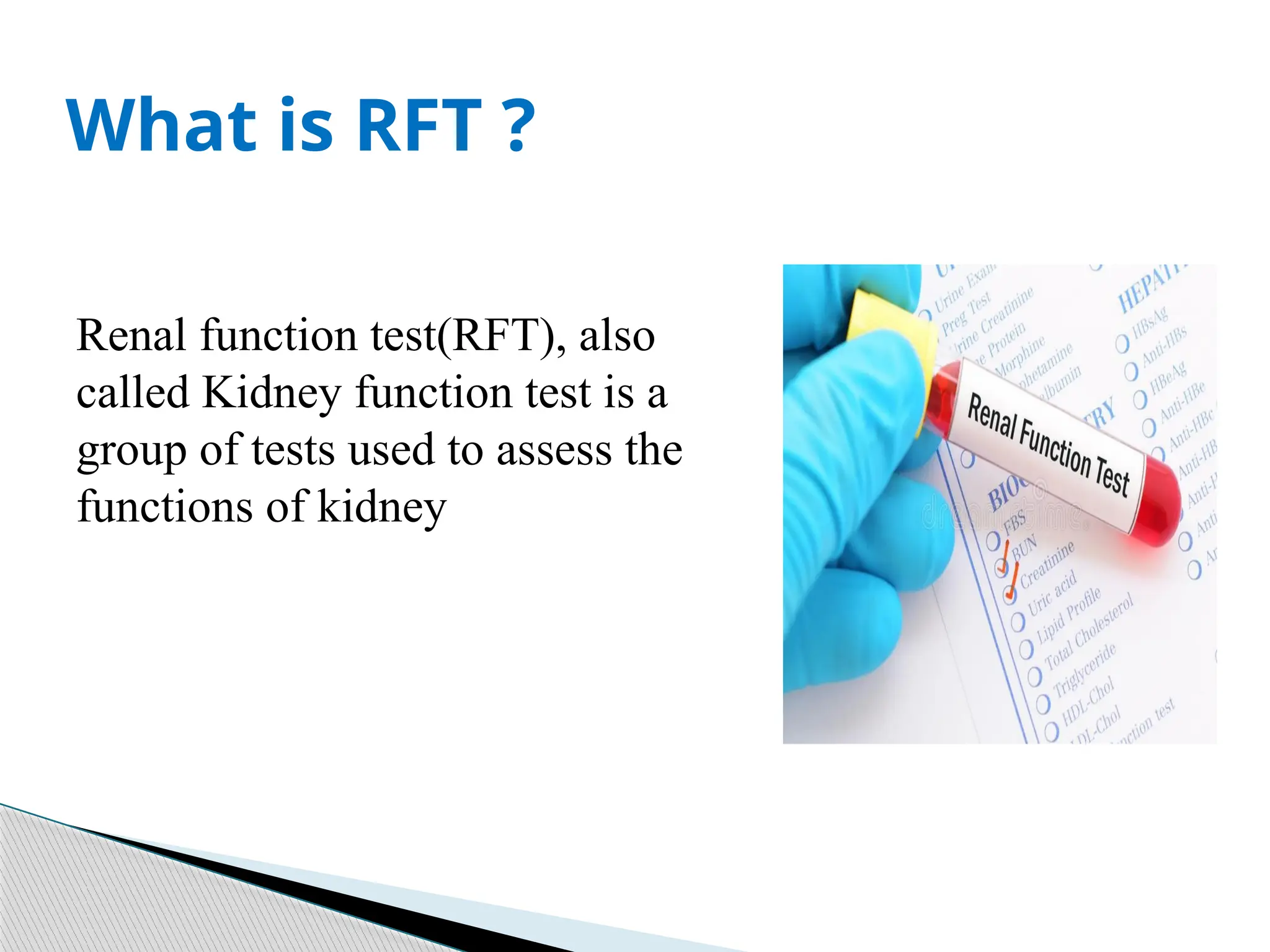
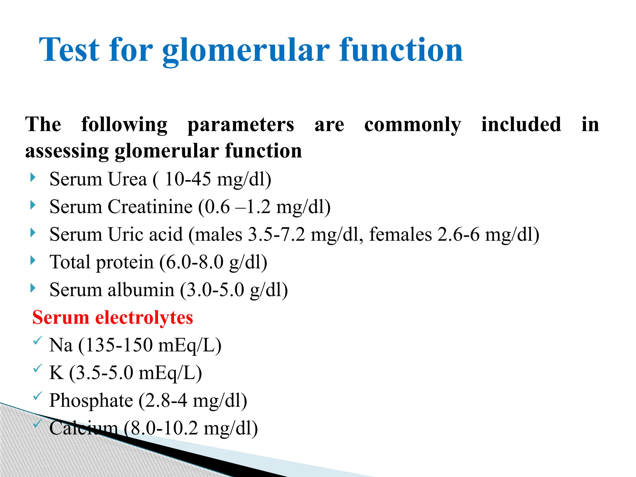
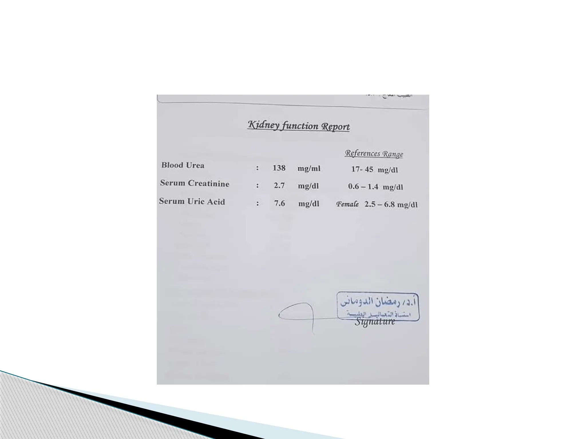
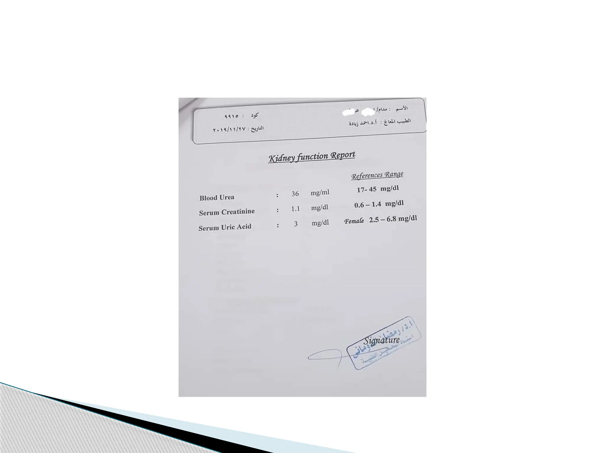
Renal Function Test
RFT
By:
Fatima Ali
Renal function test(RFT), also
called Kidney function test is a
group of tests used to assess the
functions of kidney
What is RFT ?
 To assess functional capacity of kidneys
 To diagnose renal impairment
 To assess the severity and progression of renal
impairment
 To assess the effectiveness of treatment
Why Renal Function Tests needed?
Can be divided into two categories:
 Test for glomerular function
 Serum Urea
 Serum Creatinine
 Clearance tests
 Tests for tubular function
 Urine concentration test
 Dilution test
 Para amino hippuric acid clearance test
 Acidification test
 Urine examination: Important for assessing both glomerular
and tubular function
Renal function test
The following parameters are commonly included in
assessing glomerular function
 Serum Urea ( 10-45 mg/dl)
 Serum Creatinine (0.6 –1.2 mg/dl)
 Serum Uric acid (males 3.5-7.2 mg/dl, females 2.6-6 mg/dl)
 Total protein (6.0-8.0 g/dl)
 Serum albumin (3.0-5.0 g/dl)
Serum electrolytes
 Na (135-150 mEq/L)
 K (3.5-5.0 mEq/L)
 Phosphate (2.8-4 mg/dl)
 Calcium (8.0-10.2 mg/dl)
Test for glomerular function
Measurement of urea is used to:
 evaluate renal function
 to assess hydration status
 to determine nitrogen balance
 to aid in the diagnosis of renal disease
 and to verify adequacy of dialysis.
Serum Urea
Azotemia:
elevated conc. of urea in blood.
Uremia:
very high plasma urea concentration accompanied by
renal failure. Causes of blood urea elevations are: Pre-
renal Renal and post-renal
Disease Correlations
 Anything that produces a decrease in functional blood
volume, include: Congestive heart failure, Shock,
Hemorrhage, Dehydration High protein diet or
increased catabolism (Fever, major illness, stress)
Pre-Renal Azotemia:
Decreased renal function causes increased blood urea due
to poor excretion Acute & Chronic renal failure
Glomerulonephritis Tubular necrosis & other Intrinsic
renal disease
Renal Azotemia:
Post-Renal Azotemia
Obstruction of urine flow Renal calculi Tumors of
bladder or prostate Severe infections
Decreased blood urea
Low protein dietary intake Liver disease (lack of
synthesis) Severe vomiting and/or diarrhea(loss)
 Creatinine is the waste product of creatine phosphate,
a compound found in the skeletal muscle tissue.
 It is excreted entirely by the kidneys.
 The creatinine level is affected primarily by renal
dysfunction and is thus very useful in evaluating renal
function.
 Increased levels of creatinine indicate a slowing of the
glomerular filtration rate.
 Female: 0.6–1.2 mg/dL
 Male: 0.8–1.4 mg/dL
Serum Creatinine
Possible Meanings of Abnormal Values:
 Congestive heart failure
 Glomerulonephritis
 Nephritis
 Pyelonephritis
 Renal failure
 Urinary obstruction
 Dehydration
it is filtered by the glomerulus and secreted by the distal
tubules into the urine, in which most uric acid is
reabsorbed in the proximal tubules and reused.
Serum Uric Acid
Possible Meanings of Abnormal Values:
Increased (hyperuricemia):
Congestive heart failure
Glomerulonephritis
Alcoholism
Gout
Dehydration
Trauma and surgery.
Administration of certain drugs (diuretics, cyclosporine,
etc.)
Decreased (hypouricemia)
Liver disease
Renal tubular defects
Defectivetubular reabsorption(Fanconi syndrome)
Chemotherapy with azathioprine or 6mercaptopurine
Overtreatment with allopurinol
 Sodium is decreased (hyponatremia) and potassium is
increased (hyperkalemia) in chronic kidney disease
(CKD) as kidney reabsorb sodium in exchange of
potassium
 Chloride and phosphate is increased in CKD
 Calcium is decreased as vitamin D is deficient
Serum electrolytes
1. Hypernatremia :
Its defined as an increased sodium concentration in
plasma water, and is generally diagnosed at serum
sodium levels >145 mmol/L.
Hypernatremia is always associated with an increased
effective plasma osmolality, and hence with a reduced
cell volume.
Sodium disorders:
2. Hyponatremia:
Is defined as reduced plasma sodium concentration to a
value less than 135 mmol/L.
The five most common causes of Hyponatremia are
overhydration, diuretics, SIADH, diabetic
hyperosmolarity and Addison’s disease.
Sodium disorders
1. Hypokalemia:
 Is a plasma K concentration below the lower limit of
the reference range.
 Hypokalemia can occur with GI or urinary loss of K or
with increased cellular uptake of K.
Potassium disorders:
 Hyperkalemia may be caused by one of three
mechanisms:
 (1) shift of potassium from the cells to the ECF
 (2) increased potassium intake
 (3) reduced renal potassium excretion.
Hyperkalemia:
Any question?
24
Thank you

RFT renal function Laboratory report ppt

  • 1.
  • 2.
    Renal function test(RFT),also called Kidney function test is a group of tests used to assess the functions of kidney What is RFT ?
  • 3.
     To assessfunctional capacity of kidneys  To diagnose renal impairment  To assess the severity and progression of renal impairment  To assess the effectiveness of treatment Why Renal Function Tests needed?
  • 4.
    Can be dividedinto two categories:  Test for glomerular function  Serum Urea  Serum Creatinine  Clearance tests  Tests for tubular function  Urine concentration test  Dilution test  Para amino hippuric acid clearance test  Acidification test  Urine examination: Important for assessing both glomerular and tubular function Renal function test
  • 5.
    The following parametersare commonly included in assessing glomerular function  Serum Urea ( 10-45 mg/dl)  Serum Creatinine (0.6 –1.2 mg/dl)  Serum Uric acid (males 3.5-7.2 mg/dl, females 2.6-6 mg/dl)  Total protein (6.0-8.0 g/dl)  Serum albumin (3.0-5.0 g/dl) Serum electrolytes  Na (135-150 mEq/L)  K (3.5-5.0 mEq/L)  Phosphate (2.8-4 mg/dl)  Calcium (8.0-10.2 mg/dl) Test for glomerular function
  • 8.
    Measurement of ureais used to:  evaluate renal function  to assess hydration status  to determine nitrogen balance  to aid in the diagnosis of renal disease  and to verify adequacy of dialysis. Serum Urea
  • 9.
    Azotemia: elevated conc. ofurea in blood. Uremia: very high plasma urea concentration accompanied by renal failure. Causes of blood urea elevations are: Pre- renal Renal and post-renal Disease Correlations
  • 10.
     Anything thatproduces a decrease in functional blood volume, include: Congestive heart failure, Shock, Hemorrhage, Dehydration High protein diet or increased catabolism (Fever, major illness, stress) Pre-Renal Azotemia:
  • 11.
    Decreased renal functioncauses increased blood urea due to poor excretion Acute & Chronic renal failure Glomerulonephritis Tubular necrosis & other Intrinsic renal disease Renal Azotemia:
  • 12.
    Post-Renal Azotemia Obstruction ofurine flow Renal calculi Tumors of bladder or prostate Severe infections
  • 13.
    Decreased blood urea Lowprotein dietary intake Liver disease (lack of synthesis) Severe vomiting and/or diarrhea(loss)
  • 14.
     Creatinine isthe waste product of creatine phosphate, a compound found in the skeletal muscle tissue.  It is excreted entirely by the kidneys.  The creatinine level is affected primarily by renal dysfunction and is thus very useful in evaluating renal function.  Increased levels of creatinine indicate a slowing of the glomerular filtration rate.  Female: 0.6–1.2 mg/dL  Male: 0.8–1.4 mg/dL Serum Creatinine
  • 15.
    Possible Meanings ofAbnormal Values:  Congestive heart failure  Glomerulonephritis  Nephritis  Pyelonephritis  Renal failure  Urinary obstruction  Dehydration
  • 16.
    it is filteredby the glomerulus and secreted by the distal tubules into the urine, in which most uric acid is reabsorbed in the proximal tubules and reused. Serum Uric Acid
  • 17.
    Possible Meanings ofAbnormal Values: Increased (hyperuricemia): Congestive heart failure Glomerulonephritis Alcoholism Gout Dehydration Trauma and surgery. Administration of certain drugs (diuretics, cyclosporine, etc.)
  • 18.
    Decreased (hypouricemia) Liver disease Renaltubular defects Defectivetubular reabsorption(Fanconi syndrome) Chemotherapy with azathioprine or 6mercaptopurine Overtreatment with allopurinol
  • 19.
     Sodium isdecreased (hyponatremia) and potassium is increased (hyperkalemia) in chronic kidney disease (CKD) as kidney reabsorb sodium in exchange of potassium  Chloride and phosphate is increased in CKD  Calcium is decreased as vitamin D is deficient Serum electrolytes
  • 20.
    1. Hypernatremia : Itsdefined as an increased sodium concentration in plasma water, and is generally diagnosed at serum sodium levels >145 mmol/L. Hypernatremia is always associated with an increased effective plasma osmolality, and hence with a reduced cell volume. Sodium disorders:
  • 21.
    2. Hyponatremia: Is definedas reduced plasma sodium concentration to a value less than 135 mmol/L. The five most common causes of Hyponatremia are overhydration, diuretics, SIADH, diabetic hyperosmolarity and Addison’s disease. Sodium disorders
  • 22.
    1. Hypokalemia:  Isa plasma K concentration below the lower limit of the reference range.  Hypokalemia can occur with GI or urinary loss of K or with increased cellular uptake of K. Potassium disorders:
  • 23.
     Hyperkalemia maybe caused by one of three mechanisms:  (1) shift of potassium from the cells to the ECF  (2) increased potassium intake  (3) reduced renal potassium excretion. Hyperkalemia:
  • 24.
  • 25.