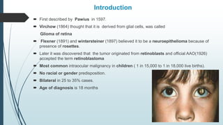
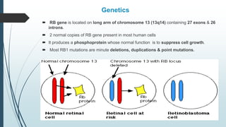
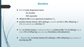
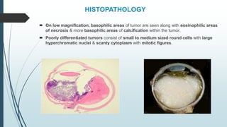
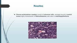
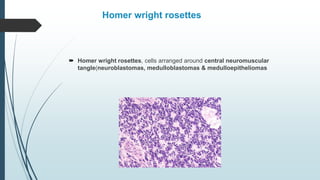
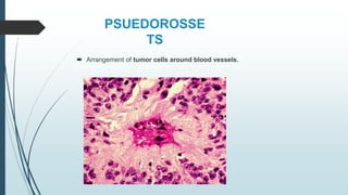
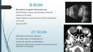
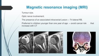
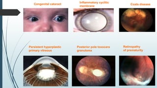
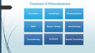
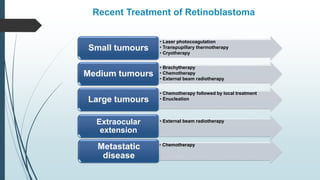
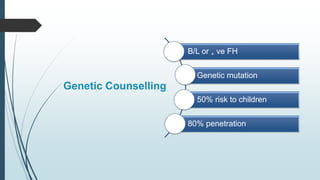
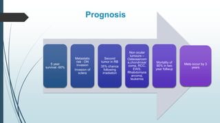
RETINOBLASTO
MA
Dr.
EYE
 First described by Pawius in 1597.
 Virchow (1864) thought that it is derived from glial cells, was called
Glioma of retina
 Flexner (1891) and wintersteiner (1897) believed it to be a neuroepithelioma because of
presence of rosettes.
 Later it was discovered that the tumor originated from retinoblasts and official AAO(1926)
accepted the term retinoblastoma
 Most common intraocular malignancy in children ( 1 in 15,000 to 1 in 18,000 live births).
 No racial or gender predisposition.
 Bilateral in 25 to 35% cases.
 Age of diagnosis is 18 months
Introduction
Genetics
 RB gene is located on long arm of chromosome 13 (13q14) containing 27 exons & 26
introns.
 2 normal copies of RB gene present in most human cells
 It produces a phosphoprotein whose normal function is to suppress cell growth.
 Most RB1 mutations are minute deletions, duplications & point mutations.
 Out of newly diagnosed cases
6% familial
94% sporadic
 Bilateral RB involve germinal mutations Pts,
 positive family history, 40% siblings would be at risk & 40% offspring of
affected patient may develop RB
 Pts, no family history, if affected child has unilateral RB, 1% of siblings are at
risk & 8% of offspring may develop Hereditary retinoblastoma
 Bilateral RB, no family history 6% siblings & 40% offspring have chance of
developing RB
Genetics
Hereditary Retinoblastoma
Heterozygous
alleles
(RB/RB+).
Only one
mutation
First hit
occurs in
utero
All cells of
body affected
Second hit
occurs in any
retinal cell
HISTOPATHOLOGY
 On low magnification, basophilic areas of tumor are seen along with eosinophilic areas
of necrosis & more basophilic areas of calcification within the tumor.
 Poorly differentiated tumors consist of small to medium sized round cells with large
hyperchromatic nuclei & scanty cytoplasm with mitotic figures.
Rosettes
 Flexner-wintersteiner rosettes consist of columnar cells arranged around a central
lumen,highly characteristic of Retinoblastoma, also seen in medulloepithelioma
Homer wright rosettes
 Homer wright rosettes, cells arranged around central neuromuscular
tangle(neuroblastomas, medulloblastomas & medulloepitheliomas
PSUEDOROSSE
TS
 Arrangement of tumor cells around blood vessels.
Fleurettes
 Fleurettes -eosinophilic structures, composed of tumor cells with pear shaped
eosinophilic processes, projecting through fenestrated membrane.
CLASSIFICATION
Common Presentations
 Leucocoria 56%
 Strabismus 20%
 Red painful eye 7%
 Poor vision 5%
 Asymptomatic 3%
 Orbital cellulitis 3%
 Unilateral mydriasis 2%
 Heterochromia iridis 1%
 Hyphema 1%
TYPES OF RB
Grades of Optic Nerve Involvment
• Superficial involvement of optic nerve
head only
Grade 1
• Involvement upto or involving
lamina cribrosa
Grade 2
• Involvement beyond lamina cribrosa
Grade 3
• Involvement upto surgical margin
Grade 4
DIAGNOSIS
 History
 Prenatal/natal/postnatal
Maternal rubella ( cong. Cat.)
Gestation period or delivery ( ROP )
Oxygen therapy ( ROP )
 Family history
 Physical examination
Rule out Tuberous sclerosis, Cong. defects asso with trisomy 13-15,
Rubella syndrome
 Ocular examination
Squint, Leukocoria, Heterochromia, Proptosis, Lymphadenopathy
Pseudohypopyon, Rubeosis iridis
Fundus examination
 Detailed Anterior segment examination
 I O P
 Indirect Ophthalmoscopy under anaesthesia with
 Chalky, white-gray retinal mass with a soft, friable consistency
Fundus drawing denoting site & extent
 Bilateral with 360 degree scleral depression
 Indirect ophthalmoscopy (diagnostic in 90% cases)
 Wide angle fundus camera ( Retcam)
B SCAN
Rounded or irregular intraocular mas
Demonstrates a mass more echogenic than the
vitreous on B mode.
Highly reflective intrinsic echoes of fine calcifications
on A mode.
Accuracy 80%
CT SCAN
Delineates extraocular extension
Can detect Asso. Pinealoblastoma
Cases with atypical manisfestations
Diagnostic dilemma Extraocular &I ntracranial tumour
extension is suspected
Magnetic resonance imaging (MRI)
Tumour size.
Optic nerve involvement.
The presence of an associated intracranial Lesion→ Tri-lateral RB.
Preferred in children younger than one year of age → avoid cancer risk that
increase with CT
FFA
 Not required in routine work up.
 Can be used to differentiate viable tumour from an avascular residue following radiotherapy or
spontaneously regressed retinoblastoma.
 Active lesion shows hypervascularity, dilated feeders and late staining
BIOCHEMICAL TESTS
AQUEOUS HUMOUR ENZYME ASSAY
Normal aqueous to plasma LDH >1.0
Increased phosphoglucoisomerase levels
Increased neuron specific enolase level
BONEMARROW ASPIRATION AND LUMBAR PUNCTURE
Useful in ruling out intracranial or distant spread
As primary mode of spread of RB is hematogenous
EXAMINATION OF SIBLINGS/PARENTS
 To detect small lesions which may otherwise go undetected in siblings.
 Parents may harbour regressed RB lesions.
 Blood specimens of patient/parents/siblings should be taken for DNA analysis
which could aid n genetic counselling
SYSTEMIC EXAMINATION
 PIneoblastoma or an ectopic intracranial retinoblastoma
 Sarcomas of bone and soft tissue
Congenital cataract
Persistent hyperplastic
primary vitreous
Inflammatory cyclitic
membrane
Coats disease
Posterior pole toxocara
granuloma
Retinopathy
of prematurity
Differential Diagnosis of Vitreous Seeds
 Pars planitis (intermediate uveitis)
 Microbial endophthalmitis or retinitis
 Leukemic infiltration
Differential Diagnosis of Discrete Retinal Tumors
 Astrocytoma of retina
 Medulloepithelioma
 Retinal capillary hemangioma/Hemangioblastoma
 Focal patches of myelinated retinal nerve fiber
Treatment of Retinoblastoma
Enucleation Cryotherapy Photocoagulation
EBRT Brachy Therapy Chemotherapy
Thermotherapy Combined Genetic Counselling
Goals of Treatment
Preserve
Vision
Salvage Eye
Minimize
Complications
Minimize side
effects of
therapy
Save life
ENUCLEATION
 Large tumors ( >50 % of globe volume) with no visual potential.
 Tumors that extend into the optic nerve.
 Blind or painful eyes.
 Group E tumors that have failed previous "globeconserving“ approaches.
 Group D tumors (Significant vitreous seeding and subretinal infiltration)
 Secondary glaucoma.
 Vitreous seeding.
 Anterior chamber invasion
Special considerations for enucleation in R.B
 Minimal manipulation
 Avoid perforation of eye
 Harvest long ( >15 mm) optic nerve stump
 Inspect the enucleated eye for macroscopic
extraocular extension & optic nerve involvement
 Harvest fresh tissue for genetic studies
 Place a primary implant
 ORBITAL IMPLANT
Promotes orbital growth
Provides better cosmesis
Enhances prosthesis motility
Adjuvant chemo after enucleation
 Considered in patients with high-risk features
Iris, ciliary body infiltration
Massive choroidal or scleral infiltration,
Invasion of the optic nerve posterior to the lamina cribrosa.
Adjuvant EBRTx should be considered in extrascleral extension.
Post-enucleation follow-up
 Monitored closely for orbital relapse in the two years after surgery
 The incidence of orbital recurrence after enucleation is 4.2%.
 85 % of patients with orbital recurrence also developes metastatic disease.
EXTERNAL BEAM
RADIOTHERAPY
Rarely utilized
Large bilateral tumours
Vitreous seeding
Thick tumours near the optic nerve or fovea in the eye
with visual potential.
Multiple tumours that are too large for cryotherapy or
laser photocoagulation.
Recurrent diseases
Progression on chemo reduction.
Radioactive Plaques
I125 brachytherapy
40 to 45 Gy - To the tumor apex
Must be <15 mm in diameter and
<10 mm thickness
Ruthenium-106 (Ru106)
For small tumors (thickness
less than 6 mm)
Reduces the radiation dose to
the uninvolved retina,
Children managed with brachytherapy should be monitored for the
development of radiation retinopathy and optic neuropathy
Tumour identified using I/O
Dummy plaque placed against
sclera
Position confirmed
Radioactive plaque placed
4000Gy total
Removed after 3-5 days of
placement
Steps
Complications
Cataract
Radiation
Retinopathy
Vitreous
Hge
Chronic Dry
Eye
Corneal
Toxicity
Second
Malignant
Neoplasm
Avascular
transluscent
mass
Calcific residue
Complete
disappearance of
tumour
Flat chorioretinal
scar
Regression
Laser Photocoagulation
Small posterior
tumours
3-4mm diameter 2mm
thick
With out vitreous
seeding , not
involving disc or
macula
Coagulate the blood
supply to tumour
Direct treatment
avoided 
Dissemination
Argon laser
Temperature 60degree
Two rows of laser
burns spot size 2mm
Power 200-400mW
Laser Photocoagulation
Successful
treatment -
Flat
chorioretinal
scar
Recurrence
20- 30%
Complications :
Transient RD
Vascular occlusion
Retinal hole
Preretinal fibrosis
CRYO THERAPY
Small equatorial or peripheral tumour 4mm
x2mm
Triple freeze thaw cycle , 2-3 session
of 1 month interval
Advantage : Preserve ILM and
bruch’s membrane  natural barrier
for tr growth
Disadvantage: Not useful for vitreous
seeding & inferior trs which pinch off
from posterior trs and form new trs
Thermotherapy
Chemothermotherapy
 HEAT FACILITATES CELLULAR UPTAKE OF PLATINUM GRP OF DRUGS
 20-30 mts of exposure to diode laser 2-3 hrs after iv carboplatin
Delivery of heat using IR radiation
Traanspupillary diode laser appli
4x2mm tumour
Iv chemotherapy
Germline mutation retinoblastoma,
Bilateral retinoblastoma
Familial retinoblastoma
Age of 4 months or younger
Suspicious evidence of early optic nerve
or choroidal invasion
Chemoreduction
Reduce tumor volume
Improves the ocular salvage rate.
Enhances the success of local therapies
Carboplatin, vincristine, and etoposide
High dose chemo ….Every 4 weeks
Group A, B, and C
Secondary acute myeloid leukemia
Intra-Arterial Chemotherapy
 Safe and effective in advanced intraocular RB.
 Catheterization of the ophthalmic artery and injection of chemotherapy, usually
melphalan (with or without topotecan)
Indications
 Nongermline mutation retinoblastoma
 Unilateral retinoblastoma
 Age greater than 4 months
 Recurrent retinoblastoma following previous IVC or plaque radiotherapy,
 Recurrent subretinal seeds involving two or more quadrants
 Recurrent vitreous seeds.
Complications
Vitreous hemorrhage (2%)
Branch retinal artery obstruction (1%)
Ophthalmic artery spasm with
reperfusion (2%)
Ophthalmic artery obstruction (2%),
Partial choroidal ischemia (2%)
Optic neuropathy
PERIOCULAR
CHEMOTHERAPY
Indications
 Bilateral advanced groups D or E in which a higher local dose of chemotherapy is
desired
 Recurrent localized tumor.
 Carboplatin or topotecan
Complications
 Orbital and eyelid edema and ecchymosis
 Orbital fat atrophy
 Muscle fibrosis leading to strabismus
 Optic atrophy
Intravitreal Chemotherapy
 Vitreous seeds nonresponsive to standard therapy
Vitreous seeds recurrent after previous therapy
 Intravitreal injection of melphalan
 Tumor seeding can occur following IVitC.
 High doses induce hypotonia and Pthysis bulbi
Recurrent Retinoblastoma
 The prognosis depends on the site and extent treatment with etoposide and
carboplatin
• Prognosis for sight and survival may be excellent
• Local ophthalmic therapy
Confined to the eye
and is small
• Prognosis for sight is poor
• Survival remains excellent.
Confined to the eye
but is extensive
• 5 year survival chances are minimal
Extra-ocular
Recent Treatment of Retinoblastoma
• Laser photocoagulation
• Transpupillary thermotherapy
• Cryotherapy
Small tumours
• Brachytherapy
• Chemotherapy
• External beam radiotherapy
Medium tumours
• Chemotherapy followed by local treatment
• Enucleation
Large tumours
• Chemotherapy
Metastatic
disease
• External beam radiotherapy
Extraocular
extension
Genetic Counselling
B/L or + ve FH
Genetic mutation
50% risk to children
80% penetration
Prognosis
5 year
survival -90%
Metastatic
risk : ON
invasion
Invasion of
sclera
Second
tumor in RB
35% chance
following
irradiation
Non ocular
tumours –
Osteosarcom
a,chondrosar
coma, RCC,
EWS,
Rhabdomyos
arcoma,
leukemia
Mortality of
90% in two
year follwup
Mets occur by 3
years
Poor Prognostic Factors
Risk of
metastatic
disease
• Optic nerve
• Choroidal
• Orbital
invasion
Delay in
diagnosis
of more
than six
months
Patients
with SMN
• Vitreous
seeding
• Extraocular
spread
Risk factors for extension into the optic N:
 Exophytic growth pattern.
 Elevated intraocular pressure.
 Tumor thickness ≥15 mm.
Risk factors for extension into the choroid:
 Elevated I.O. pressure.
 Iris neovascularization.
Follow Up
Once a year lifelong for heritable tumors
6 monthly till fifth year
4 monthly for second year
3monthly x I year
Six weeks after initial therapy
Newer treatment
Systemic SE of chemo agents
S/C carboplatin
Photosensitizing
agents = Porphyrins
Iv 2.5mg/kg
hematoporphyrin  3
days later argon laser
2mm spot 2oomW x
10 mts… Repeat at I
month if viable
Gene therapy
References
 Shields CL, Lally SE, Leahey AM, Jabbour PM, Caywood EH, Schwendeman R, Shields JA.
Targeted retinoblastoma management: when to use intravenous, intra-arterial, periocular, and
intravitreal chemotherapy. Current opinion in ophthalmology. 2014 Sep 1;25(5):374-85Podos SM,
 Yanoff M, editors. Textbook of ophthalmology. Gower Medical Pub.; 2016
 Shields CL, Douglass AM, Beggache M, Say EA, Shields JA. Intravitreous chemotherapy for
active vitreous seeding from retinoblastoma: outcomes after 192 consecutive injections. The
2015 Howard Naquin Lecture. Retina. 2016 Jun 1;36(6):1184-90.
 Yuan S, Friedman DL, Daniels AB. Evolution of chemotherapy approaches for the treatment of
intraocular retinoblastoma: a comprehensive review. International ophthalmology clinics. 2017
Jan 1;57(1):117-28.
 Intra-arterial chemotherapy for retinoblastoma. Indian journal of ophthalmology. 2017;65(9):900-
1.
 Sinha AK. Clinico-pathological observations on retinoblastoma. Indian journal of ophthalmology.
1975 Oct 1;23(3):20.

RETINOBLASTOMA.pptx

  • 1.
  • 2.
     First describedby Pawius in 1597.  Virchow (1864) thought that it is derived from glial cells, was called Glioma of retina  Flexner (1891) and wintersteiner (1897) believed it to be a neuroepithelioma because of presence of rosettes.  Later it was discovered that the tumor originated from retinoblasts and official AAO(1926) accepted the term retinoblastoma  Most common intraocular malignancy in children ( 1 in 15,000 to 1 in 18,000 live births).  No racial or gender predisposition.  Bilateral in 25 to 35% cases.  Age of diagnosis is 18 months Introduction
  • 3.
    Genetics  RB geneis located on long arm of chromosome 13 (13q14) containing 27 exons & 26 introns.  2 normal copies of RB gene present in most human cells  It produces a phosphoprotein whose normal function is to suppress cell growth.  Most RB1 mutations are minute deletions, duplications & point mutations.
  • 4.
     Out ofnewly diagnosed cases 6% familial 94% sporadic  Bilateral RB involve germinal mutations Pts,  positive family history, 40% siblings would be at risk & 40% offspring of affected patient may develop RB  Pts, no family history, if affected child has unilateral RB, 1% of siblings are at risk & 8% of offspring may develop Hereditary retinoblastoma  Bilateral RB, no family history 6% siblings & 40% offspring have chance of developing RB Genetics
  • 5.
    Hereditary Retinoblastoma Heterozygous alleles (RB/RB+). Only one mutation Firsthit occurs in utero All cells of body affected Second hit occurs in any retinal cell
  • 6.
    HISTOPATHOLOGY  On lowmagnification, basophilic areas of tumor are seen along with eosinophilic areas of necrosis & more basophilic areas of calcification within the tumor.  Poorly differentiated tumors consist of small to medium sized round cells with large hyperchromatic nuclei & scanty cytoplasm with mitotic figures.
  • 7.
    Rosettes  Flexner-wintersteiner rosettesconsist of columnar cells arranged around a central lumen,highly characteristic of Retinoblastoma, also seen in medulloepithelioma
  • 8.
    Homer wright rosettes Homer wright rosettes, cells arranged around central neuromuscular tangle(neuroblastomas, medulloblastomas & medulloepitheliomas
  • 9.
    PSUEDOROSSE TS  Arrangement oftumor cells around blood vessels.
  • 10.
    Fleurettes  Fleurettes -eosinophilicstructures, composed of tumor cells with pear shaped eosinophilic processes, projecting through fenestrated membrane.
  • 11.
  • 14.
    Common Presentations  Leucocoria56%  Strabismus 20%  Red painful eye 7%  Poor vision 5%  Asymptomatic 3%  Orbital cellulitis 3%  Unilateral mydriasis 2%  Heterochromia iridis 1%  Hyphema 1%
  • 15.
  • 17.
    Grades of OpticNerve Involvment • Superficial involvement of optic nerve head only Grade 1 • Involvement upto or involving lamina cribrosa Grade 2 • Involvement beyond lamina cribrosa Grade 3 • Involvement upto surgical margin Grade 4
  • 18.
    DIAGNOSIS  History  Prenatal/natal/postnatal Maternalrubella ( cong. Cat.) Gestation period or delivery ( ROP ) Oxygen therapy ( ROP )  Family history  Physical examination Rule out Tuberous sclerosis, Cong. defects asso with trisomy 13-15, Rubella syndrome  Ocular examination Squint, Leukocoria, Heterochromia, Proptosis, Lymphadenopathy Pseudohypopyon, Rubeosis iridis
  • 19.
    Fundus examination  DetailedAnterior segment examination  I O P  Indirect Ophthalmoscopy under anaesthesia with  Chalky, white-gray retinal mass with a soft, friable consistency Fundus drawing denoting site & extent  Bilateral with 360 degree scleral depression  Indirect ophthalmoscopy (diagnostic in 90% cases)  Wide angle fundus camera ( Retcam)
  • 20.
    B SCAN Rounded orirregular intraocular mas Demonstrates a mass more echogenic than the vitreous on B mode. Highly reflective intrinsic echoes of fine calcifications on A mode. Accuracy 80% CT SCAN Delineates extraocular extension Can detect Asso. Pinealoblastoma Cases with atypical manisfestations Diagnostic dilemma Extraocular &I ntracranial tumour extension is suspected
  • 21.
    Magnetic resonance imaging(MRI) Tumour size. Optic nerve involvement. The presence of an associated intracranial Lesion→ Tri-lateral RB. Preferred in children younger than one year of age → avoid cancer risk that increase with CT
  • 22.
    FFA  Not requiredin routine work up.  Can be used to differentiate viable tumour from an avascular residue following radiotherapy or spontaneously regressed retinoblastoma.  Active lesion shows hypervascularity, dilated feeders and late staining BIOCHEMICAL TESTS AQUEOUS HUMOUR ENZYME ASSAY Normal aqueous to plasma LDH >1.0 Increased phosphoglucoisomerase levels Increased neuron specific enolase level BONEMARROW ASPIRATION AND LUMBAR PUNCTURE Useful in ruling out intracranial or distant spread As primary mode of spread of RB is hematogenous
  • 23.
    EXAMINATION OF SIBLINGS/PARENTS To detect small lesions which may otherwise go undetected in siblings.  Parents may harbour regressed RB lesions.  Blood specimens of patient/parents/siblings should be taken for DNA analysis which could aid n genetic counselling SYSTEMIC EXAMINATION  PIneoblastoma or an ectopic intracranial retinoblastoma  Sarcomas of bone and soft tissue
  • 24.
    Congenital cataract Persistent hyperplastic primaryvitreous Inflammatory cyclitic membrane Coats disease Posterior pole toxocara granuloma Retinopathy of prematurity
  • 25.
    Differential Diagnosis ofVitreous Seeds  Pars planitis (intermediate uveitis)  Microbial endophthalmitis or retinitis  Leukemic infiltration Differential Diagnosis of Discrete Retinal Tumors  Astrocytoma of retina  Medulloepithelioma  Retinal capillary hemangioma/Hemangioblastoma  Focal patches of myelinated retinal nerve fiber
  • 26.
    Treatment of Retinoblastoma EnucleationCryotherapy Photocoagulation EBRT Brachy Therapy Chemotherapy Thermotherapy Combined Genetic Counselling
  • 27.
    Goals of Treatment Preserve Vision SalvageEye Minimize Complications Minimize side effects of therapy Save life
  • 28.
    ENUCLEATION  Large tumors( >50 % of globe volume) with no visual potential.  Tumors that extend into the optic nerve.  Blind or painful eyes.  Group E tumors that have failed previous "globeconserving“ approaches.  Group D tumors (Significant vitreous seeding and subretinal infiltration)  Secondary glaucoma.  Vitreous seeding.  Anterior chamber invasion
  • 29.
    Special considerations forenucleation in R.B  Minimal manipulation  Avoid perforation of eye  Harvest long ( >15 mm) optic nerve stump  Inspect the enucleated eye for macroscopic extraocular extension & optic nerve involvement  Harvest fresh tissue for genetic studies  Place a primary implant  ORBITAL IMPLANT Promotes orbital growth Provides better cosmesis Enhances prosthesis motility
  • 30.
    Adjuvant chemo afterenucleation  Considered in patients with high-risk features Iris, ciliary body infiltration Massive choroidal or scleral infiltration, Invasion of the optic nerve posterior to the lamina cribrosa. Adjuvant EBRTx should be considered in extrascleral extension. Post-enucleation follow-up  Monitored closely for orbital relapse in the two years after surgery  The incidence of orbital recurrence after enucleation is 4.2%.  85 % of patients with orbital recurrence also developes metastatic disease.
  • 31.
    EXTERNAL BEAM RADIOTHERAPY Rarely utilized Largebilateral tumours Vitreous seeding Thick tumours near the optic nerve or fovea in the eye with visual potential. Multiple tumours that are too large for cryotherapy or laser photocoagulation. Recurrent diseases Progression on chemo reduction.
  • 32.
    Radioactive Plaques I125 brachytherapy 40to 45 Gy - To the tumor apex Must be <15 mm in diameter and <10 mm thickness Ruthenium-106 (Ru106) For small tumors (thickness less than 6 mm) Reduces the radiation dose to the uninvolved retina, Children managed with brachytherapy should be monitored for the development of radiation retinopathy and optic neuropathy
  • 33.
    Tumour identified usingI/O Dummy plaque placed against sclera Position confirmed Radioactive plaque placed 4000Gy total Removed after 3-5 days of placement Steps
  • 34.
  • 35.
    Laser Photocoagulation Small posterior tumours 3-4mmdiameter 2mm thick With out vitreous seeding , not involving disc or macula Coagulate the blood supply to tumour Direct treatment avoided  Dissemination Argon laser Temperature 60degree Two rows of laser burns spot size 2mm Power 200-400mW
  • 36.
    Laser Photocoagulation Successful treatment - Flat chorioretinal scar Recurrence 20-30% Complications : Transient RD Vascular occlusion Retinal hole Preretinal fibrosis
  • 37.
    CRYO THERAPY Small equatorialor peripheral tumour 4mm x2mm Triple freeze thaw cycle , 2-3 session of 1 month interval Advantage : Preserve ILM and bruch’s membrane  natural barrier for tr growth Disadvantage: Not useful for vitreous seeding & inferior trs which pinch off from posterior trs and form new trs
  • 38.
    Thermotherapy Chemothermotherapy  HEAT FACILITATESCELLULAR UPTAKE OF PLATINUM GRP OF DRUGS  20-30 mts of exposure to diode laser 2-3 hrs after iv carboplatin Delivery of heat using IR radiation Traanspupillary diode laser appli 4x2mm tumour
  • 39.
    Iv chemotherapy Germline mutationretinoblastoma, Bilateral retinoblastoma Familial retinoblastoma Age of 4 months or younger Suspicious evidence of early optic nerve or choroidal invasion
  • 40.
    Chemoreduction Reduce tumor volume Improvesthe ocular salvage rate. Enhances the success of local therapies Carboplatin, vincristine, and etoposide High dose chemo ….Every 4 weeks Group A, B, and C Secondary acute myeloid leukemia
  • 41.
    Intra-Arterial Chemotherapy  Safeand effective in advanced intraocular RB.  Catheterization of the ophthalmic artery and injection of chemotherapy, usually melphalan (with or without topotecan) Indications  Nongermline mutation retinoblastoma  Unilateral retinoblastoma  Age greater than 4 months  Recurrent retinoblastoma following previous IVC or plaque radiotherapy,  Recurrent subretinal seeds involving two or more quadrants  Recurrent vitreous seeds.
  • 42.
    Complications Vitreous hemorrhage (2%) Branchretinal artery obstruction (1%) Ophthalmic artery spasm with reperfusion (2%) Ophthalmic artery obstruction (2%), Partial choroidal ischemia (2%) Optic neuropathy
  • 43.
    PERIOCULAR CHEMOTHERAPY Indications  Bilateral advancedgroups D or E in which a higher local dose of chemotherapy is desired  Recurrent localized tumor.  Carboplatin or topotecan Complications  Orbital and eyelid edema and ecchymosis  Orbital fat atrophy  Muscle fibrosis leading to strabismus  Optic atrophy
  • 44.
    Intravitreal Chemotherapy  Vitreousseeds nonresponsive to standard therapy Vitreous seeds recurrent after previous therapy  Intravitreal injection of melphalan  Tumor seeding can occur following IVitC.  High doses induce hypotonia and Pthysis bulbi
  • 45.
    Recurrent Retinoblastoma  Theprognosis depends on the site and extent treatment with etoposide and carboplatin • Prognosis for sight and survival may be excellent • Local ophthalmic therapy Confined to the eye and is small • Prognosis for sight is poor • Survival remains excellent. Confined to the eye but is extensive • 5 year survival chances are minimal Extra-ocular
  • 46.
    Recent Treatment ofRetinoblastoma • Laser photocoagulation • Transpupillary thermotherapy • Cryotherapy Small tumours • Brachytherapy • Chemotherapy • External beam radiotherapy Medium tumours • Chemotherapy followed by local treatment • Enucleation Large tumours • Chemotherapy Metastatic disease • External beam radiotherapy Extraocular extension
  • 47.
    Genetic Counselling B/L or+ ve FH Genetic mutation 50% risk to children 80% penetration
  • 48.
    Prognosis 5 year survival -90% Metastatic risk: ON invasion Invasion of sclera Second tumor in RB 35% chance following irradiation Non ocular tumours – Osteosarcom a,chondrosar coma, RCC, EWS, Rhabdomyos arcoma, leukemia Mortality of 90% in two year follwup Mets occur by 3 years
  • 49.
    Poor Prognostic Factors Riskof metastatic disease • Optic nerve • Choroidal • Orbital invasion Delay in diagnosis of more than six months Patients with SMN • Vitreous seeding • Extraocular spread
  • 50.
    Risk factors forextension into the optic N:  Exophytic growth pattern.  Elevated intraocular pressure.  Tumor thickness ≥15 mm. Risk factors for extension into the choroid:  Elevated I.O. pressure.  Iris neovascularization.
  • 51.
    Follow Up Once ayear lifelong for heritable tumors 6 monthly till fifth year 4 monthly for second year 3monthly x I year Six weeks after initial therapy
  • 52.
    Newer treatment Systemic SEof chemo agents S/C carboplatin Photosensitizing agents = Porphyrins Iv 2.5mg/kg hematoporphyrin  3 days later argon laser 2mm spot 2oomW x 10 mts… Repeat at I month if viable Gene therapy
  • 54.
    References  Shields CL,Lally SE, Leahey AM, Jabbour PM, Caywood EH, Schwendeman R, Shields JA. Targeted retinoblastoma management: when to use intravenous, intra-arterial, periocular, and intravitreal chemotherapy. Current opinion in ophthalmology. 2014 Sep 1;25(5):374-85Podos SM,  Yanoff M, editors. Textbook of ophthalmology. Gower Medical Pub.; 2016  Shields CL, Douglass AM, Beggache M, Say EA, Shields JA. Intravitreous chemotherapy for active vitreous seeding from retinoblastoma: outcomes after 192 consecutive injections. The 2015 Howard Naquin Lecture. Retina. 2016 Jun 1;36(6):1184-90.  Yuan S, Friedman DL, Daniels AB. Evolution of chemotherapy approaches for the treatment of intraocular retinoblastoma: a comprehensive review. International ophthalmology clinics. 2017 Jan 1;57(1):117-28.  Intra-arterial chemotherapy for retinoblastoma. Indian journal of ophthalmology. 2017;65(9):900- 1.  Sinha AK. Clinico-pathological observations on retinoblastoma. Indian journal of ophthalmology. 1975 Oct 1;23(3):20.