Anatomy of the trachea, bronchi, and lungs
•Download as PPTX, PDF•
0 likes•60 views

The document describes several anatomical structures in the thorax, including the trachea, esophagus, thoracic duct, azygos vein, hemiazygos vein, and thoracic sympathetic trunk. It provides details on the location, course, tributaries, relations and nerve supply of each structure. Key points include that the trachea divides into right and left principal bronchi, the esophagus passes through the mediastinum to connect the pharynx and stomach, and the thoracic duct drains lymph from the thorax and left side of the body.

Recommended
More Related Content
Similar to Anatomy of the trachea, bronchi, and lungs
Similar to Anatomy of the trachea, bronchi, and lungs (20)
Recently uploaded
Recently uploaded (20)
Anatomy of the trachea, bronchi, and lungs
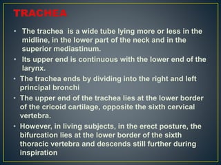
- 1. • The trachea is a wide tube lying more or less in the midline, in the lower part of the neck and in the superior mediastinum. • Its upper end is continuous with the lower end of the larynx. • The trachea ends by dividing into the right and left principal bronchi • The upper end of the trachea lies at the lower border of the cricoid cartilage, opposite the sixth cervical vertebra. • However, in living subjects, in the erect posture, the bifurcation lies at the lower border of the sixth thoracic vertebra and descends still further during inspiration
- 3. • Relations of the Thoracic Part Anteriorly • 1 Manubrium sterni • 2 Sternothyroid muscles • 3 Remains of the thymus • 4 Left brachiocephalic and inferior thyroid veins. • 5 Aortic arch, brachiocephalic and left common carotid arteries • 6 Deep cardiac plexus (see Fig. 19.6) • 7 Some lymph nodes • Posteriorly • 1 Oesophagus • 2 Vertebral column
- 5. • On the Right Side • 1 Right lung and pleura • 2 Right vagus • 3 Azygos vein (Fig. 20.2) • On the Left Side • 1 Arch of aorta, left common carotid and left subclavian arteries • 2 Left recurrent laryngeal nerve. • Arterial Supply: • Inferior thyroid arteries. • Venous drainage: Into the left brachiocephalic vein. • Lymphatic drainage: • To the pretracheal and paratracheal nodes.
- 7. Structure • The trachea has a fibroelastic wall supported by a cartilaginous skeleton formed by C-shaped rings. • The rings are about 16 to 20 in number and make the tube convex anterolaterally. • Posteriorly, there is a gap which is closed by a fibroelastic membrane and contains transversely arranged smooth muscle known as the trachealis. • The lumen is lined by ciliated columnar epithelium and contains many mucous and serous glands.
- 8. • Nerve Supply • 1 Parasympathetic: Nerves through vagi and recurrent laryngeal nerves. • It is: • a. Sensory and secretomotor to the mucous membrane. • b. Motor to the trachealis muscle. • 2 Sympathetic: Fibres from the middle cervical • ganglion reach it along the inferior thyroid arteries and are vasomotor.
- 9. OESOPHAGUS • Features • The oesophagus is a narrow muscular tube, formingn the food passage between the pharynx and stomach. • It extends from the lower part of the neck to the upper part of the abdomen. • The pharyngo-oesophageal junction is the narrowest part of the alimentary canal except for the vermiform appendix. • The oesophagus begins in the neck at the lower border of the cricoid cartilage, where it is continuous with the lower end of the pharynx. • It descends in front of the vertebral column through the superior and posterior parts of the mediastinum, • pierces the diaphragm at level of tenth thoracic vertebra. • It ends by opening into the stomach at itsncardiac end at the level of eleventh thoracic vertebra
- 10. • Curvatures • In general, the oesophagus is vertical, but shows slight curvatures in the following directions. • There are two side-to-side curvatures, both towards the left. • One is at the root of the neck and the other near the lower end. • It also has anteroposterior curvatures that correspond to the curvatures of the cervicothoracic spine.
- 12. Constrictions • Normally, the oesophagus shows four constrictions. • These are seen as indentations. 1. At its beginning, 15 cm/6-inch from the incisor teeth, where it is crossed by cricopharyngeus muscle. 2. Where it is crossed by the aortic arch, 22.5 cm/9-inch from the incisor teeth. 3. Where it is crossed by the left bronchus, 27.5 cm/11-inch from the incisor teeth (Fig. 20.9). 4. Where it pierces the diaphragm 37.5 cm/15-inch from the incisor teeth. • The distances from the incisor teeth are important in passing instruments like endoscope into the oesophagus.
- 14. • Relations of the Thoracic Part of the Oesophagus Anteriorly • 1 Trachea 2. Right pulmonary artery • 3 Left bronchus 4. Pericardium with left atrium • 5 The diaphragm (Figs 20.2 and 20.3). Posteriorly • 1 Vertebral column • 2 Right posterior intercostal arteries • 3 Thoracic duct • 4 Azygos vein with the terminal parts of the hemiazygos veins • 5 Thoracic aorta • 6 Right pleural recess • 7 Diaphragm
- 17. • Arterial Supply 1. The cervical part including the segment up to the arch • of aorta is supplied by the inferior thyroid arteries. 2. The thoracic part • is supplied by the oesophageal branches of the aorta. 3. The abdominal part is supplied by the oesophageal branches of the left gastric artery Venous Drainage upper part of the oesophagus drains into the brachiocephalic veins; • from the middle part it goes to the azygos veins; and from the lower end it goes to the left gastric vein and vena azygos via • hemiazygos vein. • lower end of the oesophagus is one of the sites of
- 18. Nerve Supply 1. Parasympathetic nerves: • The upper half of the oesophagus is supplied by the recurrent laryngeal nerves, • the lower half by the oesophageal plexus formed mainly by the two vagi. • Parasympathetic nerves are sensory, motor and secretomotor to the oesophagus. 2. Sympathetic nerves: • For upper half of oesophagus, the fibres come from middle cervical ganglion and run with inferior thyroid arteries. • For lower half, the fibres come directly from upper four thoracic ganglia, to form oesophageal plexus before supplying the oesophagus.
- 19. • The oesophageal plexus is formed mainly by the parasympathetic through vagi but sympathetic fibres are also present. • Towards the lower end of the oesophagus; the vagal fibres form the anterior and posterior gastric nerves which enter the abdomen through the oesophageal opening of the diaphragm. • Lymphatic Drainage • The cervical part drains to the deep cervical nodes. • thoracic part to the posterior mediastinal nodes; • abdominal part to the left gastric nodes
- 22. THORACIC DUCT Features • The thoracic duct is the largest lymphatic vessel in the body. • It extends from the upper part of the abdomen to the lower part of the neck, crossing the posterior and superior parts of the mediastinum. • It is about 45 cm/ 18 inch long. It has a beaded appearance because of the presence of many valves in its lumen. • Course • The thoracic duct begins as a continuation of the upper end of the cisterna chyli near the lower border of the 12th thoracic vertebra and enters the thorax through the aortic opening of the diaphragm
- 24. • It then ascends through the posterior mediastinum from level of twelfth thoracic vertebra to fifth thoracic vertebra, where it crosses from the right side to the left side. • Then it courses through the superior mediastinum along the left edge of the oesophagus and reaches the neck. • In the neck, it arches laterally at the level of the transverse process of seventh cervical vertebra. • Finally, it descends in front of the first part of the left subclavian artery and ends by opening into the angle of junction between the left subclavian and left internal jugular veins
- 26. • Relations • At the Aortic Opening of the Diaphragm Anteriorly: • Diaphragm Posteriorly: Vertebral column To the right: Azygos vein To the left: Aorta (see Fig. 12.16) In the Posterior Mediastinum • Anteriorly • 1 Diaphragm (Fig. 20.6c) • 2 Oesophagus • 3 Right pleural recess
- 27. • Posteriorly • 1 Vertebral column • 2 Right posterior intercostal arteries • 3 Terminal parts of the hemiazygos veins To the right: Azygos vein To the left: Descending thoracic aorta (Fig. 20.6c)
- 29. • In the Superior Mediastinum • Anteriorly • 1 Arch of aorta • 2 The origin of the left subclavian artery (Fig. 20.6a) • Posteriorly: • Vertebral column • To the right: Oesophagus • To the left: Pleura
- 30. • In the Neck • The thoracic duct forms an arch rising about 3–4 cm above the clavicle. The arch has the following relations. Anteriorly • 1 Left common carotid artery • 2 Left vagus • 3 Left internal jugular vein Posteriorly • 1 Vertebral artery and vein. • 2 Sympathetic trunk • 3 Thyrocervical trunk and its branches • 4 Left phrenic nerve • 5 Medial border of the scalenus anterior • 6 Prevertebral fascia covering all the structures mentioned • 7 The first part of the left subclavian artery
- 31. • Tributaries • The thoracic duct receives lymph from, roughly, both halves of the body below the diaphragm and the left half above the diaphragm (Fig. 20.12). • In the thorax, the thoracic duct receives lymph vessels from the posterior mediastinal nodes and from small intercostal nodes. • At the root of the neck, efferent vessels of the nodes in the neck form the left jugular trunk, and those from nodes in the axilla form the left subclavian trunk. • these trunks end in the thoracic duct.
- 33. • The left bronchomediastinal trunk drains lymph from the left half of the thorax and ends in the thoracic duct. • On the right side, there is right lymphatic duct into which right bronchomediastinal, right jugular and right subclavian lymph trunks drain. • The right lymphatic trunk ends in the right brachiocephalic vein at the junction of right subclavian and right internal jugular veins
- 34. AZYGOS VEIN. The term ‘azygos’ means unpaired. • The azygos vein drains the thoracic wall and the upper lumbar region. • It forms an important channel connecting the superior and inferior venae cavae. • The vein occupies the upper part of the posterior abdominal wall and the posterior mediastinum. • It also connects portal venous system, caval venous system and vertebral venous system.
- 35. Formation • The azygos vein is formed by union of the lumbar azygos, right subcostal and right ascending lumbar veins. 1. The lumbar azygos vein may be regarded as the abdominal part of the azygos vein. • It lies to the right of the lumbar vertebrae. • Its lower end communicates with the inferior vena cava. 2. The right subcostal vein accompanies the corresponding artery. 3. The ascending lumbar vein is formed by vertical anastomoses that connect the lumbar veins. • The azygos vein may be formed by union of the right subcostal and ascending lumbar veins (Fig. 14.10
- 37. Course • 1 The azygos vein enters the thorax by passing through • the aortic opening of the diaphragm (see Fig. 12.16). • 2 The azygos vein then ascends up to fourth thoracic vertebra where it arches forwards over the root of the right lung • ends by joining the posterior aspect of the superior vena cava before it pierces the pericardium (see Fig. 15.2). • Relations • Anteriorly: Oesophagus. • Posteriorly: • 1 Lower eight thoracic vertebrae • 2 Right posterior intercostal arteries
- 39. • To the right: • 1 Right lung and pleura • 2 Greater splanchnic nerve • To the left: • 1 Thoracic duct and aorta in lower part • 2 Oesophagus, trachea and vagus in the upper part
- 40. • Tributaries 1. Right superior intercostal vein formed by union of • the second, third and fourth posterior intercostal veins. 2. Fifth to eleventh right posterior intercostal veins. 3 Hemiazygos vein at the level of lower border of eighth thoracic vertebra. 4. Accessory hemiazygos vein at the level of upper border of eighth thoracic vertebra. 5. Right bronchial vein, near the terminal end of the • azygos vein. 6. Several oesophageal, mediastinal, pericardial veins
- 41. • HEMIAZYGOS VEIN • It is also called the inferior hemiazygos vein. It is the mirror image of the lower part of the azygos vein. • the hemiazygos is formed by the union of the left lumbar azygos, left ascending lumbar, and left subcostal veins . Course • Hemiazygos vein pierces the left crus of the diaphragm, ascends on the left side of the vertebra overlapped by the aorta. • At the level of eighth thoracic vertebra, it turns to the right, passes behind the oesophagus and the thoracic duct, and joins the azygos vein. Tributaries • Ninth to eleventh left posterior intercostal veins and • oesophageal veins.
- 42. ACCESSORY HEMIAZYGOS VEIN • it is also called the superior hemiazygos vein. • It is the mirror image of the upper part of the azygos vein. • Course • Accessory hemiazygos vein begins at the medial end of the fourth or fifth intercostal space, and • Descends on the left side of the vertebral column. • At the level of eighth thoracic vertebra, it turns to the right, passes behind the aorta and the thoracic duct, and joins the azygos vein.
- 43. • Tributaries • 1 Fifth to eighth left posterior intercostal veins • 2 Sometimes the left bronchial veins
- 44. THORACIC SYMPATHETIC TRUNK Features • The thoracic sympathetic trunk is a ganglionated chain • situated one on each side of the thoracic vertebral column. Superiorly, it is continuous with the cervical part of the chain and inferiorly with the lumbar part • Theoretically, the chain bears 12 ganglia corresponding to the 12 thoracic nerves. • The first thoracic ganglion is commonly fused with the inferior cervical ganglion to form the cervicothoracic, or stellate ganglion. • The remaining thoracic ganglia generally lie at the levels • of the corresponding intervertebral discs and the intercostal nerves.
- 45. • Course and Relations • The chain crosses the neck of the first rib, the heads of the second to tenth ribs, and bodies of the eleventh and twelfth thoracic vertebrae. • The whole chain descends in front of the posterior intercostal vessels and the intercostal nerves, and passes deep to the medial arcuate ligament to become continuous with the lumbar part of the sympathetic chain
- 46. • Branches Lateral Branches for the Limbs and Body Wall • Each ganglion is connected with its corresponding spinal nerve by two rami, the white (preganglionic) and grey (postganglionic) rami communicantes. • The white ramus is distal to the grey ramus • The grey rami communicantes along with spinal nerves supply structures in the skin and blood vessels of skeletal muscles of the whole body (Fig.14.14).
- 49. • Medial Branches for the Viscera 1. Medial branches from the upper 5 ganglia • are postganglionic and get distributed to the heart, The great vessels, the lungs and the oesophagus, through the following. • a. Pulmonary branches to the pulmonary plexuses • b. Cardiac branches to the deep cardiac plexus • c. Aortic branches to thoracic aortic plexus • d. Oesophageal branches which join the oesophageal plexus (Fig. 14.13
- 52. • 2 Medial branches from the lower 7 ganglia • Are preganglionic and form three splanchnic nerves. a. The greater splanchnic nerve is formed by 5 roots from ganglia 5 to 9. • It descends obliquely on the vertebral bodies, pierces the crus of the diaphragm, and ends (in the abdomen) mainly in the coeliac ganglion, and partly in the aorticorenal ganglion and the suprarenal gland. b. The lesser splanchnic nerve is formed by two roots from ganglia 10 and 11. • Its course is similar to that of the greater splanchnic nerve. • It pierces the crus of the diaphragm, and ends in the coeliac ganglion (Fig. 14.14)
- 54. • c. The least (lowest) splanchnic nerve (renal nerve) • Is tiny. It arises by one root from ganglion 12. • It pierces the corresponding crus of the diaphragm.