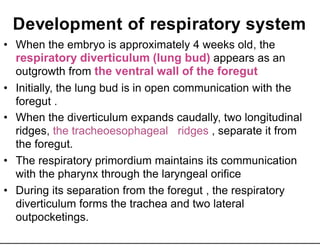
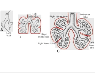
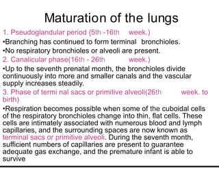
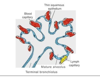
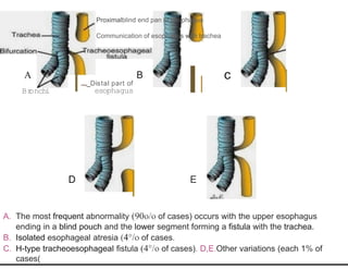
Development of respiratory system
• When the embryo is approximately 4 weeks old, the
respiratory diverticulum (lung bud) appears as an
outgrowth from the ventral wall of the foregut
• Initially, the lung bud is in open communication with the
foregut .
• When the diverticulum expands caudally, two longitudinal
ridges, the tracheoesophageal ridges , separate it from
the foregut.
• The respiratory primordium maintains its communication
with the pharynx through the laryngeal orifice
• During its separation from the foregut , the respiratory
diverticulum forms the trachea and two lateral
outpocketings.
Development of the larynx and trachea
• The upper part of the tube becomes the larynx, whereas
the lower part becomes the trachea.
• The epithelium lining of the larynx, trachea, and bronchi
and the epithelium of the alveoli develop from the
entodermal lining of the floor of the pharynx
• Just behind the developing tongue, a small opening
persists that will become the permanent opening into the
larynx.
• The cartilages and muscles of larynx originate from
mesenchyme of the fourth and sixth pharyngeal arches.
• The tube divides distally into the right and left lung buds.
Tracheoesophageal
ridge Foregut
/.
Esophagus
/
Respirator
y
diverticulu
m A B
Tuberculum imp r Lat l lingual swelling
era
-r--
Trachea
11
cecum
c
Epiglotta
l
swelling
Larynge
al
orifice
Development of the Lungs
• Each lung bud consists of an entodermal tube surrounded
by splanchnic mesoderm. Each bud grows laterally and
projects into the pleural part of the embryonic coelom.
• At the beginning of the 51 week, each of these buds
h
enlarges to form right and left main bronchi. The right forms
three secondary bronchi and the left forms two bronchi
• Each main bronchus then divides repeatedly in a
dichotomous manner, until eventually the terminal
bronchioles and alveoli are formed. The division of the
terminal bronchioles, with the formation of additional
bronchioles and alveoli, continues for some time after
birth.
Lung
buds
--: Left
bronchus
Lett upper
lobe
B
Right
middle lobe
Right lower lobe
lower
lobe
c
• By the seventh month, the capillary loops
connected with the pulmonary circulation
have become sufficiently well developed
to support life.
• With the onset of respiration at birth, the
lungs expand and the alveoli become
dilated. However, it is only after 3 or 4
days of postnatal life that the alveoli in
the periphery of each lung become fully
expanded
Development of pleura
• Each bud grows laterally and projects
into the pleural part of the embryonic
coelom.
• Each lung will receive a covering of
visceral pleura derived from the
splanchnic mesoderm and the parietal
pleura will be formed from somatic
mesoderm.
Maturation of the lungs
1. Pseudoglandular period {5th -16th week.)
•Branching has continued to form terminal bronchioles.
•No respiratory bronchioles or alveoli are present.
2. Canalicular phase(16th - 26th week.)
•Up to the seventh prenatal month, the bronchioles divide
continuously into more and smaller canals and the vascular
supply increases steadily.
3. Phase of termi nal sacs or primitive alveoli(26th week. to
birth)
•Respiration becomes possible when some of the cuboidal cells
of the respiratory bronchioles change into thin, flat cells. These
cells are intimately associated with numerous blood and lymph
capillaries, and the surrounding spaces are now known as
terminal sacs or primitive alveoli. During the seventh month,
sufficient numbers of capillaries are present to guarantee
adequate gas exchange, and the premature infant is able to
survive
A
Respirator
y
bronchiolu
s
Blood
capillarie
s
Thin
squamou
s
epithelium Flat
endothelium cell
of blood
capillary
Terminal
sacsr-
........ 'I:
Cuboidal
epithelium If
• 4. alveolar period (from 81h month of prenatal life to
childhood)
• During the last 2 months of prenatal life and for several years
thereafter , the number of terminal sacs increases steadily. In
addition, cells lining the sacs (type I alveolar epithelial cells)
become thinner, so that surrounding capillaries protrude into the
alveolar sacs. This intimate contact between epithelial and
endothelial cells makes up the blood-air barrier.
• Mature alveoli are not present before birth.
• Another cell type develops at
the end of the sixth month. These cells, type II alveolar epithelial cells,
produce surfactant , a phospholipid-rich fluid capable of lowering
surface tension at the air-alveolar interface.
• Surfactant deposited as a thin phospholipid coat on alveolar cell
membranes. Without the fatty surfactant layer, the alveoli would
collapse during expiration (atelectasis).
Thin squamous
epithelium
Blood
capillary
Mature alveolus
Terminal bronchiolus
Lymph
capillary
• Fetal breathing movements begin before birth and cause
aspiration of amniotic fluid. These movements are
important for stimulating lung development and
conditioning respiratory muscles. When respiration
begins at birth, most of the lung fluid is rapidly resorbed
by the blood and lymph capillaries, and a small amount
is probably expelled via the trachea and bronchi during
delivery
• Growth of the lungs after birth is primarily due to an
increase in the number of respiratory bronchioles and
alveoli and not to an increase in the size of the alveoli.
• New alveoli are formed during the first 10 years of
postnatal life.
Congenital anomalies
• Abnormalities in partitioning of the esophagus
and trachea by the tracheoesophageal septum
result in esophageal atresia with or without
tracheoesophageal fistulas (TEFs). Obstruction
of the esophagus prevents the child from
swallowing saliva and milk, and this
leads to aspiration into the larynx and trachea,
which usually results in pneumonia.
• With early diagnosis, it is often possible to
correct this serious anomaly surgically
Proximalblind end pan of osophagus
Communication of esophagus with trachea
A
Br
onchi
B
......_Distal part of
esophagus
D
c
E
A. The most frequent abnormality (90o/o of cases) occurs with the upper esophagus
ending in a blind pouch and the lower segment forming a fistula with the trachea.
B. Isolated esophageal atresia (4°/o of cases.
C. H-type tracheoesophageal fistula (4°/o of cases). D,E.Other variations (each 1% of
cases(
Conginital anomalies of the lung
• Respiratory distress syndrome
• Surfactant is particularly important for survival
of the premature infant. When surfactant is
insufficient, the air-water (blood) surface
membrane tension becomes high, bringing
great risk that alveoli will collapse during
expiration. As a result, respiratory distress
syndrome (RDS) develops. This is a common
cause of death in the premature infant.
• Ectopic lung lobes arising from the trachea or
esophagus. It is believed that these lobes are
formed from additional respiratory buds of the
foregut that develop independently of the main
respiratory system
• Congenital cysts of the lung, which are formed
by dilation of terminal or larger bronchi. These
cysts may be small and multiple, giving the lung
a honeycomb appearance on radiograph, or
they may be restricted to one or more larger
ones. Cystic structures of the lung usually drain
poorly and frequently cause chronic infections.

Respiratorysystem.ppt

  • 1.
    Development of respiratorysystem • When the embryo is approximately 4 weeks old, the respiratory diverticulum (lung bud) appears as an outgrowth from the ventral wall of the foregut • Initially, the lung bud is in open communication with the foregut . • When the diverticulum expands caudally, two longitudinal ridges, the tracheoesophageal ridges , separate it from the foregut. • The respiratory primordium maintains its communication with the pharynx through the laryngeal orifice • During its separation from the foregut , the respiratory diverticulum forms the trachea and two lateral outpocketings.
  • 2.
    Development of thelarynx and trachea • The upper part of the tube becomes the larynx, whereas the lower part becomes the trachea. • The epithelium lining of the larynx, trachea, and bronchi and the epithelium of the alveoli develop from the entodermal lining of the floor of the pharynx • Just behind the developing tongue, a small opening persists that will become the permanent opening into the larynx. • The cartilages and muscles of larynx originate from mesenchyme of the fourth and sixth pharyngeal arches. • The tube divides distally into the right and left lung buds.
  • 3.
    Tracheoesophageal ridge Foregut /. Esophagus / Respirator y diverticulu m AB Tuberculum imp r Lat l lingual swelling era -r-- Trachea 11 cecum c Epiglotta l swelling Larynge al orifice
  • 4.
    Development of theLungs • Each lung bud consists of an entodermal tube surrounded by splanchnic mesoderm. Each bud grows laterally and projects into the pleural part of the embryonic coelom. • At the beginning of the 51 week, each of these buds h enlarges to form right and left main bronchi. The right forms three secondary bronchi and the left forms two bronchi • Each main bronchus then divides repeatedly in a dichotomous manner, until eventually the terminal bronchioles and alveoli are formed. The division of the terminal bronchioles, with the formation of additional bronchioles and alveoli, continues for some time after birth.
  • 5.
  • 6.
    • By theseventh month, the capillary loops connected with the pulmonary circulation have become sufficiently well developed to support life. • With the onset of respiration at birth, the lungs expand and the alveoli become dilated. However, it is only after 3 or 4 days of postnatal life that the alveoli in the periphery of each lung become fully expanded
  • 7.
    Development of pleura •Each bud grows laterally and projects into the pleural part of the embryonic coelom. • Each lung will receive a covering of visceral pleura derived from the splanchnic mesoderm and the parietal pleura will be formed from somatic mesoderm.
  • 8.
    Maturation of thelungs 1. Pseudoglandular period {5th -16th week.) •Branching has continued to form terminal bronchioles. •No respiratory bronchioles or alveoli are present. 2. Canalicular phase(16th - 26th week.) •Up to the seventh prenatal month, the bronchioles divide continuously into more and smaller canals and the vascular supply increases steadily. 3. Phase of termi nal sacs or primitive alveoli(26th week. to birth) •Respiration becomes possible when some of the cuboidal cells of the respiratory bronchioles change into thin, flat cells. These cells are intimately associated with numerous blood and lymph capillaries, and the surrounding spaces are now known as terminal sacs or primitive alveoli. During the seventh month, sufficient numbers of capillaries are present to guarantee adequate gas exchange, and the premature infant is able to survive
  • 9.
    A Respirator y bronchiolu s Blood capillarie s Thin squamou s epithelium Flat endothelium cell ofblood capillary Terminal sacsr- ........ 'I: Cuboidal epithelium If
  • 10.
    • 4. alveolarperiod (from 81h month of prenatal life to childhood) • During the last 2 months of prenatal life and for several years thereafter , the number of terminal sacs increases steadily. In addition, cells lining the sacs (type I alveolar epithelial cells) become thinner, so that surrounding capillaries protrude into the alveolar sacs. This intimate contact between epithelial and endothelial cells makes up the blood-air barrier. • Mature alveoli are not present before birth. • Another cell type develops at the end of the sixth month. These cells, type II alveolar epithelial cells, produce surfactant , a phospholipid-rich fluid capable of lowering surface tension at the air-alveolar interface. • Surfactant deposited as a thin phospholipid coat on alveolar cell membranes. Without the fatty surfactant layer, the alveoli would collapse during expiration (atelectasis).
  • 11.
  • 12.
    • Fetal breathingmovements begin before birth and cause aspiration of amniotic fluid. These movements are important for stimulating lung development and conditioning respiratory muscles. When respiration begins at birth, most of the lung fluid is rapidly resorbed by the blood and lymph capillaries, and a small amount is probably expelled via the trachea and bronchi during delivery • Growth of the lungs after birth is primarily due to an increase in the number of respiratory bronchioles and alveoli and not to an increase in the size of the alveoli. • New alveoli are formed during the first 10 years of postnatal life.
  • 13.
    Congenital anomalies • Abnormalitiesin partitioning of the esophagus and trachea by the tracheoesophageal septum result in esophageal atresia with or without tracheoesophageal fistulas (TEFs). Obstruction of the esophagus prevents the child from swallowing saliva and milk, and this leads to aspiration into the larynx and trachea, which usually results in pneumonia. • With early diagnosis, it is often possible to correct this serious anomaly surgically
  • 14.
    Proximalblind end panof osophagus Communication of esophagus with trachea A Br onchi B ......_Distal part of esophagus D c E A. The most frequent abnormality (90o/o of cases) occurs with the upper esophagus ending in a blind pouch and the lower segment forming a fistula with the trachea. B. Isolated esophageal atresia (4°/o of cases. C. H-type tracheoesophageal fistula (4°/o of cases). D,E.Other variations (each 1% of cases(
  • 15.
    Conginital anomalies ofthe lung • Respiratory distress syndrome • Surfactant is particularly important for survival of the premature infant. When surfactant is insufficient, the air-water (blood) surface membrane tension becomes high, bringing great risk that alveoli will collapse during expiration. As a result, respiratory distress syndrome (RDS) develops. This is a common cause of death in the premature infant.
  • 16.
    • Ectopic lunglobes arising from the trachea or esophagus. It is believed that these lobes are formed from additional respiratory buds of the foregut that develop independently of the main respiratory system • Congenital cysts of the lung, which are formed by dilation of terminal or larger bronchi. These cysts may be small and multiple, giving the lung a honeycomb appearance on radiograph, or they may be restricted to one or more larger ones. Cystic structures of the lung usually drain poorly and frequently cause chronic infections.