Asthma is adisease characterized by:
1. episodic airway obstruction
2. airway hyperresponsiveness usually accompanied by
airway in
fl
ammation.
- In most cases, the airway obstruction is reversible, but in a
subset of asthmatics, a component of the obstruction may
become irreversible.
- In a large proportion of patients, the airway in
fl
ammation is
eosinophilic, but some patients may present with di
ff
ering
types of airway in
fl
ammation, and in some cases, there is
no obvious evidence of airway in
fl
ammation.
MANIFESTATIONS
Asthma most frequently presents as:
1. episodic shortness of breath
2. wheezing
3. cough, which can occur in relation to triggers but may also
occur spontaneously.
4. Other symptoms can include chest tightness and/or
mucus production.
- These symptoms can occur in combination or separately.
Section 2 Diseases of the Respiratory System
Asthma
3.
- These symptomscan resolve spontaneously or with
therapy.
- In some patients, wheezing and/or dyspnea can be
persistent.
- Episodes of acute bronchospasm, known as exacerbations,
may be severe enough to require emergency medical care
or hospitalization and may result in death.
4.
EPIDEMIOLOGY
- Asthma isthe most common chronic disease associated
with signi
fi
cant morbidity and mortality, with ~241 million
people a
ff
ected globally.
- Prevalence continues to increase (starting at 7.3% in 2001
in the United States)
- and is associated with transition from rural to urban living.
- Asthma is more prevalent among children (8.4%) than
adults (7.7%).
- In children, the prevalence is greatest among boys (2:1
male-to-female ratio).
- with a trend toward greater prevalence in women in
adulthood.
- In some patients, asthma resolves as they enter adulthood
only to “recur” later in life.
- In the United States, asthma is more prevalent in blacks
than Caucasians.
- black race is associated with greater case morbidity.
- The ethnicity with the greatest prevalence in the United
States is the Puerto Rican population.
- Asthma mortality increased worldwide in the 1960s,
apparently related to overuse of inhaled β2-agonists.
5.
- Reduction inmortality since then has been attributed to
increased use of inhaled corticosteroids.
- Asthma mortality declined globally from 0.44 per 100,000
people in 1993 to 0.19 in 2006, but further reduction in
mortality has not occurred since that time.
6.
PATHOPHYSIOLOGY
■ MECHANISMS LEADINGTO ACUTE AND CHRONIC
AIRWAY OBSTRUCTION
(1) Airway Hyperresponsiveness
Airway hyperresponsiveness is a hallmark of asthma.
It is de
fi
ned as an acute narrowing response of the airways
in reaction to agents that do not elicit airway responses in
non- a
ff
ected individuals
or an excess narrowing response to inhaled agents as
compared to that which would occur in nona
ff
ected
individuals.
A component of the hyperresponsiveness occurs at the level
of the airway smooth muscle itself as demonstrated by
hyperresponsiveness to direct smooth-muscle–acting agents
such as histamine or methacholine.
In many patients, the apparent hyperresponsiveness is due to
1 indirect activation of airway narrowing mechanisms as a
result of:
stimulation of in
fl
ammatory cells (which release direct
bronchoconstrictors and mediators that cause airway
edema and/or mucus secretion)
and/ or stimulation of sensory nerves that can act on the
smooth muscle or in
fl
ammatory cells.
7.
2The apparent increasedresponsiveness of the airways in
asthma may also have a structural etiology.
In asthma, airway wall thickness is associated with disease
severity and duration.
This thickening, which may result from a combination of:
• smooth-muscle hypertrophy and hyperplasia
• subepithelial collagen deposition
• airway edema
• mucosal in
fl
ammation
can result in a tendency for the airway to narrow
disproportionately in response to stimuli that elicit increased
airway muscle tension.
A major therapeutic objective in asthma is to decrease the
degree of airway hyperresponsiveness.
(2) In
fl
ammatory Cells
While airway in
fl
ammation can be precipitated by acute
exposure to inhalants, most asthmatics have evidence of
chronic in
fl
ammation in the airways.
Most commonly, this in
fl
ammation is eosinophilic in nature.
In some patients, neutrophilic in
fl
ammation may be
predominant, especially in those with more severe asthma.
Mast cells are also more frequent.
8.
(3) Airway SmoothMuscle
Airway smooth muscle can contribute to asthma in three
ways.
First, it can be hyperresponsive to stimuli, as noted above.
Second,hypertrophy and hyperplasia can lead to airway
wall thickening with consequences for
hyperresponsiveness
Lastly, airway smooth-muscle cells can produce
chemokines and cytokines that promote airway
in
fl
ammation and promote the survival of in
fl
ammatory
cells, particularly mast cells.
(4)Subepithelial Collagen Deposition and Matrix
Deposition
Thickening of the subepithelial basement membrane occurs
as a result of deposition of:
• repair-type collagens
• and tenascin, periostin,
fi
bronectin,
• and osteopontin primarily from myo
fi
broblasts under the
epithelium.
The deposition of collagen and matrix sti
ff
ens the airway and
can result in exaggerated responses to increased
circumferential tension exerted by the smooth muscle.
9.
Such deposition canalso narrow the airway lumen and
decrease its ability to relax and thus can contribute to chronic
airway obstruction.
(5) Airway Epithelium
Airway epithelium disruption takes the form of separation of
columnar cells from the basal cells.
The damaged epithelium is hypothesized to form a trophic
unit with the underlying mesenchyme.
This unit elaborates multiple growth factors thought to
contribute to airway remodeling as well as multiple cytokines
and mediators that promote asthmatic airway in
fl
ammation.
(6) Vascular Proliferation
In a subset of asthmatics, there is a signi
fi
cant degree of
angiogenesis thought to be secondary to elaboration of
angiogenic factors in the context of airway in
fl
ammation.
In
fl
ammatory mediators can result in leakage from
postcapillary venules, which can contribute to the acute and
chronic edema of the airways.
(7) Airway Edema
Submucosal edema can be present:
as an acute response in asthma
as a chronic contributor to airway wall thickening.
10.
(8) Epithelial GobletCell Metaplasia and Mucus
Hypersecretion
Chronic in
fl
ammation can result in:
the appearance and proliferation of mucus cells.
Increased mucus production can reduce the e
ff
ective
airway luminal area.
Mucus plugs can obstruct medium-size airways and can
extend into the small airways.
(9)Neuronal Proliferation
Neurotrophins, which can lead to neuronal proliferation, are
elaborated by:
• smooth-muscle cells
• epithelial cells
• in
fl
ammatory cells.
Neuronal inputs can regulate smooth-muscle tone and
mucus production, which may mediate acute bronchospasm
and potentially chronically increased airway tone.
11.
■AIRWAY INFLAMMATION
(TYPE 2AND NON–TYPE2 INFLAMMATION)
Most asthma is accompanied by airway in
fl
ammation.
In the past, asthma had been divided into:
atopic
nonatopic (or intrinsic)asthma.
The former(Atopic) was identi
fi
ed:
- as relating to allergen sensitivity and exposure
- with production of IgE
- occurring more commonly in children.
The latter(nonatopic) was identi
fi
ed:
- with or without allergies
- but frequently with eosinophilia.
- occurring in individuals with later onset asthma
Type 2 In
fl
ammation
Type 2 in
fl
ammation is an immune response involving the
innate and adaptive arms of the immune system to promote
barrier immunity on mucosal surfaces.
It is called type 2 because it is associated with the type 2
subset of CD4+ T-helper cells, which produce the cytokines
interleukin (IL) 4, IL-5, and IL-13.
12.
these cytokines canhave pleiotropic e
ff
ects.
IL-4 induces B-cell isotype switching to production of IgE.
IgE, through its binding to basophils and mast cells, results in
environmental sensitivity to allergens as a result of cross
linking of IgE on the surface of these mast cells and
basophils.
The products released from these cells include type 2
cytokines as well as direct activators of smooth-muscle
constriction and edema.
IL-5 has a critical role in regulating eosinophils. It controls
formation, recruitment, and survival of these cells.
IL-13 induces:
- airway hyperresponsiveness
- mucus hypersecretion
- goblet cell metaplasia.
✓While allergen exposure in allergic individuals can elicit a
cascade of activation of type 2 in
fl
ammation
✓it is now understood that nonallergic stimuli can elicit
production of type 2 cytokines, particularly due to
stimulation of type 2 innate lymphoid cells (ILC2).
ILC2s can be activated by epithelial cytokines known as
alarmins,which are produced in response to “nonallergic”
epithelial exposures such as irritants, pollutants, oxidative
agents, fungi, or viruses.
13.
These cells canproduce IL-5 and IL-13.
Thus, these “nonallergic” stimuli can be associated with
eosinophilia.
The development of anti–IL-5 drugs that dramatically reduce
eosinophils has allowed us to determine that, in many
asthmatics, eosinophils play a major role in asthma
pathobiology.
eosinophils may induce hyperresponsiveness through:
✓release of oxidative radicals and major basic protein, which
can disrupt the epithelium.
✓In addition, recent CT imaging has suggested that mucus
plugs, which may contain signi
fi
cant amounts of eosinophil
aggregates, may accumulate in the airways and contribute
to asthma severity.
Non–Type 2 Processes
- While type 2 in
fl
ammatory processes are most common,
non–type 2 processes can exist:
• either in combination with type 2 in
fl
ammation
• or without type 2 in
fl
ammation.
- Neutrophilic in
fl
ammation can also occur.
- This type of in
fl
ammation is more commonly seen in severe
asthma that has not responded to the common anti
in
fl
ammatory therapies, such as corticosteroids, that
usually suppress type 2 in
fl
ammation.
14.
- In somecases, it may also be associated with chronic
infection, occasionally with atypical pathogens such as
Mycoplasma, perhaps accounting for the response of
some of these patients to macrolide antibiotics.
- It is also commonly seen in reactive airway dysfunction
syndrome.
- In a small subset of asthmatics, the pathologic changes
seen in Fig. 287-2 may occur without any evidence of
tissue in
fi
ltration by in
fl
ammatory cells.
- The etiology of such pauci-granulocytic asthma is unclear.
15.
■ MEDIATORS
Many chemicalsubstances or signaling factors can
contribute to the pathobiologic picture of asthma.
(1)Cytokines
IL-4, IL-5, and IL-13 are the major cytokines associated
with type 2 in
fl
ammation.They have all been targeted
successfully in asthma therapies.
Thymic stromal lymphopoietin (TSLP), IL-25, and IL-33
also play a role in the signaling cascade and are being
actively studied as targets for treatment of asthma.
IL-9 has been implicated as well.
IL-6, IL-17, ILـ
8 , tumor necrosis factor α (TNF-α), IL-1β
have been implicated in non–type 2 in
fl
ammation.
(2)Fatty Acid Mediators
Proin
fl
ammatory mediators derived from arachidonic acid
include leukotrienes and prostaglandins.
leukotrienes
- The cysteinyl leukotrienes (leukotrienes C4, D4, and E4):
• are produced by eosinophils and mast cells.
• They are potent smooth-muscle constrictors.
• They also stimulate mucus secretion
• recruit allergic in
fl
ammatory cells
16.
• cause microvascularleakage
• modulate cytokine production
• in
fl
uence neural transmission.
- Cysteinyl leukotriene modi
fi
ers have shown clinical bene
fi
t
in asthma.
- The non-cysteinyl leukotriene, LTB4, is:
• produced primarily from neutrophils but can also be
synthesized by macrophages and epithelial cells.
• It is a potent neutrophil chemo_attractant.
Prostaglandins
are for the most part proin
fl
ammatory.
Prostaglandin D2 (PGD2) is produced by mast cells.
Receptors for PGD2(CRTH2 receptors) are present on TH2
cells, ILC2 cells, mast cells, eosinophils, macrophages, and
epithelial cells, and the activation of these receptors
upregulates type 2 in
fl
ammation.
Initial studies with drugs blocking CRTH2 have shown mild to
moderate e
ff
ectiveness in asthma.
There are several classes of fatty acid–derived mediators that
are responsible for the resolution of in
fl
ammation.These
include:
17.
the resolvins andlipoxins Several studies suggest that
de
fi
ciencies in these moieties may be responsible for the
ongoing in
fl
ammation in asthma,especially in severe asthma.
(3) Nitric Oxide
Nitric oxide is a potent vasodilator, and in vitro studies
suggest that it can increase mucus production and smooth-
muscle proliferation.
It is produced by:
• epithelial cells, especially in response to IL-13
• and by stimulated in
fl
ammatory cells including eosinophils,
mast cells, and neutrophils.
Its precise role in the asthmatic diathesis is unclear.
However, its production is increased in the airways in the
presence of asthmatic eosinophilic in
fl
ammation, and it can
be detected in exhaled breath.
(4) Reactive Oxygen Species
When allergens, pollutants, bacteria, and viruses activate
in
fl
ammatory cells in the airway,they induce respiratory bursts
that release reactive oxygen species that result in oxidative
stress in the surrounding tissue.
18.
Increases in oxidativestress have been shown to:
• a
ff
ect smooth-muscle contraction
• increase mucus secretion
• produce airway hyperresponsiveness
• result in epithelial shedding.
(5) Chemokines
A variety of chemokines are secreted by the epithelium (as
well as other in
fl
ammatory cells) and attract in
fl
ammatory
cells into the airways.
Those of particular interest include:
• eotaxin (an eosinophil chemoattractant),
• TARC and MDC (which attract TH2 cells),
• RANTES (which has pluripotent pro-phlogistic e
ff
ects).
ETIOLOGIC MECHANISMS, RISK FACTORS, TRIGGERS,
AND COMPLICATING COMORBIDITIES.
■ HERITABLE PREDISPOSITION
- Asthma has a strong genetic predisposition.
Family and twin studies suggest a 25–80% degree of
heritability.
- Genetic studies suggest complex polygenic inheritance
complicated by interaction with environmental exposures.
19.
- Further, epigeneticmodi
fi
cations related to environmental
exposures may also produce heritable patterns of asthma.
Many of the genes related to asthma have been
associated with risk for atopy.
However, there appear to be genetic modi
fi
cations that
predispose to asthma and its severity.
A signi
fi
cant proportion of the heritability of asthma
relates to the heritability of atopy.
Atopy is the genetic tendency toward speci
fi
c IgE
production in response to allergen exposure.
Serum levels of IgE correlate closely with the
development of asthma.
The National Health and Nutrition Examination Survey
(NHANES) III found that half of asthma in patients
aged 6–59 could be attributed to atopy with evidence
of allergic sensitization.
The allergens most associated with risk include house
dust mites, indoor fungi, cockroaches, and indoor
animals.
■ EXPOSURES AND RISK FACTORS
(1) Allergic Sensitization and Allergen Exposure
- Like asthma,the development of allergic sensitization
involves an interplay between heritable susceptibility and
allergen exposure.
20.
- Allergen exposureduring vulnerable developmental
periods is believed to increase the risk of development of
allergic sensitization in those with a tendency toward
atopy.
- Allergic sensitization is increased in industrialized nations.
- (exposure to bacteria and bacterial products) may
in
fl
uence the development of atopy with decreased risk for
atopy in those in rural environments.
(2)Tobacco
- Maternal smoking and secondhand smoke exposure are
associated with increased childhood asthma.
- Childhood secondhand smoke exposure increased asthma
risk twofold.
- Active smoking increase the incidence of asthma by up to
fourfold in adolescents and young adults.
(3)Air Pollution
- Early life exposure to pollution increases the risk of
development of asthma.
- Proximity to major roadways increases the risk of early
childhood asthma, thought to be attributed to levels of
nitrogen dioxide exposure.
- exposure to mixed pollutants suggest that most risk lies
with carbon monoxide and nitric dioxide, with marginal
e
ff
ects of sulfur dioxide.
21.
- Indoor airpollution from open
fi
res and use of gas stoves
has been associated with increased risk of children
developing asthma symptoms.
- Mechanistically, pollutants are thought to cause oxidative
injury to the airways, producing airway in
fl
ammation and
leading to remodeling and increased risk of airway
sensitization.
(4)Infections
- Respiratory infections clearly can precipitate asthma
deteriorations.
- Incidence and frequency of human rhinovirus and
respiratory syncytial virus infections in children are
associated with development of asthma, but whether they
play a causal role is unclear.
- Evidence of prior Mycoplasma pneumoniae infection has
been associated with the development of asthma in
Taiwanese adults.
(5)Occupational Exposures
Occupational asthma is estimated to account for 10–25% of
adult-onset asthma.
The occupations associated with the most cases in European
Community Health Surveys were nursing and cleaning.
22.
Two types ofexposures are recognized:
(1)an immunologic stimulus
- The immunologic form is associated with a latency period
between time of exposure and development of symptoms.
- (subdivided into high-molecular-weight [e.g., proteins,
fl
our] and low-molecular-weight [e.g., formal-dehyde,
diisocyanate] stimuli based on whether they act as haptens
or can directly stimulate a response).
(2) an irritative stimulus.
- The irritative form, known as reactive airway dysfunction
syndrome (RADS).
- A combination of:
• genetic predisposition (including atopy)
• timing
• intensity of exposure
• co-exposure (e.g., smoking)
in
fl
uences whether an individual will develop occupational
asthma.
(6)Diet
- There are suggestions that prenatal diet or vitamin
de
fi
ciency may alter the risk of developing asthma.
23.
- The evidenceis not yet de
fi
nitive, but vitamin D
insu
ffi
ciency may increase asthma risk in the progeny and
supplementation may decrease such risk.
- preliminary studies suggest that maternal
supplementation with vitamins C and E and zinc may
decrease asthma in children.
- One study suggested that maternal polyunsaturated fatty
acid supplementation may decrease childhood asthma
risk.
- Observational studies have suggested that increased
maternal sugar intake may increase childhood asthma risk.
(7)Obesity
Multiple studies suggest that obesity may be a risk factor for
development of childhood and adult asthma.
Adipokines and IL-6 have been thought to play a
pathobiologic role.
(8)Medications
- Use of H2 blockers and proton pump inhibitors in
pregnancy has been associated with an increased risk of
asthma in children (relative risk,1.36–1.45) another study
found a small risk for H2 blockers only.
- Con
fl
icting data on the risk of perinatal acetaminophen and
early childhood acetaminophen use.
24.
- In aprospective study, the use of acetaminophen was not
associated with an increased risk of exacerbations in
young children with asthma, when compared to ibuprofen.
(9)Prenatal and Perinatal Risk Factors
- Preeclampsia and prematurity have been associated with
increased risk of asthma in the progeny.
- Babies born by cesarean section are at higher risk for
asthma.
- Those with neonatal jaundice are also at increased risk.
- Breast-feeding reduced early wheezing but has a less clear
e
ff
ect on later incidence of asthma.
(10)Endogenous Developmental Risk Factors
Asthma is more prevalent among boys than girls,with the
di
ff
erence receding by age 20 and reversing (more prevalent
among women) by age 40.
Atopy is more prevalent among boys in childhood, and they
have reduced airway size compared with girls.
Both factors are thought to contribute to the sex discrepancy.
A subset of women develop asthma around menopause.
Such asthma tends to involve non–type 2 mechanisms.
Pregnancy may precipitate or aggravate asthma as well.
25.
(11)High-Concentration Irritant Exposureand RADS
- A solitary exposure to a high concentration of irritant
agents that rapidly (usually within hours) produces
bronchospasm and bronchial hyperactivity is known as
RADS.
- Causative agents include oxidizing and reducing agents in
an aerosol or high levels of particulates.
- The acute pathology usually involves epithelial injury with
neutrophilia.
- There is little evidence of type 2 in
fl
ammation.
- This syndrome di
ff
ers from occupational asthma in that
these patients have not become sensitized to the
provocative agent and can return to work in that
environment once they have recovered.
- However, the course of the disease may be variable, with
some series showing documented abnormalities and
persistent symptoms 10 years after exposure.
(12)Fungi and Allergic Airway Mycoses
One to 2% of patients with asthma may have an IgE
mediated sensitization to colonization of the airway by fungi,
with the most common fungus causing such a reaction being
Aspergillus fumigatus.
26.
So-called allergic bronchopulmonaryaspergillosis (ABPA) is
characterized by:
type 2 airway in
fl
ammatory response to aspergillus with IgE
>1000 IU/mL, eosinophils >500/μL, positive skin test to
Aspergillus, and speci
fi
c IgE and IgG antibodies to
Aspergillus.
Patients may have intermittent mucus plugging and central
bronchiectasis.
Up to two-thirds of patients will grow Aspergillus from the
sputum.
Treatment involves systemic antifungal treatment with
itraconazole or voriconazole and oral corticosteroids.
(13)Exercise-Induced Symptoms in Elite Athletes
Exercise induced airway narrowing in elite athletes
undertaking extreme exercise in strenuous condition.
These athletes may have little, or no, airway hyperreactivity or
asthma risk factors.
The condition may involve additional mechanisms including
direct epithelial injury.
Such a syndrome has also been reported in swimmers
possibly related to pool chlorination.
27.
■TRIGGERS OF AIRWAYNARROWING
Almost all asthmatics can identify triggers that will make their
asthma worse.
Many of them overlap with the risk factors and etiologic
factors reviewed above.
In some cases, elimination of these triggers may dramatically
reduce the impairment caused by asthma.
In a minority,abatement can lead to “remission” so that these
patients no longer require asthma medications and do not
experience bronchospasm with their daily activities and
routines.
While acute exposures to these triggers generally cause
short-lived bronchospasm, the bronchospasm may be severe
enough that treatment for an exacerbation is required.
Chronic exposure may lead to permanent deterioration in
asthma control, although this does not appear to be true for
exercise or stress.
It should be noted that evidence suggests that severe
asthma exacerbations (those requiring systemic
corticosteroids) may, in and of themselves, accelerate lung
function decline.
28.
Allergens
In patients withsensitization to particular allergens through
production of allergen-speci
fi
c IgE, exposure to those
allergens by inhalation can result in activation of mast cells
and basophils with acute production of bronchoactive
mediators.
Such exposure can produce immediate bronchospasm (early
response) and a late response (2–24 h after exposure) with
bronchial narrowing and in
fl
ammation.
These mechanisms can account for reactions to inhalation of
pollens, mold, or dust; insects (especially cockroaches);
animals; occupational materials; seasonal worsening of
asthma; and so-called “thunderstorm asthma.”Chronic
exposure may lead to persistent symptoms.
While food allergies can produce bronchospasm through
anaphylaxis, food allergies are generally not etiologically
linked to asthma.
Irritants
Many asthmatics report increased symptoms on exposure to
strong odors, smoke, combustion products, cleaning
fl
uids,
or perfumes.
In general, the e
ff
ects are short-lived, although chronic
exposure and large-quantity exposures can lead to
longlasting or permanent symptoms.
29.
Viral Infections
Most asthmaticsreport that asthma exacerbations can be
triggered by upper respiratory infections.The in
fl
ammation
that occurs may be neutrophilic as well as eosinophilic.
There is some evidence that IgE generation may reduce
production of interferon, possibly predisposing to the e
ff
ects
of upper respiratory viruses.
Increased airway reactivity after viral infections generally
persists for 4–6 weeks but, in some cases, may be
associated with permanent changes and impairment.
Exercise and Cold/Dry Air Exercise
may be a trigger to asthmatic bronchoconstriction in patients
with asthma.
Hyperventilation that occurs with exercise dries the airway
lining, changing the tonicity of lining cells and causing release
of bronchoconstrictive mediators.
This e
ff
ect is more prominent the lower the moisture content
of the air, and since cold air has a lower absolute moisture
content, the lower the temperature of the inspired air, the less
exercise is required to induce bronchoconstriction.
In addition, cold air may produce airway edema during airway
wall rewarming.
At routine levels of exercise, these e
ff
ects are short-lived.
30.
Air Pollution
Increased ratesof exacerbations have been associated with
increased ambient ozone, sulfur dioxide, and nitrogen
dioxide, among other air pollutants.
Drugs
Beta blockers may trigger bronchospasm even when used
solely in ophthalmic preparations.
While the more selective beta blockers are safe for most
asthmatics, beta blocker use may be a cause of di
ffi
cult-to-
control asthma.
Aspirin may precipitate bronchospasm in those with aspirin-
exacerbated respiratory disease.
Angiotensin-converting enzyme (ACE) inhibitors (and to a
lesser extent angiotensin receptor blockers) may cause
cough.
Occupational Exposures
In addition to RADS, episodic and/or recurrent exposures to
workplace irritants and/or substances to which one has
become sensitized can produce symptoms.
These symptoms are usually reduced when patients are away
from such exposures on weekends or vacation.
Stress Asthmatics may report increased symptoms with
stress.
The mechanisms are poorly understood.
31.
Hormonal Factors
A smallproportion of women report a regular increase in
perimenstrual symptoms, and symptoms may worsen during
perimenopause.
This may be related to rapid
fl
uctuations in estrogen levels.
Pregnancy can precipitate worsening of asthma in
approximately one-third of pregnant patients.
■ COMORBIDITIES
Comorbidities may make asthma di
ffi
cult to manage.
Obesity
Obese adults with asthma have more severe asthma
symptoms than lean adults and are two to four times more
likely to be hospitalized with an asthma exacerbation.
improvement and signi
fi
cant reduction in exacerbations after
bariatric surgery.
Gastroesophageal Re
fl
ux Disease
The presence of gastroesophageal re
fl
ux disease (GERD)
predicts poor quality of life and is an independent predictor
of asthma exacerbations.
Treatment of symptomatic re
fl
ux disease has been shown to
produce modest improvements in airway function,symptoms,
and exacerbation frequency.
Treatment of asymptomatic patients has not shown a bene
fi
t.
32.
Rhinosinusitis And/Or NasalPolyposis
Rhinosinusitis may be a manifestation of the eosinophilic
in
fl
ammation in the lower airway in asthma.
In addition, poorly controlled rhinosinusitis is believed to
aggravate asthma by several potential mechanisms including
in
fl
ammatory and irritant e
ff
ects of the secretions on the
lower airway, neural re
fl
exes, and production of in
fl
ammatory
mediators and cells that produce systemic in
fl
ammation.
Treatment with intranasal corticosteroids has been shown to
decrease airway reactivity and emergency department visits
and hospitalizations.
Evidence for the bene
fi
t of surgical therapy is inconclusive.
There is increasing evidence that biologics targeted at type 2
in
fl
ammation may also be particularly useful for asthma
associated with rhinosinusitis and polyposis in particular.
Nasal polyposis is rare in children, and its presence in adults
with asthma should raise suspicions of aspirin-exacerbated
respiratory disease.
Vocal Cord Dysfunction
Now known as inducible laryngeal obstruction, vocal cord
dysfunction involves inappropriate narrowing of the larynx,
producing resistance to air
fl
ow.
It can complicate asthma and mimic it.
33.
It is morecommonly seen in women and patients with anxiety
and depression.
De
fi
nitive diagnosis involves laryngoscopy during
symptomatic episodes or during induced obstruction.
Chronic Obstructive Pulmonary Disease (COPD)
Anxiety/Depression
Increased rates of asthma exacerbations occur in asthmatics
with anxiety, depression, or chronic stress.
Some patients may be unable to distinguish anxiety attacks
from asthma.
DIAGNOSIS AND EVALUATION
■ APPROACH
- A presumptive diagnosis of asthma can usually be made
based on a compatible history of recurrent wheezing,
shortness of breath, chest tightness, or cough related to
common bronchoconstrictor precipitants when appropriate
components of the di
ff
erential diagnosis have been
considered and/or eliminated.
- In some cases, a therapeutic trial of low-dose inhaled
corticosteroid (ICS) may be considered.
- In all but the mildest cases, the diagnosis should be
con
fi
rmed with pulmonary function testing or
demonstration of airway hyperresponsiveness.
34.
- Unfortunately, thediagnosis may be di
ffi
cult to con
fi
rm
after initiation of therapy since airway obstruction and
hyperresponsiveness may be mitigated with therapy.A trial
of tapering medications may be necessary.
- Studies have shown that more than one-third of patients
with a physician diagnosis of asthma do not meet the
criteria for the diagnosis.
- Cases that require more than a daily moderate-dose ICS
combined with a long-acting β2-agonist (LABA) (together
known as ICS/LABA) should undergo more formal
evaluation to assess comorbidities that may make asthma
di
ffi
cult to control and a reassessment of any possible
confounding diagnoses that may mimic asthma symptoms.
35.
#PRIMARY ASSESSMENT TOOLSFOR ESTABLISHING A
DIAGNOSIS
History
- most commonly complain of:
• episodes of wheezing
• shortness of breath, chest tightness, mucus production, or
cough upon exposure to triggers
• Symptoms may be worse on arising in the morning.
• Some may have nocturnal symptoms alone.
- However, such patients should be evaluated for:
• postnasal drip
• GERD if that is their sole presenting symptom.
- frequently complain of symptoms with rapid changes of
temperature or humidity.
- Exercise-induced symptoms are common with increased
sensitivity to cold air.
- As compared to cardiac sources of dyspnea, exercise
symptoms tend to develop more slowly after initiation of
exercise and tend to resolve more slowly unless a β2-
agonist is administered after the onset of symptoms.
36.
- A carefulexposure history should be obtained for home
(e.g., pets, molds, dust, direct or secondhand smoke),
work (work environment and exposure to occupational
sensitizers), and recreational (e.g., hobbies, recreational
inhalants) exposures.
- Allergen-sensitized patients may complain of symptoms on
exposure to known allergens such as animals and may
complain of increased symptoms during speci
fi
c pollen
seasons.
- Up to two-thirds of patients with asthma will be atopic and
almost half will have a history of rhinitis, with many
complaining of intermittent sinusitis.
- occupational history should be obtained
- history of reactions to (NSAIDs) or use of new medications,
such as beta blockers (including ophthalmic
preparations)and ACE inhibitors (due to potential cough),
should be elicited.
Physical Examination
- Many patients will have evidence of allergic rhinitis with
pale nasal mucus membranes
- Five percent or more of patients may have nasal polyps,
with increased frequency in those with more severe
asthma and aspirin-exacerbated respiratory disease.
37.
- In betweenacute attacks:
• physical
fi
ndings may be normal.
- During acute attacks:
• Some patients will have wheezing on expiration (less so on
inspiration).
• a prolonged expiratory phase, is common during attacks.
• patients present with tachypnea and tachycardia, and use
of accessory muscles can be observed.
• but as the severity of airway obstruction progresses, the
chest may become “silent” with loss of breath sounds.
Pulmonary Function Tests
- E
ff
ective reduction of the airway lumen in asthma
produces increased resistance to air
fl
ow, which can be
detected as a reduction in expiratory air
fl
ow during forced
expiratory maneuvers.
• The peak expiratory
fl
ow rate (PEFR).
• forced expiratory volume in 1 s (FEV1).
• the FEV1/forced vital capacity (FVC) ratio are reduced
below the lower limit of normal.
- The
fl
ow-volume loop may show a characteristic
scalloping.
38.
- These
fi
ndings maynot be present:
• during acute attacks
• on therapy (especially after recent use of bronchodilators).
- Reversibility is de
fi
ned as:
• a ≥12% increase in the FEV1 and
• an absolute increase of ≥200 mL at least 15 min after
administration of a β2-agonist or after several weeks of
corticosteroid therapy.
- Diurnal peak
fl
ow variability of >20% has also been
proposed as an indicator of reversible airways disease, but
it is less reliable due to di
ffi
culties with quality control and
variability of home assessments.
- Lung volumes and di
ff
using capacity should be normal in
uncomplicated asthma.
Assessment of Airway Responsiveness
In cases where pulmonary function tests are noncon
fi
rmatory
and the diagnosis remains in doubt
>>>testing to demonstrate increased reactivity to
provocative stimuli in the laboratory can be undertaken.
1. Methacholine, a cholinergic agonist, inhaled in increasing
concentrations is most commonly used.A provocative
dose producing a 20% drop in FEV1 (PD20) is calculated,
with a value ≤400 μg indicative of airway reactivity.
39.
2. Mannitol isused as well, and occasionally,
3. hypertonic saline may be used.
4. Challenge with exercise and/or cold, dry air can be
performed, with a positive response recorded if there is a
≥10% drop in FEV1 from baseline.
5. speci
fi
c allergen challenges may be undertaken in highly
specialized labs In the case of suspected environmental
occupational exposures.
■ ADJUNCTIVE ASSESSMENT TOOLS
Eosinophil Counts
- A large proportion of asthma patients not treated with oral
or high-dose ICSs will have eosinophil counts ≥300 cells/
μL.
- Eosinophil counts correlate with severity of disease in
population studies.
- Their presence in patients with severe asthma indicates a
likelihood that the patient would respond to medications
targeted at type 2 in
fl
ammation.
- Extremely elevated levels should prompt consideration of
eosinophilic granulomatosis with polyangiitis or primary
eosinophilic disorders.
40.
IgE, Skin Tests,and Radioallergosorbent Tests
- Total serum IgE levels are useful in considering whether
patients with severe asthma would be eligible for anti-IgE
therapy.
- Levels >1000 IU/mL should prompt consideration of ABPA.
- Skin tests can be useful in con
fi
rming atopy and
suggesting allergic rhinitis, which can complicate asthma
management.
- Allergy investigations may be useful, when correlated with
a history of reactions, in identifying environmental
exposures that may be aggravating asthma.
Exhaled Nitric Oxide
- Fraction of exhaled nitric oxide (FeNO) in exhaled breath is
an >> approximate indicator of eosinophilic in
fl
ammation
in the airways.
- It is easily suppressed by ICSs and,thus, can be used to
assess adherence in patients in whom it was initially
elevated.
- Elevated levels (>35–40 ppb) in untreated patients are
indicative of eosinophilic in
fl
ammation.
- Levels >20–25 ppb + severe asthma + on moderate to
high-dose ICS >> indicate either poor adherence or
persistent type 2 in
fl
ammation despite therapy.
41.
■ ADDITIONAL EVALUATIONIN SEVERE/POORLY
RESPONSIVE ASTHMA
including:
sinus radiographic studies (even in those who have no
symptoms of sinus disease) and
esophageal studies in those who have symptoms of re
fl
ux.
many obtain a serum α1 antitrypsin level In patients with
nonreversible disease.
Chest Radiography
Chest CT can be useful to assess for the presence of:
• bronchiectasis
• other structural abnormalities that could produce airway
obstruction.
Sputum
- Induced sputum may be used in more specialized centers
to help characterize type 2 and non–type 2 in
fl
ammation
by detection of eosinophils and neutrophils, respectively.
- In severe asthma,evidence that some patients may have
localized persistent eosinophilic airway in
fl
ammation
despite lack of peripheral eosinophils on blood analysis.
42.
TREATMENT
GOALS OF ASTHMATHERAPY AND ASSESSMENT OF
CONTROL
- Goals of asthma therapy:
1. achieving control of symptoms
2. reducing risk (frequency of asthma exacerbations) by
avoiding and reducing asthma triggers and, if necessary,
3. the adjunctive use of medications.
- Asthma medications are primarily divided into:
those that relax smooth muscle and produce a fairly rapid
relief of acute symptoms (reliever)
and those that target in
fl
ammation or mediator production.
(controller).
>>>REDUCING TRIGGERS
๏Mitigation
- those with occupational exposures, removal from the
o
ff
ending environment may sometimes result in complete
resolution of symptoms or signi
fi
cant improvement.
- Secondhand smoke exposure and frequent exposure to
combustion products of cannabis are remediable
environmental exposures as well.
43.
- The removalof pets that are clearly associated with
symptoms can reduce symptoms(those with evidence of
IgE-mediated sensitivity (skin test or IgE RAST).
- The e
ff
ect of dust or mold control in reducing asthma
symptoms has been more variable.
- There is moderate evidence that dust control (impermeable
mattress and pillowcase covers) in those patients with
symptoms and sensitization may be e
ff
ective in reducing
symptoms.
๏Allergen Immunotherapy
Allergen immunotherapy reduces IgE-mediated reactions to
the allergens administered.
It reduces the symptoms of allergic rhinitis and thus reducing
this comorbidity.
variable e
ff
ectiveness in isolated asthma in those who are
sensitized and have clinical symptoms.
Due to the risk of anaphylaxis, guidelines recommend
immunotherapy only in patients whose asthma is under
control and who have mild to moderate asthma.
44.
๏Vaccination
Respiratory infections area major cause of asthma
exacerbations.Patients with asthma are strongly advised to
receive:
• both types of pneumococcal vaccines.
• yearly in
fl
uenza vaccines.
• COVID-19 vaccination is advised.
>>>MEDICATIONS
1Bronchodilators:Bronchodilators relax airway smooth
muscle.There are three major classes of bronchodilators:
• β2-agonists
• anticholinergics
• theophylline.
2Controller (Anti-In
fl
ammatory/Antimediator) Therapies
• Corticosteroids
• Leukotriene Modi
fi
ers
• Cromolyn Sodium
• Anti-IgE
• IL-5–Active Drugs
• Anti–IL-4/13
3Bronchial Thermoplasty
4Alternative Therapies
5Therapies Under Development
45.
β2-agonists
- Available ininhaled or oral form, these agents activate β2-
receptors present on airway smooth muscle.
- β2-receptors are G protein–coupled receptors that activate
adenyl cyclase to produce cyclic AMP, which results in
relaxation of smooth muscle.
- Such receptors are also present on mast cells, but they
contribute little to the e
ffi
cacy of these agents in asthma.
Use
- β2-Agonists are primarily used in inhaled forms to provide
relief of bronchospasm or to reduce the degree of
bronchospasm anticipated in response to exercise or other
provocative stimuli.
- Regular use associated with tachyphylaxis of the
bronchoprotective e
ff
ect and possible increased airway
reactivity.This may be more common in patients with a
polymorphism at the 16th amino acid position of the β2-
receptor.
- Frequent short-acting β-2 agonist use has been associated
with increased asthma mortality resulting in decreased
enthusiasm for use in isolation without inhaled
corticosteroids.
46.
Short-Acting a2-Agonists
Albuterol (alsoknown as salbutamol) is the most commonly
used agent.
Bronchodilation begins within 3–5 min of inhalation, and
e
ff
ects generally last 4–6 h.
It is most commonly administered by metered-dose inhaler.
Solutions for nebulization are also used, especially for relief
of bronchospasm in children.
Oral forms are available but are not commonly used.
Long-Acting B2-Agonists
Salmeterol and formoterol are the two available LABAs.
They have an ~12-h duration of action.
Formoterol has a quick onset comparable to the short-acting
β2-agonists.
Salmeterol has a slower onset of action.
1. These agents can be used for prophylaxis of exercise-
induced bronchospasm.
2. in chronic obstructive pulmonary disease (COPD), these
agents can be used as monotherapy.
3. Their use in asthma is generally restricted to use in
combination with an ICS.
47.
Ultra-Long-Acting B2-Agonists
These agents(indacaterol, olodaterol, and vilanterol) have a
24-h e
ff
ect.
They are only used in combination with ICSs in the treatment
of asthma.
Safety
1. β2-Agonists are fairly speci
fi
c for the β2-receptors, but in
at higher doses, they can produce tremor, tachycardia,
palpitations, and hypertension.
2. hypokalemia ,They promote potassium reentry into cells,
and at high doses.
3. Type B (nonhypoxic) lactic acidosis can also occur and is
thought to be secondary to increased glycogenolysis and
glycolysis and increased lipolysis, leading to a rise in fatty
acid levels, which can inhibit conversion of pyruvate to
acetyl-coenzyme A.
4. Increased use of β2-agonists for relief of bronchospasm
is a clear marker of poor asthma control and has been
associated with increased mortality.
48.
Anticholinergics
Cholinergic nerve–induced smooth-muscleconstriction plays
a role in asthmatic bronchospasm.
- Anticholinergic medications can produce smooth-muscle
relaxation by antagonizing this mechanism of airway
narrowing.
- Agents pharmacologically designed to be less systemically
absorbed so as to minimize their systemic anticholinergic
e
ff
ects.
- The long-acting agents are known as long-acting
muscarinic antagonists (LAMAs).
- Use The short-acting agents can be used alone for acute
bronchodilation.
- They appear to be somewhat less e
ff
ective than β2-
agonists and have a slower onset of action as well.
Safety
1. Dry mouth.
2. acute glaucoma and urinary retention have been
reported,At higher doses and in the elderly.
3. not signi
fi
cant di
ff
erence in mortality in African
Americans treated with ICS/LAMA versus ICS/LABA for
asthma.
49.
Theophylline
an oral compoundthat increases cyclic AMP levels by
inhibiting phosphodiesterase, is now rarely used for asthma
due to:
its narrow therapeutic window
drug-drug interactions
reduced bronchodilation as compared to other agents.
Controller (Anti-In
fl
ammatory/Antimediator) Therapies
So-called “controller” therapies:
• reduce asthma exacerbations
• improve long-term control
• decreasing the need for intermittent use of bronchodilator
therapies.
• Not prevent progression of airway remodeling or the more
rapid decline in lung function that can occur in a subset of
asthma patients.
Corticosteroids
Corticosteroids are particularly e
ff
ective in reducing type 2
in
fl
ammation and airway hyperresponsiveness.
- Corticosteroids bind to a cytoplasmic glucocorticoid
receptor to form a complex that translocates to the
nucleus.
50.
- The complexbinds to positive and negative response
elements that result in inhibition of T-cell activation;
eosinophil function, migration, and proliferation; and
proin
fl
ammatory cytokine elaboration and activation of
nuclear factor-κB.
- It also attaches to other transcription factors, resulting in
deactivation of other proin
fl
ammatory pathways.
Use
• Corticosteroids reduce airway hyperresponsiveness
• improve airway function
• prevent asthma exacerbations
• improve asthma symptoms.
• Corticosteroid use by inhalation (ICSs) minimizes systemic
toxicity and represents a cornerstone of asthma treatment.
ICS and ICS/LABA
- ICSs are the cornerstone of asthma therapy.
- They are generally used regularly twice a day as
fi
rst-line
therapy for all forms of persistent asthma.
- Doses are increased, and they are combined with LABAs
to control asthma of increasing severity.
- European guidelines now recommend their intermittent use
even in intermittent asthma.
51.
- ICS withLABAs permits e
ff
ective control at lower ICS
dose.
- Longer-acting preparations permitting once-a-day use are
available.
- Their e
ff
ects can be noticeable in several days.
- the majority of improvement evident within the
fi
rst month
of regular use.
- continued improvement occur over months of therapy.
- Adherence to regular therapy is generally poor, with as few
as 25% of total annual prescriptions being re
fi
lled.
- Very high doses are sometimes used to reduce oral
corticosteroid requirements.
- Not all patients respond to ICS.
- Increasing evidence suggests that the most responsive
patients are those with signi
fi
cant type 2–mediated
asthma.
Oral Corticosteroids
- Chronic oral corticosteroids (OCSs) at the lowest doses
possible (due to side e
ff
ects) are used in patients who
cannot achieve acceptable asthma control without them.
- Alternate-day dosing may be preferred, and pneumocystis
pneumonia prophylaxis should be administered for those
maintained on a daily prednisone dose of ≥20 mg.
52.
- OCSs arealso used to treat asthma exacerbations,
frequently at a dose of 40–60 mg/d of prednisone or
equivalent for 1–2 weeks.
- Since they are well absorbed, they may also be used for
managing hospitalized patients.
Intravenous Corticosteroids
- Intravenous preparations are frequently used in
hospitalized patients.
- Patients are rapidly transitioned to OCS once their
condition has stabilized.
Intramuscular Corticosteroids
In high-risk, poorly adherent patients, intramuscular
triamcinolone acetonide has been used to achieve asthma
control and reduce exacerbations.
Safety
Chronic administration of systemic corticosteroids is
associated with a plethora of side e
ff
ects including:
1. diabetes
2. osteoporosis
3. cataracts and glaucoma
4. bruising
5. weight gain
53.
6. truncal obesity
7.hypertension
8. ulcers
9. depression
10. accelerated cardiac risk, among others.
11. Appropriate monitoring and infectious (pneumocystis
pneumonia prophylaxis for those treated chronically with
≥20 mg prednisone/d) and bone health prophylaxis are
necessary.
12. Local e
ff
ects include thrush, which can be reduced by
use of a spacer and gargling.
13. Hoarseness may be the result of a direct myopathic e
ff
ect
on the vocal cords.
14. Children may experience growth suppression.
- Intermittent “bursts” of systemic corticosteroids
associated with reduced side e
ff
ects, but the cumulative
dose over time is associated with deleterious side e
ff
ects.
- ICSs have dramatically reduced side e
ff
ects as compared
to OCSs.
- At higher doses, bruising occurs and osteoporosis can
accelerate.
- Rare patients exhibit side e
ff
ects even at moderate doses
of ICS.
54.
Leukotriene Modi
fi
ers Agents
thatinhibit production of leukotrienes (zileuton, an inhibitor
of 5-lipoxygenase)
Or the action of leukotrienes at the CysLT1 receptor
(montelukast and za
fi
rlukast) are moderately e
ff
ective in
asthma.
1. They can improve airway function and reduce
exacerbations but not to the same degree as
bronchodilators or ICS, respectively.
2. They are also e
ff
ective in reducing symptoms of allergic
rhinitis and asthma with concomitant Allergic rhinitis.
3. children with mild asthma due to concerns of ICS-related
growth suppression.
4. e
ff
ective in preventing exercise-induced
bronchoconstriction without the tachyphylactic e
ff
ects
that occur with regular use of LABAs.
5. particularly e
ff
ective in aspirin-exacerbated respiratory
disease,which is characterized by signi
fi
cant leukotriene
overproduction.
6. modest e
ff
ect as add-on therapy in patients poorly
controlled on high-dose ICS/LABA.
7. Montelukast use may decrease due to safety warnings
regarding depression with this compound.
55.
CysLT1 Antagonists
Montelukast *1and za
fi
rlukast *2 , orally.
The onset of e
ff
ect is rapid(hours).
majority of chronic e
ff
ectiveness seen within1 month.
5-Lipoxygenase Inhibition
Zileuton in its extended form *2
Safety
- Montelukast is well tolerated ,but an association with
suicidal ideation has now resulted in a warning label from
the FDA
- Zileuton increases liver function tests (transaminases) in
3% of patients.Intermittent monitoring is suggested.
- It inhibits CYP1A2, and appropriate dose adjustments of
concomitant medications may be necessary.
Cromolyn Sodium
- inhaled agent believed to stabilize mast cells.
- It is only available by nebulization and must be
administered two to four times a day.
- It is mildly to modestly e
ff
ective ,helpful for exercise-
induced bronchospasm.
- Used primarily in pediatrics With concerned about ICS side
e
ff
ects.
56.
Anti-IgE
- Omalizumab, amonoclonal antibody to the Fc
portion of the IgE molecule, prevents the binding of
IgE to mast cells and basophils.
- Reduction in free IgE that can bind to e
ff
ector cells blocks
antigen-related signaling, which is responsible for
production or release of many of the mediators and
cytokines critical to asthma pathobiology.
- In addition, through feedback mechanisms, reduction in
IgE production occurs as well.
- Anti-IgE increase interferon production in rhinovirus
infections, decrease viral-induced asthma exacerbations,
and reduce duration and peak viral shedding.This e
ff
ect is
believed to be due to IgE’s ability to reduce interferon γ
production in response to viral infections.
Use
1. It is generally used in patients not responsive to
moderate- to high-dose ICS/LABA.
2. It reduces exacerbations by25–50% and can reduce
asthma symptoms but has minimal e
ff
ect on lung
function.
- Anti-IgE is dosed based on body weight and circulating
IgE S.C every 2–4 weeks depending on the calculated
dose.
57.
- the maximumdose is 300 mg every 2 weeks, which
generally restricts the drug to those with a body weight
≤150 kg.
- Most e
ff
ects are generally seen in 3–6 months.
- exhaled nitric oxide approximately ≥20 ppb or circulating
eosinophils ≥260/μL have the greatest response as
ascertained by reduction in exacerbations.
- FeNO is slightly reduced by treatment,but circulating IgE,
as measured by available clinical tests, is not a
ff
ected
since these tests measure total circulating IgE, not free
IgE.
Safety
The incidence of side e
ff
ects is low.
Anaphylaxis reported in 0.2% of patients receiving the drug.
IL-5–Active Drugs
Mepolizumab and reslizumab are monoclonal antibodies that
bind to IL-5, and benralizumab binds to the IL-5 receptor.
They rapidly (within a day) reduce circulating eosinophils.
Use
1 symptomatic pts on moderate- to high-dose ICS/LABA,
2 with two or more exacerbations that require OCS per year
3 and with an eosinophil count of ≥300/μL >>>IL-5–active
drugs reduce exacerbations by about half or more.
58.
FEV1 and symptomsimprove moderately as well.
- In patients who are not on chronic OCSs, these drugs are
less e
ff
ective in those with eosinophil counts <300/μL.
- They are also e
ff
ective in reducing the need for chronic
OCSs regardless of circulating eosinophil count
(presumably due to the fact that many of those patients
have type 2 in
fl
ammation but their circulating eosinophils
have been suppressed by the systemic OCS).
- FeNO and IgE are relatively una
ff
ected by these drugs.
- Most clinical e
ff
ects are usually seen within 3–6 months.
Safety
minimal side e
ff
ects.
Mepolizumab and benralizumab are approved for home
administration.
Anti–IL-4/13
The IL-4 and IL-13 receptors are heterodimers that share a
common subunit, IL-4 receptor α.
Dupilumab binds to this subunit and, thus, blocks signaling
through both receptors.
1. e
ff
ective in the phenotype of patients who respond to
anti–IL-5 therapies.
59.
2. poorly controlledpatients on moderate- to high-dose
ICS/LABA with an FeNO of 20–25 ppb also appear to
respond to dupilumab even if their peripheral eosinophils
are not elevated.
3. Dupilumab reduces exacerbations by ≥50%, decreases
symptoms, and may produce more of an e
ff
ect on FEV1
than anti–IL-5 drugs.
4. It gradually reduces FeNO and IgE levels.
5. Paradoxically, circulating eosinophil counts may initially
temporarily increase.
6. Most e
ff
ects are seen by 3–6 months of therapy.
Safety
minimal S.E but cases of serious systemic eosinophilia
associated with the reduction of oral corticosteroids have
been noted.
This drug is also approved for home administration and is
also approved for atopic dermatitis.
Bronchial Thermoplasty
radiofrequency ablation of the airway smooth muscle in the
major airways administered through a series of three
bronchoscopies for patients with severe asthma.
There is some evidence that it may reduce exacerbations in
very select patients| signi
fi
cant morbidity| do not recommend
it other than in the context of clinical trials.
61.
Alternative Therapies
Alternative therapiessuch as acupuncture and yoga have not
been shown to improve asthma in controlled trials.
Studies with placebo have demonstrated that there may be a
signi
fi
cant response to placebo.
Therapies in Development Trials
the cornerstone of preferred therapy is the intensi
fi
cation of
ICS therapy in conjunction with the use of a LABA to achieve
greater control at lower ICS doses.
Since formoterol is a LABA with a rapid onset, combination
ICS/formoterol has been used as a single agent as needed
without background therapy in milder asthma, and as needed
twice daily ICS/formoterol in more severe asthma.
asthma mortality can occur even in mild asthma.
62.
Asthma Attacks TX
-Asthma deteriorations of mild to moderate severity can be
initially treated with:
7. β2-agonist administered up to every 1 h.
8. Increasing the dose of ICSs by four- to
fi
vefold may be
helpful as well.
- If patients:
fail to achieve adequate control and
continue to require β2-agonists hourly for several hours
>>> they should be referred for urgent care.
- In the urgent care setting, PEFR or FEV1 should be
assessed, and patients are usually treated with:
#Those with PEFR >60% of predicted will frequently
respond to β2-agonists alone.
nebulized β2-agonists up to every 20 min.
If they fail to respond in 1–2 h,
intravenous corticosteroids should be administered.
Supplemental oxygen is usually administered to correct
hypoxemia.
An LTRA and magnesium are sometimes given as well.
63.
Nebulized anticholinergics canbe administered to produce
additional bronchodilation.
#Failure to achieve PEFR >60% or persistent severe
tachypnea over 4–6 h
>>> should prompt consideration of admission to the
hospital,In-hospital treatment may include:
1. continuous bronchodilator nebulization.
2. Noninvasive positive-pressure ventilation to assist with
respiratory exhaustion is sometimes used to prevent a
need for intubation, and helium-oxygen mixtures may be
used to decrease the work of breathing.
3. Antibiotics should be administered only if there are signs
of infection.
4. Mechanical ventilation may be di
ffi
cult in patients with
status asthmaticus due to high positive pressures in the
setting of high resistance to air
fl
ow due to airway
obstruction.
Most patients with asthma attacks present with hypocapnia
due to a high respiratory rate.
Normal or near-normal Pco2 + asthma in respiratory distress
should raise concerns of impending respiratory failure and
need for mechanical ventilation.
64.
Mechanical ventilation shouldaim for low respiratory rates
and/or ventilation volumes to decrease peak airway
pressures.
This can frequently be achieved by “permissive hypercapnia”
—allowing the Pco2 to rise and, if necessary, temporarily
correcting critical acidosis with administration of
fl
uids to
increase the pH.
5. Neuromuscular paralysis may some-times be bene
fi
cial.
6. Bronchoscopy to clear mucus plugs has been described
but may be dangerous in the setting of di
ffi
culties with
mechanical ventilation.
■ EXERCISE-INDUCED SYMPTOMS
- In many cases, the degree of exercise intolerance may
re
fl
ect poor asthma control.
- patients may report that they cannot under-take the level
of exercise they desire.
- Some increase in exercise capacity can be achieved by
starting at lower levels of exercise (warming up) and by
using a mask in colder weather to condition the air.
- Pretreatment with an SABA can increase the threshold of
ventilation required to induce bronchoconstriction.
- LABAs may extend the period of protection, but their use
alone in asthma is to be discouraged.
65.
1. For occasionalexercise, ICS/LABA can be used, but
regular use may expose the patient to unnecessary doses
of ICS.
2. If regular exercise is undertaken, then LTRAs may provide
protection and can be used regularly.
3. A SABA (or ICS/formoterol) should always be available for
quick relief.
4. conditioning of incoming air may be of major assistance.
5. Ipratropium has been reported to be of utility as well.
Exercise-induced airway narrowing in elite athletes may be
related to direct epithelial injury.
■ PREGNANCY
Asthma may improve, deteriorate, or remain unchanged
during pregnancy.
Poor asthma control, especially exacerbations, is associated
with poor fetal outcomes.
The general principles of asthma management and its goals
are unchanged.
However, it is clear that poorly controlled asthma during
pregnancy carries greater risk to the fetus and mother than
drugs e
ff
ects.
There should be no hesitancy in administering routine
pharmacotherapy for acute exacerbations.
66.
1. There isextensive experience suggesting the safety of
inhaled albuterol, beclomethasone, budesonide, and
fl
uticasone, with reassuring information on formoterol and
salmeterol in pregnancy.
2. Animal studies have not suggested toxicity for
montelukast, za
fi
rlukast, omalizumab, and ipratropium.
3. Antibodies cross the placenta, and there are few human
data on the safety of IL-5–active drugs or anti–IL-4Rα.
4. Chronic use of OCS has been associated with neonatal
adrenal insu
ffi
ciency, preeclampsia, low birth weight, and
a slight increase in the frequency of cleft palate.
5. Initiation of allergen immunotherapy or omalizumab
during pregnancy is not recommended.
6. Incases where prostaglandins are needed to manage
pregnancy, PGF2-α should be avoided since it is
associated with bronchoconstriction.
■ ASPIRIN-EXACERBATED RESPIRATORY DISEASE
(5–10%) present in adulthood with di
ffi
cult-to-control asthma
and type 2 in
fl
ammation with eosinophilia, sinusitis,nasal
polyposis, and severe asthma exacerbations that are
precipitated by ingesting inhibitors of cyclooxygenase, with
aspirin being the most prominent of such inhibitors.
67.
Such patients, classi
fi
edas having aspirin-exacerbated
respiratory disease, overproduce leukotrienes in response to
inhibition of cyclooxygenase-1, probably secondary to
inhibition of PGE2.
1. These patients should avoid inhibitors of
cyclooxygenase-1, (aspirin and NSAIDs) but can generally
tolerate inhibitors of cyclooxygenase-2 and
acetaminophen.
2. They should be treated with leukotriene modi
fi
ers.
3. Aspirin desensitization can be undertaken to decrease
upper respiratory symptoms and to allow chronic
administration of aspirin or NSAIDs for those that require
it.
4. Dupilumab and the IL-5–active biologics appear to be
particularly helpful and appear to be superseding aspirin
desensitization in management except when chronic
administration of aspirin or NSAIDs is required for another
therapeutic indication.
■ SEVERE ASTHMA
Severe and di
ffi
cult-to-treat asthma, which composes
~5-10% of asthma, is de
fi
ned as:
asthma that, having undergone appropriate evaluation for
comorbidities and mimics, education, and trigger
mitigation,remains uncontrolled on step 5 therapy or requires
step 5 therapy for its control.
68.
Cause
1. A signi
fi
cantproportion of these patients have trouble
with adherence and/or inhaler technique, and these
factors need to be investigated vigorously.
2. Almost half of these patients have evidence of persistent
eosinophilic in
fl
ammation as evidenced by peripheral
blood eosinophils and/or induced sputum.
Those with recurrent exacerbations have a substantially
increased likelihood of responding to the type 2 targeted
biologics.
Treatment for those with mixed in
fl
ammation, isolated
neutrophilic in
fl
ammation, or pauci-granulocytic in
fl
ammation
remains to be determined.
Some data suggest that many of these patients may have
aberrations in the pathways responsible for resolution of
in
fl
ammation.
A rare patient may have biochemical abnormalities that
interfere with steroid response pathways.
Macrolides are of use in a subset.
■ ELDERLY PATIENTS WITH ASTHMA
- Asthma may present at or persist into older age.
- The mortality of asthma in those >65 years old is
fi
ve times
greater than that of younger.
69.
- Many ofthese patients had asthma as children, some with
quiescent periods as they entered adulthood.
- Of those with new-onset asthma, almost half were
smokers or are currently smoking.
- One-quarter of adult-onset asthma is believed to be due to
occupational exposure.
- Patients presenting with eosinophilic in
fl
ammation appear
to have more severe asthma.
■ ASTHMA-COPD OVERLAP
- Most clinicians agree that asthma-COPD overlap is not a
syndrome.
- From an asthma perspective, recognition that COPD and
smoking can alter the response to asthma therapies may
be important.
- Smoking can blunt the response to ICS.
70.
- Additionally, inpatients with both diseases, earlier initiation
of anticholinergics may be considered.
- Further, it has been di
ffi
cult to demonstrate the
e
ff
ectiveness of biologic agents targeted at type 2
in
fl
ammation in patients with COPD despite the presence
of ≥300 circulating eosinophils/μL.
Hypersensitivity Pneumonitis and
Pulmonary In
fi
ltrates with
Eosinophilia
HYPERSENSITIVITY PNEUMONITIS
71.
■ INTRODUCTION ANDDEFINITION
Hypersensitivity pneumonitis (HP)=extrinsic allergic alveolitis
Is a pulmonary disease that occurs due to inhalational
exposure to a variety of antigens leading to an in
fl
ammatory
response of the alveoli and small airways.
- Although sensitization to an inhaled antigen (as manifested
by speci
fi
c circulating IgG antibodies) is necessary for the
development of HP,sensitization alone is not su
ffi
cient as a
de
fi
ning characteristic, because many sensitized
individuals do not develop HP.
- The incidence and prevalence of HP are variable,
depending on geography, occupation, avocation, and
environment of the cohort being studied.
- decreased risk of developing HP in smokers (unexplained).
■ OFFENDING ANTIGENS
73.
- Antigens derivedfrom fungal, bacterial, mycobacterial,
bird-derived, and chemical sources have all been
implicated in causing HP.
- Categories of individuals at particular risk include farmers,
bird owners, industrial workers, and hot tub users.
- Occupational exposure to birds may also cause HP, as is
seen in poultry worker’s lung.
- Chemical worker’s lung is provoked by exposure to
occupational chemical antigens such as diphenylmethane
diisocyanate and toluene diisocyanate.
- Mycobacteria may cause HP rather than frank infection, a
phenomenon observed in hot tub lung and in HP due to
metalworking
fl
uid.
74.
■ PATHOPHYSIOLOGY
it hasbeen established that HP is an immune-mediated
condition that occurs in response to inhaled antigens that are
small enough to deposit in distal airways and alveoli.
1. From an immunologic perspective, HP is characterized by
dysregulated TH1 and TH17 immune responses.
2. A prominent role for adaptive immunity By the presence
of precipitating IgG antibodies against speci
fi
c antigens in
HP
3. Innate immune mechanisms likely also make an important
contribution.
4. This is highlighted by the observation that Toll-like
receptors and downstream signaling proteins such as
MyD88 are activated in HP, leading to neutrophil
recruitment.
5. Although no clear genetic basis for HP has been
established, in speci
fi
c cohorts, polymorphisms in genes
involved in antigen processing and presentation,
including TAP1 and major histocompatibility complex
type II, have been observed.
6. In chronic HP, bone marrow–derived
fi
brocytes may
contribute to lung in
fl
ammation and
fi
brosis.
75.
■CLINICAL PRESENTATION
the presentationof HP is variable BCZ of variability in
o
ff
ending antigens, and di
ff
erences in the intensity and
duration of exposure to antigen.
❖HP has been traditionally categorized: acute, subacute,
and chronic forms.
Acute HP
1. usually manifests itself 4–8 h following exposure to the
inciting antigen, often intense in nature.
2. Systemic symptoms,including fevers, chills, and malaise,
are prominent and are accompanied by dyspnea.
3. Symptoms resolve within hours to days if no further
exposure to the o
ff
ending antigen occurs.(Reversible)
subacute HP
1. resulting from ongoing antigen exposure, the onset of
respiratory and systemic symptoms is typically more
gradual over the course of weeks.
2. A similar presentation may occur as a culmination of
intermittent episodes of acute HP.
3. antigen avoidance generally results in resolution of the
symptoms, but with a slower time course, on the order of
weeks to months, than that seen with acute HP.
(reversible)
76.
Chronic HP
1. moregradual onset of symptoms than subacute HP
2. with progressive dyspnea, cough, fatigue, weight loss,
and clubbing of the digits.The insidious onset of
symptoms and frequent lack of an anteceding episode of
acute HP make diagnosing chronic HP a challenge.
3. there can be an irreversible component to the respiratory
impairment that is not responsive to removal of the
responsible antigen from the patient’s environment.
4. The disease progression of chronic HP >>
lung
fi
brosis with honeycombing on chest imaging
hypoxemic respiratory failure can mirror that seen in
idiopathic pulmonary
fi
brosis (IPF), with a similar prognosis.
Diagnostic uncertainty between these two entities is not
uncommon.
Fibrotic lung disease is a feature of chronic HP due to
exposure to bird antigens
Whereas an emphysematous phenotype may be seen in
farmer’s lung.
1. HP that has not progressed to chronic lung disease has a
more favorable outcome with likely resolution if antigen
avoidance can be achieved.
77.
2. chronic HPresulting in lung
fi
brosis has a poorer
prognosis, with patients with chronic pigeon breeder’s
lung having demonstrated a similar mortality as seen in
IPF.
❖bipartite fashion
- with one group featuring recurrent systemic signs and
symptoms.
- And the other featuring more severe respiratory
fi
ndings.
❖Some experts have argued for a reclassi
fi
cation of HP:
- into acute/in
fl
ammatory
- and chronic/
fi
brotic categories.
■ DIAGNOSIS
1 No accepted criteria for the diagnosis of HP
2 diagnosis depends foremost on establishing an
occupational history and home exposure to an o
ff
ending
antigen that correlates with respiratory and systemic
symptoms.
If necessary by a clinician visit to the work or home
environment.
3 establishing an immunologic and physiologic response to
inhalational antigen exposure (When HP is suspected by
history) with chest imaging, pulmonary function testing (PFT),
serologic studies, bronchoscopy, and lung biopsy.
78.
4 Re-exposure tothe o
ff
ending environment may be
performed to aid in con
fi
rming the diagnosis of HP.
■Investigations
Chest Imaging
1. Chest x-ray
fi
ndings in HP are nonspeci
fi
c.
2. In cases of acute and subacute HP,
fi
ndings may be
transient and can include ill-de
fi
ned micronodular
opacities or hazy ground-glass airspace
opacities.Findings on chest x-ray will often resolve with
removal from the o
ff
ending antigen,although the time
course of resolution may vary.
3. With chronic HP, the abnormalities seen on the chest
radiograph are frequently more
fi
brotic in nature and may
be di
ffi
cult to distinguish from IPF.
High-resolution computed tomography (HRCT)
1. is a common component in the diagnostic workup for HP.
2. Although the HRCT may be normal in acute transient
forms of HP (not always performed)this may be due to
lack of temporal correlation between exposure to the
o
ff
ending antigen and obtaining the imaging.
3. In subacute forms of the disease,
ground-glass airspace opacities are characteristic, as is the
presence of centrilobular nodules.
79.
4. Expiratory imagesmay show areas of air trapping that are
likely caused by involvement of the small airways.
Pulmonary Function Testing
1. Either restrictive or obstructive PFTs can be present in HP,
so the pattern of PFT change is not useful in establishing
the diagnosis of HP.
2. However, obtaining PFTs is of use in characterizing the
physiologic impairment of an individual patient and in
gauging the response to antigen avoidance and/or
corticosteroid therapy.
3. Di
ff
usion capacity for carbon monoxide may be
signi
fi
cantly impaired, particularly in cases of chronic HP
with
fi
brotic pulmonary parenchymal changes.
Serum Precipitins
- Assaying for IgG antibodies against speci
fi
c antigens,
either through precipitin testing or direct serum
measurement, can be a useful adjunct in the diagnosis of
HP.
- the presence of an immunologic response alone is not
su
ffi
cient for establishing the diagnosis, because many
asymptomatic individuals with high levels of exposure to
antigen may display speci
fi
c IgG, as has been observed in
farmers and in pigeon breeders.
- It should also be noted that panels that test for several
speci
fi
c antigens often provide false-negative results
80.
Bronchoscopy
- with bronchoalveolarlavage (BAL) may be used in the
evaluation of HP.
- Although not a speci
fi
c
fi
nding, BAL lymphocytosis is
characteristic of HP.
- However, in active smokers, a lower threshold should be
used to establish BAL lymphocytosis, because smoking
will result in lower lymphocyte percentages.
- Most cases of HP have a CD4+/CD8+ lymphocyte ratio of
<1, but again, this is not a speci
fi
c
fi
nding.
Lung Biopsy
- Tissue samples may be obtained by a bronchoscopic
approach using transbronchial biopsy.
- A common histologic feature in HP is the presence of
noncaseating granulomas(loose and poorly de
fi
ned in
nature) in the vicinity of small airways.
- As opposed to pulmonary sarcoidosis, in which
noncaseating granulomas are well de
fi
ned.
- Within the alveolar spaces and in the interstitium, a mixed
cellular in
fi
ltrate with a lymphocytic predominance is
observed that is frequently patchy in distribution.
- Bronchiolitis with the presence of organizing exudate is
also often observed.
- Fibrosis in chronic HP.
81.
- Fibrotic changesmay be focal but can be di
ff
use and
severe with honeycombing in advanced cases, similar to
fi
ndings in IPF.
■ DIFFERENTIAL DIAGNOSIS
- Di
ff
erentiating HP from other conditions requires an
increased index of suspicion based on obtaining a history
of possible exposure to an o
ff
ending antigen.
- Presentations of acute or subacute HP can be mistaken for
respiratory infection.
1. In cases of chronic disease, HP must be di
ff
erentiated
from interstitial lung disease, such as IPF or nonspeci
fi
c
interstitial pneumonitis (NSIP); this can be a di
ffi
cult task
even with lung biopsy.
2. sarcoidosis.
3. Other inhalational syndromes, such as organic toxic dust
syndrome (OTDS), can be misdiagnosed as HP.
OTDS occurs with exposure to organic dusts, including those
produced by grains or mold silage, but neither requires prior
antigen sensitization nor is characterized by positive speci
fi
c
IgG antibodies.
82.
■TREATMENT
The mainstay oftreatment for HP is antigen avoidance, if
possible. (Identify and its location to modify the environment
to minimize patient exposure).
This may be accomplished with measures such as:
1. removal of birds, removal of molds, and improved
ventilation.
2. Personal protective equipment including respirators and
ventilated helmets can be used but may not provide
adequate protection for sensitized individuals.
3. In some cases, fully avoiding speci
fi
c environments may
be necessary, although such a recommendation must be
balanced against the e
ff
ects to an individual’s lifestyle or
occupation.
1. Because acute HP is generally a self-limited disease after
a discrete exposure to an o
ff
ending antigen,
pharmacologic therapy is generally not necessary.
83.
2. subacute HP
Withsevere symptoms antigen avoidance may be
insu
ffi
cient after establishing the diagnosis.
glucocorticoids
>> do not change the long-term outcome in these patients,
they can accelerate the resolution of symptoms.
>>prednisone therapy
initiated at 0.5–1 mg/kg
(not exceed 60 mg/d or alternative glucocorticoid
equivalent) over a duration of 1–2 weeks
followed by a taper over the next 2–6 weeks.
3. In chronic HP
a similar trial of corticosteroids may be used, although a
variable component of
fi
brotic disease may be irreversible.
lung transplantation In advanced cases of chronic HP with
lung
fi
brosis.
PULMONARY INFILTRATES WITH
EOSINOPHILIA
84.
■ CLASSIFYING PULMONARYINFILTRATES WITH
EOSINOPHILIA AND GENERAL APPROACH
the
fi
rst step in classifying pulmonary eosinophilic syndromes
is distinguishing between:
- primary pulmonary eosinophilic lung disorders
- and those with eosinophilia that are secondary to a
speci
fi
c cause such as a drug reaction, an infection, a
malignancy, or another pulmonary condition such as
asthma(Secondary pulm.eosinophilic dis).
1. A detailed history
- onset, timing, and precipitants of speci
fi
c symptoms.
- pharmacologic, occupational, and environmental
exposures is instructive, and family and travel history are
crucial.
- details about the sinuses and lungs.
- systemic manifestations and assess for physical
fi
ndings
of cardiac, gastrointestinal (GI), neurologic, dermatologic,
and genitourinary involvement, all of which may give clues
to speci
fi
c diagnoses.
86.
2. laboratory testing
-(including measurements of blood eosinophils, cultures,
and markers of in
fl
ammation)
- spirometry
- radiographic imaging can help distinguish between
di
ff
erent diseases.
- Often, however, BAL, transbronchial, or open lung biopsies
are required.
- In many cases, biopsies or noninvasive diagnostic studies
of other organs (e.g., echocardiogram, electromyogram, or
bone marrow biopsy) can be helpful.
87.
■ PATHOPHYSIOLOGY
- Pathologically,the pulmonary eosinophilic syndromes are
characterized by tissue in
fi
ltration by eosinophils.
- In eosinophilic granulomatosis with polyangiitis (EGPA),
extravascular granulomas and necrotizing vasculitis may
occur in the lungs, as well as in the heart, skin, muscle,
liver, spleen, and kidneys, and may be associated with
fi
brinoid necrosis and thrombosis.
>>>The exact etiology of the various pulmonary eosinophilic
syndromes is unknown; however, it is felt that these
syndromes result from:
1. dysregulated eosinophilopoiesis
2. or an autoimmune process because of the prominence of
allergic features and the presence of immune complexes
heightened T-cell immunity
altered humoral immunity as evidenced by:
• elevated IgE, IgG4
• rheumatoid factor.
>>>interleukin 5 (IL-5) play an etiologic role Because of its
integral involvement in eosinophilopoiesis.
>>>Antineutrophil cytoplasmic antibodies (ANCAs) are
present in about one-third to two-thirds of patients with
EGPA; binding of ANCAs to vascular walls likely contributes
to vascular in
fl
ammation and injury as well as chemotaxis of
in
fl
ammatory cells.
88.
Monoclonal antibodies againstIL-5 are:
• the treatment of eosinophilic asthma and eosinophilic
granulomatosis with polyangiitis.
• under investigation for other conditions characterized by
pulmonary in
fi
ltrates with eosinophilia.
■ ACUTE EOSINOPHILIC PNEUMONIA
Acute eosinophilic pneumonia is a syndrome characterized
by:
fevers
acute respiratory failure that often requires mechanical
ventilation.
di
ff
use pulmonary in
fi
ltrates.
pulmonary eosinophilia in a previously healthy individual.
89.
Clinical Features andEtiology
At presentation, acute eosinophilic pneumonia is often
mistaken for acute lung injury or acute respiratory distress
syndrome (ARDS),
until a BAL is performed and reveals >25% eosinophils.
>>> predominant symptoms of acute eosinophilic pneumonia
are:
• cough
• dyspnea
• malaise
• myalgias
• night sweats
• pleuritic chest pain
>>> physical examination
fi
ndings include:
• high fevers
• basilar rales, and rhonchi on forced expiration.
most often a
ff
ects males between age 20 and 40 with no
history of asthma.
>>> Causes:
• no clear etiology has been identi
fi
ed
• several case reports have linked to recent initiation of
tobacco smoking or even electronic cigarette inhalation
(vaping), or exposure to other environmental stimuli
including dust from indoor renovations.
90.
>>> Investigations
1. thekey to establishing a diagnosis of acute eosinophilic
pneumonia is the presence of >25% eosinophilia on BAL
fl
uid.
2. While lung biopsies show eosinophilic in
fi
ltration with
acute and organizing di
ff
use alveolar damage, it is
generally not necessary to proceed to biopsy to establish
a diagnosis.
3. Although patients present with an elevated white blood
cell count, in contrast to other pulmonary eosinophilic
syndromes, acute eosinophilic pneumonia is often not
associated with peripheral eosinophilia upon presentation.
between 7 and 30 days of disease onset, peripheral
eosinophilia often occurs with mean eosinophil counts of
1700.
4. Erythrocyte sedimentation rate (ESR)
5. C-reactive protein
6. IgE levels All three are high but nonspeci
fi
c.
7. HRCT is always abnormal with bilateral random patchy
ground-glass or reticular opacities and small pleural
e
ff
usions in as many as two-thirds of patients.
8. Pleural
fl
uid is characterized by a high pH with marked
eosinophilia.
91.
>>> Clinical Courseand Response to Therapy
1. some patients improve spontaneously.
most patients require admission to an intensive care unit and
respiratory support with either invasive (intubation) or
noninvasive mechanical ventilation.
2. Absence of organ dysfunction or multisystem organ
failure other than respiratory failure, that what
distinguishes acute eosinophilic pneumonia from both
other cases of acute lung injury as well as some of the
other pulmonary eosinophilic syndromes.
3. One of the characteristic features of acute eosinophilic
pneumonia is the high degree of corticosteroid
responsiveness and the excellent prognosis.
4. Another distinguishing feature of acute eosinophilic
pneumonia is that complete clinical and radiographic
recovery without recurrence or residual sequelae occurs
in almost all patients within several weeks of initiation of
therapy.
■ CHRONIC EOSINOPHILIC PNEUMONIA.
In contrast to acute eosinophilic pneumonia, chronic
eosinophilic pneumonia is a more indolent syndrome that is
characterized by:
pulmonary in
fi
ltrates
And eosinophilia in both the tissue and blood.
92.
- Most patientsare female nonsmokers.
- mean age of 45.
- patients do not usually develop the acute respiratory failure
and signi
fi
cant hypoxemia appreciated in acute
eosinophilic pneumonia.
- A majority have asthma, with many having a history of
allergies Similar to EGPA.
Symptoms:
Patients present with a subacute illness over weeks to
months with
• cough
• low-grade fevers
• progressive dyspnea
• weight loss
• wheezing
• malaise
• night sweats
Nonrespiratory manifestations are uncommon but
• arthralgias
• neuropathy
• and skin and GI symptoms have all been reported
their presence may suggest EGPA or a hypereosinophilic
syndrome
93.
Investigations:
1 A chestx-ray
migratory bilateral peripheral opacities OR pleural-based
opacities.
“photographic negative pulmonary edema” appearance on
chest x-ray and chest CT is pathognomonic of chronic
eosinophilic pneumonia, <25% of patients present with this
fi
nding.
Other
fi
ndings:
atelectasis
pleural e
ff
usions
lymphadenopathy, and septal line thickening.
2 peripheral eosinophilia in Almost 90% of patients, with
mean eosinophil counts of over 30% of total white blood cell
count.
3 BAL eosinophilia is also an important distinguishing
feature with mean BAL eosinophil counts of ~60%.
4 increased ESR, C-reactive protein, platelets, and IgE.
5 Lung biopsy is also often not required to establish a
diagnosis but may show accumulation of eosinophils and
histiocytes in the lung parenchyma and interstitium, as well
as cryptogenic organizing pneumonia, but with minimal
fi
brosis.
94.
Treatment
- Rapid responseto corticosteroids with quick resolution of
peripheral and BAL eosinophilia and improvement in
symptoms.
- >50% of patients relapse, and many require prolonged
courses of corticosteroids for months to years In contrast
to acute eosinophilic pneumonia.
95.
■ EOSINOPHILIC GRANULOMATOSISWITH
POLYANGIITIS (EGPA)
- Previously known as allergic angiitis granulomatosis or
Churg-Strauss syndrome.
- this complex syndrome is characterized by:
I. the presence of eosinophilic vasculitis,
II. in the setting of asthma,
III. and involvement of multiple end organs (a feature it
shares with hypereosinophilic syndrome)
• the lungs
• heart
• skin
• GI tract
• nervous system.
Above three are the primary features that distinguish EGPA
from other pulmonary eosinophilic syndromes.
- peripheral and pulmonary eosinophilia with in
fi
ltrates on
chest x-ray.
- In last few years increased incidence of this disease
particularly in association with various asthma therapies,
including:
leukotriene modi
fi
ers
and anti-IGE therapy with omalizumab, possibly due to
concurrent systemic corticosteroid withdrawal (forme
fruste EGPA).
96.
- The primaryfeatures of EGPA include:
5. asthma
6. peripheral eosinophilia
7. neuropathy
8. pulmonary in
fi
ltrates
9. paranasal sinus abnormality,
10. and presence of eosinophilic vasculitis.
- The mean age at diagnosis is 48 years,
- with a range of 14–74 years; the average length of time
between diagnosis of asthma and vasculitis is 9 years.
- EGPA typically occurs in several phases:
๏The prodromal phase is characterized by:
asthma and allergic rhinitis
and usually begins when the individual is in his or her
twenties or thirties, typically persisting for many years.
๏The eosinophilic in
fi
ltrative phase is characterized by:
peripheral eosinophilia
and eosinophilic tissue in
fi
ltration of various organs
including the lungs and GI tract.
97.
๏The third phaseis the
vasculitic phase
and may be associated with constitutional signs and
symptoms including fever, weight loss, malaise, and
fatigue.
>>>This phasic progression supports the hypothesis that
there is a pathophysiologic continuum between eosinophilic
asthma, chronic eosinophilic asthma, and EGPA.
# constitutional symptoms are very common in EGPA and
include:
weight loss of 10–20 lb
fevers
di
ff
use myalgias
migratory polyarthralgias.
Myositis may be present with evidence of vasculitis on
muscle biopsies.
# In contrast to the eosinophilic pneumonias, EGPA involves
many organ systems including the lungs, skin, nerves, heart,
GI tract, and kidneys.
98.
Symptoms and ClinicalManifestations
• RESPIRATORY
- Most EGPA patients have asthma that arises later in life
and in individuals who have no family history of atopy.
- The asthma can often be severe, and oral corticosteroids
are often required to control symptoms but may lead to
suppression of vasculitic symptoms.
- In addition to the more common symptoms of cough,
dyspnea, sinusitis, and allergic rhinitis, alveolar
hemorrhage and hemoptysis may also occur.
• NEUROLOGIC
Over three-fourths of EGPA patients have neurologic
manifestations.
Mono_neuritis multiplex most commonly involves the
peroneal nerve, but also involves the ulnar, radial, internal
popliteal, and occasionally, cranial nerves.
Cerebral hemorrhage and infarction may also occur and are
important causes of death.
Despite treatment, neurologic sequelae often do not
completely resolve.
99.
• DERMATOLOGIC
- Approximatelyhalf of EGPA patients develop dermatologic
manifestations.
- These include:
• palpable purpura
• skin nodules
• urticarial rashes
• and livedo.
• CARDIOVASCULAR
- The heart is a primary target organ in EGPA, and cardiac
involvement often portends a worse prognosis.
• cardiomyopathy and heart failure may be seen in up to
half of all patients but are often at least partially
reversible.
• Acute pericarditis
• constrictive pericarditis
• myocardial infarction, and other electrocardiographic
changes all may occur.
- Granulomas, vasculitis, and widespread myocardial
damage may be found on biopsy or at autopsy.
100.
• GI
- GIsymptoms are common in EGPA.
- represent an eosinophilic gastroenteritis characterized by:
• abdominal pain
• diarrhea
• GI bleeding, and colitis.
- In association with EGPA and usually portend a worse
prognosis:
• Ischemic bowel
• pancreatitis
• cholecystitis
• RENAL
- Renal involvement is more common than once thought
- ~25% of patients have some degree of renal involvement.
- This may include:
• proteinuria
• glomerulonephritis
• renal insu
ffi
ciency, and rarely, renal infarct.
101.
Lab Abnormalities
- Systemiceosinophilia:
• is the hallmark laboratory
fi
nding in patients with EGPA.
• re
fl
ects the likely pathogenic role that the eosinophil plays
in this disease.
• Eosinophilia >10% is one of the de
fi
ning features of this
illness and may be as high as 75% of the peripheral WBC
count.
• It is present at the time of diagnosis in >80% of patients
• may respond quickly (often within 24 h) to initiation of
systemic corticosteroid therapy.
• Even in the absence of systemic eosinophilia, tissue
eosinophilia may be present.
- Antineutrophil cytoplasmic antibodies (ANCAs):
not speci
fi
c to EGPA but are present in approximately
one-third to two-thirds of patients, mostly in a perinuclear
staining pattern, with speci
fi
c antibodies against
myeloperoxidase detected.
- Non speci
fi
c lab
fi
ndings:
• marked elevation in ESR
• a normochromic normocytic anemia
• an elevated IgE
• hypergammaglobulinemia
• positive rheumatoid factor and antinuclear antibodies
(ANA).
102.
- BAL oftenreveals signi
fi
cant eosinophilia, but this may be
seen in other eosinophilic lung diseases.
- PFT often reveals an obstructive defect similar to asthma.
Radiographic Features
- Chest x-ray abnormalities are extremely common in EGPA
and consist of:
• bilateral, nonsegmental, patchy in
fi
ltrates that often
migrate and may be interstitial or alveolar in appearance.
• Reticulonodular and nodular disease without cavitation
can be seen
• pleural e
ff
usions and hilar adenopathy.
- The most common CT
fi
ndings include:
• bilateral ground-glass opacity and airspace
consolidation that is predominantly subpleural.
• bronchial wall thickening
• hyperin
fl
ation
• interlobular septal thickening
• lymph node enlargement
• pericardial and pleural e
ff
usions.
- Angiography may be used diagnostically and may show
signs of vasculitis in the:
• coronary,
• central nervous system,
• and peripheral vasculature.
103.
Treatment and Prognosisof EGPA
Prognosis
- Recent data suggest that clinical remission may be
obtained in >90% of treated patients.
- ~25% of those patients may relapse, often due to
corticosteroid tapering, with a rising eosinophil count
heralding the relapse
- survival does not di
ff
er between those treated or untreated
with cyclophosphamide
- Common causes of death include:
• heart failure
• renal failure
• cerebral hemorrhage
• and GI bleeding.
Treatment
# Most patients diagnosed with EGPA have previously been
diagnosed with asthma, rhinitis, and sinusitis, and have
received treatment with inhaled or systemic corticosteroids.
# Corticosteroids to treat severe asthma (in EGPA pts ) may
delay the diagnosis of EGPA because signs of vasculitis may
be masked.
104.
1l Corticosteroids
- initialtreatment of choice for EGPA patients.
- dramatically alter the course of EGPA: up to 50% Of
untreated pts die within 3 months of diagnosis,whereas
treated patients have a 6-year survival of >70%.
2l treatment with higher doses of corticosteroids or the
addition of cytotoxic agents such as cyclophosphamide
in poor prognosis pts with:
Myocardial, GI, and renal involvement
is often warranted ,cyclophosphamide is associated with a
reduced incidence of relapse and an improved clinical
response to treatment
3l Recent studies of anti-IL-5 therapy with mepolizumab
compared with placebo have shown promise, indicating that
mepolizumab is a safe and e
ff
ective corticosteroid sparing
agent that can reduce relapses
4l Other therapies include azathioprine, methotrexate,
rituximab, omalizumab, intravenous gamma globulin, and
interferon α that have been used successfully in the
management of EGPA.
5l Plasma exchange has not been shown to provide any
additional bene
fi
t.
105.
■ HYPEREOSINOPHILIC SYNDROMES
Hypereosinophilicsyndromes (HES) constitute a
heterogeneous group of disease entities manifest by:
1. persistent eosinophilia >1500 eosinophils/μL
2. in association with end organ damage or dysfunction
3. in the absence of secondary causes of eosinophilia.
HES subtypes
1. familial
2. unde
fi
ned
3. overlap syndromes with incomplete criteria
4. the myeloproliferative
5. lymphocytic variants.
4 and 5 are the most predominant subtypes.
>>The myeloproliferative variants may have
- acquired genetic abnormalities, including of:
platelet-derived growth factor receptor α (PDGFRα),
attributed to a constitutively activated tyrosine kinase fusion
protein (Fip1L1-PDGFRα) due to a chromosomal deletion on
4q12; this variant is often responsive to imatinib.
- also be associated with mutations involving
platelet_derived growth factor β (PDGFRβ), Janus kinase 2
(JAK2), and
fi
broblast growth factor receptor 1 (FGFR1).
106.
- Chronic eosinophilicleukemia with demonstrable
cytogenetic abnormalities and/or blasts on peripheral
smear is often categorized with the myeloproliferative HES.
- Clinical and laboratory
fi
ndings in myeloproliferative HES
may include:
• dysplastic peripheral eosinophils
• increased serum vitamin B12
• increased tryptase
• anemia, thrombocytopenia
• splenomegaly, bone marrow cellularity >80%,
• spindle-shaped mast cells, and myelo
fi
brosis.
>>lymphocytic HES evaluation includes searching for
abnormal T-cell clonal populations.
IgE may be elevated in lymphocytic HES variants.
107.
Extrapulmonary Manifestations ofHES
- In More common in men than in women
- occurs between the ages of 20 and 50
- is characterized by signi
fi
cant extrapulmonary involvement,
including:
in
fi
ltration of the heart, GI tract, kidney, liver, joints, and skin.
#Cardiac involvement includes:
• myocarditis
• and/or endomyocardial
fi
brosis
• A restrictive cardiomyopathy.
Pulmonary Manifestations of HES:
- Lung involvement occurs in 40% of these patients and is
characterized by:
• cough
• dyspnea
• as well as pulmonary in
fi
ltrates.
- Although it is often di
ffi
cult to discern the pulmonary
in
fi
ltrates and e
ff
usions seen on chest x-ray from
pulmonary edema resulting from cardiac involvement
- CT scan
fi
ndings include:
• interstitial in
fi
ltrates
• ground-glass opacities
• small nodules.
- HES are typically not associated with ANCA.
108.
Course and Responseto Therapy
- less than half of patients with these HES respond to
corticosteroids as
fi
rst-line therapy Unlike the other
pulmonary eosinophilic syndromes.
- Although other treatment options include:
• hydroxyurea
• cyclosporine
• interferon
- the tyrosine kinase inhibitor imatinib has emerged as an
important therapeutic option for patients with the
myeloproliferative variant, particularly in individuals with
the Fip1L1-PDGFRA gene fusion.
- Anti-IL-5 therapy with mepolizumab or benralizumab also
holds promise for these patients and is currently being
investigated.
109.
■ ALLERGIC BRONCHOPULMONARYASPERGILLOSIS
(ABPA) is an eosinophilic pulmonary disorder that occurs in
response to allergic sensitization to antigens from Aspergillus
species fungi.
#The predominant clinical presentation of ABPA is:
1. an asthmatic phenotype
2. often accompanied by cough with production of brownish
plugs of mucus.
3. ABPA also well described as a complication of cystic
fi
brosis.
#A workup for ABPA may be bene
fi
cial in patients who carry
a diagnosis of asthma but have proven refractory to usual
therapy.
#ABPA is a distinct diagnosis from simple asthma
characterized by:
1. prominent peripheral eosinophilia
2. elevated circulating levels of IgE (often >1000 IU/mL).
3. Establishing a diagnosis of ABPA also requires
establishing sensitivity to Aspergillus antigens by skin test
reactivity and/or direct measurement of circulating
speci
fi
c IgE to Aspergillus.
4. Positive serum precipitins for Aspergillus or direct
measurement of circulating speci
fi
c IgG to Aspergillus
can be used as an adjunct diagnostic criterion.
110.
5. Classic
fi
nding onchest imaging in ABPA Is Central
bronchiectasis but is not necessary for making a
diagnosis. Possible c. xray >>patchy in
fi
ltrates and
evidence of mucus impaction.
Treatment
1l Systemic glucocorticoids
• used in the treatment of ABPA that is persistently
symptomatic despite the use of inhaled therapies for
asthma.
• Courses of should be tapered over 3–6 months
• their use must be balanced against the risks of prolonged
steroid therapy.
2l Antifungal agents
such as itraconazole and voriconazole given over a 4-month
course reduce the antigenic stimulus in ABPA and may
therefore modulate disease activity in selected patients.
Newer azole agents may be used as well.
3l The use of monoclonal antibody against IgE (omalizumab)
described in treating severe ABPA, particularly in individuals
with ABPA as a complication of cystic
fi
brosis.
4l Other monoclonal antibodies used in severe eosinophilic
asthma, such as those targeting IL-5 (or its receptor) or
targeting IL-4-receptor-alpha, may be considered as well in
refractory cases.
111.
#ABPA-like syndromes
• havebeen reported as a result of sensitization to several
non-Aspergillus species fungi.
• rarer than ABPA
• present in a signi
fi
cant proportion of patients with
refractory asthma.
■INFECTIOUS PROCESSES
- Infectious etiologies of pulmonary eosinophilia are largely
due to helminths.
- particular importance in the evaluation of pulmonary
eosinophilia in tropical environments and in the developing
world.
- considered in recent travelers to endemic regions.
112.
#Lo
ffl
er syndrome refersto transient pulmonary in
fi
ltrates
with eosinophilia that occurs:
in response to passage of helminthic larvae through the
lungs, most commonly larvae of Ascaris species
(roundworm).
in response to hookworm infection with Ancylostoma
duodenale or Necator americanus.
Symptoms are generally self-limited and may include:
• dyspnea
• cough
• wheeze
• hemoptysis.
#Chronic Strongyloides stercoralis infection
- can lead to recurrent respiratory symptoms with peripheral
eosinophilia between
fl
ares.
- In immunocompromised hosts, including patients on
glucocorticoids, a severe, potentially fatal, hyperinfection
syndrome can result from Strongyloides infection.
#Paragonimiasis,
fi
lariasis, and visceral larval migrans
can all cause pulmonary eosinophilia as well.
113.
■ DRUGS ANDTOXINS
- A host of medications are associated with the
development of pulmonary in
fi
ltrates with peripheral
eosinophilia.
- Therefore, drug reaction must always be included in the
di
ff
erential diagnosis of pulmonary eosinophilia.
- the list of medications associated with pulmonary
eosinophilia is ever expanding, common culprits include:
• nonsteroidal anti-in
fl
ammatory medications
• systemic antibiotics.
- various and diverse environmental exposures also
cause pulmonary eosinophilia such as:
• particulate metals
• scorpion stings
• inhalational drugs of abuse
- Radiation therapy for breast cancer has been linked with
eosinophilic pulmonary in
fi
ltration as well.
- The mainstay of treatment is
• removal of the o
ff
ending exposure
• although glucocorticoids may be necessary if respiratory
symptoms are severe.
114.
■GLOBAL CONSIDERATIONS
- Inthe United States, drug-induced eosinophilic
pneumonias are the most common cause of eosinophilic
pulmonary in
fi
ltrates.
- A travel history or evidence of recent immigration >>
parasite-associated disorders.
- Tropical eosinophilia is usually caused by
fi
larial infection.
Chronic Obstructive Pulmonary
disease
115.
Chronic obstructive pulmonarydisease (COPD)
Is de
fi
ned as a disease state
characterized by:
• persistent respiratory symptoms
• and air
fl
ow obstruction.
#COPD includes:
1l emphysema
an anatomically de
fi
ned condition characterized by
destruction of the lung alveoli with air space enlargement.
2l chronic bronchitis
A clinically de
fi
ned condition with chronic cough and phlegm.
3l and/or small airway disease
a condition in which small bronchioles are narrowed and
reduced in number.
#The classic de
fi
nition of COPD requires:
1. the presence of chronic air
fl
ow obstruction, determined
by spirometry,
2. usually
- occurs in the setting of noxious environmental exposures
most commonly:
• products of combustion
• cigarette smoking in the United States
• biomass fuels in some other countries.
- Host factors such as abnormal lung development and
genetics can lead to COPD.
116.
Emphysema, chronic bronchitis,and small airway disease are
present in varying degrees in di
ff
erent COPD patients.
#Patients with
• history of cigarette smoking
• without chronic air
fl
ow obstruction
>> may have chronic bronchitis, emphysema, and dyspnea.
>> Although these patients are not included within the classic
de
fi
nition of COPD, they may have similar disease processes.
#Respiratory symptoms and other features of COPD can
occur in subjects who do not meet a de
fi
nition of COPD
based only on air
fl
ow obstruction determined by spirometric
population thresholds of normality.
PATHOGENESIS
> Air
fl
ow obstruction, the physiologic marker of COPD, can
result from airway disease and/or emphysema.
> Small airways may become narrowed by:
• cells (hyperplasia and accumulation)
• mucus, and
fi
brosis
• extensive small airway destruction (hallmark of COPD).
117.
The current dominantparadigm for the pathogenesis of
emphysema comprises a series of four interrelated events:
(1) Chronic exposure to cigarette smoke in genetically
susceptible individuals triggers in
fl
ammatory and immune
cell recruitment within large and small airways and in the
terminal air spaces of the lung.
(2) In
fl
ammatory cells release proteinases that damage the
extracellular matrix supporting airways, vasculature, and gas
exchange surfaces of the lung.
(3) Structural cell death occurs through oxidant-induced
damage, cellular senescence, and proteolytic loss of cellular-
118.
matrix attachments leadingto extensive loss of smaller
airways, vascular pruning, and alveolar destruction.
(4) Disordered repair of elastin and other extracellular matrix
components contributes to air space enlargement and
emphysema.
PATHOGENESIS
>INFLAMMATION AND EXTRACELLULAR MATRIX
PROTEOLYSIS
- Elastin, the principal component of elastic
fi
bers, is a
highly stable component of the extracellular matrix that is
critical to the integrity of the lung.
- Elastin degradation and disordered repair are thought to
be primary mechanisms in the development of
emphysema.
119.
- the balanceof elastin-degrading enzymes and their
inhibitors determines the susceptibility of the lung to
destruction, resulting in air space enlargement.
1l This hypothesis > patients with genetic de
fi
ciency in α1
antitrypsin (α1AT) (the inhibitor of the serine proteinase
neutrophil elastase) > were at increased risk of emphysema.
antielastase hypothesis remains a prevailing mechanism for
the development of emphysema.
2l A complex network of immune and in
fl
ammatory cells and
additional biological mechanisms contribute to emphysema.
3l Upon exposure to oxidants from cigarette smoke:
-lung macrophages and epithelial cells become activated,
producing proteinases and chemokines that attract other
in
fl
ammatory and immune cells.
- CD8+ T cells are also recruited in response to cigarette
smoke and release interferon-inducible protein-10 (IP-10,
CXCL-7), which in turn leads to macrophage production of
macrophage elastase (matrix metalloproteinase-12
[MMP-12])
-Matrix metalloproteinases and serine proteinases, most
notably neutrophil elastase, work together by degrading the
inhibitor of the other, leading to lung destruction.
-Concomitant cigarette smoke–induced loss of cilia in the
airway epithelium and impaired macrophage phagocytosis
predispose to bacterial infection with neutrophilia.
-In end-stage lung disease, long after smoking cessation,
there remains an exuberant in
fl
ammatory response,
suggesting that cigarette smoke–induced in
fl
ammation both
initiates the disease and, in susceptible individuals,
establishes a chronic process that can continue disease
progression even after smoking cessation.
120.
4l Oxidative stressis a key component of COPD
pathobiology
- the transcription factor NRF2, a major regulator of oxidant-
antioxidant balance
- SOD3, a potent antioxidant, have been implicated in
emphysema pathogenesis by animal models.
- Mitochondrial dysfunction in COPD may worsen oxidative
stress.
- One mechanism of macrophage activation occurs via
oxidant-induced inactivation of histone deacetylase-2
(HDAC2), shifting the balance toward acetylated or open
chromatin, exposing nuclear factor-κB sites, and resulting
in transcription of matrix metalloproteinases and
proin
fl
ammatory cytokines such as interleukin 8 (IL-8) and
tumor necrosis factor α (TNF-α); this leads to neutrophil
recruitment.
5l There is some evidence that autoimmune mechanisms
may promote the progression of disease:
- Increased B cells and lymphoid follicles are present around
the airways of COPD patients, particularly those with
advanced disease.
- Antibodies have been found against elastin fragments as
well; IgG autoantibodies with avidity for pulmonary
epithelium and the potential to mediate cytotoxicity have
been detected.
> Ine
ff
ective Repair
121.
• The abilityof the adult lung to replace lost smaller airways
and microvasculature and to repair damaged alveoli
appears limited.
• Uptake of apoptotic cells by macrophages normally
results in production of growth factors and dampens
in
fl
ammation,promoting lung repair.
• Cigarette smoke impairs macrophage uptake of apoptotic
cells, limiting repair.
• It is unlikely that the intricate and dynamic process of
septation that is responsible for alveologenesis during
lung development can be reinitiated in the adult human
lung.
PATHOLOGY
Cigarette smoke exposure may a
ff
ect:
- the large airways
- small airways (≤2 mm diameter)
- alveoli.
- Changes in large airways cause cough and sputum
production
- while changes in small airways and alveoli are responsible
for physiologic alterations.
#Airway in
fl
ammation, destruction, and the development of
emphysema are present in most persons with COPD;
122.
however, they appearto be relatively independent processes,
and their relative contributions to obstruction vary from one
person to another.
1l The early stages of COPD
- appear to be primarily associated with medium and small
airway disease
- with the majority of Global Initiative for Chronic
Obstructive Lung Disease (GOLD) spirometric air
fl
ow
obstruction stage 1 and stage 2 subjects.
- demonstrating little or no emphysema.
- The early development of chronic air
fl
ow obstruction is
driven by small airway disease.
2l Advanced stages of COPD (GOLD stages 3 and 4 are
typically characterized by:
- extensive emphysema, although there are a small number
of subjects with very severe (GOLD stage 4) obstruction
with virtually no emphysema.
The subjects at greatest risk of progression in COPD are
those with both aggressive airway disease and emphysema.
Thus,
fi
nding emphysema (by chest computed tomography
[CT]) either early or late in the disease process suggests
enhanced risk for disease progression.
123.
■ LARGE AIRWAYSPATHOLOGY
Cigarette smoking often results in:
- mucus gland enlargement and goblete cell hyperplasia,
leading to cough and mucus production that de
fi
ne
chronic bronchitis, but these abnormalities are not directly
related to air
fl
ow obstruction.
- goblet cells increase not only in number but also in extent
through the bronchial tree.
- Bronchi also undergo squamous metaplasia, predisposing
to carcinogenesis and disrupting mucociliary clearance.
- smooth-muscle hypertrophy and bronchial hyperreactivity
leading to air
fl
ow obstruction (not as prominent as in
asthma).
- Neutrophil in
fl
ux has been associated with purulent
sputum during respiratory tract infections. Independent of
124.
its proteolytic activity,neutrophil elastase is among the
most potent secretagogues identi
fi
ed.
■ SMALL AIRWAYS PATHOLOGY
The major site of increased resistance in most individuals
with COPD is in airways ≤2 mm diameter.
Characteristic cellular changes include:
- goblet cell metaplasia, with these mucus-secreting cells
replacing surfactant-secreting Club cells.
- Smooth-muscle hypertrophy may also be present.
- Luminal narrowing can occur by
fi
brosis, excess mucus,
edema, and cellular in
fi
ltration.
- Reduced surfactant may increase surface tension at the
air-tissue interface, predisposing to airway narrowing or
collapse.
Respiratory bronchiolitis with mononuclear in
fl
ammatory cells
collecting in distal airway tissues may cause proteolytic
destruction of elastic
fi
bers in the respiratory bronchioles and
alveolar ducts where the
fi
bers are concentrated as rings
around alveolar entrances.
#Advanced COPD associated with
- a loss of many of the smaller airways
- a similar signi
fi
cant loss of the lung microvasculature.
125.
■ LUNG PARENCHYMAPATHOLOGY
Emphysema is characterized by destruction of gas
exchanging air spaces,
i.e., the respiratory bronchioles, alveolar ducts, and alveoli.
- Large numbers of macrophages accumulate in respiratory
bronchioles of essentially all smokers.
- Neutrophils, B lymphocytes, and T lymphocytes,
particularly CD8+ cells, are also increased in the alveolar
space of smokers.
#Alveolar walls become perforated and later obliterated with
coalescence of the delicate alveolar structure into larger
emphysematous air spaces.
#Emphysema is classi
fi
ed into distinct pathologic types,
which include:
centrilobular, panlobular, and paraseptal.
1l Centrilobular emphysema
- most frequently associated with cigarette smoking.
- is characterized by enlarged air spaces found (initially) in
association with respiratory bronchioles.
- usually most prominent in the upper lobes and superior
segments of lower lobes and is often quite focal.
126.
2l Panlobular emphysema
-commonly observed in patients with α1AT de
fi
ciency,
which has a predilection for the lower lobes.
- refers to abnormally large air spaces evenly distributed
within and across acinar units.
3l Paraseptal emphysema
- occurs in 10–15% of cases.
- distributed along the pleural margins with relative sparing
of the lung core or central regions.
- It is commonly associated with signi
fi
cant airway
in
fl
ammation and with centrilobular emphysema.
127.
PATHOPHYSIOLOGY
Persistent reduction inforced expiratory
fl
ow rates is the
classic de
fi
nition of COPD.
- Hyperin
fl
ation with increases in the residual volume,
- increase in the residual volume/total lung capacity ratio,
- nonuniform distribution of ventilation,
- ventilation-perfusion mismatching also occur.
1l■ AIRFLOW OBSTRUCTION
The hallmark of COPD is air
fl
ow obstruction.
The degree of air
fl
ow obstruction is an important prognostic
factor in COPD and is the basis for the GOLD spirometric
severity classi
fi
cation.
Air
fl
ow obstruction, also known as air
fl
ow limitation, is
typically determined for clinical purposes by spirometry,
which involves maximal forced expiratory maneuvers after
the subject has inhaled to total lung capacity.
Key parameters obtained from spirometry include:
the volume of air exhaled within the
fi
rst second of the forced
expiratory maneuver (FEV1) and the total volume of air
exhaled during the entire spirometric maneuver (forced vital
capacity [FVC]).
Patients with air
fl
ow obstruction related to COPD have a
chronically reduced ratio of FEV1/FVC.
128.
In contrast toasthma, the reduced FEV1 in COPD seldom
shows large improvements to inhaled bronchodilators,
although improvements up to 15% are common.
2l■ HYPERINFLATION
- In COPD, there is often “air trapping” (increased residual
volume and increased ratio of residual volume to total lung
capacity)
And progressive hyperin
fl
ation (increased total lung capacity)
in more advanced disease.
- Hyperin
fl
ation of the thorax during tidal breathing
preserves maximum expiratory air
fl
ow, because:
-as lung volume increases,
-elastic recoil pressure increases,
-and airways enlarge
-so that airway resistance decreases.
- Despite compensating for airway obstruction,
hyperin
fl
ation can push the diaphragm into a
fl
attened
position with a number of adverse e
ff
ects.
First, by decreasing the zone of apposition between the
diaphragm and the abdominal wall, positive abdominal
pressure during inspiration is not applied as e
ff
ectively to the
chest wall, hindering rib cage movement and impairing
inspiration.
129.
Second, because themuscle
fi
bers of the
fl
attened
diaphragm are shorter than those of a more normally curved
diaphragm, they are less capable of generating inspiratory
pressures than normal.
Third, the
fl
attened diaphragm must generate greater tension
to develop the transpulmonary pressure required to produce
tidal breathing.
Fourth, the thoracic cage is distended beyond its normal
resting volume, and during tidal breathing, the inspiratory
muscles must do work to overcome the resistance of the
thoracic cage to further in
fl
ation instead of gaining the normal
assistance from the chest wall recoiling outward toward its
resting volume.
3l■ GAS EXCHANGE
- The partial pressure of oxygen in arterial blood Pao2
usually remains near normal until the FEV1 is decreased to
below 50% of predicted,
- and even much lower FEV1 values can be associated with
a normal Pao2, at least at rest.
- An elevation of arterial level of carbon dioxide (Paco2) is
not expected until the FEV1 is <25% of predicted and
even then may not occur.
- Pulmonary arterial hypertension severe enough to cause
cor pulmonale and right ventricular failure due to COPD
typically occurs in individuals who have
-marked decreases in FEV1 (<25% of predicted)
-And chronic hypoxemia (Pao2 <55 mmHg);
130.
-however, some patientsdevelop signi
fi
cant pulmonary
arterial hypertension independent of COPD severity.
- Nonuniform ventilation and ventilation-perfusion
mismatching are characteristic of COPD, re
fl
ecting the
heterogeneous nature of the disease process within the
airways and lung parenchyma.
- Ventilation-perfusion mismatching accounts for essentially
all of the reduction in Pao2 that occurs in COPD; shunting
is minimal.
- This
fi
nding explains the e
ff
ectiveness of modest
elevations of inspired oxygen in treating hypoxemia due to
COPD and therefore the need to consider problems other
than COPD when hypoxemia is di
ffi
cult to correct with
modest levels of supplemental oxygen.
RISK FACTORS
1l■ CIGARETTE SMOKING
- cigarette smoking was a major risk factor for mortality from
chronic bronchitis and emphysema.
- accelerated decline in FEV1 in a dose-response
relationship to the intensity of cigarette smoking, which is
typically expressed as pack-years (average number of
packs of cigarettes smoked per day x the total number of
years of smoking) the higher prevalence rates of COPD
with increasing age.
131.
- Pack-years ofcigarette smoking is the most highly
signi
fi
cant predictor of FEV1, but only 15% of the
variability in FEV1 is explained by pack-years
- This
fi
nding suggests that additional environmental and/or
genetic factors contribute to the impact of smoking on the
development of chronic air
fl
ow obstruction.
- higher rate of smoking among males is the likely
explanation for the higher prevalence of COPD among
males,
- the prevalence of COPD among females is increasing as
the gender gap in smoking rates has diminished in the
past 50 years.
- relationship between cigarette smoking and the
development of COPD has been absolutely proved, there
is considerable individual variability in the response to
smoking.
#many patients with a history of cigarette smoking with
normal spirometry have evidence for:
- worse health-related quality of life,
- reduced exercise capacity
- and emphysema and/or airway disease on chest CT
evaluation; thus, they have not escaped the harmful
e
ff
ects of cigarette smoking.
132.
#The impact ofelectronic cigarettes and vaping on the
development and progression of COPD has not yet been
determined.
2l■ AIRWAY RESPONSIVENESS AND COPD
- A tendency for increased bronchoconstriction in response
to a variety of exogenous stimuli, including methacholine
and histamine, is one of the de
fi
ning features of asthma.
- However, many patients with COPD also share this feature
of airway hyperresponsiveness.
- The origin of asthma is viewed in many patients as an
allergic disease while COPD is thought to primarily result
from smoking-related in
fl
ammation and damage; however,
they likely share common environmental and genetic
factors and the chronic form in older subjects can present
similarly.
- This is particularly relevant for childhood asthmatic
subjects who become chronic smokers.
133.
- increased airwayresponsiveness is clearly a signi
fi
cant
predictor of subsequent decline in pulmonary function.
- Asthmatics with reduced lung function early in life were
more likely to meet spirometric criteria for COPD in early
adulthood.
>>Both asthma and airway hyperresponsiveness are risk
factors for COPD.
3l■ RESPIRATORY INFECTIONS
- The impact of adult respiratory infections on decline in
pulmonary function is controversial, but signi
fi
cant long-
term reductions in pulmonary function are not typically
seen following an individual episode of acute bronchitis or
pneumonia.
- However, respiratory infections are important causes of
COPD exacerbations.
- COPD exacerbations are associated with increased loss of
lung function, particularly among those individuals with
better baseline lung function levels.
- recent studies have suggested that childhood pneumonia
may lead to increased risk for COPD later in life.
4l■ OCCUPATIONAL EXPOSURES
134.
-Increased respiratory symptomsand air
fl
ow obstruction
result from exposure to dust and fumes.
-Several speci
fi
c occupational exposures, including:
-coal mining
-gold mining
-cotton textile dust
have been implicated as risk factors for chronic air
fl
ow
obstruction.
- nonsmokers in these occupations can develop some
reductions in FEV1.
- the importance of dust exposure as a risk factor for COPD,
independent of cigarette smoking, is not certain for most
of these exposures.
- coal mine dust exposure was a signi
fi
cant risk factor for
emphysema in both smokers and nonsmokers.
- the magnitude of these occupational exposures on COPD
risk is likely substantially less important than the e
ff
ect of
cigarette smoking.
5l■ AMBIENT AIR POLLUTION
- increased respiratory symptoms in those living in urban
compared to rural areas, which may relate to increased
pollution in the urban settings.
- However, the relationship of air pollution to chronic air
fl
ow
obstruction remains unproved.
135.
- Prolonged exposureto smoke produced by biomass
combustion—a common mode of cooking in some
countries—also appears to be a signi
fi
cant risk factor for
COPD, particularly among women.
6l■ PASSIVE,OR SECOND-HAND, SMOKING EXPOSURE
Exposure of children to maternal smoking results in:
- signi
fi
cantly reduced lung growth.
- In utero, signi
fi
cant reductions in postnatal pulmonary
function.
7l■ GENETIC CONSIDERATIONS
Although cigarette smoking is the major environmental risk
factor for the development of COPD, the development of
air
fl
ow obstruction in smokers is highly variable.
Severe α1AT de
fi
ciency is a proven genetic risk factor for
COPD; there is increasing evidence that other genetic
determinants also exist.
α1 Antitrypsin De
fi
ciency
Many variants of the protease inhibitor (PI or SERPINA1)
locus that encodes α1AT have been described.
- The common M allele is associated with normal α1AT
levels.
- The S allele, associated with slightly reduced α1AT levels,
136.
- the Zallele, associated with markedly reduced α1AT
levels,
occur >1% in most white populations.
- Rare individuals inherit null alleles, absence of any α1AT
production through a heterogeneous collection of
mutations.
#Individuals with two Z alleles or one Z + one null allele are
referred to as Piz, most common form of severe α1AT
de
fi
ciency.
only ~1% of COPD patients are found to have severe α1AT
de
fi
ciency as a contributing cause of COPD,
- PiZ individuals often develop early-onset COPD.
- The clinical laboratory test used most frequently to screen
for α1AT de
fi
ciency is measurement of the immunologic
level of α1AT in serum.
- cigarette smokers with severe α1AT de
fi
ciency are more
likely to develop COPD at early ages.
- However, the development of COPD in PiZ subjects, even
among current or ex-smokers, is not absolute.
- Asthma and male gender also appear to increase the risk
of COPD in PiZ subjects.
- Other genetic and/or environmental factors likely
contribute to this variability.
137.
- Speci
fi
c treatmentin the form of α1AT augmentation
therapy is available for severe α1AT de
fi
ciency as a weekly
IV infusion.
- α1AT augmentation therapy is not recommended for use in
PiMZ subjects.
8l■Other Genetic Risk Factors
Familial aggregation of air
fl
ow obstruction within families of
COPD patients has also been demonstrated.
- HHIP gene on chromosome 4
- a cluster of genes on chromosome 15 (including
components of the nicotinic acetylcholine receptor
- IREB2, related to mitochondrial iron regulation), and a
region within a gene of unknown function (FAM13A).
NATURAL HISTORY
- the intensity of smoking exposure
- the timing of smoking exposure during growth and
development, and the baseline lung function of the
individual
- other environmental factors may have similar e
ff
ects on
the pulmonary function.
- Most individuals follow a steady trajectory of increasing
pulmonary function with growth during childhood and
adolescence, followed by a plateau in early adulthood, and
then gradual decline with aging.
138.
- The riskof eventual mortality from COPD is closely
associated with reduced levels of FEV1.
- The rate of decline in pulmonary function can be modi
fi
ed
by changing environmental exposures (i.e.quitting
smoking),at an earlier age providing a more bene
fi
cial
e
ff
ect than smoking cessation after marked reductions in
pulmonary function have already developed.
- The absolute annual loss in FEV1 tends to be highest in
mild COPD and lowest in very severe COPD.
>>In chronic smokers, substantial chest CT changes
(emphysema and airway wall thickening) have been identi
fi
ed
in subjects with normal physiology (normal FEV1 and FEV1/
FVC).
#COPD in these subjects commonly progresses in two
primary patterns.
1l Subjects with an emphysema-
predominant pattern show emphysema early and classically
progress through GOLD 1 to GOLD 2–4.
2l Subjects with an airway disease–
- predominant pattern typically show initial evidence of
airway in
fl
ammation
139.
- and progresswith little emphysema early as FEV1 falls
while retaining a normal FEV1/FVC ratio.
- This is termed preserved ratio–impaired spirometry
(PRISm) physiology.
- These subjects tend to develop emphysema late and can
progress directly to GOLD 3 and 4 with severe, end-stage
COPD.
CLINICAL PRESENTATION
■ HISTORY
- The three most common symptoms in COPD are:
-cough
-sputum production
-exertional dyspnea.
- Many patients have such symptoms for months or years
before seeking medical attention.
140.
- the developmentof air
fl
ow obstruction is a gradual
process.
- A careful history, usually reveals the presence of
respiratory symptoms prior to the acute exacerbation.
#The development of exertional dyspnea, often described as
increased e
ff
ort to breathe, heaviness, air hunger, or gasping,
can be insidious.
-history focused on typical physical activities and how the
patient’s ability to perform them has changed.
-Activities involving signi
fi
cant arm work, particularly at or
above shoulder level, are particularly di
ffi
cult for many
patients with COPD.
-Conversely, activities that allow the patient to brace the
arms and use accessory muscles of respiration are better
tolerated.
-Examples of such activities include pushing a shopping
cart or walking on a treadmill.
- As COPD advances, the principal feature is worsening
dyspnea on exertion with increasing intrusion on the ability
to perform vocational or avocational activities.
- In the most advanced stages, patients are breathless
doing basic activities of daily living.
141.
- Patients mayalso develop resting hypoxemia and require
institution of supplemental oxygen.
■ PHYSICAL FINDINGS
>>In the early stages of COPD
- usually an entirely normal physical examination.
- Current smokers may have signs of active smoking,
including an odor of smoke or nicotine staining of
fi
ngernails.
>>In patients with more severe disease
- prolonged expiratory phase
- expiratory wheezing.
- signs of hyperin
fl
ation include:
a barrel chest
enlarged lung volumes with poor diaphragmatic
excursion as assessed by percussion.
- exhibit use of accessory muscles of respiration
- sitting in the characteristic “tripod” position to facilitate the
actions of the sternocleidomastoid, scalene, and
intercostal muscles.
- cyanosis, visible in the lips and nail beds.
- patients with predominant emphysema, termed “pink
pu
ff
ers,” are thin, noncyanotic at rest, and have prominent
use of accessory muscles, and
142.
- patients withchronic bronchitis are more likely to be heavy
and cyanotic (“blue bloaters”).
>>Advanced disease may be accompanied by:
cachexia
signi
fi
cant weight loss and di
ff
use loss of subcutaneous
adipose tissue.
This syndrome has been associated with both:
inadequate oral intake
elevated levels of in
fl
ammatory cytokines (TNF-α).
Such wasting is an independent poor prognostic factor in
COPD.
paradoxical inward movement of the rib cage with
inspiration (Hoover’s sign), the result of alteration of the
vector of diaphragmatic contraction on the rib cage due
to chronic hyperin
fl
ation.
Signs of overt right heart failure, termed cor pulmonale,
are relatively infrequent since the advent of supplemental
oxygen therapy.
#Clubbing of the digits is not a sign of COPD, and its
presence should alert the clinician to initiate an investigation
for causes of clubbing.
In COPD patients, the development of lung cancer is the
most likely explanation for newly developed clubbing.
#most patients have elements of both chronic bronchitis and
emphysema and that the physical examination does not
reliably di
ff
erentiate the two entities.
143.
■ LABORATORY FINDINGS
1lPulmonary function testing shows:
- air
fl
ow obstruction with > reduction in FEV1 and FEV1/FVC
- With ⬆ dis severity >increase in total lung capacity
Increase functional residual capacity
Increase residual volume.
- the di
ff
using capacity may be reduced In patients with
emphysema re
fl
ecting the lung parenchymal destruction
characteristic of the disease.
2l Arterial blood gases and oximetry
- may demonstrate resting or exertional hypoxemia.
144.
- evaluation ofpatients presenting with symptoms of an
exacerbation.
- provide additional information about alveolar ventilation
and acid-base status by measuring arterial Pco2 and pH.
- The change in pH with Pco2 is;
0.08 units/10 mmHg acutely
0.03 units/10 mmHg in the chronic state.
- allows the classi
fi
cation of ventilatory failure,(de
fi
ned as
Pco2 >45 mmHg) into acute or chronic conditions with
acute respiratory failure being associated with acidemia.
- the presence of chronic hypoxemia:
An elevated hematocrit.
the presence of signs of right ventricular hypertrophy.
3l C.Xray
- may assist in the classi
fi
cation of the type of COPD.
- hyperin
fl
ation By:
Increased lung volumes
fl
attening of the diaphragm but do not provide
information about chronicity of the changes.
- The presence of emphysema:
Obvious bullae
paucity of parenchymal markings, or hyperlucency
on chest x-ray.
4l Chest CT scan
- is the current de
fi
nitive test for establishing:
the presence or absence of emphysema
145.
the pattern ofemphysema
the presence of signi
fi
cant disease involving
medium and large airways.
- It also enables the discovery of coexisting interstitial lung
disease and bronchiectasis.
- Smokers with COPD are at high risk for development of
lung cancer, which can be identi
fi
ed on a chest CT scan.
- In advanced COPD, CT scans can help determine the
possible value of surgical therapy.
5l testing for α1AT de
fi
ciency by Recent guidelines
in all subjects with COPD or asthma with chronic air
fl
ow
obstruction.
Measurement of the serum α1AT level is a reasonable initial
test.
For subjects with low α1AT levels, the de
fi
nitive diagnosis of
α1AT de
fi
ciency requires PI type determination.
6l Molecular genotyping
can be performed for the common PI alleles (M, S, and Z),
and DNA sequencing can detect other rare de
fi
ciency
variants.
146.
#It has beenshown that a multifactorial index (BODE),
incorporating air
fl
ow obstruction, exercise performance,
dyspnea, and body mass index, is a better predictor of
mortality.
147.
The BODE indexranges from 0 to 10, and higher scores are
associated with a greater death risk.
Treatment
STABLE PHASE COPD
#The two main goals of therapy are to:
-provide symptomatic relief (reduce respiratory symptoms,
improve exercise tolerance, and improve health status)
-reduce future risk (prevent disease progression, prevent and
treat exacerbations, and reduce mortality).
#Three interventions—have been demonstrated to improve
survival of patients with COPD:
-smoking cessation
-oxygen therapy in chronically hypoxemic patients
148.
-lung volume reductionsurgery (LVRS) in selected patients
with emphysema.
#triple inhaled therapy reduces mortality in selected patients
with COPD.
-long-acting beta agonist bronchodilator
-long-acting muscarinic antagonist bronchodilator
-inhaled corticosteroid.
PHARMACOTHERAPY
1l Smoking Cessation
-middle-aged smokers who stop smoking experienced a
signi
fi
cant improvement in the rate of decline in pulmonary
function, often returning to annual changes similar to that of
nonsmoking patients.
-improves survival.
-three principal pharmacologic approaches to the problem:
- nicotine replacement therapy available as gum,
transdermal patch, lozenge, inhaler, and nasal spray.
- bupropion
- varenicline, a nicotinic acid receptor agonist/antagonist.
-all adult, nonpregnant smokers considering quitting be
o
ff
ered pharmacotherapy, in the absence of any
contraindication to treatment.
149.
2l Bronchodilators
bronchodilators arethe primary treatment for almost all
patients with COPD and are used for symptomatic bene
fi
t
and to reduce exacerbations.
The inhaled route is preferred for medication delivery,
because side e
ff
ects are less than with systemic medication
delivery.
3l Muscarinic Antagonists
-Short-acting ipratropium bromide improves symptoms with
acute improvement in FEV1.
-Long-acting muscarinic antagonists improve symptoms and
reduce exacerbations.
(LAMA, including aclidinium, glycopyrrolate, glycopyrronium,
revefenacin, tiotropium, and umeclidinium)
-Side e
ff
ects are minor; dry mouth is the most frequent side
e
ff
ect.
4l Beta Agonists
-Short-acting beta agonists ease symptoms with acute
improvements in lung function.
-Long-acting beta agonists (LABAs) provide symptomatic
bene
fi
t and reduce exacerbations, though to a lesser extent
than an LAMA.
150.
-Currently available long-actinginhaled beta agonists are
arformoterol, formoterol, indacaterol, olodaterol, salmeterol,
and vilanterol.
-The main side e
ff
ects are tremor and tachycardia.
5l Combinations of Beta Agonist–Muscarinic Antagonist
provide improvement in lung function that is greater than
either agent alone and reduces exacerbations.
6l Inhaled Corticosteroids
-The main role of ICS is to reduce exacerbations.
-patients with an eosinophil count of <100 cells per microliter
do not bene
fi
t, while bene
fi
t increases as eosinophil counts
rise above 100 In population studies.
-ICS are never used alone in COPD due to little symptomatic
bene
fi
t but, rather, are combined with a LABA or used with a
LABA and LAMA.
-Their use associated with increased rates of:
oropharyngeal candidiasis
pneumonia
increased rate of loss of bone density
development of cataracts.
-A trial of ICS should be considered in patients with frequent
exacerbations, de
fi
ned as two or more per year or in
patients hospitalized with one exacerbation.
151.
-In stable patientswithout exacerbations, ICS withdrawal
may be considered.
-Although ICS withdrawal does not lead to an increase in
exacerbations, there may be a small decline in lung function
-Patients who continue to smoke cigarettes do not bene
fi
t as
greatly from ICS use.
7l Oral Glucocorticoids
-The chronic use of oral glucocorticoids for treatment of
COPD is not recommended because of an unfavorable
bene
fi
t/risk ratio.
-The chronic use of oral glucocorticoids is associated with
signi
fi
cant side e
ff
ects including:
• osteoporosis
• weight gain
• cataracts
• glucose intolerance
• increased risk of infection.
#A study demonstrated that patients tapered o
ff
chronic
low-dose prednisone (~10 mg/d) did not experience any
adverse e
ff
ect on the frequency of exacerbations, health-
related quality of life, or lung function.
152.
8l Theophylline
-Theophylline producesmodest improvements in air
fl
ow and
vital capacity, but is not
fi
rst-line therapy due to
side e
ff
ects and drug interactions.
-side e
ff
ect:
Nausea is a commom S.E.
tachycardia and tremor have also been reported.
-Monitoring of blood theophylline levels is required to
minimize toxicity.
9l PDE4 Inhibitors
-The selective (PDE4) inhibitor ro
fl
umilast to reduce
exacerbation frequency in patients with
severe COPD
chronic bronchitis
a prior history of exacerbations
-its e
ff
ects on air
fl
ow obstruction and symptoms are modest,
-side e
ff
ects (including nausea, diarrhea, and weight loss) are
common.
10l Antibiotics
-bacterial infection as a precipitant of a substantial portion of
exacerbations.
-A randomized clinical trial of azithromycin, chosen for both
its anti-in
fl
ammatory and antimicrobial properties,
153.
- administered dailywith a history of exacerbation in the
past 6 months
- demonstrated a reduced exacerbation frequency and
longer time to
fi
rst exacerbation in the macrolide-treated
cohort .
- most e
ff
ective in older patients and milder GOLD stages;
there was little bene
fi
t in current smokers.
11l Oxygen
-Supplemental O2 is the only pharmacologic therapy
demonstrated to decrease mortality in patients with COPD.
-For patients with resting hypoxemia
• resting O2 saturation ≤88% in any patient or
• ≤89% + signs of pulmonary arterial hypertension
right heart failure
erythrocytosis.
-Patients meeting these criteria should be on continuous
oxygen supplementation because the mortality bene
fi
t is
proportional to the number of hours per day oxygen is used.
-A recent study failed to demonstrate mortality bene
fi
ts to
COPD patients with moderate hypoxemia at rest or with
hypoxemia only with activity.
12l α1AT augmentation Therapy
154.
-severe α1AT de
fi
ciency>Speci
fi
c treatment in the form of IV.
-Despite sterilization procedures for these blood-derived
products and the absence of reported cases of viral
infection from therapy, some physicians recommend
hepatitis B vaccination prior to starting augmentation
therapy.
-the bene
fi
ts of α1AT augmentation therapy are
controversial.
-A randomized study suggested a reduction in emphysema
progression in patients receiving α1AT augmentation
therapy.
-Eligibility for α1AT augmentation therapy requires a serum
α1AT level <11 μM (~50 mg/dL).
NONPHARMACOLOGIC THERAPIES
-Patients with COPD should receive the in
fl
uenza vaccine
annually.
-Pneumococcal vaccines and vaccination for Bordetella
pertussis are recommended.
1l Pulmonary Rehabilitation
-This refers to a comprehensive treatment program that
incorporates:
exercise + education + psychosocial and nutritional
counseling.
-improve: health-related quality of life
dyspnea
exercise capacity.
155.
-Reduce rates ofhospitalization over a 6 to 12-month period.
2l Lung Volume Reduction Surgery
-In carefully selected patients with emphysema, surgery to
remove the most emphysematous portions of lung
improves:
exercise capacity
lung function
survival
-The anatomic distribution of emphysema and
postrehabilitation exercise capacity are important
prognostic characteristics.
>>Patients with upper lobe predominant emphysema and a
low postrehabilitation exercise capacity are most likely to
bene
fi
t from LVRS.
^^Patients with an FEV1 <20% of predicted and either
di
ff
usely distributed emphysema on CT scan or (DlCO) <20%
of predicted have increased mortality after the procedure,
and thus are not candidates for LVRS.
3l Lung Transplantation
-COPD is currently the second leading indication for lung
transplantation.
-candidates:
- very severe air
fl
ow obstruction
- severe disability despite maximal medical therapy
156.
- Be freeof signi
fi
cant comorbid conditions such as liver,
renal, or cardiac disease.
EXACERBATIONS OF COPD
-Exacerbations are episodic acute worsening of respiratory
symptoms, including increased dyspnea, cough, wheezing,
and/ or change in the amount and character of sputum.
-They may or may not be accompanied by other signs of
illness, including fever, myalgias, and sore throat.
-The strongest single predictor of exacerbations is a history
of a previous exacerbation.
-The frequency of exacerbations increases as air
fl
ow
obstruction worsens;
- patients with severe (FEV1 <50% predicted)
- very severe air
fl
ow obstruction (FEV1 <30% predicted)
on average have 1–3 episodes per year.
-Other factors, such as an elevated ratio of the diameter of
the pulmonary artery to aorta on chest CT and
gastroesophageal re
fl
ux, are also associated with increased
risk of COPD exacerbations.
157.
Precipitating Causes andStrategies to Reduce
Frequency of Exacerbations
-bacterial infection superinfection is involved in >50% of
exacerbations.
-Viral respiratory infections are present in approximately one
third of COPD exacerbations.
-In a signi
fi
cant minority of instances (20–35%), no speci
fi
c
precipitant can be identi
fi
ed.
Patient Assessment
-the single greatest risk factor for hospitalization with an
exacerbation is a history of previous hospitalization.
-Speci
fi
c attention should be focused on:
- tachycardia, tachypnea,
- use of accessory muscles,
- signs of perioral or peripheral cyanosis,
- the ability to speak in complete sentences,
- the patient’s mental status.
-Patients with severe underlying COPD, who are in moderate
or severe distress, or those with focal
fi
ndings should have
a chest x-ray or chest CT scan.
158.
-Approximately 25% ofx-rays in this clinical situation will be
abnormal, with the most frequent
fi
ndings being pneumonia
and congestive heart failure.
-Advanced COPD + arterial blood gas measurement.
- a history of hypercarbia
- mental status changes (confusion, sleepiness)
- those in signi
fi
cant distress
-The presence of hypercarbia, de
fi
ned as a Pco2 >45 mmHg,
has important implications for treatment.
-measurement of pulmonary function has not been
demonstrated to be helpful in the diagnosis or management
of exacerbations of COPD In contrast to its utility in the
management of exacerbations of asthma.
-Pulmonary embolus (PE) should also be considered, as the
incidence of PE is increased in COPD exacerbations.
-The need for inpatient treatment of exacerbations is
suggested by:
- the presence of respiratory acidosis and hypercarbia
- new or worsening hypoxemia
- severe underlying disease
- those whose living situation is not conducive to careful
observation and the delivery of prescribed treatment.
159.
TREATMENT OF ACUTEEXACERBATIONS
1l Bronchodilators
-inhaled beta agonists and muscarinic antagonists.
-administered separately or together
-the frequency of administration depends on the severity of
the exacerbation.
-Patients are often treated initially with nebulized therapy,
bcz its often easier to administer in those in respiratory
distress.
-conversion to metered dose inhalers is e
ff
ective when
accompanied by education and training of patients and sta
ff
with economic bene
fi
ts and also allows an easier transition
to outpatient care.
-The addition of methylxanthines (theophylline) to this
regimen can be considered, although convincing proof of its
e
ffi
cacy is lacking.
2l Antibiotics
-Patients with COPD are frequently colonized with potential
respiratory pathogens, and it is often di
ffi
cult to identify
conclusively a speci
fi
c species of bacteria responsible for a
particular clinical event.
# Bacteria frequently implicated in COPD exacerbations
include:
-Streptococcus pneumoniae
160.
-Haemophilus in
fl
uenzae
-Moraxella catarrhalis
-Chlamydiapneumoniae.
# The choice of antibiotic should be based on local patterns
of antibiotic susceptibility of the above bacterial pathogens
as well as the patient’s clinical condition.
-Patients with moderate or severe exacerbations are usually
treated with antibiotics, even in the absence of data
implicating a speci
fi
c pathogen.
# In patients admitted to the hospital, the use of systemic
glucocorticoids:
- reduces the length of stay
- hastens recovery
- reduces the chance of subsequent exacerbation or
relapse.
#Current recommendations suggest 30–40 mg of oral
prednisolone or its equivalent typically for a period of 5–10
days in outpatients.
Hyperglycemia, particularly in patients with preexisting
diagnosis of diabetes, is the most frequently acute
complication of glucocorticoid treatment.
161.
3l Oxygen
-maintain oxygensaturation ≥90%.
-patients with both acute and chronic hypercarbia, the
administration of supplemental O2 does not reduce minute
ventilation.
-It does, in some patients, result in modest increases in
arterial Pco2, chie
fl
y by altering ventilation-perfusion
relationships within the lung.
-This should not deter practitioners from providing the
oxygen needed to correct hypoxemia.
4l Mechanical Ventilatory Support
-The initiation of noninvasive positive-pressure ventilation
(NIPPV) in patients with respiratory failure, de
fi
ned as Paco2
>45 mmHg, results in:
• a signi
fi
cant reduction in mortality rate
• ⬇need for intubation
• ⬇complications of therapy,
• ⬇hospital length of stay.
-Contraindications to NIPPV include:
• cardiovascular instability
162.
• impaired mentalstatus
• inability to cooperate
• copious secretions or the inability to clear secretions,
craniofacial abnormalities
• trauma precluding e
ff
ective
fi
tting of mask
• extreme obesity
• signi
fi
cant burns.
-Invasive (conventional) mechanical ventilation via an
endotracheal tube is indicated:
• severe respiratory distress despite initial therapy
• life-threatening hypoxemia
• severe hypercarbia and/or acidosis
• markedly impaired mental status
• respiratory arrest
• hemodynamic instability, or other complications.
The goal of mechanical ventilation is to correct the
aforementioned conditions.
163.
-patients aged >65admitted to the ICU for treatment >> MR
doubles over the next year to 60%, regardless of whether
164.
mechanical ventilation wasrequired.
-Following a hospitalization for COPD
• 20% of pts are rehospitalized in the subsequent 30 days
• 45% are hospitalized in the next year.
-Mortality following hospital discharge is about 20% in the
following year.
Occupational and Environmental Lung
Disease
-Occupational and environmental lung diseases are di
ffi
cult
to distinguish from those of nonenvironmental origin.
-the etiology of many diseases may be multifactorial;
occupational and environmental factors may interact with
other factors (such as smoking and genetic risk).
-Why is knowledge of occupational or environmental etiology
so important?
Patient management and prognosis are a
ff
ected signi
fi
cantly
by such knowledge.
165.
-Establishment of causemay have signi
fi
cant legal and
fi
nancial implications for a patient who no longer can work
in his or her usual job.
-new associations between exposure and disease may be
identi
fi
ed (e.g., nylon
fl
ock worker’s lung disease and
diacetyl-induced bronchiolitis obliterans).
-15–20% of the burden of adult asthma and(COPD) has been
estimated to be due to occupational factors.
■HISTORY AND EXPOSURE ASSESSMENT
- Inquiry into speci
fi
c work practices should include
questions about:
• the speci
fi
c contaminants involved,
• the presence of visible dusts,chemical odors,
• the size and ventilation of workspaces,
• the use of respiratory protective equipment,
• whether coworkers have similar complaints.
- must be questioned about alternative sources of exposure
to potentially toxic agents, including:
166.
hobbies, home characteristics,exposure to secondhand
smoke, and proximity to tra
ffi
c or industrial facilities.
- Short-term and long-term exposures to potential toxic
agents in the distant past also must be considered.
- Employers must provide speci
fi
c information about
potential hazardous agents in products being used through
Safety Data Sheets as well as training in personal
protective equipment and environmental control
procedures.
- with a suspected work-related illness, a visit to the work
site can be very instructive.
- Because chronic diseases may result from exposure over
many years >> current environmental measurements +
work histories to arrive at estimates of past exposure.
■ LABORATORY TESTS
-Exposures to inorganic and organic dusts > cause >
interstitial lung disease that presents with
a restrictive pattern
and a decreased di
ff
using capacity.
-Similarly, exposures to a number of dusts or chemical
agents >may result in >occupational asthma or COPD that
is characterized by airway obstruction.
167.
1l Measurement ofchange in forced expiratory volume in 1 s
(FEV1) before and after a working shift can be used to detect
an acute bronchoconstrictive response.
2l The chest radiograph is useful in detecting and
monitoring the pulmonary response to mineral dusts, certain
metals, and organic dusts capable of inducing
hypersensitivity pneumonitis.
#The Inter-national Labour Organisation (ILO) classi
fi
es chest
radiographs by:
• the nature and size of opacities seen
• the extent of involvement of the parenchyma.
>>In general,
• small rounded opacities are seen in silicosis or coal
worker’s pneumoconiosis
• small linear opacities are seen in asbestosis.
>With dusts causing rounded opacities;
the degree of involvement on the C.ray may be extensive,
whereas pulmonary function may be only minimally impaired.
>In contrast, in pneumoconiosis causing linear, irregular
opacities like those seen in asbestosis;
the radiograph may lead to underestimation of the severity of
the impairment until relatively late in the disease.
168.
>patients with ahistory of asbestos exposure >> (CT) is more
sensitive for the detection of pleural thickening, and high-
resolution CT (HRCT) improves the detection of asbestosis.
3l skin prick testing or speci
fi
c IgE antibody titers for
>evidence of immediate hypersensitivity to agents capable of
inducing occupational asthma (e.g.,
fl
our antigens in bakers),
>speci
fi
c IgG precipitating antibody titers for agents capable
of causing hypersensitivity pneumonitis (e.g., pigeon antigen
in bird handlers),
>and assays for speci
fi
c cell-mediated immune responses
(e.g., beryllium lymphocyte proliferation testing in nuclear
workers or tuberculin skin testing in health care workers).
4l bronchoscopy to obtain transbronchial biopsies of lung
tissue may be required;for histologic diagnosis (chronic
beryllium disease [CBD]).
5l video-assisted thoracoscopic surgery rarely to obtain a
larger sample of lung tissue may be required to determine the
speci
fi
c diagnosis of environmentally induced lung disease
(hypersensitivity pneumonitis or giant cell interstitial
pneumonitis due to cobalt exposure).
■ DETERMINANTS OF INHALATIONAL EXPOSURE
#Water-soluble gases such as ammonia and sulfur dioxide
are absorbed in the lining
fl
uid of the upper and proximal
airways and thus tend to produce irritative and
bronchoconstrictive responses.
169.
#In contrast, nitrogendioxide and phosgene, which are less
soluble, may penetrate to the bronchioles and alveoli in
su
ffi
cient quantities to produce acute chemical pneumonitis
Particle size of air contaminants must also be considered.
#~particles >10–15 μm in diameter do not penetrate beyond
the nose and throat.
~Particles <10 μm in size are deposited below the larynx.
These particles are divided into three size fractions on the
basis of their size characteristics and sources.
~Particles ~2.5–10 μm (coarse-mode fraction) contain
crustal elements such as silica, aluminum, and iron.
These particles mostly deposit relatively high in the
tracheobronchial tree.
~potential toxic agents can deposit and be carried to the
lower airways, is dominated by particles <2.5 μm (
fi
ne-mode
fraction).
These
fi
ne particles are created primarily by the burning of
fossil fuels or high temperature industrial processes resulting
in condensation products from gases, fumes, or vapors.
~particles <0.1 μm in size, represent the ultra
fi
ne fraction
and make up the largest number of particles; they tend to
remain in the airstream and deposit in the lung only on a
170.
random basis asthey come into contact with the alveolar
walls.
OCCUPATIONAL EXPOSURES AND
PULMONARY DISEASE
■1l ASBESTOS-RELATED DISEASES
Asbestos is a generic term for several di
ff
erent mineral
silicates, including: chrysolite, amosite, anthophyllite, and
crocidolite.
171.
workers involved inthe production of asbestos products
(mining, milling, and manufacturing).
many workers in the ship building and construction trades,
including pipe
fi
tters and boilermakers, were occupationally
exposed because asbestos was widely used during the
twentieth century for its thermal and electrical insulation
properties.
Asbestos also was used in the manufacture of:
-
fi
re-resistant textiles
- in cement and
fl
oor tiles
- in friction materials such as brake and clutch linings.
Exposure to asbestos is not limited to persons who directly
handle the material.
Cases of asbestos-related diseases have been
encountered in individuals with only bystander exposure,
such as painters and electricians who worked alongside
insulation workers in a shipyard.
Community exposure resulted from the use of asbestos-
containing mine and mill tailings as land
fi
ll, road surface,
and playground material
(e.g.,Libby, MT, the site of a vermiculite mine in which the ore
was contaminated with asbestos).
172.
exposure can occurfrom the disturbance of naturally
occurring asbestos (e.g., from increasing residential
development in the foothills of the Sierra Mountains in
California).
>>Asbestos replaced in the developed world with synthetic
mineral
fi
bers such as
fi
berglass and refractory ceramic
fi
bers, but it continues to be used in the developing world.
#The major health e
ff
ects from exposure to asbestos are:
- pleural and pulmonary
fi
brosis
- cancers of the respiratory tract
- pleural and peritoneal mesothelioma.
Asbestosis is a di
ff
use interstitial
fi
brosing disease of
the lung that is directly related to the intensity and duration of
exposure.
- The disease resembles other forms of di
ff
use interstitial
fi
brosis.
173.
- Usually, exposurehas taken place for at least 10 years
before the disease becomes manifest.
- The mechanisms by which asbestos
fi
bers induce lung
fi
brosis are:
• not completely understood
• known to involve oxidative injury due to the generation of
reactive oxygen species by the transition metals on the
surface of the
fi
bers as well as from cells engaged in
phagocytosis.
#Past exposure to asbestos is speci
fi
cally
1l indicated by pleural plaques on chest radiographs,
characterized by either thickening or calci
fi
cation along the
parietal pleura, particularly along the lower lung
fi
elds, the
diaphragm, and the cardiac border.
2l Without additional manifestations, pleural plaques imply
only exposure, not pulmonary impairment.
3l Benign pleural e
ff
usions also may occur.
4l Irregular or linear opacities that usually are
fi
rst noted in
the lower lung
fi
elds are the chest radiographic hallmark of
asbestosis.
5l An indistinct heart border or a “ground-glass” appearance
in the lung
fi
elds may be seen.
#HRCT
may show distinct changes of subpleural curvilinear lines 5–
10 mm in length that appear to be parallel to the pleural
surface.
174.
#Pulmonary function testingin asbestosis
reveals a restrictive pattern with a decrease in both lung
volumes and di
ff
using capacity.
There may also be evidence of mild air
fl
ow obstruction due
to peribronchiolar
fi
brosis.
#treatment
• no speci
fi
c therapy is available for asbestosis
• supportive care is the same as that given to any patient
with di
ff
use interstitial
fi
brosis of any cause.
#progression
Lung cancer is the most common cancer associated with
asbestos exposure.
signi
fi
cant interactive e
ff
ect of smoking and asbestos
exposure that results in greater risk than what would be
expected from the additive e
ff
ect of each factor.
Mesotheliomas Both pleural and peritoneal, are also
associated with asbestos exposure.
- In contrast to lung cancers, Mesothelioma do not appear
to be associated with smoking.
175.
- short termasbestos exposures of ≤1–2 years, occurring up
to 40 years in the past, have been associated with the
development of mesotheliomas.
- the risk of mesothelioma is much less than that of lung
cancer among asbestos-exposed workers.
• >80% of mesotheliomas associated with asbestos
exposure
• documented mesothelioma in a patient with occupational
or environmental exposure to asbestos may be
compensable.
■ 2l SILICOSIS
176.
one of theoldest known occupational pulmonary hazards.
free silica (SiO2), or crystalline quartz, is still a major cause
of disease.
less hazardous silicates include:
• fuller’s earth,
• kaolin, mica,
• diatomaceous earths,
• silica gel,
• soapstone,
• carbonate dusts, and cement dusts.
The production of
fi
brosis in workers exposed to these
agents is believed to be related:
> either to the free silica content of these dusts.
> or, for substances that contain no free silica, to the
potentially large dust loads to which these workers may be
exposed.
Some silicates, including talc and vermiculite, may be
contaminated with asbestos.
Fibrosis of lung or pleura, lung cancer, and mesothelioma
have been associated with chronic exposure to talc and
vermiculite dusts.
#The disease may be quite severe and progressive despite
the discontinuation of exposure.
#The major occupational exposures include:
177.
• mining;
• stonecutting;
•sand blasting;
• glass and cement manufacturing;
• foundry work;
• packing of silica
fl
our;
• quarrying, particularly of granite.
- Most often, pulmonary
fi
brosis due to silica exposure
(silicosis) occurs in a dose-response fashion after many
years of exposure.
- Two recent out-breaks of silicosis have involved
sandblasting of denim jeans to make them look “used,”
and manufacture and installation of arti
fi
cial stone (“faux
granite”) kitchen countertops.
- Workers heavily exposed through sandblasting in con
fi
ned
spaces may develop acute silicosis with as little as 10
months of exposure:
tunneling through rock with a high quartz content (15–
25%)
or the manufacture of abrasive soaps.
#The clinical and pathologic features of acute silicosis are:
similar to those of pulmonary alveolar proteinosis.
#chest radiograph may show
178.
• profuse miliaryin
fi
ltration or consolidation
• small rounded opacities in the upper lobes may appear on
the chest radiograph after 15–20 years of exposure, usually
without associated impairment of lung function (simple
silicosis).
• Calci
fi
cation of hilar nodes may occur in as many as 20%
of cases and produces a characteristic “eggshell” pattern.
• The nodular
fi
brosis may be progressive in the absence of
further exposure, with coalescence and formation of
nonsegmental conglomerates of irregular masses >1 cm in
diameter (complicated silicosis).
>>These masses can become quite large, and when this
occurs, the term progressive massive
fi
brosis (PMF) is
applied.
179.
#HRCT pattern knownas “crazy paving”
Silicotic nodules may be identi
fi
ed more readily by HRCT.
#PFT Signi
fi
cant functional impairment with both restrictive
and obstructive components may be associated with PMF.
#Whole-lung lavage may provide symptomatic relief and slow
the progression.
#Because silica causes alveolar macrophage dysfunction,
patients with silicosis are at greater risk of acquiring lung
infections that involve these cells as a primary defense
(Mycobacterium tuberculosis, atypical mycobacteria, and
fungi).
Because of the increased risk of active tuberculosis, the
recommended treatment of latent tuberculosis in these
patients is longer.
#complication
1l Silica has immunoadjuvant properties, and another
potential clinical complication of silicosis is autoimmune
connective tissue disorders such as rheumatoid arthritis and
scleroderma.
2l silica as a probable lung carcinogen.
180.
■3l COAL WORKER’SPNEUMOCONIOSIS (CWP)
Occupational exposure to coal dust can lead to CWP, which
has enormous social, economic, and medical signi
fi
cance in
every nation in which coal mining is an important industry.
#Simple radiographically identi
fi
ed CWP is seen in:
• ~10% of all coal miners
• as many as 50% of anthracite miners with >20 years of
work on the coal face.
• lower prevalence in workers in bituminous coal mines.
#coal dust can cause:
• chronic bronchitis
• COPD
• CWP
• additive e
ff
ects to those of cigarette smoking
#c.Xray
• small, rounded opacities similar to those of silicosis may
develop With prolonged exposure to coal dust (i.e., 15–20
years).
• As in silicosis, the presence of these nodules (simple CWP)
usually is not associated with pulmonary impairment.
• chest radiograph appearance of nodules ≥1 cm in diameter
generally con
fi
ned to the upper half of the lungs in
Complicated CWP.
• can progress to PMF that is accompanied by severe lung
function de
fi
cits and associated with premature mortality
As in silicosis.
181.
Despite improvements intechnology to protect coal miners,
cases of PMF still occur.
#Caplan syndrome is the combination of:
• pneumoconiotic nodules
• seropositive rheumatoid arthritis.
-
fi
rst described in coal miners
- subsequently in patients with silicosis.
■4l CHRONIC BERYLLIUM DISEASE
Beryllium is a lightweight metal with tensile strength, good
electrical conductivity, and value in the control of nuclear
reactions through its ability to quench neutrons.
#beryllium may produce:
• an acute pneumonitis
• more commonly associated with a chronic granulomatous
in
fl
ammatory disease that is similar to sarcoidosis.
#occupational exposures to beryllium in :
• the manufacture of alloys
• ceramics
• high-technology electronics
in a patient with sarcoidosis, one may miss entirely the
etiologic relationship to the occupational exposure.
182.
#What distinguishes CBDfrom sarcoidosis is
>>evidence of a speci
fi
c cell-mediated immune response
(i.e., delayed hypersensitivity) to beryllium.
>>The test that usually provides this evidence is the beryllium
lymphocyte proliferation test (BeLPT).
>>The BeLPT compares the in vitro proliferation of
lymphocytes from blood or bronchoalveolar lavage in the
presence of beryllium salts with that of unstimulated cells.
Proliferation is usually measured by lymphocyte uptake of
radiolabeled thymidine.
#Chest imaging
fi
ndings
-are similar to those of sarcoidosis (nodules along septal
lines)
-except that hilar adenopathy is somewhat less common.
#pulmonary function test as with sarcoidosis,results may
show:
-restrictive and/or obstructive ventilatory de
fi
cits
-decreased di
ff
using capacity.
With early disease, both chest imaging studies and
pulmonary function tests may be normal.
183.
#Fiberoptic bronchoscopy withtransbronchial lung
biopsy.
usually is required to make the diagnosis of CBD.
-the presence of noncaseating granulomas or monocytic
in
fi
ltration in lung tissue establishes the diagnosis In a
beryllium-sensitized individual.
-Accumulation of beryllium-speci
fi
c CD4+ T cells occurs in
the granulomatous in
fl
ammation seen on lung biopsy.
Susceptibility to CBD is highly associated with human
leukocyte antigen DP (HLA-DP) alleles that have a glutamic
acid in position 69 of the β chain.
■5l OTHER METALS
Aluminum and titanium dioxide have been rarely associated
with a sarcoid-like reaction in lung tissue.
Exposure to dust containing tungsten carbide, also known as
“hard metal,” may produce giant cell interstitial pneumonitis.
The most common exposures to tungsten carbide occur in
tool and dye, saw blade, and drill bit manufacture.
>>Cobalt is a constituent of tungsten carbide and is the likely
etiologic agent of both the interstitial pneumonitis and the
occupational asthma that may occur.
Diamond polishing may also involve exposure to cobalt dust.
184.
#In patients withinterstitial lung disease, always inquire
about exposure to metal fumes and/or dusts.
Especially when sarcoidosis appears to be the diagnosis, one
should always consider possible CBD.
■6l OTHER INORGANIC DUSTS
Most of the inorganic dust are associated with the production
of either dust macules or interstitial
fi
brotic changes in the
lung.
#Other inorganic and organic dusts (289-1), along with some
of the dusts are associated with:
• chronic mucus hypersecretion (chronic bronchitis)
• with or without reduction of expiratory
fl
ow rates.
-Cigarette smoking is the major cause of these conditions.
-disease related to occupational and environmental
exposures must take cigarette smoking into account.
-The pattern of the irritant dust e
ff
ect is similar to that of
cigarette smoking, suggesting that small airway
in
fl
ammation may be the initial site of pathologic response
in those cases and continued exposure may lead to chronic
bronchitis and COPD.
185.
■7l ORGANIC DUSTS
Manyof these diseases are named for the speci
fi
c setting in
which they are found, e.g., farmer’s lung, malt worker’s
disease, and mushroom worker’s disease.
>Three occupational exposures discussed because they
a
ff
ect the largest proportions of workers.
*Cotton Dust (Byssinosis)
Workers occupationally exposed to cotton dust (but also to
fl
ax, hemp, or jute dust) in the production of yarns for textiles
and rope making are at risk for an asthma-like syndrome
known as byssinosis.
#Byssinosis is characterized clinically as
• occasional (early-stage) and then regular (late-stage) chest
tightness toward the end of the
fi
rst day of the workweek
(“Monday chest tightness”).
• Exposed workers may show a signi
fi
cant drop in FEV1 over
the course of a Monday work-shift.
• Initially the symptoms do not recur on subsequent days of
the week, but in a subset of workers, chest tightness may
recur or persist throughout the workweek.
• After >10 years of exposure, workers with recurrent
symptoms are more likely to have an obstructive pattern on
pulmonary function testing.
186.
#The risk ofbyssinosis is associated with both:
• cotton dust
• endotoxin levels in the workplace environment.
#Prevention Dust exposure can be reduced by:
1. the use of exhaust hoods
2. general increases in ventilation, and wetting procedures
3. respiratory protective equipment may be required during
certain operations.
4. Regular surveillance of pulmonary function in cotton dust–
exposed workers using spirometry before and after the
workshift is required by OSHA.
5. All workers with persistent symptoms or signi
fi
cantly
reduced levels of pulmonary function should be moved to
areas of lower risk of exposure.
*Grain Dust
many farmers and workers in grain storage facilities are
exposed to grain dust.
The presentation of obstructive airway disease in grain dust–
exposed workers is virtually identical to the characteristic
fi
ndings in cigarette smokers
• persistent cough.
• mucus hypersecretion.
• wheeze and dyspnea on exertion.
• reduced FEV1 and FEV1/FVC (forced vital capacity) ratio.
187.
#As in byssinosis,endotoxin may play a role in grain dust–
induced chronic bronchitis and COPD.
-The e
ff
ect of grain dust exposure is additive to that of
cigarette smoking, with ~50% of workers who smoke
having symptoms.
-Smoking grain dust–exposed workers are more likely to
have obstructive ventilatory de
fi
cits on pulmonary function
testing.
*Farmer’s Lung
exposure to moldy hay containing spores of thermophilic
actinomycetes that produce a hypersensitivity pneumonitis.
#A patient with acute farmer’s lung presents 4–8 h after
exposure with:
• fever
• chills
• malaise
• cough
• dyspnea without wheezing.
-The history of exposure is obviously essential to distinguish
this disease from in
fl
uenza or pneumonia with similar
symptoms.
-In the chronic form of the disease, the history of repeated
attacks after similar exposure is important in di
ff
erentiating
this syndrome from other causes of patchy
fi
brosis (e.g.,
sarcoidosis).
188.
■8l TOXIC CHEMICALS
Exposureto toxic chemicals a
ff
ecting the lung generally
involves gases and vapors.
- A common accident (victim is trapped in a con
fi
ned space)
where (the chemicals have accumulated to harmful levels).
- the victim often sustains considerable anoxia, which can
play a dominant role in determining whether the individual
survives.
- All these agents in su
ffi
cient concentrations a
ff
ect the
lower airways and disrupt alveolar architecture, either
acutely or as a result of chronic exposure.
1. Fire
fi
ghters and
fi
re victims are at risk of smoke inhalation,
an important cause of acute cardiorespiratory failure.
Smoke inhalation kills more
fi
re victims than does thermal
injury.
2. Carbon monoxide poisoning with resulting signi
fi
cant
hypoxemia can be life-threatening.
3. Synthetic materials (plastic, polyurethanes), when burned,
may release a variety of other toxic agents (such as
cyanide and hydrochloric acid).
Exposed victims may have some degree of lower respiratory
tract in
fl
ammation and/or pulmonary edema.
189.
4. Exposure tocertain highly reactive, low-molecular-weight
agents used in the manufacture of synthetic polymers,
paints, and coatings (diisocyanates in polyurethanes,
aromatic amines and acid anhydrides in epoxies) is
associated with a high risk of occupational asthma.
Although this occupational asthma manifests clinically as if
sensitization has occurred, an IgE antibody–mediated
mechanism is not necessarily involved.
5. Hypersensitivity pneumonitis–like reactions also have been
described in diisocyanate and acid anhydride–exposed
workers.
6. Fluoropolymers such as Te
fl
on, which at normal
temperatures produce no reaction, become volatilized
upon heating.
7. Occupational exposure to nylon
fl
ock has been shown to
induce a lymphocytic bronchiolitis.
8. workers exposed to diacetyl, which is used to provide
“butter”
fl
avor in the manufacture of micro-wave popcorn
and other foods, have developed bronchiolitis obliterans
#The inhaled agents cause a characteristic syndrome of
• fever
• chills
• malaise
• occasionally mild wheezing, leading to the diagnosis of
polymer fume fever.
190.
#acute exposure tofumes containing zinc oxide, typically
from welding of galvanized steel
self-limited, in
fl
uenza-like syndrome metal fume fever.
#These inhalational fever syndromes may:
• begin several hours after work
• resolve within 24 h
• only to return on repeated exposure.
■9l OCCUPATIONAL RESPIRATORY CARCINOGENS
-Exposures at work contribute 10% of all lung cancer cases.
-In addition to asbestos, other agents either proven or
suspected to be respiratory carcinogens include:
acrylonitrile, arsenic compounds, beryllium, bis(chloromethyl)
ether, chromium (hexavalent), formaldehyde (nasal),
isopropanol (nasal sinuses), mustard gas, nickel carbonyl
(nickel smelting), polycyclic aromatic hydrocarbons (coke
oven emissions and diesel exhaust), secondhand tobacco
smoke, silica (both mining and processing), talc (possible
asbestos contamination in both mining and milling), vinyl
chloride (sarcomas), wood (nasal), and uranium.
#risk of radiation-related lung cancer include:
• mining or processing uranium
• those exposed in underground mining operations of other
ores where radon daughters may be emitted from rock
formations.
192.
■ ASSESSMENT OFDISABILITY
Disability: describe the decreased ability to work due to the
e
ff
ects of a medical condition.
#For respiratory impairment:
1. resting pulmonary function tests (spirometry and di
ff
using
capacity) are used as the initial assessment tool,
2. cardiopulmonary exercise testing (to assess maximal
oxygen consumption) used if the results of the resting
tests do not correlate with the patient’s symptoms.
3. Methacholine challenge (to assess airway reactivity) can
also be useful in patients with asthma who have normal
spirometry when evaluated.
GENERAL ENVIRONMENTAL EXPOSURES
■ OUTDOOR AIR POLLUTION
-Pollutants are generated from both stationary sources
(power plants and industrial facilities) and mobile sources
(motor vehicles).
-The symptoms and diseases associated with air pollution
are the same as conditions commonly associated with
cigarette smoking.
-decreased growth of lung function and asthma associated
with chronic exposure to only modestly elevated levels of
tra
ffi
c-related air pollution.
193.
-The inhalation of
fi
neparticles from combustion sources
generates oxidative stress followed by local injury and
in
fl
ammation in the lungs that in turn lead to autonomic and
systemic in
fl
ammatory responses.
■ INDOOR EXPOSURES
Secondhand tobacco smoke, radon gas, wood smoke, and
other biologic agents generated indoors must be considered.
Exposure to radon gas in homes is a risk factor for lung
cancer.
• Other indoor exposures of concern are bioaerosols that
contain antigenic material (fungi, cockroaches, dust mites,
and pet danders) associated with an increased risk of atopy
and asthma.
• Indoor chemical agents include strong cleaning agents
(bleach, ammonia), formaldehyde, perfumes, pesticides,
and oxides of nitrogen from gas appliances.
• “tight-building syndrome,” perhaps better termed “building-
associated illness,” in which no particular agent has been
implicated, have included a wide variety of complaints,
among them respiratory symptoms that are relieved only by
avoiding exposure in the building in question.
• household air pollution from cooking or heating with solid
fuels (wood, dung, crop residues, charcoal, coal) is
estimated to be responsible for ~2.7% of worldwide
disability-adjusted life-years (DALYs) lost, due to acute
lower respiratory infections in children, COPD and lung
cancer in women, and cardiovascular disease among men.
194.
• Kerosene (similarto diesel fuel) is often used for lighting
and sometimes cooking in the rural areas of developing
countries.
• associations between exposure to biomass smoke and
both chronic bronchitis and COPD.
• improved stoves that reduce biomass smoke exposure can
reduce risk of respiratory illness in both children and adults.
• Household air pollution (HAP) from domestic use of solid
fuels also contributes substantially to outdoor air pollution.
• Contributions from HAP, coal-
fi
red power plants without
emission scrubbers, and increased tra
ffi
c congestion
involving motor vehicles without pollution controls can lead
to high concentrations of outdoor air pollution.
Bronchiectasis
195.
Bronchiectasis refers toan irreversible airway dilation that
involves the lung in either a focal or a di
ff
use manner.
#classically categorized as:
- cylindrical or tubular (the most common form)
- varicose, or cystic.
■ ETIOLOGY
Bronchiectasis can arise from infectious or noninfectious
causes.
Clues to the underlying etiology often are provided by the
pattern of lung involvement.
> Focal bronchiectasis refers to:
• bronchiectatic changes in a localized area of the lung.
• Can be a consequence of obstruction of the airway.
—either extrinsic (e.g., due to compression by adjacent
lymphadenopathy or parenchymal tumor mass)
—or intrinsic (e.g., due to an airway tumor or aspirated
foreign body, a scarred/stenotic airway, or bronchial atresia
from congenital underdevelopment of the airway,obstruction
as a result of erosion of a calci
fi
ed lymph node through the
airway wall (e.g., broncholithiasis).
197.
> Di
ff
use bronchiectasisis characterized by:
• widespread bronchiectatic changes throughout the lung.
• often arises from an underlying systemic or infectious
disease process.
1l More pronounced involvement of the upper lung
fi
elds is
most common in CF.
2l postradiation
fi
brosis, corresponding to the lung region
encompassed by the radiation port.
3l Bronchiectasis with predominant involvement of the lower
lung
fi
elds usually has its source in
✦ chronic recurrent aspiration (e.g,due to esophageal
motility disorders like those in scleroderma)
✦ end-stage
fi
brotic lung disease (e.g., traction
bronchiectasis from idiopathic pulmonary
fi
brosis),
✦ or recurrent immunode
fi
ciency-associated infections (e.g.,
hypogammaglobulinemia).
4l Bronchiectasis resulting from infection by nontuberculous
mycobacteria (NTM), most commonly the Mycobacterium
avium-intracellulare complex (MAC), often preferentially
a
ff
ects the midlung
fi
elds.
5l Congenital causes of bronchiectasis with predominant
midlung
fi
eld involvement include the dyskinetic/ immotile
cilia syndrome.
198.
6l Congenital causesof central airway–predominant
bronchiectasis resulting from cartilage de
fi
ciency include:
• tracheobronchomegaly (Mounier-Kuhn syndrome)
• Williams-Campbell syndrome.
7l predominant involvement of the central airways is
reported in association with allergic bronchopulmonary
aspergillosis (ABPA), in which an immune-mediated reaction
to Aspergillus damages the bronchial wall.
8l In many cases, the etiology of bronchiectasis is not
determined.
9l as many as 25–50% of patients referred for bronchiectasis
have idiopathic disease.
199.
■ EPIDEMIOLOGY
• prevalenceof bronchiectasis recently increased
• varies greatly with the underlying etiology.
• the incidence of bronchiectasis increases with age.
• Bronchiectasis is more common among women than
among men.
#patients born with CF often develop signi
fi
cant clinical
bronchiectasis in late adolescence or early adulthood,
although atypical presentations of CF in adults in their thirties
and forties are also possible.
#In contrast, bronchiectasis resulting from MAC infection
classically a
ff
ects nonsmoking women >50 years of age.
•bronchiectasis more frequently occurs as a sequela of
granulomatous infection In areas where tuberculosis is
prevalent.
•Especially in reactivated tuberculosis, parenchymal
destruction from infection can result in areas of more
di
ff
use bronchiectasis.
•Apart from TB cases,increased incidence of non-CF
bronchiectasis with an unclear underlying mechanism
reported as a signi
fi
cant problem in developing nations.
#Bronchiectasis may also frequently be co-diagnosed with
chronic obstructive pulmonary disease (COPD) or asthma.
#It has been suggested that the high incidence of
malnutrition in certain areas may predispose to immune
dysfunction and development of bronchiectasis.
200.
■ PATHOGENESIS ANDPATHOLOGY
1l The most widely mechanism of infectious
bronchiectasis is the “vicious cycle hypothesis,” in which:
• susceptibility to infection
• poor mucociliary clearance result in microbial colonization
of the bronchial tree.
-Some organisms, such as Pseudomonas aeruginosa,
exhibit a particular propensity for colonizing damaged
airways and evading host defense mechanisms.
-Impaired mucociliary clearance can result from inherited
conditions such as CF or dyskinetic cilia syndrome.
-it has been proposed that a single severe infection (e.g.,
pneumonia caused by Bordetella pertussis or Mycoplasma
pneumoniae) can result in signi
fi
cant airway damage and
poor secretion clearance.
-The presence of the microbes incites :
• continued chronic in
fl
ammation,
• with consequent damage to the airway wall,
• continued impairment of secretions and microbial
clearance,
• and ongoing propagation of the infectious/in
fl
ammatory
cycle.
-mediators released directly from bacteria can interfere with
mucociliary clearance.
201.
-pathology of bronchiectasisdemonstrated:
• signi
fi
cant small-airway wall in
fl
ammation
• larger-airway wall destruction as well as dilation, with loss
of elastin, smooth muscle, and cartilage.
-in
fl
ammatory cells in the small airways release proteases
and other mediators, such as reactive oxygen species and
proin
fl
ammatory cytokines, that damage the larger airway
walls.
-Furthermore, the ongoing in
fl
ammatory process in the
smaller airways results in air
fl
ow obstruction.
-It is thought that antiproteases, such as α1 antitrypsin, play
an important role in neutralizing the damaging e
ff
ects of
neutrophil elastase and in enhancing bacterial killing.
#Bronchiectasis and emphysema have been observed in
patients with α1 antitrypsin de
fi
ciency.
2l Proposed mechanisms for noninfectious
bronchiectasis include:
-immune-mediated reactions that damage the bronchial wall
(e.g., those associated with systemic autoimmune
conditions such as Sjögren’s syndrome and rheumatoid
arthritis).
-Recent studies suggest that there might exist a new
bronchiectasis endophenotype of patients with sensitization
to multiple environmental allergens.
202.
3l Traction bronchiectasisrefers to dilated airways arising
from parenchymal distortion as a result of lung
fi
brosis
(e.g., postradiation
fi
brosis or idiopathic pulmonary
fi
brosis).
■CLINICAL MANIFESTATIONS
>>The most common clinical presentation is:
• A persistent productive cough with ongoing production of
thick, tenacious sputum.
• Acute exacerbations of bronchiectasis usually are
characterized by:
changes in the nature of sputum production, with
increased volume and purulence.
• typical signs and symptoms of lung infection, such as
fever and new in
fi
ltrates, may not be present.
>>Physical
fi
ndings frequently include:
• crackles and wheezing on lung auscultation.
• clubbing of the digits in some patients.
>>The diagnosis usually is based on presentation with:
• a persistent chronic cough and sputum production
• accompanied by consistent radiographic features.
203.
>>Investigations
1l pulmonary functiontest
• Mild to moderate air
fl
ow obstruction.
• overlapping with that seen at presentation with other
conditions, such as COPD.
2l chest radiographs
• lack sensitivity
• the presence of “tram tracks” indicating dilated airways is
consistent with bronchiectasis.
3l Chest CT
• more speci
fi
c for bronchiectasis
• the imaging modality of choice for con
fi
rming the
diagnosis.
• CT
fi
ndings include:
—airway dilation
(detected as parallel “tram tracks” or as the “signet-ring
sign”—a cross-sectional area of the airway with a diameter at
least 1.5 times that of the adjacent vessel)
—lack of bronchial tapering
(including the presence of tubular structures within 1 cm from
the pleural surface)
—bronchial wall thickening in dilated airways
—inspissated secretions (e.g., the “tree-in-bud” pattern)
or cysts emanating from the bronchial wall (especially
pronounced in cystic bronchiectasis).
204.
#APPROACH TO THEPATIENT
Evaluation of a patient with bronchiectasis:
• clinical history
• chest imaging
• workup to determine the underlying etiology.
#Evaluation of focal bronchiectasis
almost always requires bronchoscopy to exclude airway
obstruction by an underlying mass or foreign body.
#A workup for di
ff
use bronchiectasis includes:
analysis for the major etiologies (Table 290-1)
with an initial focus on excluding CF.
■TREATMENT
>Treatment of infectious bronchiectasis is directed at:
• Control of active infection.
• Improvements in secretion clearance and bronchial hygiene
so as to decrease the microbial load within the airways and
minimize the risk of repeated infections.
205.
1l ANTIBIOTIC TREATMENT
>>Antibioticstargeting the causative or presumptive
pathogen (with Haemophilus in
fl
uenzae and P. aeruginosa
isolated commonly)
should be administered in acute exacerbations
minimum of 7–10 days and perhaps for as long as 14 days.
>>NTM infection
-Decisions about treatment of NTM infection can be di
ffi
cult.
-prolonged treatment course often is not well tolerated.
-diagnostic criteria for true clinical infection with NTM should
be considered in patients with:
1.symptoms
2.and radiographic
fi
ndings of lung disease
3.who have at least two sputum samples positive on culture;
• at least one bronchoalveolar lavage (BAL)
fl
uid
sample positive on culture;
• a biopsy sample displaying histopathologic
features of NTM infection
(e.g., granuloma or a positive stain for acid-fast
bacilli) along with one positive sputum culture;
• or a pleural
fl
uid sample (or a sample from another
sterile extrapulmonary site) positive on culture.
206.
>>MAC strains arethe most common NTM pathogens, and
the recommended regimen for HIV-negative patients infected
with macrolide-sensitive MAC includes a macrolide
combined with rifampin and ethambutol.
2l BRONCHIAL HYGIENE
The numerous approaches used to enhance secretion
clearance in bronchiectasis include:
• hydration
• mucolytic administration
• aerosolization of bronchodilators and hyperosmolar agents
(e.g., hypertonic saline)
• chest physiotherapy (e.g., postural drainage, traditional
mechanical chest percussion via hand clapping to the
chest, or use of devices such as an oscillatory positive
expiratory pressure
fl
utter valve or a high-frequency chest
wall oscillation vest).
• Pulmonary rehabilitation and a regular exercise program
may assist with secretion clearance.
• The mucolytic dornase (DNase) is recommended routinely
in CF-related bronchiectasis but not in non-CF
bronchiectasis, given concerns about lack of e
ffi
cacy and
potential harm in the non-CF population.
207.
3l ANTI-INFLAMMATORY THERAPY
-controlof the in
fl
ammatory response may be of bene
fi
t in
bronchiectasis, and relatively alleviated dyspnea, decreased
need for inhaled β-agonists, and reduced sputum
production with inhaled glucocorticoids.
-no signi
fi
cant di
ff
erences in lung function or bronchiectasis
exacerbation rates.
-Risks of immunosuppression and adrenal suppression must
be carefully considered with use of anti-in
fl
ammatory
therapy in infectious bronchiectasis.
#administration of oral/systemic glucocorticoids may be
important due to certain etiologies, such as:
ABPA, or of noninfectious bronchiectasis due to underlying
conditions, especially that in which an autoimmune condition
is believed to be active (e.g., rheumatoid arthritis or Sjögren’s
syndrome).
Patients with ABPA also may bene
fi
t from a prolonged course
of treatment with the oral antifungal agent itraconazole.
4l REFRACTORY CASES
In select cases, surgery can be considered, with resection of
a focal area of suppuration.
In advanced cases, lung transplantation can be considered.
208.
■ COMPLICATIONS
• microbialresistance to antibiotics due to severe
recurrent infections and repeated courses of antibiotics.
In certain cases, combinations of antibiotics that have
independent toxicity pro
fi
les may be necessary to treat
resistant organisms.
• Recurrent infections can result in injury to super
fi
cial
mucosal vessels, with bleeding and, in severe cases, life-
threatening hemoptysis.
Management of massive hemoptysis:
- usually requires intubation to stabilize the patient
- identi
fi
cation of the source of bleeding
- protection of the nonbleeding lung.
- bronchial artery embolization
- in severe cases, surgery.
■ PROGNOSIS
vary widely with;
• the underlying etiology
• comorbid conditions
• frequency of exacerbations
• (in infectious cases) the speci
fi
c pathogens involved (with
worse outcomes associated with P. aeruginosa
colonization).
209.
■ PREVENTION
-Reversal ofan underlying immunode
fi
cient state
• (e.g., by administration of gamma globulin for
immunoglobulin-de
fi
cient patients)
• vaccination of patients with chronic respiratory
conditions (e.g., in
fl
uenza and pneumococcal vaccines)
can decrease the risk of recurrent infections.
-smoking cessation.
-the use of suppressive antibiotics to minimize the microbial
load and reduce the frequency of exacerbations has been
proposed After resolution of an acute infection in patients
with recurrences (e.g., ≥3 episodes per year).
• Although there is less consensus about this approach in
non-CF-associated bronchiectasis than in CF-related
bronchiectasis, small studies have supported bene
fi
ts of
selected therapies.
#Possible suppressive treatments include:
(1) administration of an oral antibiotic (e.g., cipro
fl
oxacin)
daily for 1–2 weeks per month;
(2) use of a rotating schedule of oral antibiotics (to minimize
the risk of development of drug resistance);
(3) administration of a macrolide antibiotic daily or three
times per week (with mechanisms of possible bene
fi
t
related to non-antimicrobial properties, such as anti-
in
fl
ammatory e
ff
ects and reduction of gram-negative
bacillary bio
fi
lms);
210.
(4) inhalation ofaerosolized antibiotics (e.g., tobramycin
inhalation solution) for select patients on a rotating
schedule (e.g., 30 days on, 30 days o
ff
), with the goal of
decreasing the microbial load without eliciting the side
e
ff
ects of systemic drug administration;
inhaled aztreonam and inhaled cipro
fl
oxacin
formulations have shown con
fl
icting results,
subpopulations of patients with bronchiectasis
who might bene
fi
t from speci
fi
c therapies
(5) intermittent administration of IV antibiotics (e.g., “clean-
outs”) for patients with more severe bronchiectasis and/or
resistant pathogens.
#In non-CF bronchiectasis support a bene
fi
t of long-term
macrolides (6–12 months of azithromycin or erythromycin) in
decreasing rates of bronchiectasis exacerbation, mucus
production, and decline in lung function.
*studies reported increased macrolide resistance in
commensal pathogens, dampening enthusiasm for universal
use of macrolides in this setting and raising the question of
whether there might be select non-CF bronchiectasis
patients with higher morbidity for whom bene
fi
ts of long-term
macrolides might outweigh the risks of emergence of
antibiotic resistance.
*development of macrolide-resistant NTM is a potential
concern, making treatment of those pathogens much more
di
ffi
cult.
*patients with di
ff
erent patterns of microbial colonization may
not all experience similar bene
fi
ts with macrolide therapy
211.
#Therefore, before chronicmacrolide therapy is considered:
• rule out NTM infection
• electrocardiogram to rule out a prolonged QT interval that
might place the patient at increased risk of arrhythmias.
• Bronchial hygiene can promote secretion clearance and
decrease the microbial load in the airways.
Interstitial Lung Disease
212.
Interstitial Lung Disease
>>Di
ff
useparenchymal lung diseases include:
a large number (>200) of heterogeneous conditions that
a
ff
ect the lung parenchyma with varying degrees of
in
fl
ammation and
fi
brosis.
-remodeling of the interstitial space (the region between the
epithelium and endothelium) tends to be the dominant site
of involvement for most of the interstitial lung diseases
(ILDs).
-prominent role of the alveolar epithelium and endothelial
cells (including both airways and vessels) in the
pathogenesis of these ILDs.
>>most patients ultimately diagnosed with an ILD report
progressive exertional dyspnea or a persistent dry cough.
>>some ILDs are part of multisystem disorders,identi
fi
ed
based on nonrespiratory symptomatology:
(e.g., skin thickening in the setting of systemic sclerosis,
(e.g., ulnar deviation of the
fi
ngers in the setting of [RA].
>>identi
fi
ed incidentally based on the results of:
abnormal pulmonary function tests, chest x-rays (CXRs),
computed tomography (CT) studies of both the chest and
abdomen (which can both visualize, at least a portion, of the
lung parenchyma), and positron emission tomography (PET)
scans.
213.
-ILDs can beassociated with high rates of morbidity and
mortality.
-prognosis depends on both: disease extent and speci
fi
city.
-ILDs can be di
ffi
cult to diagnose :Owing to a variety of
clinical presentations, as well as overlapping imaging and
histopathologic
fi
ndings.
-jointly required to make a con
fi
dent diagnosis:
- laboratory studies
- pulmonary function testing
- imaging
fi
ndings
- histopathology (if obtained).
-No single piece of data confers a diagnosis alone,For
example, a lung biopsy demonstrating a usual interstitial
214.
pneumonia (UIP) patternis helpful in diagnosing a patient
with idiopathic pulmonary
fi
brosis (IPF) but can also be
present in some connective tissue diseases (CTDs) (e.g.,
RA-associated ILD.
-multidisciplinary approach to the diagnosis (and in some
cases for the management) of ILDs.
-genetic studies demonstrate that a signi
fi
cant portion of
familial pulmonary
fi
brosis and IPF
[classically described as diseases of unknown cause]
may be explained, in part, by genetic factors).
-Known causes of ILD include:
occupational exposures (e.g., asbestosis)
medications (e.g., nitrofurantoin)
underlying systemic disease (e.g., cryptogenic organizing
pneumonia [COP] in the setting of polymyositis).
-Unknown causes of ILD include:
-groups of rare disorders often with classic presentations
(e.g., a spontaneous pneumothorax in a young female with
di
ff
use cystic changes on a chest CT might suggest
lymphangioleiomyomatosis [LAM],
the most common group of ILDs, the idiopathic interstitial
pneumonias (IIPs).
215.
-Granulomatous lung diseases:
‣known (e.g., hypersensitivity pneumonitis [HP] due to
chronic bird exposure
‣ and unknown (e.g., sarcoidosis,) causes
and are often separated due to their unique presentations,
imaging
fi
ndings, and diagnostic evaluation.
#group of dis are among the most common forms of ILD:
- IPF
- sarcoidosis
- ILDs related to Connective tissue diseases.
216.
DIAGNOSTIC APPROACH
The initialdiagnostic approach to di
ff
use parenchymal lung
disease is often broader than a focus on ILD.
evaluation for alternate causes including:
- cardiovascular disease (e.g., heart failure)
- di
ff
use infections (e.g., pneumocystis pneumonia)
- malignancy (e.g., bronchoalveolar cell carcinoma)
■ HISTORY
>Age
-Age at presentation has a strong in
fl
uence on the pretest
IPF.
-IPF most common age >60
-quite rare among patients aged <50.
-in patients aged >65
-without strong evidence for an alternate diagnosis
-atypical chest CT
fi
ndings are still more likely to result in a
histopathologic diagnosis of UIP (a pathologic hallmark
of IPF)
-than they are to result in an alternate Idiopathic interstitial
pneumonia diagnosis.
-tend to present between the ages of 20 and 40:
- Other common ILDs, such as sarcoidosis
- CTD-associated ILD
217.
- less commonILDs, such as Lymphangioleiomyomatosis
(LAM) and pulmonary Langerhans cell histiocytosis (PLCH).
>Sex
• LAM (and the related disorder tuberous sclerosis) frequently
diagnosed in young women.
• Many CTD-associated ILDs are more common among
women with the exception of RA-associated ILD, which is
more common among men.
• IPF and occupational/exposure-related ILDs (tend to di
ff
er
between men and women) are more common among men.
>Duration of Symptoms
๏Acute
- ILDs that can present acutely include:
• eosinophilic pneumonia
• acute interstitial pneumonia (AIP)
• HP
• granulomatosis with polyangiitis (GPA).
- Acute presentations (days to weeks) of ILD are:
• unusual
• commonly misdiagnosed as more common diseases
such as:
pneumonia
(COPD) exacerbation
heart failure.
218.
- An acuteexacerbation of IPF as the initial presentation of
this disease should also be a consideration given its
prevalence.
๏subacute presentations (weeks to months) can occur in
most of the ILDs, suggest :
sarcoidosis
CTD-associated ILD
drug-induced ILD, or COP.
๏chronic indolent presentation most commonly (months to
years) typi
fi
ed by IPF.
>Respiratory Symptoms
1l Progressive dyspnea
• most frequently noted with exertion
• most common complaint in patients presenting with an ILD.
• research studies; asymptomatic patient referrals with
abnormal chest CT imaging patterns demonstrated that
some patients, even those with more extensive disease,
may not report dyspnea.
2l Cough
particularly a dry cough
common and can be the most prominent symptom in
patients with IPF.
Cough is often reported in other ILDs, particularly those that
have prominent airway involvement including sarcoidosis and
HP.
219.
3l Cough withhemoptysis
rare
suggest : -ILD associated with di
ff
use alveolar hemorrhage
(DAH) (e.g., Goodpasture’s syndrome)
-GPA,or LAM.
-secondary pulmonary infection that can be seen in
patients with traction bronchiectasis and in those receiving
immunosuppressive therapy.
4l Chest pain
rare in most of the ILDs with the exception of sarcoidosis
where chest discomfort is not uncommon.
5l Fatigue is common to all of the ILDs.
Past Medical History
- personal history of a CTD or a history of symptoms
commonly associated with a CTD (e.g., Raynaud’s
phenomena).
- ILD associated with a CTD can be the initial presenting
symptom of the disease and can precede the development
of additional symptomatology by many years.
- A history of malignancy is important because some
malignancies can be associated with dermatomyositis-
associated COP and sarcoid-like reactions.
220.
- A historyof asthma and allergic rhinitis might suggest a
diagnosis of eosinophilic GPA.
Medications
- many medications commonly used to treat in
fl
ammatory
and granulomatous lung disease are also associated with
ILD development.
• methotrexate
• azathioprine
• rituximab
• tumor necrosis factor α–blocking agents.
- Speci
fi
c medications in many classes are also known to
cause ILD, including:
• antibiotics (nitrofurantoin)
• antiarrhythmics (e.g., amiodarone)
• many of the antineoplastic agents (e.g., bleomycin).
Family History
-The percentage of pulmonary
fi
brosis that is familial, as
opposed to idiopathic, varies by study, with estimates
ranging from <5% to as high as 20%.
-the presence of a close relative with an IIP is among the
strongest risk factors for IPF.
-Family studies have consistently noted familial aggregation
of diverse forms of IIP (such as IPF, nonspeci
fi
c interstitial
221.
pneumonia [NSIP], andDIP running in the same family) and,
in some cases, other forms of ILD.
-genetic factors for pulmonary
fi
brosis (a promoter variant of
a mucin gene [MUC5B]) and variants in the telomerase
reverse transcriptase gene [TERT]) appear to be associated
with both familial and idiopathic forms of pulmonary
fi
brosis
similarly.
Social History
-A history of smoking is nearly always present in some forms
of ILD (e.g., respiratory bronchiolitis and desquamative
interstitial pneumonia [DIP]—sometimes referred by
pathologists jointly as smoking-related ILD) where it is felt to
be causative.
-A history of smoking noted in approximately three-quarters
of IPF patients.
-Occupational and environmental exposure histories are also
important to obtain>> as they might identify exposures
known to cause pulmonary
fi
brosis (e.g., signi
fi
cant
asbestos exposure) or HP (pigeon breeder’s lung).
222.
■ PHYSICAL EXAMINATION
1lEnd-inspiratory
fi
ne crackles, or rales,at the lung bases are
found in most patients with IPF and may be one of the
earliest signs of the disease. However, rales are nonspeci
fi
c
and can be found in many forms of ILD.
2l Wheezing is uncommon in most forms of ILD but can be
present in some disorders, such as
• sarcoidosis
• HP
• eosinophilic GPA.
3l Signs of advanced disease include cyanosis,digital
clubbing, and corpulmonale.
■ LABORATORY STUDIES
- tests can reveal the presence of an underlying CTD as the
cause of an ILD (e.g.,a positive anti-cyclic citrullinated
peptide [anti-CCP] antibody for RA) even when no other
symptomatology or physical examination
fi
ndings
suggestive of the disorder are present.
- the cost-e
ff
ectiveness and long list of autoantibody tests
that could be ordered.
223.
■ PULMONARY FUNCTIONTESTS
• Most forms of ILD will eventually result in a restrictive
de
fi
cit on pulmonary function testing.
• A restrictive de
fi
cit is typi
fi
ed by;
1. reduced total lung capacity (TLC)
2. symmetrically reduced forced expiratory volume in 1 s
(FEV1) and forced vital capacity (FVC).
3. A reduction in the di
ff
using capacity of the lung for
carbon monoxide (DlCO) is also common and may
precede a reduction in lung volumes, however, variability
in DlCO measurement and the test is less speci
fi
c for
ILD.
#A reduced FEV1 to FVC ratio
( which is diagnostic of airway obstruction)
- is unusual in many forms of ILD
- can be present as an isolated
fi
nding or in conjunction with
an additional restrictive de
fi
cit in ILDs involving the airways
such as
• sarcoidosis
• HP
• LAM.
• pulmonary function testing is rarely diagnostic.
• But reductions in lung function help to characterize:
the extent of disease
224.
evidence for declinein repeated measures of
pulmonary function (e.g., FVC) has been correlated
with an elevated rate of mortality.
■CHEST IMAGING STUDIES
Chest X-Ray
- Findings on CXR can be the
fi
rst clinical indication that an
ILD might be present.
>For example, enlarged hilar lymph nodes and a
pattern of central nodular opacities in the mid to upper
lung zones can suggest sarcoidosis.
>A basilar reticular pattern, with small cystic spaces,
in the absence of clinical evidence for heart failure,
might suggest IPF.
- With a few exceptions, CXR alone rarely leads to a speci
fi
c
diagnosis.
Chest CT High-resolution CT (HRCT)
• HRCT can be diagnostic for some ILDs (e.g., IPF) and spare
the patient the risk of, a lung biopsy.
• HRCT also helps to:
- de
fi
ne the extent of the ILD
- determine the presence of more concerning features
suggestive of advanced disease (e.g., honeycombing)
- provide information on coexisting diseases (e.g.,
emphysema and lung cancer)
225.
- provide themost useful locations for obtaining lung
biopsy specimens.
■ LUNG BIOPSY
Fiberoptic Bronchoscopy
establishing a speci
fi
c ILD diagnosis, and can help to
establish an alternate diagnosis, in select cases.
In general bronchoscopically obtained tissue samples are
often felt to be insu
ffi
cient to diagnose most of the IIPs.
>Examination of serial lavage
fl
uid can be helpful in
establishing DAH, which can be present in ILDs with
vasculitis (e.g., GPA)
>cellular examination can suggest a speci
fi
c diagnosis
(eosinophilia >25% in chronic eosinophilic pneumonia or fat
globules in macrophages in lipoid pneumonia).
>Transbronchial lung biopsies and lymph node biopsies
(in sarcoidosis in particular) can lead to a con
fi
dent diagnosis
in patients with likely granulomatous lung disease
(e.g., sarcoidosis and HP).
Surgical Lung Biopsy
• help solidify the diagnosis of ILD.
226.
• obtained througha video-assisted thoracoscopic (VATS)
approach which tends to reduce the length of operative
times and hospital stays.
• weighed against the risks, which can include a short-term
mortality rate of as high as 5%.
• higher risks in biopsies of patients ultimately diagnosed
with IPF and in those presenting acutely.
■INDIVIDUAL FORMS OF ILD
>numerous ILDs commonly listed as“unknown cause” have
been determined to have signi
fi
cant genetic underpinnings
(e.g., IPF and LAM)
while
>pathophysiologic processes result in ILDs of “known
cause” (e.g., CTD) remain incompletely understood.
IDIOPATHIC INTERSTITIAL
PNEUMONIAS
1. IDIOPATHIC PULMONARY FIBROSIS
227.
2. NONSPECIFIC INTERSTITIALPNEUMONIA
3. SMOKING-RELATED ILD
4. CRYPTOGENIC ORGANIZING PNEUMONIA
■1l IDIOPATHIC PULMONARY FIBROSIS
Clinical Manifestations
• IPF is the most common ILD of unknown cause.
• Prevalence increases with age.
• IPF is commonly diagnosed in the
fi
fth or sixth decade in
life
• a
ff
ects men more than women
• frequently associated with a history of smoking or other
environmental exposures.
• IPF is a variably progressive disease that carries a poor
prognosis with an estimated 50% 3- to 5-year survival.
HRCT Image Findings
- subpleural reticulation with a posterior basal predominance
usually including more advanced
fi
brotic features, such as
honeycombing and traction bronchiectasis.
- these imaging
fi
ndings are referred to as a UIP pattern.
- should raise suspicion for an alternative diagnosis in The
presence of:
• extensive ground-glass opacities
• bronchovascular changes
228.
• micronodules mosaicattenuation, or an upper lung
predominance.
Histopathology Diagnostic
- VATS biopsy
fi
ndings include subpleural reticulation
associated with honeycomb changes and
fi
broblast foci
(subepithelial collections of myo
fi
broblasts and collagen).
- These
fi
brotic changes alternate with areas of preserved
normal alveolar architecture consistent with temporal and
spatial heterogeneity.
- Collectively, these pathologic
fi
ndings are referred to as
UIP.
Treatment
IPF was felt to be refractory to medical therapy with lung
transplantation the only viable therapeutic option.
1l Physical therapy and supplemental oxygen, when
indicated, can improve exercise tolerance and reduce
likelihood of developing pulmonary hypertension.
2l anti
fi
brotic therapy (pirfenidone and nintedanib)
- can slow decline of lung function in IPF patients
- may also improve survival.
- e
ff
ective in other forms of progressive pulmonary
fi
brosis as well.
229.
3l Immunosuppression therapycommonly prescribed to
many IPF patients, now(in some cases) associated with
increased morbidity and mortality.
4l Lung transplantation can extend survival and improve the
quality of life in a subset of IPF patients who meet criteria to
undergo transplant.
■2l NONSPECIFIC INTERSTITIAL PNEUMONIA
Clinical Manifestations
-commonly observed in patients with CTD and less
frequently with familial interstitial pneumonia, drug toxicity,
and infection.
-commonly diagnosed in nonsmoking females in their
fi
fth
decade of life.
-Positive serologic tests for CTD are frequently observed
-Idiopathic NonSpeci
fi
c Interstitial Pneumonia is a distinct
clinical entity with characteristic clinical, radiologic, and
pathologic features.
-Idiopathic NSIP has a relatively good prognosis, with a 5-
year survival of >80%.
-patients with a predominant cellular NSIP pattern have a
more favorable prognosis than those with a
fi
brosing NSIP
pattern.
HRCT Image Findings:
230.
- Di
ff
use subpleural,symmetric, groundglass, and reticular
opacities are common.
- Volume loss and traction bronchiectasis involving the lower
lung zones can also be found.
- Occasionally subpleural sparing is noted, while
peribronchiolar thickening and honeycombing are
uncommon.
Histopathology
-Diagnostic lung biopsy
fi
ndings include varying amounts of
interstitial in
fl
ammation and
fi
brosis with a uniform
appearance.
-Honeycomb changes are usually absent and
fi
broblast foci
are rare.
-NSIP is often referred to histopathologically as being either:
>predominantly cellular (and potentially more
responsive to medical therapy) or
>
fi
brotic (and potentially less likely to resolve with
medical therapy).
Treatment
- Pulmonary
fi
brosis associated with CTD is commonly
treated with immunosuppression.
- Idiopathic NSIP is often treated with:
• oral steroids (prednisone)
• cytotoxic agents (mycophenolate, azathioprine, and
cyclophosphamide), or biologics (rituximab).
231.
- anti
fi
brotic therapymay bene
fi
t NSIP patients with
progressive pulmonary
fi
brosis.
- patients with progressive disease:
• Oxygen therapy
• pulmonary rehabilitation
• lung transplantation
■3l SMOKING-RELATED ILD
-commonly result of active or prior tobacco smoke exposure.
including:
•respiratory bronchiolitis with interstitial lung disease.
(RB-ILD)
•and DIP are frequently sub-classi
fi
ed with the IIPs
•along with Pulmonary Langerhans cell histiocytosis
(PLCH), an ILD.
-DIP known to occur in children with familial pulmonary
fi
brosis (FPF).
-Smokers, particularly elderly smokers, frequently have
radiologic (centrilobular) interstitial abnormalities.
often incidentally found on routine CXR or chest CT
studies in asymptomatic or minimally symptomatic
individuals.
-in some cases, these imaging
fi
ndings can progress to more
advanced radiologic changes where more di
ff
use signs of
interstitial pneumonia tend to be present.
Clinical Manifestations
232.
-predominantly occur inactive, and in many cases heavy,
smokers who are typically between 40 and 50 years of age.
-RB-ILD or DIP present w:
• dyspnea
• And cough are relatively common
• symptomatic wheezing is not rare.
-account for <10% of the IIPs.
-prolonged survival can be expected in most patients with
RB-ILD and death secondary to progressive ILD is felt to be
rare.
HRCT Image Findings
-Prominent and common
fi
ndings in RB-ILD include:
• central bronchial wall thickening
• peripheral bronchial wall thickening
• centrilobular nodules
• ground-glass opacities.
• Septal lines and a reticular pattern are also not
uncommon.
• Honeycombing is generally felt to be rare (and indicates a
worse prognosis).
-Similar
fi
ndings are noted in patients with DIP where di
ff
use
(or patchy) bilateral symmetric ground-glass opacities tend
to be even more prominent.
Histopathology
Common features of RB-ILD include:
-the accumulation of pigmented macrophages within the
lumens of respiratory bronchioles and alveolar ducts,
233.
-accompanied by chronicin
fl
ammation of the respiratory
bronchiolar walls and both bronchiolar and peri-bronchiolar
alveolar
fi
brosis causing architectural distortion.
-These features are patchy and con
fi
ned to the
peribronchiolar region.
-DIP tends to include similar changes but has a more di
ff
use
pattern characterized by pigmented macrophage
accumulation, pneumocyte hyperplasia, and prominent
interstitial thickening.
Treatment
-smoking cessation.
-immunosuppressive (e.g., prednisone)
-and cytotoxic (e.g., azathioprine, and cyclophosphamide)
agents
-and, in some cases, bronchodilators.
>>no strong evidence that these therapies result in signi
fi
cant
improvements in symptoms or measures of pulmonary
function or prevent clinical deterioration.
■4l CRYPTOGENIC ORGANIZING PNEUMONIA
Clinical Manifestations
-typically involves patients in their 50–60s.
-Commonly mistaken for pneumonia.
often presents as a subacute
fl
ulike illness
cough,
dyspnea,
234.
fever,
and fatigue.
Inspiratory ralesare often present on examination,
>> most patients are noted to have restrictive lung de
fi
cits on
pulmonary function testing with hypoxemia.
• this syndrome can occur in isolation
• secondary to an underlying CTD (e.g., polymyositis) or
medications, or can result from an underlying malignancy.
Laboratory testing for various CTDs is helpful as testing can
both be diagnostic and suggest the need for prolonged
medical therapy.
HRCT Image Findings
-The most common imaging
fi
ndings include :
-patchy, sometimes migratory, subpleural consolidative
opacities often with associated ground-glass opacities.
-Peribronchiolar or perilobar opacities can be present,
-and sometimes a rim of subpleural sparing (often referred to
as a reversed halo or atoll sign) can be seen, which can aid
in the diagnosis.
Histopathology
Surgical lung biopsy specimens tend to reveal
patchy regions of organizing pneumonia with granulation
tissue that commonly involves the small airways, alveolar
ducts, and alveoli with surrounding in
fl
ammation that can
involve the alveolar walls.
Treatment
-Corticosteroids can result in:
235.
• substantial clinicalimprovement
• need to be continued for at least 6 months as relapse
rates are high.
-Evidence is growing that alternate cytotoxic
(e.g., mycophenolate, cyclophosphamide) or biologic (e.g.,
rituximab) therapies can be helpful in both treating the
disease and reducing the need for steroids.
-In some patients with secondary forms of the disease, long-
term therapy may be needed.
ACUTE OR SUBACUTE IIPS
■ACUTE INTERSTITIAL PNEUMONIA
(HAMMAN-RICH SYNDROME)
Clinical Manifestations
- AIP is a rare and often fatal lung disorder that is
characterized by:
• an acute onset of respiratory distress and hypoxemia.
• A prodromal period of symptoms consistent with an
acute upper respiratory infection is common.
-The MR within 6 months of presentation (>50%)
-recurrences are common.
-In those who recover, lung function improvement can be
substantial.
-AIP can be di
ffi
cult to distinguish from acute respiratory
distress syndrome (ARDS) and an acute exacerbation of an
unsuspected underlying pulmonary
fi
brotic process.
236.
HRCT Image Findings
Themost common imaging
fi
ndings are:
• patchy bilateral ground-glass opacities.
• Dependent regions of air-space consolidation are also
common.
Histopathology
Similar to ARDS and acute exacerbations of underlying
pulmonary
fi
brosis, AIP presents histopathologically as
di
ff
use alveolar damage (DAD) demonstrated on a surgical
lung biopsy.
Treatment
Treatment is mostly supportive and often includes:
• mechanical ventilation.
• There is no proven drug therapy for AIP.
• Glucocorticoids are often given, but they are not clearly
e
ff
ective and data on their use in other forms of DAD (e.g.,
ARDS) is controversial.
■ ACUTE EXACERBATIONS OF IIPS
Clinical Manifestations
-Acute exacerbations are accelerated phase of lung injury
that can occur in any ILD >> resulting in pulmonary
fi
brosis.
-most commonly described and most severe in patients with
known IPF.
-characterized by:
• an acute onset (<30 days) of respiratory distress and
hypoxemia
237.
• occurring ina patient with underlying pulmonary
fi
brosis
not explained by an alternate cause (e.g., pneumonia,
left heart failure).
-mortality rates are very high (>85%)
-mean survival periods range from as little as days to
months.
HRCT Image Findings
The most common imaging
fi
ndings include:
patchy bilateral ground-glass opacities and dependent
regions of air-space consolidation.
Sometimes these new changes can be appreciated on the
background of the imaging
fi
ndings typi
fi
ed by the underlying
IIP, although sometimes they obscure the preceding imaging
fi
ndings.
Histopatholog
Acute exacerbations of underlying pulmonary
fi
brosis present
histopathologically as DAD, although sometimes organizing
pneumonia can also be demonstrated on a surgical lung
biopsy.
Treatment
• mostly supportive.
• Mechanical ventilation, when not being used as a bridge to
lung transplantation, is controversial as the survival rate in
these patients tends to be poor.
238.
• There issome evidence that drug therapy (e.g., nintedanib)
may reduce the rate of acute exacerbations in patients with
IPF.
Drug therapy, in the context of an acute exacerbation,
is also controversial.
• Immunosuppressive (e.g., prednisone) and cytotoxic (e.g.,
cyclophosphamide) therapies are commonly used without
proven bene
fi
t.
ILD ASSOCIATED WITH CONNECTIVE
TISSUE DISEASE
1. SYSTEMIC SCLEROSIS
2. RHEUMATOID ARTHRITIS
3. DERMATOMYOSITIS/POLYMYOSITIS
4. GRANULOMATOUS ILDS
5. GENETICS AND ILD
-ILD is a common disease manifestation of many CTDs.
-Disease progression, response to therapy, and survival are
variable.
-ILD occurs most commonly in patients with:
• scleroderma (systemic sclerosis form, or SSc)
• RA
239.
• polymyositis/dermatomyositis
• lessfrequently Sjögren’s syndrome and systemic
lupus erythematosus (SLE).
-ILD may precede the development of extrapulmonary
manifestations of a speci
fi
c CTD or may present as part of a
poorly de
fi
ned CTD.
-In rare cases, lung manifestations may be the sole feature of
the patient’s clinical presentation.
■1l SYSTEMIC SCLEROSIS
Clinical Manifestations
-ILD occurs in ~50% of SSc patients with di
ff
use disease
and in ~30% of patients with limited disease.
-Pulmonary hypertension can occur separately or
concomitantly with more frequent in patients with limited
SSc.
HRCT Image Findings
-Similar imaging
fi
ndings noted in both patients with NSIP
and IPF can be present, although
fi
ndings consistent with
COP and DAD may also be present.
-dilated esophagus and pulmonary artery enlargement.
Histopathology
-histopathologic changes commonly noted in patients with
NSIP and IPF are frequently identi
fi
ed.
-Additionally, aspiration related to esophageal dysmotility is
common in SSc, and in these patients, histopathologic
240.
-
fi
ndings consistent withCOP and DAD may be observed.
Treatment
-Cyclophosphamide has a modest bene
fi
t in preservation of
lung function and is associated with signi
fi
cant toxicity.
-Mycophenolate have similar e
ffi
cacy and improved
tolerability.
-anti
fi
brotic therapy (e.g., nintedanib) may bene
fi
t patients
with systemic sclerosis associated pulmonary
fi
brosis.
-Minimizing the risk of re
fl
ux by using high-dose proton
pump inhibitors or antire
fl
ux surgery should be considered
in SSc with progressive ILD.
-Lung transplantation can potentially be o
ff
ered to select
patients without signi
fi
cant aspiration or chest wall
restriction.
■2l RHEUMATOID ARTHRITIS
Clinical Manifestations
- A common extraarticular complication of RA is ILD.
- RA is more common in females
- RA-ILD is more frequent in males and in patients with a
history of tobacco exposure.
- In a small subset of patients, ILD is the
fi
rst disease
manifestation of RA.
- Clinically evident RA-ILD occurs in nearly 10% of the RA
population
- up to 40–50% of RA patients have radiologic abnormalities
on chest CT, suggesting that ILD in the con-text of RA may
be underdiagnosed.
HRCT Image Findings
241.
-The most commonimaging pattern of ILD in patients with
RA is a UIP pattern, although NSIP patterns are not
uncommon.
-There is evidence that survival in patients with RA is
decreased in patients with a UIP pattern and among those
with more extensive
fi
brosis in general.
Histopathology
UIP and NSIP are most common.
Some studies suggest that UIP in the context of RA (as
compared to IPF) may present with a reduced number of
fi
broblastic foci and an increased amount of germinal
centers.
Comparable to the imaging
fi
ndings, UIP (and DAD) patterns
in patients with RA are associated with reduced survival
Treatment
-In contrast with SSc, there are no randomized clinical trials
testing the role of immune suppression in RA-ILD.
-immunosuppressive (e.g.,prednisone) and cytotoxic (e.g.,
mycophenolate, azathioprine, cyclophosphamide, and
calcineurin inhibitors) agents have been used with variable
success.
-Clinical trials testing anti
fi
brotic therapies (pirfenidone and
nintedanib) are presently being conducted.
242.
-Lung transplantation isa viable therapeutic approach for
eligible patients with progressive disease that is not
responsive to medical therapy.
■3l DERMATOMYOSITIS/POLYMYOSITIS
Clinical Manifestations
-The idiopathic in
fl
ammatory myopathies are disorders
characterized by immune-mediated destruction and
dysfunction of muscle;
-these disorders can a
ff
ect the skin, joints, cardiovascular
system, and lung.
-ILD is present in up to 45% of patients with positive anti-
synthetase antibodies.
-The anti-synthetase syndrome is characterized by:
• positive anti-synthetase antibodies
• myositis
• fever
• Raynaud’s phenomenon
• mechanic’s hands
• arthritis, and progressive ILD.
243.
-a subset ofanti–Jo-1 antibody–positive individuals who can
develop a rapidly progressive form of ILD consistent with an
acute exacerbation.
-ILD may be even more common in those with other
antibodies (e.g., anti-PL-12).
-Dermatomyositis/ polymyositis can occur:
• an isolated CTD
• or as a process associated with an underlying malignancy.
HRCT Image Findings
-Common imaging patterns of ILD in patients with
dermatomyositis/polymyositis include:
-those consistent with NSIP with or without evidence for
COP.
-A UIP pattern can also occur.
-UIP pattern may be more common among those with anti-
PL-12 antibodies.
Histopathology
-The anti-synthetase syndrome is associated with multiple
histopathologic subtypes including NSIP, COP, and UIP.
-DAD, a histopathologic pattern observed in AIP and acute
exacerbations, is associated with rapidly progressive ILD in
myositis patients.
244.
Treatment
-Immunosuppressive (e.g., prednisone)and cytotoxic (e.g.,
mycophenolate, azathioprine, cyclophosphamide, and
calcineurin inhibitors) agents are often used in patients with
progressive ILD.
-Some patients (particularly those with less
fi
brosis) noted to
have improved or resolved ILD in response to medical
therapy.
-relapses>more common in patients treated with prednisone
alone.
-Patients who fail immune-suppressive therapy can bene
fi
t
from lung transplantation.
■4l GRANULOMATOUS ILDS
I. Sarcoidosis is The most common granulomatous ILD, a
multisystem disorder of unknown cause where lung
involvement is often the most dominant feature.
II. HP, a granulomatous reaction due to inhalation of:
• organic (e.g., bird fancier’s lung secondary to exposure
to bird feathers)
• and inorganic dusts(e.g., coal worker’s pneumoconiosis
secondary to exposure to coal dusts).
III. Granulomatous Vasculitides
-These disorders are characterized by:
• blood vessels with in
fl
ammatory in
fi
ltrates
• and associated granulomatous lesions with or without
the presence of tissue necrosis.
245.
The lungs arecommonly involved.
hemoptysis is the unique feature of these disorders and can
be a presenting symptom.
-biopsies of involved tissue can be essential for making the
diagnosis.
I.GPA, also referred to as Wegener’s disease, is an example
of a granulomatous vasculitis that commonly a
ff
ects the
lung (including in
fl
ammatory in
fi
ltrates in small to medium-
sized vessels), ears, nose, throat, and kidney (resulting in
glomerulonephritis).
Common imaging abnormalities of GPA include:
• nodules, patchy ground-glass
• and consolidative opacities that can be migratory
• and hilar lymphadenopathy.
II.Eosinophilic GPA
(EG; also referred to as Churg-Strauss syndrome) is another
example of a granulomatous vasculitis that a
ff
ects the lung
(including eosinophilic in
fi
ltrates in small to medium-sized
vessels) and can result in numerous clinical manifestations
but frequently includes:
246.
• chronic sinusitis
•asthma
• and peripheral blood eosinophilia.
Common imaging abnormalities of EG include:
peripheral consolidative opacities that can be migratory and
small pleural e
ff
usions.
■5l GENETICS AND ILD
genetic epidemiology understanding of ILD:
First, studies of families with Familial pulmonary
fi
brosis have
demonstrated that unique IIPs can cosegregate with speci
fi
c
genetic variants known to be associated with IPF.
This suggests that many genetic variants appear to
predispose to interstitial lung injury patterns more broadly
than to unique diagnoses speci
fi
cally.
Second, most of the genetic variants known to be
associated with FPF are also associated with more sporadic
forms of the disease.
Third, at least one of the genetic factors most strongly
associated with FPF and IPF is both common and confers a
large increase in the risk of these diseases.
At least one copy of a mucin 5B (MUC5B) promoter variant is
present in ~20% of Caucasian populations and 35–45% of
patients with IPF and confers an approximate sixfold increase
in the risk of this disease.
247.
Fourth, studies ofgeneral population samples demonstrate
that imaging abnormalities suggestive of an early stage of
pulmonary
fi
brosis in research participants without known
ILD are not uncommon (occurring in ~7–9% of adults) and
are also associated with the same genetic variants known to
be associated with IPF (e.g., the MUC5B promoter variant).
This latter
fi
nding suggests a path forward toward an early
detection of IPF.
Additional genetic
fi
ndings demonstrating replicable
associations with pulmonary
fi
brosis include
numerous genetic variants in, and adjacent to, genes known
to be involved in the regulation of telomere length
e.g., the TERT gene,the regulator of telomere elongation
helicase 1 [RTEL1] gene) and surfactant protein genes (e.g.,
surfactant protein A2 [SFTPA2] gene).
Some of these disease-associated variants in sarcoidosis fall
in human leukocyte antigen (HLA) regions, in regions of
genes involved in immune regulation (e.g., interleukin 12B
[IL12B]), and in regions of genes that are less well understood
(butyrophilin-like 2 [BTNL2]) but also appear to be involved in
T-cell activation.
LAM is often associated with genetic variants in the tuberous
sclerosis complex genes (e.g., TSC1 and TSC2), consistent
with the known evidence that this disease can occur in
isolation but also in patients with known tuberous sclerosis.
Many genetic factors for rare diseases such as
Hermansky-Pudlak syndrome (a rare autosomal recessive
disorder that results in pulmonary
fi
brosis but also includes
oculocutaneous albinism, bleeding diatheses, and horizontal
nystagmus) have also been discovered (e.g., HSP1, and
HSP3-7).
248.
Disorders of thePleura
■ PLEURAL EFFUSION
■ PNEUMOTHORAX
■ PLEURAL EFFUSION
The pleural space lies between the lung and the chest wall
and normally contains a very thin layer of
fl
uid, which serves
as a coupling system.
A pleural e
ff
usion is present when there is an excess quantity
of
fl
uid in the pleural space.
Etiology
-Pleural
fl
uid accumulates when pleural
fl
uid formation
exceeds pleural
fl
uid absorption.
-Normally,
fl
uid enters the pleural space from:
• the capillaries in the parietal pleura and is removed via the
lymphatics in the parietal pleura.
• the interstitial spaces of the lung via the visceral pleura
• the peritoneal cavity via small holes in the diaphragm.
-The lymphatics have the capacity to absorb 20 times more
fl
uid than is formed normally.
249.
-pleural e
ff
usion maydevelop when there is:
• excess pleural
fl
uid formation (from the interstitial spaces
of the lung, the parietal pleura, or the peritoneal cavity)
• or when there is decreased
fl
uid removal by the
lymphatics.
Diagnostic Approach
1. Patients suspected of having a pleural e
ff
usion should
undergo chest imaging to diagnose its extent.
2. Chest ultrasound has replaced the lateral decubitus x-ray
in the evaluation of suspected pleural e
ff
usions and as a
guide to thoracentesis.
3. When a patient is found to have a pleural e
ff
usion, an e
ff
ort
should be made to determine the cause.
4. The
fi
rst step is to determine whether the e
ff
usion is a
transudate or an exudate.
-A transudative pleural e
ff
usion occurs when systemic
factors that in
fl
uence the formation and absorption of
pleural
fl
uid are altered.
>>The leading causes of transudative pleural e
ff
usions are
• left ventricular failure
• cirrhosis.
-An exudative pleural e
ff
usion occurs when local factors that
in
fl
uence the formation and absorption of pleural
fl
uid are
altered.
>>The leading causes of exudative pleural e
ff
usions are
• bacterial pneumonia
• malignancy
• viral infection
• pulmonary embolism.
251.
#The primary reasonfor making this di
ff
erentiation is that
additional diagnostic procedures are indicated with exudative
e
ff
usions to de
fi
ne the cause of the local disease.
#Transudative and exudative pleural e
ff
usions are
distinguished by measuring:
• the lactate dehydrogenase (LDH)
• protein levels in the pleural
fl
uid.
#Exudative pleural e
ff
usions meet at least one of the
following criteria, whereas transudative pleural e
ff
usions
meet none:
1. Pleural
fl
uid protein/serum protein >0.5
2. Pleural
fl
uid LDH/serum LDH >0.6
3. Pleural
fl
uid LDH more than two-thirds the normal upper
limit for serum.
-These criteria misidentify ~25% of transudates as exudates.
>>If a patient has an exudative pleural e
ff
usion the
following tests on the pleural
fl
uid should be obtained:
• description of the appearance of the
fl
uid
• glucose level
• di
ff
erential cell count
• microbiologic studies
252.
• cytology.
>>the di
ff
erencebetween the protein levels in the serum
and the pleural
fl
uid should be measured If:
• one or more of the exudative criteria are met
• and the patient is clinically thought to have a condition
producing a transudative e
ff
usion.
>>If this gradient is >31 g/L (3.1 g/dL):
the exudative categorization by these criteria can be ignored
because almost all such patients have a transudative pleural
e
ff
usion.
E
ff
usion Due to Heart Failure
The most common cause of pleural e
ff
usion is left ventricular
failure.
Cause:
-The e
ff
usion occurs because the increased amounts of
fl
uid
in the lung interstitial spaces exit in part across the visceral
pleura;
-this overwhelms the capacity of the lymphatics in the
parietal pleura to remove
fl
uid.
Ix:
-diagnostic thoracentesis should be performed In patients
with heart failure, if:
• the e
ff
usions are not bilateral and comparable in size
• the patient is febrile
• the patient has pleuritic chest pain to verify that the
patient has a transudative e
ff
usion.
253.
-A pleural
fl
uid (NT-proBNP)level >1500 pg/mL is virtually
diagnostic of an e
ff
usion that is secondary to congestive
heart failure.
Tx:
-the patient’s heart failure is treated.
-If the e
ff
usion persists despite therapy, a diagnostic
thoracentesis should be performed.
Hepatic Hydrothorax
-occur in ~5% of patients with cirrhosis and ascites.
-The predominant mechanism is the direct movement of
peritoneal
fl
uid through small openings in the diaphragm
into the pleural space.
-The e
ff
usion is usually right-sided and frequently is large
enough to produce severe dyspnea.
Parapneumonic E
ff
usion
-are probably the most common cause of exudative pleural
e
ff
usion.
-associated with:
• bacterial pneumonia
• lung abscess
• bronchiectasis.
-Empyema refers to a grossly purulent e
ff
usion.
#Patients with aerobic bacterial pneumonia and pleural
e
ff
usion present with:
• an acute febrile illness consisting of chest pain
• sputum production
• leukocytosis.
254.
#Patients with anaerobicinfections present with:
• a subacute illness with weight loss
• a brisk leukocytosis
• mild anemia
• a history of some factor that predisposes them to
aspiration.
#Pneumonia + pl.e
ff
usion >> The possibility of a
parapneumonic e
ff
usion should be considered whenever a
patient with bacterial pneumonia is initially evaluated.
The presence of free pleural
fl
uid can be demonstrated with a
lateral decubitus radiograph, computed tomography (CT) of
the chest, or ultrasound.
IX and TX:
If the free
fl
uid separates the lung from the chest wall by
>10 mm, a therapeutic thoracentesis should be
performed.
If the
fl
uid recurs after the initial therapeutic thoracentesis
and if any of these characteristics is present, a repeat
thoracentesis should be performed.
Factors indicating the likely need for a procedure more
invasive than a thoracentesis (in increasing order of
importance) include the following:
1. Loculated pleural
fl
uid
2. Pleural
fl
uid pH <7.20
3. Pleural
fl
uid glucose <3.3 mmol/L (<60 mg/dL)
4. Positive Gram stain or culture of the pleural
fl
uid
5. Presence of gross pus in the pleural space.
255.
If the
fl
uid cannotbe completely removed with the
therapeutic thoracentesis >>
• inserting a chest tube
• and instilling the combination of a
fi
brinolytic agent
(e.g.,tissue plasminogen activator, 10 mg) and
deoxyribonuclease (5 mg)
• or performing a thoracoscopy with the breakdown of
adhesions.
Decortication should be considered when these measures
are ine
ff
ective.
E
ff
usion Secondary to Malignancy
Malignant pleural e
ff
usions secondary to metastatic disease
are the second most common type of exudative pleural
e
ff
usion.
The three tumors that cause ~75% of all malignant pleural
e
ff
usions are:
• lung carcinoma
• breast carcinoma
• lymphoma.
Symptoms:
Most patients complain of dyspnea, which is frequently out of
proportion to the size of the e
ff
usion and The only symptom
that can be attributed to the e
ff
usion itself.
Ix:
256.
1l The pleural
fl
uidex is an exudate and its glucose level
may be reduced if the tumor burden in the pleural space is
high.
2l cytology of the pleural
fl
uid is usually made for the dx
3l thoracoscopy is the best next procedure if malignancy is
strongly suspected If the initial cytologic examination is
negative.
4l A procedure such as pleural abrasion should be
performed to e
ff
ect a pleurodesis at the time of
thoracoscopy
5l CT- or ultrasound-guided needle biopsy of pleural
thickening or nodules > An alternative to thoracoscopy.
TX:
I. Patients with a malignant pleural e
ff
usion are treated
symptomatically for the most part, since the presence of
the e
ff
usion indicates disseminated disease and most
malignancies associated with pleural e
ff
usion are not
curable with chemotherapy.
II. If the patient’s lifestyle is compromised by dyspnea and if
the dyspnea is relieved with a therapeutic thoracentesis,
one of the following procedures should be considered:
• insertion of a small indwelling catheter or
• tube thoracostomy with the instillation of a sclerosing
agent such as doxycycline (500 mg).
Mesothelioma
257.
-Malignant mesotheliomas areprimary tumors that arise from
the mesothelial cells that line the pleural cavities; most are
related to asbestos exposure.
-Patients with mesothelioma present with chest pain and
shortness of breath.
-The chest radiograph reveals:
• pleural e
ff
usion
• generalized pleural thickening
• shrunken hemithorax.
-The diagnosis is usually established with image-guided
needle biopsy or thoracoscopy.
E
ff
usion Secondary to Pulmonary Embolization
The diagnosis most commonly overlooked in the di
ff
erential
diagnosis of a patient with an undiagnosed pleural e
ff
usion is
pulmonary embolism.
Dyspnea is the most common symptom.
The pleural
fl
uid is almost always an exudate.
The diagnosis is established by spiral CT scan or pulmonary
arteriography.
Treatment is the same as it is for any patient with pulmonary
emboli.
#If the pleural e
ff
usion increases in size after anticoagulation,
>>the patient probably has
• recurrent emboli
• another complication, such as a hemothorax or a pleural
infection.
258.
Tuberculous Pleuritis
the mostcommon cause of an exudative pleural e
ff
usion is
tuberculosis (TB).
Cause:
-Tuberculous pleural e
ff
usions usually are associated with
primary TB.
-thought to be due primarily to a hypersensitivity reaction to
tuberculous protein in the pleural space.
Symptoms:
• fever
• weight loss
• dyspnea
• and/or pleuritic chest pain.
Ix
-The pleural
fl
uid is an exudate with predominantly small
lymphocytes.
-The diagnosis is established by demonstrating high levels of
TB markers in the pleural
fl
uid
(adenosine deaminase >40 IU/L or interferon γ >140 pg/mL).
-Alternatively, the diagnosis can be established by culture of
the pleural
fl
uid, needle biopsy of the pleura, or
thoracoscopy.
TX:
The recommended treatments of pleural and pulmonary TB
are identical.
259.
E
ff
usion Secondary toViral Infection
-Viral infections are probably responsible for a sizable
percentage of undiagnosed exudative pleural e
ff
usions.
-No diagnosis is established for ~20% of exudative e
ff
usions
in many cases and these e
ff
usions resolve spontaneously
with no long-term residua.
-The importance of these e
ff
usions is that one should not be
too aggressive in trying to establish a diagnosis for the
undiagnosed e
ff
usion, particularly if the patient is improving
clinically.
Chylothorax
A chylothorax occurs when the thoracic duct is disrupted and
chyle accumulates in the pleural space.
Cause:
The most common cause of chylothorax is:
• trauma (most frequently thoracic surgery).
• may result from tumors in the mediastinum.
Symptoms:
dyspnea, and a large pleural e
ff
usion is present on the chest
radiograph.
260.
IX:
-Thoracentesis reveals milky
fl
uid.
-biochemicalanalysis reveals a triglyceride level that
exceeds 1.2 mmol/L (110 mg/dL).
-Lymphangiogram and a mediastinal CT scan to assess the
mediastinum for lymph nodes in Patients with chylothorax
and no obvious trauma.
Tx:
-The treatment of choice for most chylothoraces is insertion
of a chest tube plus the administration of octreotide.
-percutaneous transabdominal thoracic duct blockage
e
ff
ectively controls most chylothoraces If these modalities
fail.
-An alternative treatment is ligation of the thoracic duct.
-Patients with chylothoraces should not undergo prolonged
tube thoracostomy with chest tube drainage because this
will lead to malnutrition and immunologic incompetence.
Hemothorax
When a diagnostic thoracentesis reveals bloody pleural
fl
uid,
>>a hematocrit should be obtained on the pleural
fl
uid.
‣If the hematocrit is more than one-half of that in the
peripheral blood, the patient is considered to have a
hemothorax.
Cause:
• Most are the result of trauma
• rupture of a blood vessel
• tumor.
Tx:
262.
-Most patients withhemothorax should be treated with tube
thoracostomy(chest tube) which allows continuous
quanti
fi
cation of bleeding.
-If the bleeding emanates from a laceration of the pleura,
apposition of the two pleural surfaces is likely to stop the
bleeding.
-If the pleural hemorrhage exceeds 200 mL/h, consideration
should be given to angiographic coil embolization,
thoracoscopy, or thoracotomy.
Miscellaneous Causes of Pleural E
ff
usion
Key features of some causes of pl.e
ff
usion:
-Intraabdominal abscess should be considered.
• If the patient is febrile
• predominantly polymorphonuclear cells in the pleural
fl
uid
• has no pulmonary parenchymal abnormalities.
-The diagnosis of esophageal rupture or pancreatic
disease is likely >>>
• If the pleural
fl
uid amylase level is elevated.
-The diagnosis of an asbestos pleural e
ff
usion is one of
exclusions.
-Benign ovarian tumors
can produce ascites and a pleural e
ff
usion (Meigs’ syndrome)
-ovarian hyperstimulation syndrome.
-Several drugs can cause pleural e
ff
usion
the associated
fl
uid is usually eosinophilic.
263.
-after coronary arterybypass surgery Pleural e
ff
usions
commonly occur.
E
ff
usions occurring within the
fi
rst weeks are typically:
• left-sided and bloody
• with large numbers of eosinophils,
• and respond to one or two therapeutic thoracenteses.
E
ff
usions occurring after the
fi
rst few weeks are typically:
• left-sided and clear yellow,
• with predominantly small lymphocytes,
• and tend to recur.
-Other medical manipulations that induce pleural
e
ff
usions include:
• abdominal surgery
• radiation therapy
• liver, lung, or heart transplantation
• intravascular insertion of central lines.
■ PNEUMOTHORAX
Pneumothorax is the presence of gas in the pleural space.
-A spontaneous pneumothorax is one that occurs without
antecedent trauma to the thorax.
-A primary spontaneous pneumothorax occurs in the
absence of underlying lung disease, whereas a secondary
pneumothorax occurs in its presence.
-A traumatic pneumothorax results from penetrating or
nonpenetrating chest injuries.
-A tension pneumothorax is a pneumothorax in which the
pressure in the pleural space is positive throughout the
respiratory cycle.
Primary Spontaneous Pneumothorax
264.
-Primary spontaneous pneumothoracesare usually due to:
• rupture of apical pleural blebs
• small cystic spaces that lie within or immediately under
the visceral pleura.
-Primary spontaneous pneumothoraces occur almost
exclusively in smokers; this suggests that these patients
have subclinical lung disease.
Approximately one-half of patients will have a recurrence.
• The initial recommended treatment for primary
spontaneous pneumothorax is simple aspiration.
• If the lung does not expand with aspiration or if the patient
has a recurrent pneumothorax, thoracoscopy with stapling
of blebs and pleural abrasion is indicated.
• Thoracoscopy or thoracotomy with pleural abrasion is
almost 100% successful in preventing recurrences.
Secondary Pneumothorax
-Most secondary pneumothoraces are due to:
• chronic obstructive pulmonary disease
• have been reported with virtually every lung disease.
#Pneumothorax inpatients with lung disease is more life-
threatening than it is in normal individuals because of the lack
of pulmonary reserve in these patients.
• Nearly all patients with secondary pneumothorax should be
treated with tube thoracostomy.
• Most should also be treated with thoracoscopy or
thoracotomy with the stapling of blebs and pleural
abrasion.
• If the patient is not a good operative candidate or refuses
surgery, pleurodesis should be attempted by the
265.
intrapleural injection ofa sclerosing agent such as
doxycycline.
Traumatic Pneumothorax
-Traumatic pneumothoraces can result from both penetrating
and nonpenetrating chest trauma.
-Traumatic pneumothoraces should be treated with tube
thoracostomy unless they are very small.
-If a hemopneumothorax is present, one chest tube should
be placed in the superior part of the hemithorax to evacuate
the air and another should be placed in the inferior part of
the hemithorax to remove the blood.
-Iatrogenic pneumothorax is a type of traumatic
pneumothorax that is becoming more common.
-The leading causes are transthoracic needle aspiration,
thoracentesis, and the insertion of central intravenous
catheters.
-Most can be managed with supplemental oxygen or
aspiration, but if these measures are unsuccessful, a tube
thoracostomy should be performed.
Tension Pneumothorax
-This condition usually occurs:
• during mechanical ventilation
• or resuscitative e
ff
orts.
-The positive pleural pressure is life-threatening both
because ventilation is severely compromised and because
the positive pressure is transmitted to the mediastinum,
resulting in decreased venous return to the heart and
reduced cardiac output.
-Di
ffi
culty in ventilation during resuscitation or high peak
inspiratory pressures during mechanical ventilation strongly
suggest the diagnosis.
266.
#The diagnosis ismade by physical examination showing
an
• enlarged hemithorax with no breath sounds
• hyperresonance to percussion
• shift of the mediastinum to the contralateral side.
-Tension pneumothorax must be treated as a medical
emergency.
-If the tension in the pleural space is not relieved, the patient
is likely to die from inadequate cardiac output or marked
hypoxemia.
-A large-bore needle should be inserted into the pleural
space through the second anterior intercostal space.
-If large amounts of gas escape from the needle after
insertion, the diagnosis is con
fi
rmed.
-The needle should be left in place until a thoracostomy tube
can be inserted.
Disorders of the Mediastinum
The mediastinum is the region between the pleural sacs.
It is separated into three compartments.
1lThe anterior mediastinum extends from the sternum
anteriorly to the pericardium and brachiocephalic vessels
posteriorly.
-It contains:
• the thymus gland
• the anterior mediastinal lymph nodes
• the internal mammary arteries and veins
267.
2lThe middle mediastinumlies between the anterior and
posterior mediastina and contains:
• the heart
• the ascending and transverse arches of the aorta
• the venae cavae
• the brachiocephalic arteries and veins
• the phrenic nerves
• the trachea
• the main bronchi, and their contiguous lymph nodes;
• the pulmonary arteries and veins.
3lThe posterior mediastinum is bounded by the pericardium
and trachea anteriorly and the vertebral column posteriorly.
-It contains:
• the descending thoracic aorta
• the esophagus
• the thoracic duct
• the azygos and hemiazygos veins
• the posterior group of mediastinal lymph nodes
■ MEDIASTINAL MASSES
The
fi
rst step in evaluating a mediastinal mass is to:
>>place it in one of the three mediastinal compartments,
since each has di
ff
erent characteristic lesions.
• Computed tomography (CT) scanning is the most
valuable imaging technique for evaluating mediastinal
masses and is the only imaging technique that should be
done in most instances.
• Barium studies of the gastrointestinal tract are indicated
in many patients with posterior mediastinal lesions,
268.
because hernias, diverticula,and achalasia are readily
diagnosed in this manner.
• An iodine-131 scan can e
ffi
ciently establish the diagnosis
of intrathoracic goiter.
• mediastinoscopy or anterior mediastinotomy for de
fi
nite
diagnosis in many patients with masses in the anterior or
middle mediastinal compartments.
• percutaneous
fi
ne-needle aspiration biopsy or endoscopic
transesophageal or endobronchial ultrasound-guided
biopsy of mediastinal masses in most cases In which
diagnosis can be established without thoracotomy.
• An alternative way to establish the diagnosis is video-
assisted thoracoscopy.
In many cases, the diagnosis can be established and the
mediastinal mass removed with video-assisted thoracoscopy.
■ ACUTE MEDIASTINITIS
Causes:
• due to esophageal perforation
• after median sternotomy for cardiac surgery,
• infections descending from the neck, oral cavity, or facial
area.
Symptoms:
• Patients with esophageal rupture are acutely ill with chest
pain and dyspnea due to the mediastinal infection.
• Patients most commonly present with wound drainage.
269.
• Other presentationsinclude sepsis and a widened
mediastinum.
>>The esophageal rupture can occur:
• spontaneously
• as a complication of esophagoscopy
• the insertion of a Blakemore tube.
Appropriate treatment consists of exploration of the
mediastinum with primary repair of the esophageal tear and
drainage of the pleural space and the mediastinum.
>>The incidence of mediastinitis after median sternotomy is
0.4–5.0%:
The diagnosis usually is established with mediastinal needle
aspiration.
Treatment includes immediate drainage, debridement, and
parenteral antibiotic therapy, but the mortality rate still
exceeds 20%.
■ CHRONIC MEDIASTINITIS
-The spectrum of chronic mediastinitis ranges from:
granulomatous in
fl
ammation of the lymph nodes to
fi
brosing
mediastinitis.
-Most cases are due to:
• histoplasmosis
• tuberculosis
• sarcoidosis
• silicosis
• other fungal diseases are at times causative.
270.
TX:
-Patients with granulomatousmediastinitis are usually
asymptomatic.
-Those with
fi
brosing mediastinitis usually have signs of
compression of a mediastinal structure such as:
• the superior vena cava or large airways,
• phrenic or recurrent laryngeal nerve paralysis,
• or obstruction of the pulmonary artery
• or proximal pulmonary veins.
If veins or arteries are involved, the placement of stents has
relieved the symptoms in many patients.
■ PNEUMOMEDIASTINUM
-In this condition, there is gas in the interstices of the
mediastinum.
-The three main causes are:
(1) alveolar rupture with dissection of air into the
mediastinum;
(2) perforation or rupture of the esophagus, trachea, or
main bronchi; and
271.
(3) dissection ofair from the neck or the abdomen into the
mediastinum.
Symptoms:
-Typically, severe substernal chest pain with or without
radiation into the neck and arms.
-The physical examination usually reveals:
• subcutaneous emphysema in the suprasternal notch
• Hamman’s sign, which is a crunching or clicking noise
synchronous with the heartbeat and is best heard in the
left lateral decubitus position.
Tx:
-The diagnosis is con
fi
rmed with the chest radiograph.
-Usually no treatment is required, but the mediastinal air will
be absorbed faster if the patient inspires high
concentrations of oxygen.
-If mediastinal structures are compressed, the compression
can be relieved with needle aspiration.
Not done
Disorders of Ventilation
Diagnostic Procedures in respa dis
Cystic Fibrosis
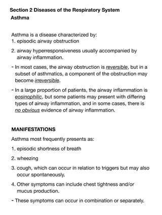
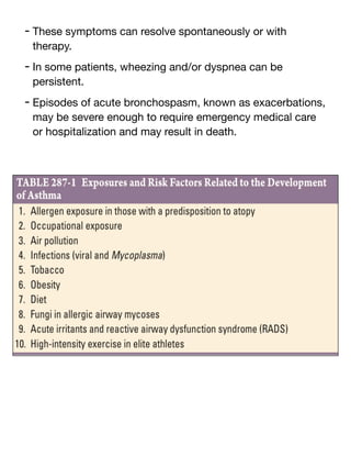
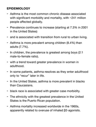
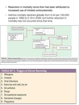
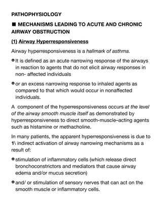
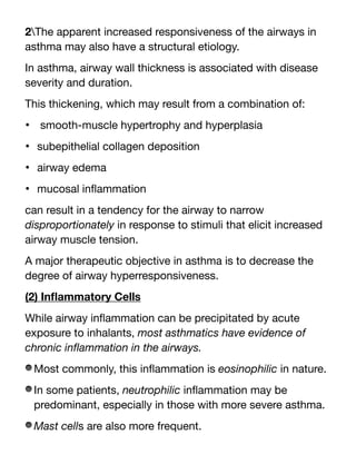
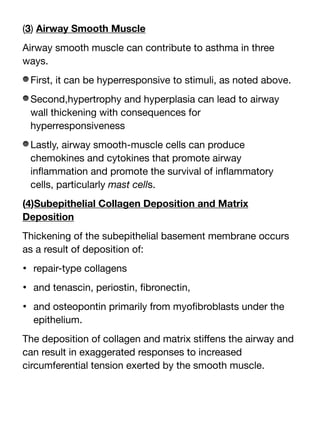
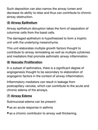
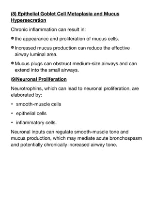
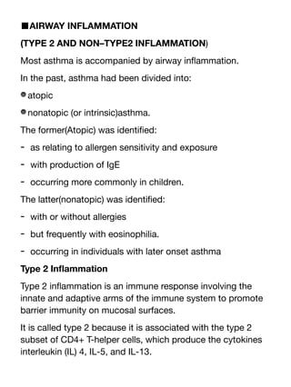
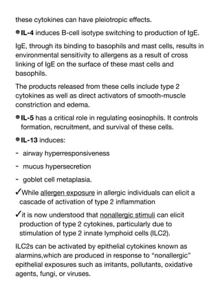
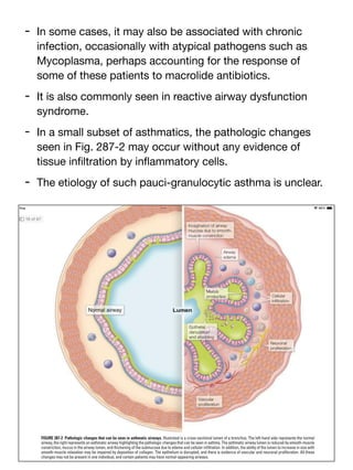
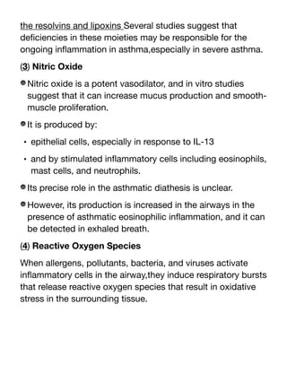
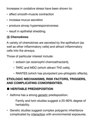
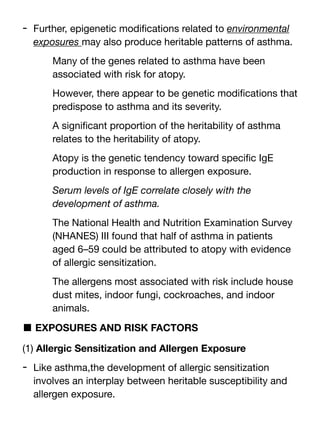
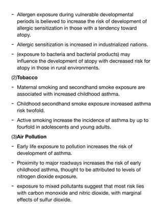
![Two types of exposures are recognized:
(1)an immunologic stimulus
- The immunologic form is associated with a latency period
between time of exposure and development of symptoms.
- (subdivided into high-molecular-weight [e.g., proteins,
fl
our] and low-molecular-weight [e.g., formal-dehyde,
diisocyanate] stimuli based on whether they act as haptens
or can directly stimulate a response).
(2) an irritative stimulus.
- The irritative form, known as reactive airway dysfunction
syndrome (RADS).
- A combination of:
• genetic predisposition (including atopy)
• timing
• intensity of exposure
• co-exposure (e.g., smoking)
in
fl
uences whether an individual will develop occupational
asthma.
(6)Diet
- There are suggestions that prenatal diet or vitamin
de
fi
ciency may alter the risk of developing asthma.](https://image.slidesharecdn.com/respa2-250609204832-e7f67f1f/85/Respiratory-system-harrison-textbook-part-2-pdf-22-320.jpg)
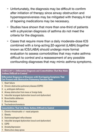
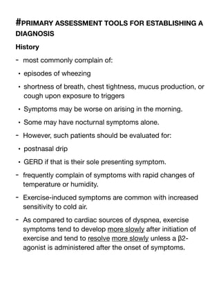
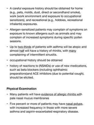
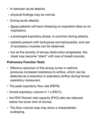
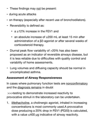
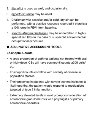
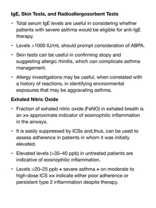
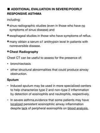
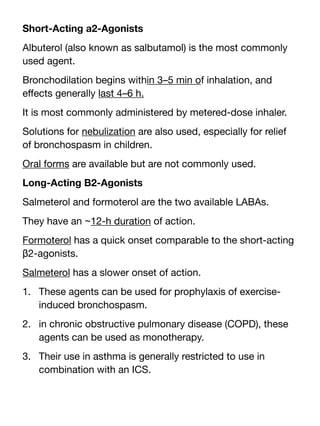
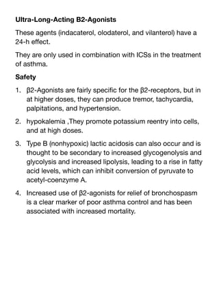
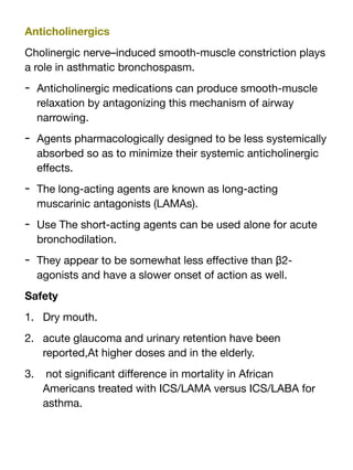
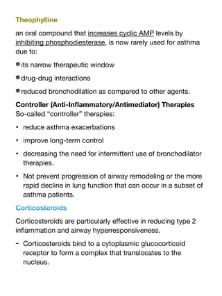
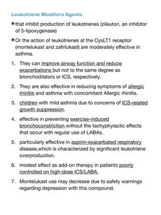
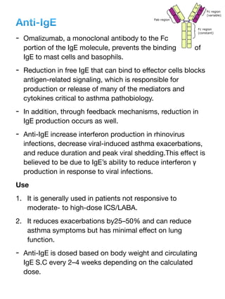

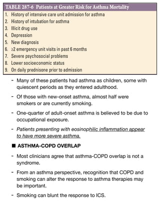
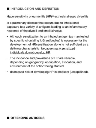
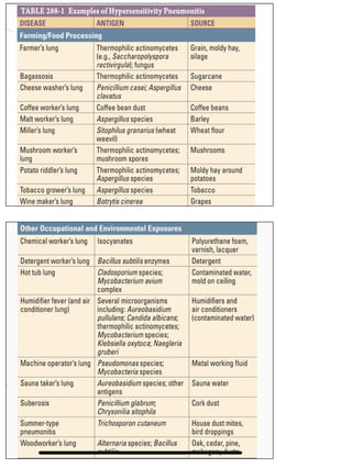
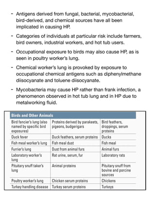

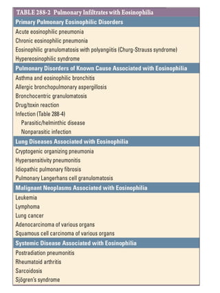
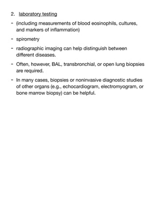
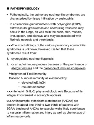
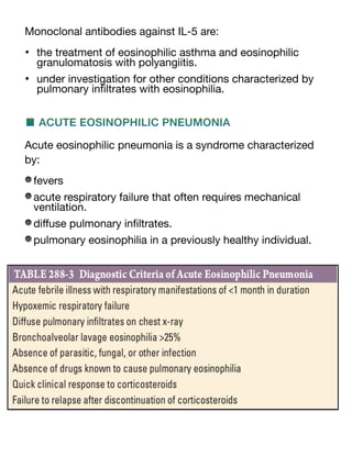
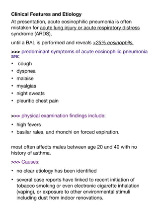
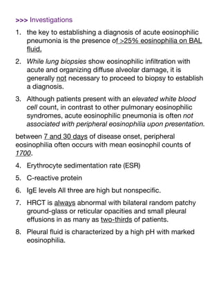
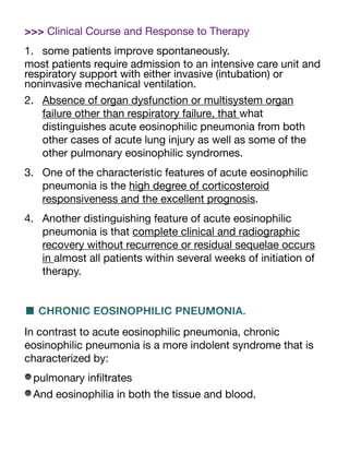
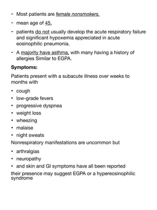
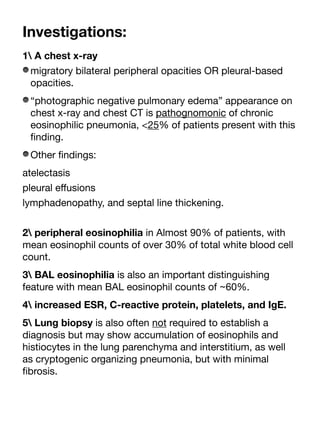
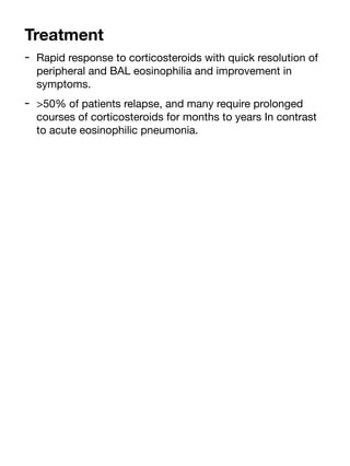
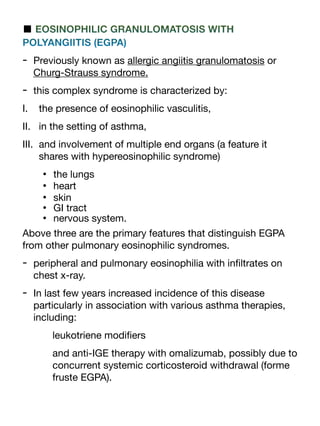
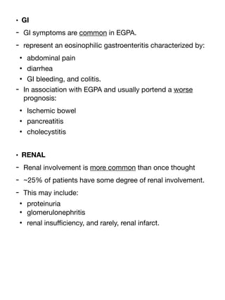
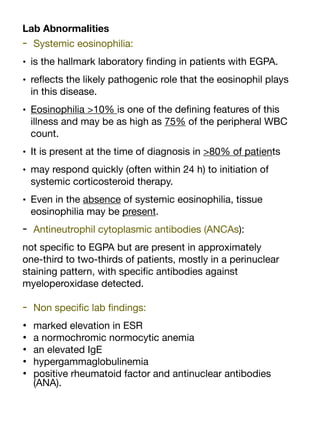
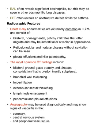
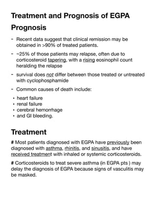
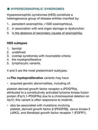
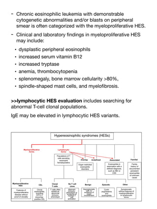
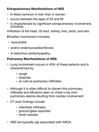

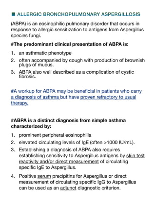
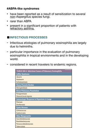
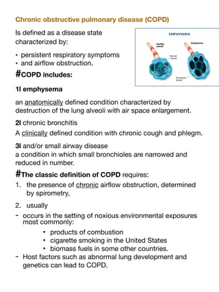
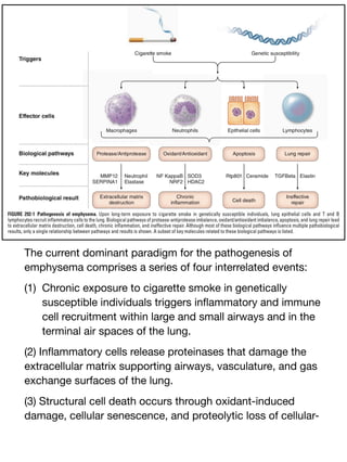
![- the balance of elastin-degrading enzymes and their
inhibitors determines the susceptibility of the lung to
destruction, resulting in air space enlargement.
1l This hypothesis > patients with genetic de
fi
ciency in α1
antitrypsin (α1AT) (the inhibitor of the serine proteinase
neutrophil elastase) > were at increased risk of emphysema.
antielastase hypothesis remains a prevailing mechanism for
the development of emphysema.
2l A complex network of immune and in
fl
ammatory cells and
additional biological mechanisms contribute to emphysema.
3l Upon exposure to oxidants from cigarette smoke:
-lung macrophages and epithelial cells become activated,
producing proteinases and chemokines that attract other
in
fl
ammatory and immune cells.
- CD8+ T cells are also recruited in response to cigarette
smoke and release interferon-inducible protein-10 (IP-10,
CXCL-7), which in turn leads to macrophage production of
macrophage elastase (matrix metalloproteinase-12
[MMP-12])
-Matrix metalloproteinases and serine proteinases, most
notably neutrophil elastase, work together by degrading the
inhibitor of the other, leading to lung destruction.
-Concomitant cigarette smoke–induced loss of cilia in the
airway epithelium and impaired macrophage phagocytosis
predispose to bacterial infection with neutrophilia.
-In end-stage lung disease, long after smoking cessation,
there remains an exuberant in
fl
ammatory response,
suggesting that cigarette smoke–induced in
fl
ammation both
initiates the disease and, in susceptible individuals,
establishes a chronic process that can continue disease
progression even after smoking cessation.](https://image.slidesharecdn.com/respa2-250609204832-e7f67f1f/85/Respiratory-system-harrison-textbook-part-2-pdf-119-320.jpg)
![however, they appear to be relatively independent processes,
and their relative contributions to obstruction vary from one
person to another.
1l The early stages of COPD
- appear to be primarily associated with medium and small
airway disease
- with the majority of Global Initiative for Chronic
Obstructive Lung Disease (GOLD) spirometric air
fl
ow
obstruction stage 1 and stage 2 subjects.
- demonstrating little or no emphysema.
- The early development of chronic air
fl
ow obstruction is
driven by small airway disease.
2l Advanced stages of COPD (GOLD stages 3 and 4 are
typically characterized by:
- extensive emphysema, although there are a small number
of subjects with very severe (GOLD stage 4) obstruction
with virtually no emphysema.
The subjects at greatest risk of progression in COPD are
those with both aggressive airway disease and emphysema.
Thus,
fi
nding emphysema (by chest computed tomography
[CT]) either early or late in the disease process suggests
enhanced risk for disease progression.](https://image.slidesharecdn.com/respa2-250609204832-e7f67f1f/85/Respiratory-system-harrison-textbook-part-2-pdf-122-320.jpg)
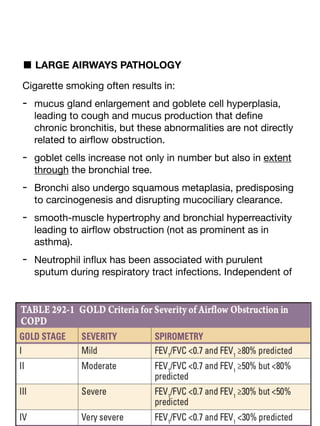
![PATHOPHYSIOLOGY
Persistent reduction in forced expiratory
fl
ow rates is the
classic de
fi
nition of COPD.
- Hyperin
fl
ation with increases in the residual volume,
- increase in the residual volume/total lung capacity ratio,
- nonuniform distribution of ventilation,
- ventilation-perfusion mismatching also occur.
1l■ AIRFLOW OBSTRUCTION
The hallmark of COPD is air
fl
ow obstruction.
The degree of air
fl
ow obstruction is an important prognostic
factor in COPD and is the basis for the GOLD spirometric
severity classi
fi
cation.
Air
fl
ow obstruction, also known as air
fl
ow limitation, is
typically determined for clinical purposes by spirometry,
which involves maximal forced expiratory maneuvers after
the subject has inhaled to total lung capacity.
Key parameters obtained from spirometry include:
the volume of air exhaled within the
fi
rst second of the forced
expiratory maneuver (FEV1) and the total volume of air
exhaled during the entire spirometric maneuver (forced vital
capacity [FVC]).
Patients with air
fl
ow obstruction related to COPD have a
chronically reduced ratio of FEV1/FVC.](https://image.slidesharecdn.com/respa2-250609204832-e7f67f1f/85/Respiratory-system-harrison-textbook-part-2-pdf-127-320.jpg)
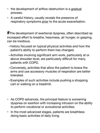
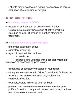
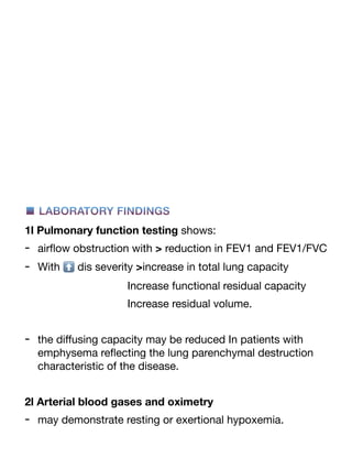
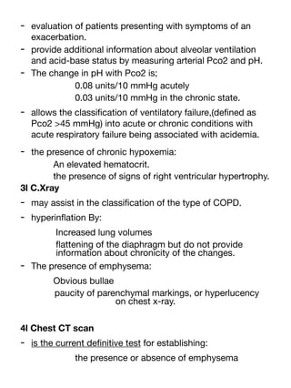
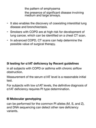
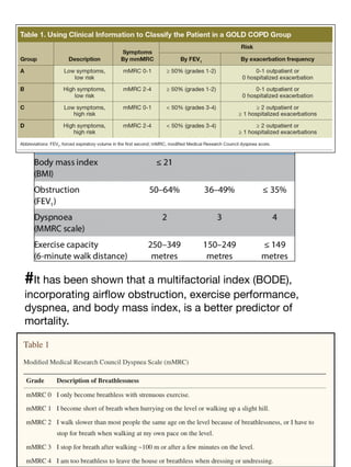
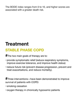

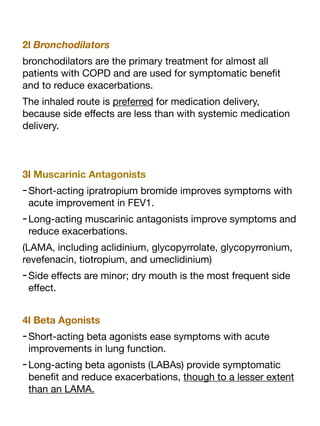
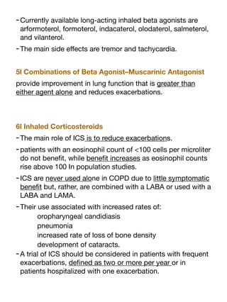
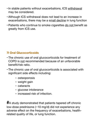
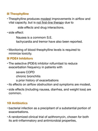
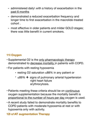
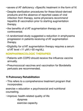
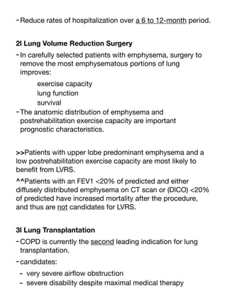
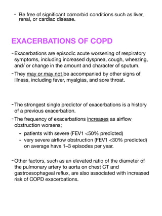

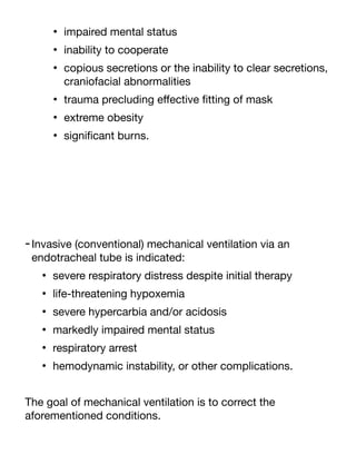
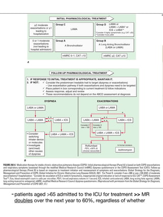
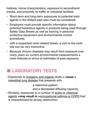
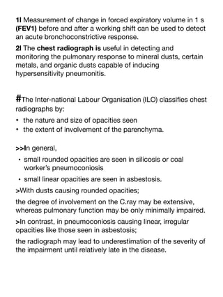
![>patients with a history of asbestos exposure >> (CT) is more
sensitive for the detection of pleural thickening, and high-
resolution CT (HRCT) improves the detection of asbestosis.
3l skin prick testing or speci
fi
c IgE antibody titers for
>evidence of immediate hypersensitivity to agents capable of
inducing occupational asthma (e.g.,
fl
our antigens in bakers),
>speci
fi
c IgG precipitating antibody titers for agents capable
of causing hypersensitivity pneumonitis (e.g., pigeon antigen
in bird handlers),
>and assays for speci
fi
c cell-mediated immune responses
(e.g., beryllium lymphocyte proliferation testing in nuclear
workers or tuberculin skin testing in health care workers).
4l bronchoscopy to obtain transbronchial biopsies of lung
tissue may be required;for histologic diagnosis (chronic
beryllium disease [CBD]).
5l video-assisted thoracoscopic surgery rarely to obtain a
larger sample of lung tissue may be required to determine the
speci
fi
c diagnosis of environmentally induced lung disease
(hypersensitivity pneumonitis or giant cell interstitial
pneumonitis due to cobalt exposure).
■ DETERMINANTS OF INHALATIONAL EXPOSURE
#Water-soluble gases such as ammonia and sulfur dioxide
are absorbed in the lining
fl
uid of the upper and proximal
airways and thus tend to produce irritative and
bronchoconstrictive responses.](https://image.slidesharecdn.com/respa2-250609204832-e7f67f1f/85/Respiratory-system-harrison-textbook-part-2-pdf-168-320.jpg)
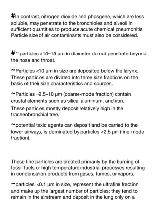
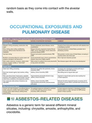
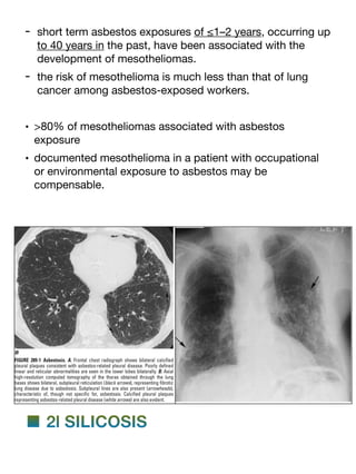
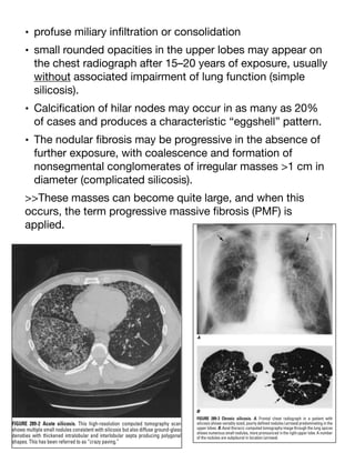

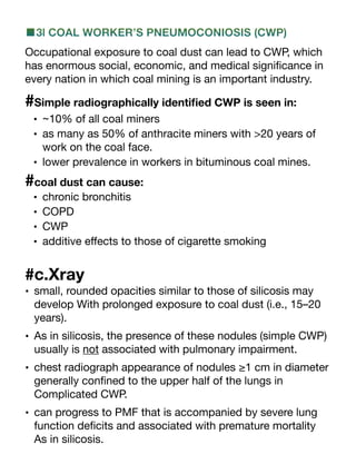
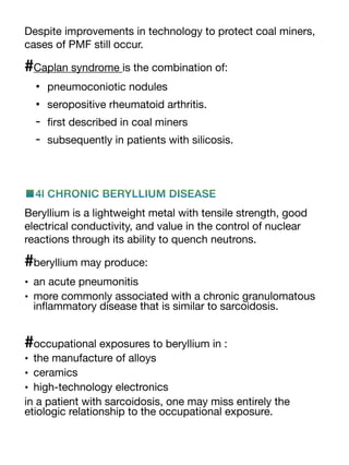
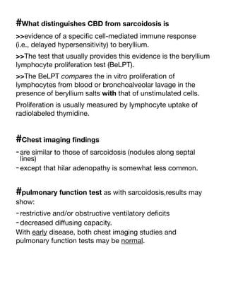
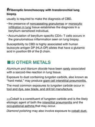

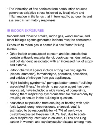
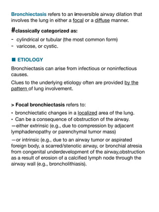
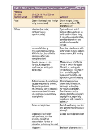
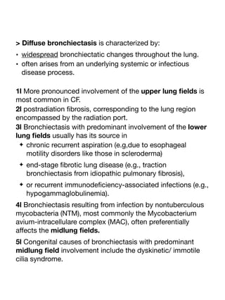
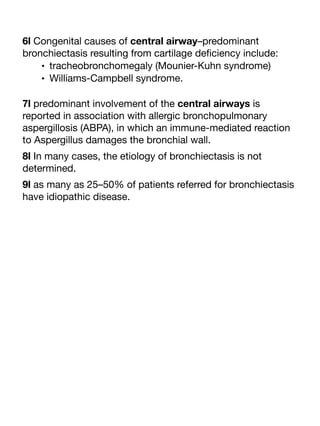
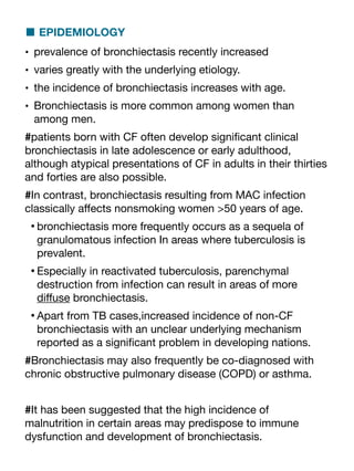
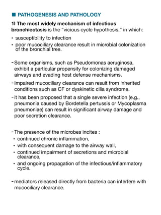
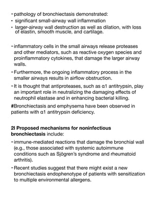
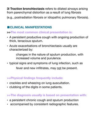
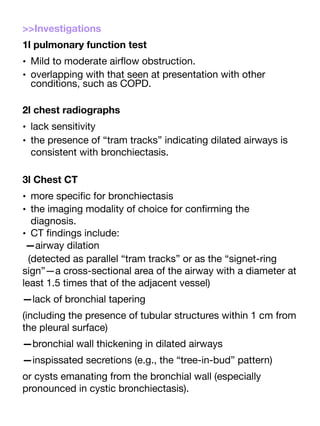
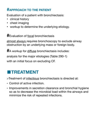
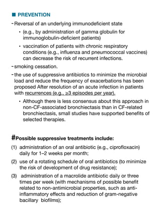
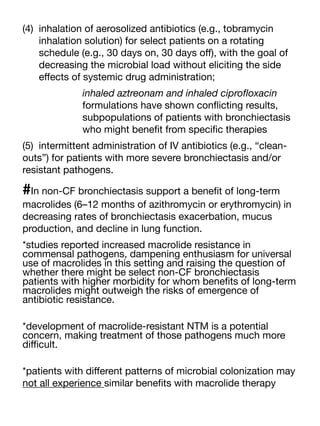
![Interstitial Lung Disease
>>Di
ff
use parenchymal lung diseases include:
a large number (>200) of heterogeneous conditions that
a
ff
ect the lung parenchyma with varying degrees of
in
fl
ammation and
fi
brosis.
-remodeling of the interstitial space (the region between the
epithelium and endothelium) tends to be the dominant site
of involvement for most of the interstitial lung diseases
(ILDs).
-prominent role of the alveolar epithelium and endothelial
cells (including both airways and vessels) in the
pathogenesis of these ILDs.
>>most patients ultimately diagnosed with an ILD report
progressive exertional dyspnea or a persistent dry cough.
>>some ILDs are part of multisystem disorders,identi
fi
ed
based on nonrespiratory symptomatology:
(e.g., skin thickening in the setting of systemic sclerosis,
(e.g., ulnar deviation of the
fi
ngers in the setting of [RA].
>>identi
fi
ed incidentally based on the results of:
abnormal pulmonary function tests, chest x-rays (CXRs),
computed tomography (CT) studies of both the chest and
abdomen (which can both visualize, at least a portion, of the
lung parenchyma), and positron emission tomography (PET)
scans.](https://image.slidesharecdn.com/respa2-250609204832-e7f67f1f/85/Respiratory-system-harrison-textbook-part-2-pdf-212-320.jpg)
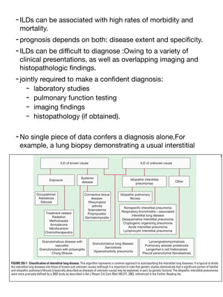
![pneumonia (UIP) pattern is helpful in diagnosing a patient
with idiopathic pulmonary
fi
brosis (IPF) but can also be
present in some connective tissue diseases (CTDs) (e.g.,
RA-associated ILD.
-multidisciplinary approach to the diagnosis (and in some
cases for the management) of ILDs.
-genetic studies demonstrate that a signi
fi
cant portion of
familial pulmonary
fi
brosis and IPF
[classically described as diseases of unknown cause]
may be explained, in part, by genetic factors).
-Known causes of ILD include:
occupational exposures (e.g., asbestosis)
medications (e.g., nitrofurantoin)
underlying systemic disease (e.g., cryptogenic organizing
pneumonia [COP] in the setting of polymyositis).
-Unknown causes of ILD include:
-groups of rare disorders often with classic presentations
(e.g., a spontaneous pneumothorax in a young female with
di
ff
use cystic changes on a chest CT might suggest
lymphangioleiomyomatosis [LAM],
the most common group of ILDs, the idiopathic interstitial
pneumonias (IIPs).](https://image.slidesharecdn.com/respa2-250609204832-e7f67f1f/85/Respiratory-system-harrison-textbook-part-2-pdf-214-320.jpg)
![-Granulomatous lung diseases:
‣ known (e.g., hypersensitivity pneumonitis [HP] due to
chronic bird exposure
‣ and unknown (e.g., sarcoidosis,) causes
and are often separated due to their unique presentations,
imaging
fi
ndings, and diagnostic evaluation.
#group of dis are among the most common forms of ILD:
- IPF
- sarcoidosis
- ILDs related to Connective tissue diseases.](https://image.slidesharecdn.com/respa2-250609204832-e7f67f1f/85/Respiratory-system-harrison-textbook-part-2-pdf-215-320.jpg)
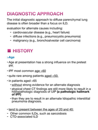
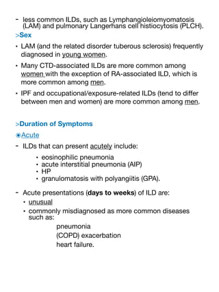
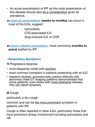
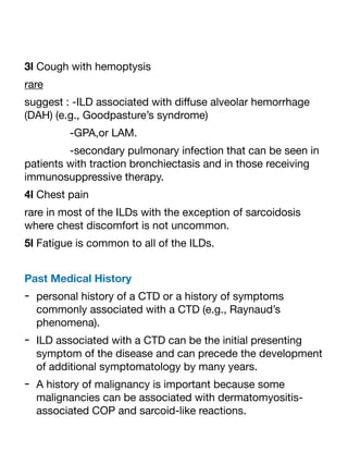
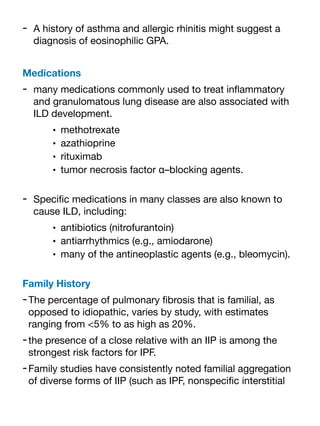
![pneumonia [NSIP], and DIP running in the same family) and,
in some cases, other forms of ILD.
-genetic factors for pulmonary
fi
brosis (a promoter variant of
a mucin gene [MUC5B]) and variants in the telomerase
reverse transcriptase gene [TERT]) appear to be associated
with both familial and idiopathic forms of pulmonary
fi
brosis
similarly.
Social History
-A history of smoking is nearly always present in some forms
of ILD (e.g., respiratory bronchiolitis and desquamative
interstitial pneumonia [DIP]—sometimes referred by
pathologists jointly as smoking-related ILD) where it is felt to
be causative.
-A history of smoking noted in approximately three-quarters
of IPF patients.
-Occupational and environmental exposure histories are also
important to obtain>> as they might identify exposures
known to cause pulmonary
fi
brosis (e.g., signi
fi
cant
asbestos exposure) or HP (pigeon breeder’s lung).](https://image.slidesharecdn.com/respa2-250609204832-e7f67f1f/85/Respiratory-system-harrison-textbook-part-2-pdf-221-320.jpg)
![■ PHYSICAL EXAMINATION
1l End-inspiratory
fi
ne crackles, or rales,at the lung bases are
found in most patients with IPF and may be one of the
earliest signs of the disease. However, rales are nonspeci
fi
c
and can be found in many forms of ILD.
2l Wheezing is uncommon in most forms of ILD but can be
present in some disorders, such as
• sarcoidosis
• HP
• eosinophilic GPA.
3l Signs of advanced disease include cyanosis,digital
clubbing, and corpulmonale.
■ LABORATORY STUDIES
- tests can reveal the presence of an underlying CTD as the
cause of an ILD (e.g.,a positive anti-cyclic citrullinated
peptide [anti-CCP] antibody for RA) even when no other
symptomatology or physical examination
fi
ndings
suggestive of the disorder are present.
- the cost-e
ff
ectiveness and long list of autoantibody tests
that could be ordered.](https://image.slidesharecdn.com/respa2-250609204832-e7f67f1f/85/Respiratory-system-harrison-textbook-part-2-pdf-222-320.jpg)
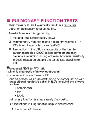
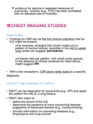
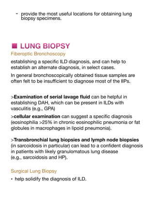
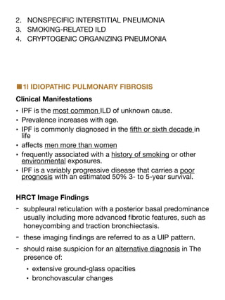
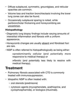
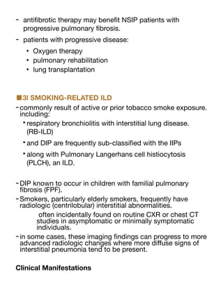
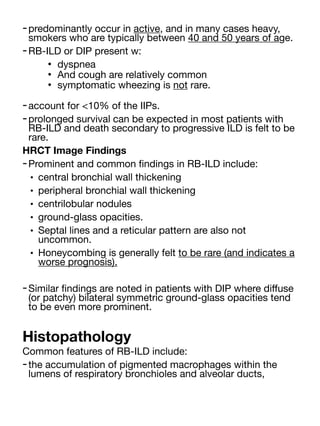
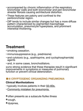
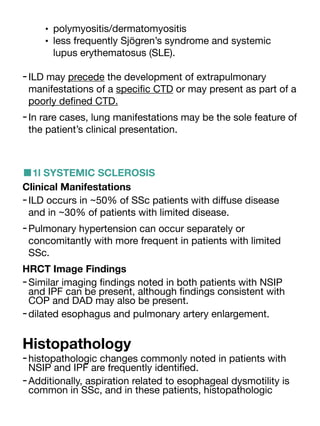
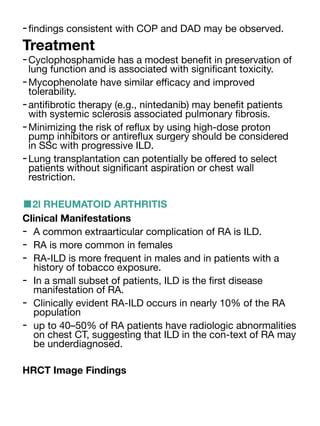
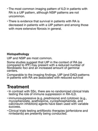
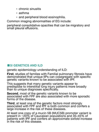
![Fourth, studies of general population samples demonstrate
that imaging abnormalities suggestive of an early stage of
pulmonary
fi
brosis in research participants without known
ILD are not uncommon (occurring in ~7–9% of adults) and
are also associated with the same genetic variants known to
be associated with IPF (e.g., the MUC5B promoter variant).
This latter
fi
nding suggests a path forward toward an early
detection of IPF.
Additional genetic
fi
ndings demonstrating replicable
associations with pulmonary
fi
brosis include
numerous genetic variants in, and adjacent to, genes known
to be involved in the regulation of telomere length
e.g., the TERT gene,the regulator of telomere elongation
helicase 1 [RTEL1] gene) and surfactant protein genes (e.g.,
surfactant protein A2 [SFTPA2] gene).
Some of these disease-associated variants in sarcoidosis fall
in human leukocyte antigen (HLA) regions, in regions of
genes involved in immune regulation (e.g., interleukin 12B
[IL12B]), and in regions of genes that are less well understood
(butyrophilin-like 2 [BTNL2]) but also appear to be involved in
T-cell activation.
LAM is often associated with genetic variants in the tuberous
sclerosis complex genes (e.g., TSC1 and TSC2), consistent
with the known evidence that this disease can occur in
isolation but also in patients with known tuberous sclerosis.
Many genetic factors for rare diseases such as
Hermansky-Pudlak syndrome (a rare autosomal recessive
disorder that results in pulmonary
fi
brosis but also includes
oculocutaneous albinism, bleeding diatheses, and horizontal
nystagmus) have also been discovered (e.g., HSP1, and
HSP3-7).](https://image.slidesharecdn.com/respa2-250609204832-e7f67f1f/85/Respiratory-system-harrison-textbook-part-2-pdf-247-320.jpg)
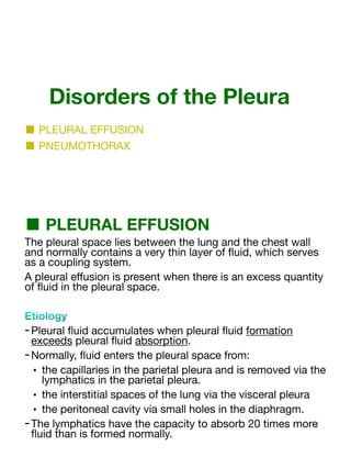
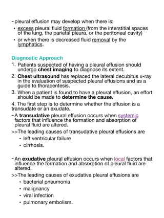
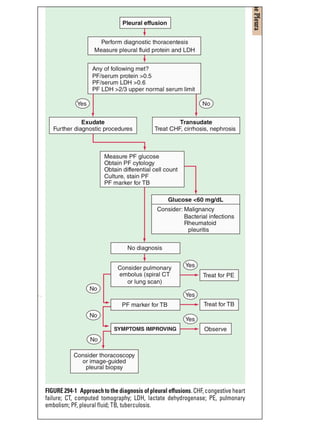
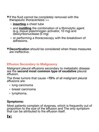
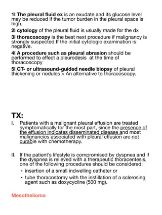
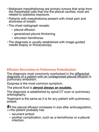
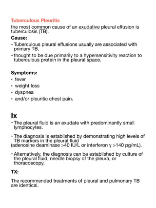

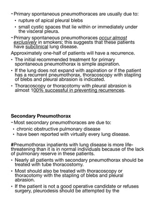
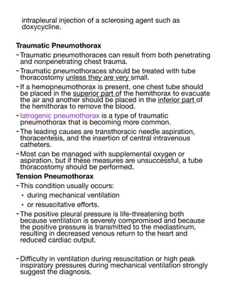
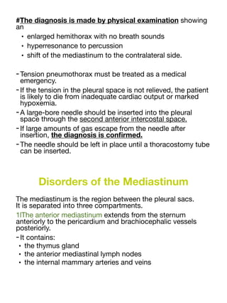
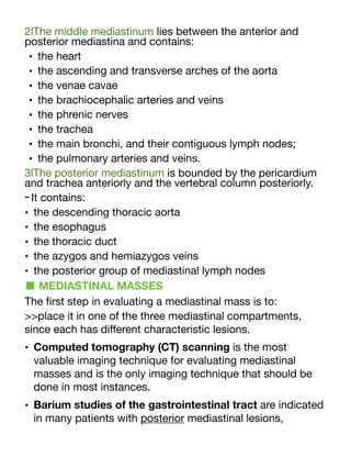
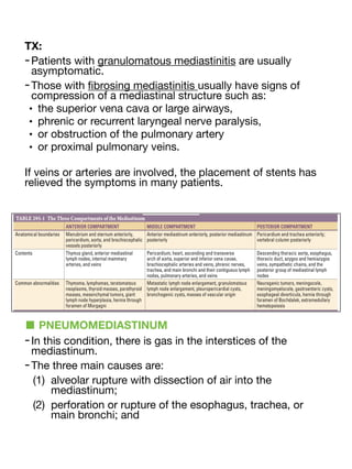


![Hypothalamus short ppt by Dr. Neha [PT].pptx](https://cdn.slidesharecdn.com/ss_thumbnails/hypothalamusbydr-260124145759-b9f94a93-thumbnail.jpg?width=640&height=640&fit=bounds)


