Research Day Poster Spring 15
•Download as PPT, PDF•
1 like•236 views

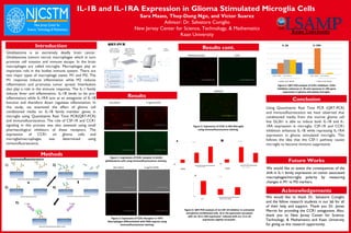
This study examined how glioblastoma tumors influence microglia cells' expression of IL-1B and IL-1RA genes through secreted factors. Results showed that glioma cell conditioned media induced both IL-1B and IL-1RA expression in microglia. Inhibiting CSF-1R or CCR1 signaling in microglia enhanced IL-1B expression while repressing IL-1RA, suggesting glioma signals cause an anti-inflammatory microglia phenotype. Future work will assess how these changes in IL-1 family expression impact microglia polarization.

More Related Content
What's hot
What's hot (20)
Similar to Research Day Poster Spring 15
Similar to Research Day Poster Spring 15 (20)
Research Day Poster Spring 15
- 1. IL-1B and IL-1RA Expression in Glioma Stimulated Microglia Cells Sara Maass, Thuy-Dung Ngo, and Victor Suarez Advisor: Dr. Salvatore Coniglio New Jersey Center for Science, Technology, & Mathematics Kean University Introduction Glioblastoma is an extremely deadly brain cancer. Glioblastoma tumors recruit macrophages which in turn promote cell invasion and immune escape. In the brain macrophages are called microglia. Macrophages play an important role in the bodies immune system. There are two major types of macrophage states: M1 and M2. The M1 response induces inflammation while M2 reduces inflammation and promotes tumor spread. Interleukins also play a role in the immune response. The IL-1 family induces fever and inflammation. IL-1B tends to be pro- inflammatory while IL-1RA acts as an antagonist of IL-1B function and therefore down regulates inflammation. In this study, we examined the effect of glioma cell conditioned media on IL-1B family member genes in microglia using Quantitative Real Time PCR(QRT-PCR) and immunofluorescence. The role of CSF-1R and CCR1 signaling in this process was also assessed using small pharmacological inhibitors of these receptors. The expression of CCR1 on glioma cells and microglia/macrophages was determined using immunofluorescence. Methods Results Results cont. Conclusion Using Quantitative Real Time PCR (QRT-PCR) and immunofluorescence we have observed that conditioned media from the murine glioma cell line GL261 is able to induce both IL-1B and IL- 1RA expression in microglia. CSF-1R and CCR1 inhibition enhances IL-1B while repressing IL-1RA expression in glioma stimulated microglia. This follows the idea that the CSF-1 pathway causes microglia to become immuno suppressive. We would like to thank Dr. Salvatore Coniglio and the fellow research students in our lab for all of their help and support. Thank you Dr. James Merritt for providing the CCR1 antagonists. Also, thank you to New Jersey Center for Science, Technology, & Mathematics and Kean University for giving us this research opportunity. Figure 1: Expression of CCR1 receptor in GL261 glioblastoma cells using immunofluorescence staining. 4 ug/ml aCCR12ary Alone Figure 2: Expression of CCR1 Receptor in THP1 Macrophages differentiated with PMA express using immunofluorescence staining. OVERLAY Figure 3: Expression of CCR1 in MG Microglia using immunofluorescence staining. Phalloidin (Actin) CCR1 Future Works Acknowledgements We would like to assess the consequences of the shift in IL-1 family expression on tumor associated macrophages/microglia polarity by measuring changes in M1 vs M2 markers. QRT-PCR 2ary Alone 4 ug/ml aCCR1 Immunofluorescence Figure 4: QRT-PCR analysis of JnJ CSF-1R inhibitor in untreated and glioma conditioned cells. A) IL-1B expression increased with JnJ. B) IL-1RA expression reduced with JnJ. C) IL-18 expression slightly increased. 0123456 IL-1B JnJ: - - + Glioma Cond. Media A IL-1RA JnJ: - - + Glioma Cond. Media B 0123456 Untx Untx 00.20.40.60.811.2 IL-18 JnJ: - - + Untx Glioma Cond. Media C IL-1B IL-1RA + GL261 Cond. Media + GL261 Cond. Media DMSO DMSO CCR1 inhibitors DMSO DMSO CCR1 inhibitors Figure 5: QRT-PCR analysis of CCR1 inhibition. CCR1 inhibition enhances IL-1B and represses IL-1RA gene expression in glioma stimulated microglia. 1) 2) 3) 4) 5) UV Light UV Light