The document describes a new method called the mutagenic chain reaction (MCR) that uses the CRISPR/Cas9 system to efficiently generate homozygous loss-of-function mutations from an initial heterozygous mutation. The researchers developed an MCR construct containing Cas9, a guide RNA targeting a genomic sequence of interest, and flanking homology arms. This construct inserts into the target site and expresses Cas9 and guide RNA to cleave the opposite chromosome, converting it to homozygosity via homology-directed repair. Testing this method in Drosophila, they found it efficiently spread mutations from the initial chromosome to the homologous one in somatic and germline cells, demonstrating the potential of MCR for
![27. D. L. Swenson et al., Vaccine 23, 3033–3042 (2005).
28. K. L. Warfield et al., J. Immunol. 175, 1184–1191 (2005).
29. E. S. Kempner, J. Pharm. Sci. 90, 1637–1646 (2001).
ACKNOWLEDGMENTS
We thank E. Ollmann-Saphire (Scripps Research Institute, La Jolla,
CA) for purified EBOV NP. We also thank S. Watson for editing the
manuscript, T. Armbrust for excellent technical assistance, and
staff of the Rocky Mountain Veterinary Branch for assistance with
animal work. Y.K. and G.N. are inventors on a patent (held by the
University of Wisconsin Alumni Research Foundation) for EBOV
reverse genetics; therefore, a Material Transfer Agreement (MTA)
is required to obtain this system. Funding for this research was
provided by the Region V “Great Lakes” Regional Center of
Excellence (GLRCE; U54 AI 57153) and by Health and Labour
Sciences Research Grants, Japan. The study was partially funded
by the Intramural Research Program of the National Institute of
Allergy and Infectious Diseases, NIH. Raw data can be found at
https://docs.google.com/spreadsheets/d/1dBgzt5_z4rp-
qOuxXcI_FbUz8wNqMvHy6kVP_tpW0MY/edit?usp=sharing.
SUPPLEMENTARY MATERIALS
www.sciencemag.org/content/348/6233/439/suppl/DC1
Materials and Methods
Tables S1 to S3
Figs. S1 and S2
14 December 2014; accepted 13 March 2015
Published online 26 March 2015;
10.1126/science.aaa4919
GENOME EDITING
The mutagenic chain reaction: A
method for converting heterozygous
to homozygous mutations
Valentino M. Gantz* and Ethan Bier*
An organism with a single recessive loss-of-function allele will typically have a wild-type
phenotype, whereas individuals homozygous for two copies of the allele will display a
mutant phenotype. We have developed a method called the mutagenic chain reaction
(MCR), which is based on the CRISPR/Cas9 genome-editing system for generating
autocatalytic mutations, to produce homozygous loss-of-function mutations. In Drosophila,
we found that MCR mutations efficiently spread from their chromosome of origin to the
homologous chromosome, thereby converting heterozygous mutations to homozygosity
in the vast majority of somatic and germline cells. MCR technology should have broad
applications in diverse organisms.
I
t is often desirable to generate recessive loss-
of-function mutations in emergent model
organisms; however, identifying such muta-
tions in the heterozygous condition is chal-
lenging. Taking advantage of the CRISPR/
Cas9 genome-editing method (1, 2), we have
developed a strategy to convert a Drosophila
heterozygous recessive mutation into a homozy-
gous condition manifesting a mutant phenotype.
Wereasonedthatautocatalyticinsertional mutants
could be generated with a construct having three
components: (i) A Cas9 gene (expressed in both
somatic and germline cells), (ii) a guide RNA
(gRNA) targeted to a genomic sequence of in-
terest, and (iii) homology arms flanking the
Cas9-gRNA cassettes that match the two ge-
nomic sequences immediately adjacent to either
side of the target cut site (Fig. 1A). In such a
tripartite construct, Cas9 should cleave the ge-
nomic target at the site determined by the gRNA
(Fig. 1A) and then insert the Cas9-gRNA cassette
into that locus via homology-directed repair (HDR)
(Fig. 1, B and C). Cas9 and the gRNA produced
from the insertion allele should then cleave the
opposing allele (Fig. 1D), followed by HDR-
driven propagation of the Cas9-gRNA cassette
to the companion chromosome (Fig. 1, E and F).
We refer to this trans-acting mutagenesis scheme
as a mutagenic chain reaction (MCR).
We expected that autocatalytic allelic conver-
sion by MCR should be very efficient in both
somatic and germline precursor cells, given the
high frequency and specificity of mutagenesis (3)
442 24 APRIL 2015 • VOL 348 ISSUE 6233 sciencemag.org SCIENCE
RESEARCH | REPORTS
Section of Cell and Developmental Biology, University of
California, San Diego, La Jolla, CA 92095, USA.
*Corresponding author. E-mail: vgantz@ucsd.edu (V.M.G.);
ebier@ucsd.edu (E.B.)
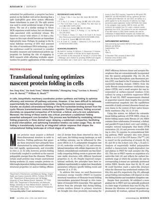
Fig. 1. Scheme outlining the mutagenic chain
reaction (MCR). (A to C) A plasmid consisting of a
core cassette carrying a Cas9 transgene, a gRNA
targeting a genomic sequence of interest, and
flanking homology arms corresponding to genomic
sequences abutting the target cleavage site (A)
inserts the core Cas9-gRNA cassette into the
targeted locus via HDR [(B) and (C)]. (D to F) In
turn, the inserted cassette expresses both Cas9
and the gRNA, leading to cleavage (D) and HDR-
mediated insertion of the cassette into the second
allele, thereby rendering the mutation homozygous
[(E) and (F)]. HA1 and HA2 denote the two
homology arms that directly flank the gRNA-
directed cut site.
HA1
genome
HA2Cas9 gRNA
HA1
Homology Directed Repair (HDR)
HA2Cas9 gRNA
HA1 HA2
Cas9 gRNA
Cas9 gRNA
Homology Directed Repair (HDR)
Cas9 gRNAHA1 HA2
Cas9 gRNA
Cas9 gRNA
Cas9 gRNA
second allele
onDecember10,2015www.sciencemag.orgDownloadedfromonDecember10,2015www.sciencemag.orgDownloadedfromonDecember10,2015www.sciencemag.orgDownloadedfrom](https://image.slidesharecdn.com/6b435ea2-dce4-4dfc-ae4b-a749874dd523-170214003738/85/Science-2015-Gantz-442-4-1-320.jpg)
![27. D. L. Swenson et al., Vaccine 23, 3033–3042 (2005).
28. K. L. Warfield et al., J. Immunol. 175, 1184–1191 (2005).
29. E. S. Kempner, J. Pharm. Sci. 90, 1637–1646 (2001).
ACKNOWLEDGMENTS
We thank E. Ollmann-Saphire (Scripps Research Institute, La Jolla,
CA) for purified EBOV NP. We also thank S. Watson for editing the
manuscript, T. Armbrust for excellent technical assistance, and
staff of the Rocky Mountain Veterinary Branch for assistance with
animal work. Y.K. and G.N. are inventors on a patent (held by the
University of Wisconsin Alumni Research Foundation) for EBOV
reverse genetics; therefore, a Material Transfer Agreement (MTA)
is required to obtain this system. Funding for this research was
provided by the Region V “Great Lakes” Regional Center of
Excellence (GLRCE; U54 AI 57153) and by Health and Labour
Sciences Research Grants, Japan. The study was partially funded
by the Intramural Research Program of the National Institute of
Allergy and Infectious Diseases, NIH. Raw data can be found at
https://docs.google.com/spreadsheets/d/1dBgzt5_z4rp-
qOuxXcI_FbUz8wNqMvHy6kVP_tpW0MY/edit?usp=sharing.
SUPPLEMENTARY MATERIALS
www.sciencemag.org/content/348/6233/439/suppl/DC1
Materials and Methods
Tables S1 to S3
Figs. S1 and S2
14 December 2014; accepted 13 March 2015
Published online 26 March 2015;
10.1126/science.aaa4919
GENOME EDITING
The mutagenic chain reaction: A
method for converting heterozygous
to homozygous mutations
Valentino M. Gantz* and Ethan Bier*
An organism with a single recessive loss-of-function allele will typically have a wild-type
phenotype, whereas individuals homozygous for two copies of the allele will display a
mutant phenotype. We have developed a method called the mutagenic chain reaction
(MCR), which is based on the CRISPR/Cas9 genome-editing system for generating
autocatalytic mutations, to produce homozygous loss-of-function mutations. In Drosophila,
we found that MCR mutations efficiently spread from their chromosome of origin to the
homologous chromosome, thereby converting heterozygous mutations to homozygosity
in the vast majority of somatic and germline cells. MCR technology should have broad
applications in diverse organisms.
I
t is often desirable to generate recessive loss-
of-function mutations in emergent model
organisms; however, identifying such muta-
tions in the heterozygous condition is chal-
lenging. Taking advantage of the CRISPR/
Cas9 genome-editing method (1, 2), we have
developed a strategy to convert a Drosophila
heterozygous recessive mutation into a homozy-
gous condition manifesting a mutant phenotype.
Wereasonedthatautocatalyticinsertional mutants
could be generated with a construct having three
components: (i) A Cas9 gene (expressed in both
somatic and germline cells), (ii) a guide RNA
(gRNA) targeted to a genomic sequence of in-
terest, and (iii) homology arms flanking the
Cas9-gRNA cassettes that match the two ge-
nomic sequences immediately adjacent to either
side of the target cut site (Fig. 1A). In such a
tripartite construct, Cas9 should cleave the ge-
nomic target at the site determined by the gRNA
(Fig. 1A) and then insert the Cas9-gRNA cassette
into that locus via homology-directed repair (HDR)
(Fig. 1, B and C). Cas9 and the gRNA produced
from the insertion allele should then cleave the
opposing allele (Fig. 1D), followed by HDR-
driven propagation of the Cas9-gRNA cassette
to the companion chromosome (Fig. 1, E and F).
We refer to this trans-acting mutagenesis scheme
as a mutagenic chain reaction (MCR).
We expected that autocatalytic allelic conver-
sion by MCR should be very efficient in both
somatic and germline precursor cells, given the
high frequency and specificity of mutagenesis (3)
442 24 APRIL 2015 • VOL 348 ISSUE 6233 sciencemag.org SCIENCE
RESEARCH | REPORTS
Section of Cell and Developmental Biology, University of
California, San Diego, La Jolla, CA 92095, USA.
*Corresponding author. E-mail: vgantz@ucsd.edu (V.M.G.);
ebier@ucsd.edu (E.B.)
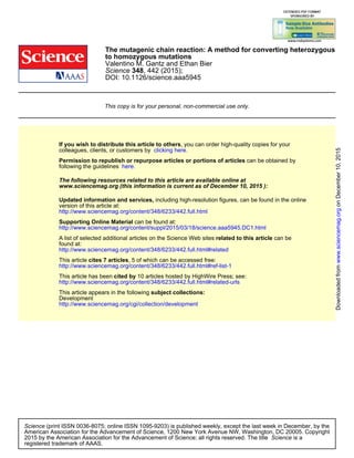
Fig. 1. Scheme outlining the mutagenic chain
reaction (MCR). (A to C) A plasmid consisting of a
core cassette carrying a Cas9 transgene, a gRNA
targeting a genomic sequence of interest, and
flanking homology arms corresponding to genomic
sequences abutting the target cleavage site (A)
inserts the core Cas9-gRNA cassette into the
targeted locus via HDR [(B) and (C)]. (D to F) In
turn, the inserted cassette expresses both Cas9
and the gRNA, leading to cleavage (D) and HDR-
mediated insertion of the cassette into the second
allele, thereby rendering the mutation homozygous
[(E) and (F)]. HA1 and HA2 denote the two
homology arms that directly flank the gRNA-
directed cut site.
HA1
genome
HA2Cas9 gRNA
HA1
Homology Directed Repair (HDR)
HA2Cas9 gRNA
HA1 HA2
Cas9 gRNA
Cas9 gRNA
Homology Directed Repair (HDR)
Cas9 gRNAHA1 HA2
Cas9 gRNA
Cas9 gRNA
Cas9 gRNA
second allele
onDecember10,2015www.sciencemag.orgDownloadedfromonDecember10,2015www.sciencemag.orgDownloadedfromonDecember10,2015www.sciencemag.orgDownloadedfrom](https://image.slidesharecdn.com/6b435ea2-dce4-4dfc-ae4b-a749874dd523-170214003738/75/Science-2015-Gantz-442-4-1-2048.jpg)