
Recommended
More Related Content
Similar to REPLICATION OF DNA.pdf
Similar to REPLICATION OF DNA.pdf (20)
More from ANAKHA JACOB
More from ANAKHA JACOB (10)
Recently uploaded
Recently uploaded (20)
REPLICATION OF DNA.pdf
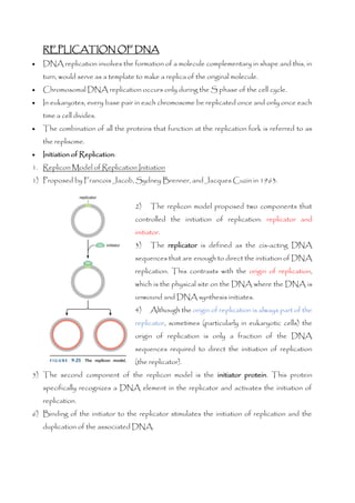
- 1. REPLICATION OF DNA • DNA replication involves the formation of a molecule complementary in shape and this, in turn, would serve as a template to make a replica of the original molecule. • Chromosomal DNA replication occurs only during the S phase of the cell cycle. • In eukaryotes, every base pair in each chromosome be replicated once and only once each time a cell divides. • The combination of all the proteins that function at the replication fork is referred to as the replisome. • Initiation of Replication: 1. Replicon Model of Replication Initiation 1) Proposed by Francois Jacob, Sydney Brenner, and Jacques Cuzin in 1963. 2) The replicon model proposed two components that controlled the initiation of replication: replicator and initiator. 3) The replicator is defined as the cis-acting DNA sequences that are enough to direct the initiation of DNA replication. This contrasts with the origin of replication, which is the physical site on the DNA where the DNA is unwound and DNA synthesis initiates. 4) Although the origin of replication is always part of the replicator, sometimes (particularly in eukaryotic cells) the origin of replication is only a fraction of the DNA sequences required to direct the initiation of replication (the replicator). 5) The second component of the replicon model is the initiator protein. This protein specifically recognizes a DNA element in the replicator and activates the initiation of replication. 6) Binding of the initiator to the replicator stimulates the initiation of replication and the duplication of the associated DNA.
- 2. 7) All the known initiator proteins are regulated by ATP binding and hydrolysis and share a common core AAA+ ATP-binding motif related to, but distinct from, that used by sliding DNA clamp loaders. 8) The initiator protein is the only sequence-specific DNA binding protein involved in the initiation of replication. Indeed, for many eukaryotic cells, even the initiator protein does not show sequence-specific DNA-binding activity. 9) In E. coli, the initiator protein is DnaA 10)The DNA sequences of known replicators share two common features: ❖ they include a binding site for the initiator protein. ❖ they include a stretch of AT-rich DNA that unwinds readily but not spontaneously. 11) The single replicator required for E. coli chromosomal replication is called oriC. 12)Two repeated motifs are critical for the oriC function: the 9-mer motif & 13-mer motif. 13)The 9-mer motif is the binding site for the E. coli initiator, DnaA, and is repeated five times at oriC. 14)The 13-mer motif, repeated three times, is the initial site of ssDNA formation during initiation. 15)Initiator proteins perform at least two different functions during the initiation of replication. ❖ These proteins bind to the replicator DNA, often via a specific binding site. ❖ Initiator proteins interact with additional factors required for replication initiation, thus recruiting them to the replicator. ❖ Some but not all initiator proteins perform a third function: They distort or unwind a region of DNA adjacent to their binding site to facilitate the initial opening of the DNA duplex.
- 3. 16) E. coli initiator protein, DnaA. 17) DnaA includes two DNA-binding domains: ❖ One domain binds the repeated 9-mer elements in oriC in their double-stranded form. ❖ When bound to ATP (but not ADP), DnaA also interacts with DNA in the region of the repeated 13-mer repeats of oriC. These additional interactions involve a distinct single-stranded DNA-binding site in DnaA and result in the separation of the DNA strands over more than 20 bp within the 13-mer repeat region. 18)Once bound to DnaA, the single-stranded DNA is held in a conformation that prevents the formation of more than three continuous base pairs, ensuring that the DNA remains single-stranded. This unwound DNA provides an ssDNA template for additional replication proteins to begin the RNA and DNA synthesis steps of replication. 19)A special type of type II topoisomerase, DNA gyrase introduces negative supercoils. This negative supercoiling facilitates the unwinding of the DNA duplex, which stimulates many reactions of DNA including the initiation of DNA replication. 20) DnaA recruits additional replication proteins to the ssDNA formed at the replicator including the DNA helicase. 21)E. coli DNA Replication Is Regulated by DnaA.ATP Levels and SeqA. 22)Once the initiator binds to the replicator, the remaining steps in the initiation of replication are largely driven by protein-protein interactions and protein–DNA interactions that are sequence-independent. 23)After the initiator (DnaA) has bound to oriC and unwound the 13-mer DNA, the combination of ssDNA and DnaA recruits a complex of two proteins: the DNA helicase (DnaB) and helicase loader (DnaC). 24) Binding to the helicase loader inactivates the DNA helicase, preventing it from functioning at inappropriate sites. 25)DnaC is an ATP-utilizing AAA+ protein. 26)DNA helicase bind to and moves directionally along ssDNA using the energy of nucleoside triphosphate (usually ATP) binding and hydrolysis to displace any DNA strand that is annealed to the bound ssDNA. 27)DNA helicase (DnaB) separates the two DNA strands by breaking the hydrogen bonds harnessing the energy of ATP for this.
- 4. ❖ DNA helicases that act at replication forks are hexameric proteins that assume the shape of a ring. These ring-shaped protein complexes encircle one of the two single strands at the replication fork adjacent to the single-stranded: double-stranded junction. ❖ The ssDNA is encircled by the six subunits of the helicase. Each subunit has a “hairpin” protein loop that binds a phosphate of the DNA backbone and its two adjacent ribose components. These DNA-binding loops are found in a right-handed spiral staircase, each binding the next phosphate along the ssDNA, the top of the staircase is associated with the 5’ end and the bottom with the 3’ end of the ssDNA. ❖ DNA helicases act processively. ❖ Polarity of DNA helicase can be either 3’-5’ or 5’-3’. This direction is always defined according to the strand of DNA bound. ❖ Each subunit passes through 3 conformations; top, middle, and bottom the top conformation is in an ATP-bound state, the middle is in an ADP-bound state, and the bottom lacks a bound nucleotide. ❖ A single subunit binds, hydrolyses, and releases ATP, it will cycle through the top, middle, and bottom conformations. 28) The junction between the newly separated template strands and the unreplicated duplex DNA is known as the replication fork. 29) DNA is synthesized only by elongating a 3’ end, only one of the two exposed templates can be replicated continuously as the replication fork moves. On this template strand, the polymerase simply “chases” the moving replication
- 5. fork. The newly synthesized DNA strand directed by this template is knownas the leading strand. 30)The other ssDNA template directs the DNA polymerase to move in the opposite direction of the replication fork. The new DNA strand directed by this template is known as the lagging strand. 31)Helicase recruits DNA primase to the origin DNA, resulting in the synthesis of an RNA primer on each strand of the origin. ❖ All DNA polymerases require a primer with a free 30 -OH. ❖ Primase is a specialized RNA polymerase dedicated to making short RNA primers (5–10 nucleotides long) on an ssDNA template. ❖ Each leading strand requires only a single RNA primer. In contrast, the discontinuous synthesis of the lagging strand means that new primers are needed for each Okazaki fragment. ❖ Primases prefer to initiate RNA synthesis using an ssDNA template containing a specific trimer (GTA in the case of Escherichia coli primase). 32)At an interval of about once per second, primase associates with the helicase and SSB- coated ssDNA and synthesizes a new RNA primer. Although the interaction between the DNA helicase and primase is relatively weak, this interaction strongly stimulates primase function (about 1000-fold). After an RNA primer is synthesized, the primase is released from the DNA helicase into the solution. 33)The relatively weak interaction between the E. coli primase and DNA helicase is important for regulating the length of Okazaki fragments. A tighter association would result in more frequent primer synthesis on the lagging strand and therefore shorter Okazaki fragments. Similarly, a weaker interaction would result in longer Okazaki fragments. 34)In addition to generating the primers for the leading DNA strands, this event also causes the release of the helicase loader and, therefore, the activation of the helicase. 35) The DNA Pol III holoenzyme is brought to the origins through interactions with the primer: template junction and the helicase. ❖ The DNA Pol III holoenzyme includes three copies of the “core” DNA Pol III enzyme and one copy of the five-subunit sliding clamp loader. Although present in only one copy
- 6. in the holoenzyme sliding clamp loader includes three copies of t protein, each of which binds one DNA Pol III core enzyme. 36)Once the holoenzyme is present, sliding clamps are assembled on the RNA primers, and the leading-strand polymerases are engaged. ❖ One key to the high processivity of the DNA polymerases that act at replication forks is their association with proteins called sliding DNA clamps. ❖ These proteins are composed of multiple identical subunits that assemble in the shape of a “doughnut.” ❖ The hole in the center of the clamp is large enough to encircle the DNA double helix and leave room for a layer of one or two water molecules between the DNA and the protein. These properties allow the clamp proteins to slide along the DNA without dissociating from it. ❖ Sliding DNA clamps also bind tightly to DNA polymerases bound to primer: template junctions. The resulting complex between the polymerase and the sliding clamp moves efficiently along the DNA template during DNA synthesis. ❖ In the absence of the sliding clamp, a DNA polymerase dissociates and diffuses away from the template DNA on average once every 20–100 bp synthesized. In the presence of the sliding clamp, the DNA polymerase still disengages its active site from the 3’ -OH end of the DNA frequently, but the association with the sliding clamp prevents the polymerase from diffusing away from the DNA. By keeping the DNA polymerase in close proximity to the DNA, the sliding clamp ensures that the DNA polymerase rapidly rebinds the same primer: template junction, vastly increasing the processivity of the DNA polymerase. ❖ Sliding clamp loaders, catalyze the opening and placement of sliding clamps on the DNA. These enzymes couple ATP binding and hydrolysis to the placement of the sliding clamp around the primer: template junctions on the DNA.
- 7. ❖ The clamp loader also removes sliding clamps from the DNA when they are no longer in use, although this does not require ATP hydrolysis. ❖ These enzymes alter the conformation of their target (the sliding clamp) but not its chemical composition. ❖ Sliding clamp loaders and DNA polymerases cannot interact with a sliding clamp at the same time because they have overlapping binding sites on the sliding clamp. Thus, a sliding clamp that is bound to a DNA polymerase is not subject to removal from the DNA. ❖ Similarly, nucleosome assembly factors, Okazaki fragment repair proteins, and other DNA-repair proteins all interact with the same region of the sliding clamp as the clamp loader. Thus, sliding clamps are only removed from the DNA once all the enzymes that interact with them have completed their function. 37)As the helicase unwinds the DNA at the replication fork, the leading-strand template is exposed and acted on immediately by one DNA Pol III core enzyme, which synthesizes a continuous strand of complementary DNA. In contrast, the lagging-strand template is not immediately acted on by DNA polymerase. Instead, it is spooled out as ssDNA that is rapidly bound by SSBs (ssDNA-binding proteins). ❖ Binding of one SSB promotes the binding of another SSB to the immediately adjacent ssDNA. This is called cooperative binding and occurs because SSB molecules bound to immediately adjacent regions of ssDNA also bind to each other. This SSB–SSB interaction strongly stabilizes SSB binding to ssDNA and makes sites already occupied by one or more SSB molecules preferred SSB-binding sites. ❖ Cooperative binding ensures that ssDNA is rapidly coated by SSB as it emerges from the DNA helicase. ❖ Once coated with SSBs, ssDNA is held in an elongated state that facilitates its use as a template for DNA or RNA primer synthesis. SSBs interact with ssDNA in a sequence-independent manner. SSBs primarily contact ssDNA through electrostatic interactions with the phosphate backbone and stacking interactions with the DNA bases. In contrast to sequence-specific DNA-binding proteins, SSBs make few, if any, hydrogen bonds to the ssDNA bases. ❖ The E. coli SSB is composed of four identical subunits, each with a mw of 18,843 Da. 38)Intermittently, primase interacts with the DNA helicase and is activated to synthesize a new RNA primer on the lagging-strand template.
- 8. 39) The resulting RNA: DNA hybrid is recognized as a primer: template junction by the sliding DNA clamp loader, a sliding clamp is assembled at this site, and a second DNA Pol III enzyme initiates lagging-strand synthesis. 40)An ordered series of events occurs each time the DNA polymerase adds a nucleotide to the growing DNA chain. The incoming nucleotide base pairs with the next available template base. 41)This interaction causes the fingers of the polymerase to close around the base-paired dNTP. This conformation of the enzyme places the critical catalytic metal ions in a position to catalyze the formation of the next phosphodiester bond. 42)Attachment of the base-paired nucleotide to the primer leads to the re-opening of the fingers and the movement of the primer: template junction by one base pair. The polymerase is then ready for the next cycle of addition. 43)Each of these events is strongly stimulated by correct base pairing between the incoming dNTP and the template. 44)It is the initial binding of polymerase to the primer: template junction that is rate-limiting for DNA synthesis. In a typical DNA polymerase reaction, it takes 1 sec for the DNA polymerase to locate and bind a primer: template junction. A highly processive polymerase increases the overall rate of DNA synthesis by as much as 1000-fold compared with a non-processive enzyme. 45)Processivity is facilitated by the sliding of DNA polymerases along the DNA template. 46)Removal of these incorrectly base-paired nucleotides is mediated by proofreading exonuclease, which degrades DNA starting from a 3’ DNA end. 47)When a mismatched nucleotide is added, it both decreases the rate of new nucleotide addition and increases the rate of proofreading exonuclease activity. 48)The DNA polymerase active site binds such a mismatched template poorly, but the exonuclease active site has a 10-fold higher affinity for single-stranded 30 ends. 49)As one lagging-strand DNA polymerase synthesizes an Okazaki fragment, additional ssDNA is generated by the helicase and a new RNA primer is synthesized on this template. As with the previous lagging-strand primer, the new RNA primer is recognized by the sliding clamp loader. 50)The third DNA Pol III initiates the synthesis of a new Okazaki fragment as soon as a sliding DNA clamp is assembled on the RNA primer, likely before the completion of the
- 9. previous Okazaki fragment. Thus, a second Okazaki fragment is thought to be initiated before the release of the polymerase synthesizing the previous Okazaki fragment. 51)When each Okazaki fragment is completed, the responsible DNA polymerase is released from the template. 52) As the release of the DNA polymerase from the sliding clamp is a slower process than DNA synthesis, having a second DNA polymerase dedicated to lagging-strand DNA synthesis ensures that lagging strand synthesis is continuous even during this slow polymerase release event. 53) Because the released DNA polymerase III core enzyme remains tethered to the helicase via the subunit of the sliding clamp loader, this polymerase is in an ideal position to bind the next RNA primer: template junction immediately after the addition of a sliding clamp. 54)The model described is known as the “trombone model” in reference to the changing size of the ssDNA loop formed between the DNA polymerase(s) and the DNA helicase on the lagging-strand template.
- 10. 55)The coupling of helicase activity to the presence of DNA Pol III, which is mediated by the subunit of the clamp loader component of the holoenzyme, holds the helicase and the DNA Pol III holoenzyme together, prevents the helicase from “running away” from the DNA Pol III holoenzyme and thus serves to coordinate these two key replication fork enzymes. 56) Finishing Replication
- 11. 1. As the strands of DNA are separated at the replication fork, the dsDNA in front of the fork becomes increasingly positively supercoiled. This accumulation of supercoils is the result of DNA helicase eliminating the base pairs between the two strands. 2. After replication of a circular chromosome is complete, the resulting daughter DNA molecules remain linked together as catenanes. 3. To segregate these chromosomes into separate daughter cells, the two circular DNA molecules must be “decatenated.” This separation is accomplished by the action of type II topoisomerases. 4. Decatenation of two covalently closed, circular DNA molecules requires passage of the two DNA strands of one molecule through a double-strand break in the second DNA molecule. 5. However, if at least one of the two catenated DNA molecules carries a nick or a gap, then a type I enzyme may also unlink the two molecules. 6. A type II topoisomerase can“untie” a knot in duplex DNA. If the DNA molecule is nicked or gapped, then a type I enzyme also can do this job. 7. Topoisomerase function requires that DNA cleavage, strand passage, and DNA rejoining all occur in a highly coordinated manner.
- 12. 8. Topoisomerases can promote both DNA cleavage and rejoining without the assistance of other proteins or high-energy co-factors (e.g., ATP) because they use a covalent- intermediate mechanism. 9. One of the DNA strands binds in a cleft in the enzyme that places it near the active-site tyrosine. 10.DNA cleavage occurs when a tyrosine residue in the active site of the topoisomerase attacks a phosphodiester bond in the backbone of the target DNA in which two strands are melted. 11.Melting of the DNA strands is favoured in highly negatively supercoiled DNA, making this DNA an excellent substrate for relaxation. 12.This attack generates a break in the DNA, whereby the topoisomerase is covalently joined to one of the broken ends via a phospho-tyrosine linkage. Thus, a covalent DNA- tyrosine intermediate is generated. 13.The other end of the DNA terminates with a free OH group. The phospho-tyrosine linkage conserves the energy of the phosphodiester bond that was cleaved. 14.After cleavage, the topoisomerase undergoes a large conformational change to open up a gap in the cleaved strand, with the enzyme bridging the gap. 15.The second (uncleaved) DNA strand then passes through the gap and binds to a DNA- binding site in an internal “donut-shaped” hole in the protein. 16.After strand passage occurs, a second conformational change in the topoisomerase– DNA complex brings the cleaved DNA ends back together, rejoining of the DNA strand occurs by the attack of the OH end on the phospho-tyrosine bond. 17.After rejoining, the enzyme must open up one final time to release the DNA. This product DNA is identical to the starting DNA molecule, except that the linking number has been increased by 1. 18.The type II enzymes, however, are dimeric (or in some cases, tetrameric). Two topoisomerase subunits, with their active-site tyrosine residues, are required to cleave the two DNA strands and make the double-stranded DNA break that is an essential feature of the type II topoisomerase mechanism. 19.Although type II topoisomerases require ATP hydrolysis for activity, the energy released by this hydrolysis is used to promote conformational changes in the topoisomerase–DNA complex rather than to cleave or rejoin DNA.
- 13. 20) To replace the RNA primers with DNA, an enzyme called RNase H recognizes and removes most of each RNA primer. This enzyme specifically degrades RNA that is base- paired with DNA (the H in its name stands for “hybrid” in RNA: DNA hybrid). RNase H removes all the RNA primers except the ribonucleotide directly linked to the DNA end. This is because RNase H can only cleave bonds between two ribonucleotides. 21) Final ribonucleotide is removed by a 5’ exonuclease that degrades RNA or DNA from their 5’ ends. 22) Removal of the RNA primer leaves a gap in the dsDNA that is an ideal substrate for DNA polymerase—a primer: template junction. DNA polymerase fills this gap until every nucleotide is base-paired, leaving a DNA molecule that is complete except for a break in the phosphodiester backbone between the 3’ -OH and 5’ -phosphate of the repaired strand. This “nick” in the DNA can be repaired by an enzyme called DNA ligase. DNA ligases use high-energy co-factors (such as ATP) to create a phosphodiester bond between an adjacent 5’ -phosphate and 3’ -OH.
- 14. 23) End Replication Problem: As the lagging-strand replication machinery reaches the end of the chromosome, at some point, primase no longer has enough space to synthesize a new RNA primer. This results in incomplete replication and a short ssDNA region at the 3’ end of the lagging-strand DNA product. When this DNA product is replicated inthe next round, one of the two products will be shortened and will lack the region that was not fully copied in the previous round of replication. 24) Organisms solve the end replication problem in a variety of ways: a) Use a protein instead of an RNA as the primer for the last Okazaki fragment at each end of the chromosome. In this situation, the “priming protein” binds to the lagging- strand template and uses an amino acid to provide an OH (typically a tyrosine) that replaces the 3’ -OH normally provided by an RNA primer. By priming the last lagging strand, the priming protein becomes covalently linked to the 5’ end of the chromosome. By binding to the DNA polymerase and to the 3’ end of the template, a priming protein provides the priming hydroxyl group to initiate DNA synthesis. Terminally attached replication proteins of this kind are found at the end of the linear chromosomes of certain species of bacteria (most bacteria have circular
- 15. chromosomes) and at the ends of the linear chromosomes of certain bacterial and animal viruses. b) Most eukaryotic cells use an entirely different solution to replicate their chromosome ends. The ends of eukaryotic chromosomes are called telomeres, and they are generally composed of head-to-tail repeats of a TG-rich DNA sequence. For example, human telomeres consist of many head-to-tail repeats of the sequence 5’ -TTAGGG-3’. Although many of these repeats are double-stranded, the 3’ end of each chromosome extends beyond the 5’ end as ssDNA. This unique structure acts as a novel origin of replication that compensates for the end replication. problem. This origin does not interact with the same proteins as other eukaryotic origins, but it instead recruits a specialized DNA polymerase called telomerase. ❖ Telomerase is a remarkable enzyme that includes multiple protein subunits and an RNA component (“telomerase RNA” (TER)) and is, therefore, a ribonucleoprotein. ❖ Telomerase acts to extend the 3’ end of its DNA substrate. But unlike most DNA polymerases, telomerase does not need an exogenous DNA template to direct the addition of new dNTPs. Instead, the RNA component of telomerase serves as the template for adding the telomeric sequence to the 3’ terminus at the end of the chromosome. ❖ Telomerase specifically elongates the 3’ -OH of telomeric ssDNA sequences using its own RNA as a template. As a result of this unusual mechanism, the newly synthesized DNA is single-stranded. ❖ Depending on the organism, TER varies in size from 150 to 1300 bases. In all organisms, the sequence of the RNA includes a short region that encodes about 1.5 copies of the complement of the telomere sequence (for humans, this sequence is 5’ - AAUCCCAAUC-3’). This region of the RNA can anneal to the ssDNA at the 3’ end of the telomere. Annealing occurs in such a way that a part of the RNA template remains single-stranded, creating a primer: template junction that can be acted on by telomerase.
- 16. ❖ One of the protein subunits of telomerase is a member of a class of DNA polymerases that use RNA templates called “reverse transcriptases” (this subunit is called “telomerase reverse transcriptase,” or TERT). ❖ These enzymes “reverse- transcribe” RNA into DNA instead of the more conventional transcription of DNA into RNA. Using the associated RNA template, TERT synthesizes DNA to the end of the TER template region but cannot continue to copy the RNA beyond that point. ❖ At this point, the RNA template disengages from the DNA product, reanneals to the last four nucleotides of the telomere, and then repeats this process. ❖ The characteristics of telomerase are in some ways distinct and in other ways similar to those of other DNA polymerases. The inclusion of an RNA component, the lack of a requirement for an exogenous template, and the ability to use an entirely ssDNA substrate to produce an ssDNA product sets telomerase apart from other DNA polymerases. ❖ Telomerase includes an RNA.DNA helicase activity. The telomerase must have the ability to displace its RNA template from the DNA product to allow repeated rounds of template-directed synthesis. ❖ Like all other DNA polymerases, telomerase requires a template to direct nucleotide addition, can only extend a 3’ -OH end of DNA, uses the same nucleotide precursors,
- 17. and acts in a processive manner, adding many sequence repeats each time it binds to a DNA substrate. ▪ When telomerase acts on the 3’ end of the telomere, it extends only one of the two strands of the chromosome. ▪ By providing an extended 3’ end, telomerase provides an additional template for the lagging-strand replication machinery. By synthesizing and extending RNA primers using the telomerase extended 3’ end as a template, the cell can effectively increase the length of the 5’ end of the chromosome as well. ▪ Even after the action of the lagging-strand machinery, there remains a short ssDNA region at the end of the chromosome. Indeed, the presence of a 3’ overhang may be important for the end protection function of the telomere. ▪ Because of the repetitive and non-protein-coding nature of the telomeric DNA, variations in the length of the telomere are easily tolerated by the cell. ▪ Proteins bound to the double-strand regions of the telomere regulate telomere length. In S. cerevisiae cells, proteins bound to the telomere act as weak inhibitors of telomerase activity. ▪ When there are relatively few copies of the telomere sequence repeat, few of these proteins are bound to the telomere, and telomerase can extend the 3’ -OH end of the telomere. As the telomere becomes longer, more of the telomere-binding proteins accumulate and inhibit telomerase extension of the 3’ -OH end of the telomere. This simple negative-feedback loop mechanism (longer telomeres inhibit telomerase) is a robust method to maintain a similar telomere length at the ends of all chromosomes.
- 18. ▪ Proteins that recognize the single-stranded form of the telomere can also modulate telomerase activity. In S. cerevisiae cells, the Cdc13 protein binds to single-stranded regions of the telomere. It recruits telomerase to the telomeres. Thus, Cdc13 is a positive activator of telomerase. ▪ Incontrast, the humanproteinthat binds to single-stranded telomeric DNA, POT1, acts in the opposite manner—that is, as an inhibitor of telomerase activity. ▪ POT1 binding to single-stranded telomere DNA inhibits telomere activity. Cells that lack this protein show dramatically increased telomere DNA length. This proteininteracts indirectly with the double-strand telomere binding proteins in human cells. It has been proposed that as telomeres increase in length, more POT1 is recruited, thereby increasing the likelihood that it binds to the ssDNA ends of the telomere and inhibits telomerase. ▪ In addition to their role in regulating telomerase function, telomere-binding proteins also play a crucial role in protecting the ends of chromosomes. Ordinarily, in a cell, the presence of a DNA end is considered the sign of a double-stranded break in the DNA, which is targeted by the DNA repair machinery. ▪ Whereas this response is appropriate for random DNA breaks, it would be
- 19. disastrous for the telomeres to participate in the same events. Attempts to repair telomeres in the same manner as double-stranded DNA breaks would lead to chromosome fusion events, which eventually result in random chromosome breaks. ▪ Proteins bound at the telomere distinguish telomeres from other DNA ends in the cell. Elimination of these proteins leads to the recognition of the telomeres as normal DNA breaks. ▪ It has been proposed that by forming a t-loop by the 3’ ssDNA, the end of the telomere is masked and cannot be recognized as a normal DNA end. Purified TRF2 can direct t- loop formation with purified telomere DNA. ▪ The t-loop structure may also be relevant to telomere length control. Just as the loop structure may protect the telomere from DNA repair enzymes, it is also likely that telomerase cannot recognize this form of the telomere, because it lacks an obvious single- strand 3’ end. It has been proposed that as telomeres shorten, they would have an increasingly difficult time forming the t-loop, thereby allowing increased access to the 3’ end of the telomere. INITIATION OF REPLICATION IN EUKARYOTES 1. Chromosomal DNA replication occurs only during the S phase of the cell cycle. 2. During this time, all the DNA in the cell must be duplicated exactly once. 3. Incomplete replication of any part of a chromosome causes inappropriate links between daughter chromosomes. 4. Segregation of linked chromosomes causes chromosome breakage or loss. 5. Re-replication of even limited amounts of eukaryotic DNA leads to DNA lesions that are difficult for the cell to repair. 6. Attempts to repair such lesions frequently result in amplification of the associated DNA, which can inappropriately increase the expression of the associated genes. 7. The addition of even one or two more copies of critical regulatory genes can lead to catastrophic defects in gene expression, cell division, or the response to environmental signals. Thus, it is critical that every base pair in each chromosome be replicated once and only once each time a eukaryotic cell divide. The need to replicate the DNA once and only once is a challenge for eukaryotic chromosomes because they each have many origins of replication.
- 20. 8. Origins are typically separated by 30 kb, thus even a small eukaryotic chromosome may have more than 10 origins and a large human chromosome may have thousands. 9. Enough of these origins must be activated to ensure that each chromosome is fully replicated during each S phase. Typically, not all replicators need to be activated to complete replication, but if too few are activated, regions of the genome will escape replication. 10. On the other hand, although some potential origins may not be used in any given round of cell division, no replicator can initiate after it has been replicated. Thus, whether a replicator is activated to cause its own replication or replicated by a replication fork derived from an adjacent replicator, it must be inactivated until the next round of cell division. If these conditions were not true, the DNA associated with anorigincould be replicated twice in the same cell cycle, breaking the “once and only once” rule of eukaryotic DNA replication. 11.The origin of the eukaryotic virus SV40 is composed of four pentamer binding sites (P) for the initiator protein called large T antigen and a 20-bp early palindrome (EP) that is the site of DNA unwinding. 12. Three elements are commonly found in S. cerevisiae replicators. The A and B1 elements bind to the initiator ORC. The B2 element facilitates the binding of the DNA helicase to the origin.
- 21. 13.Recent studies suggest that reduced local nucleosome density and nearby transcription are important replicator determinants. 14.Eukaryotic cells also have multiple DNA polymerases, with a typical cell having more than 15. Of these, three are essential to duplicate the genome: DNA Pol , DNA Pol , and DNA Pol /primase. 15.Each of these eukaryotic DNA polymerases is composed of multiple subunits. 16. DNA Pol a/primase is specifically involved in initiating new DNA strands. This four-subunit protein complex consists of a two-subunit DNA Pol and a two-subunit primase. 17. After the primase synthesizes an RNA primer, the resulting RNA primer: template junction is immediately handed off to the associated DNA Pol to initiate DNA synthesis. 18. Because of its relatively low processivity, DNA Pol /primase is rapidly replaced by the highly processive DNA Pol and Pol . The process of replacing DNA Pol /primase with DNA Pol or Pol is called polymerase switching and results in three different DNA polymerases functioning at the eukaryotic replication fork. 19.DNA Pol and are specialized to synthesize different strands at the replication fork, with DNA Pol 1 synthesizing the leading strand and DNA Pol the lagging strand. 20.The temporal separation of helicase loading from helicase activation and replisome assembly during the eukaryotic cell cycle ensures that each chromosome is replicated only once during each cell cycle. 21.Helicase loading occurs at all replicators during G1 (before the S phase). 22.Replicator or origin activation, including helicase activation and replisome assembly, only occurs after cells enter S phase.
- 22. 23.The separation of helicase loading, and origin activation is different from the situation in prokaryotic cells, where binding of the initiator to the replicator DNA directly leads to DNA unwinding, helicase loading, and replisome assembly. 24.Eukaryotic helicase loading requires four separate proteins to act at each replicator. 25.The first step in helicase loading is the recognition of the replicator by the eukaryotic initiator, ORC, bound to ATP. 26.As cells enter the G1 phase of the cell cycle, ORC bound to the origin recruits two helicase loading proteins (Cdc6 and Cdt1) and two copies of the Mcm2-7 helicase to the origin. 27.Several ORC subunits and the Cdc6 protein are members of the AAA+ family of proteins like DnaC and the subunits of the sliding clamp loaders. 28.Like the sliding clamp loader, ATP binding by ORC and Cdc6 is required for ORC- DNA binding and the stable recruitment of the helicase and helicase loading proteins. 29.ATP hydrolysis by Cdc6 results in the loading of a head-to-head dimer of the Mcm2-7 complex such that they encircle the double-stranded origin DNA. 30.During this event, Cdt1 and Cdc6 are released from the origin. 31.ORC-ATP hydrolysis is thought to reset the process and allow a new round of Mcm2-7 loading to be initiated upon ATP binding to the ORC. 32.Consistent with the Mcm2–7 complex encircling dsDNA instead of ssDNA, eukaryotic helicase loading does not lead to the immediate unwinding of origin DNA. Instead, helicases that are loaded during G1 are only activated to unwind DNA and initiate replication after cells pass from the G1 to the S phase of the cell cycle. 33.Loaded helicases are activated by two protein kinases: CDK (cyclin-dependent kinase) and DDK (Dbf4-dependent kinase). Protein kinases are proteins that covalently attach phosphate groups to target proteins. These kinases are activated when cells enter S phase. 34.Once activated, DDK targets the loaded helicase, and CDK targets two other replication proteins. Phosphorylation of these proteins results in the Cdc45 and GINS proteins binding to the Mcm2-7 helicase.
- 23. 35. Importantly, Cdc45 and GINS strongly stimulate the Mcm2-7 ATPase and helicase activities and together form the Cdc45–Mcm2-7–GINS (CMG) complex, which is the active form of the Mcm2-7 DNA helicase. 36. Although the helicase is initially loaded around dsDNA as a head-to-head dimer, at the replication fork it is thought to act as a single Mcm2-7 hexamer encircling ssDNA. Thus, during the activation events, one strand of DNA must be ejected from the central channel of each helicase, and the interactions between the two Mcm2-7 complexes must be disrupted. 37. The three eukaryotic DNA polymerases assemble at the origin in a defined order. DNA Pol associates with the origin at the same time as Cdc45 and the GINS before DNA unwinding. In contrast, DNA Pol and DNA Pol /primase both require DNA unwinding before their recruitment to the origin. This order ensures that all three DNA polymerases are present at the origin before the synthesis of the first RNA primer (by DNA Pol a/primase). 38.Only a subset of the proteins that assemble at the origin goes on to function as part of the eukaryotic replisome. 39.The CMG complex and the three DNA polymerases become part of the replication fork machinery. Similar to the E. coli DNA helicase loader (DnaC), other factors are only required to assemble the replication fork proteins (such as Cdc6 and Cdt1) and are released or destroyed after their role is complete.
- 24. Regulation: ▪ During G1, cells are in the helicase loading phase and are competent for helicase loading but unable to activate the loaded helicases. ▪ Upon entry into S phase and continuing throughout G2 and M phase, helicases loaded during G1 can be activated, but new helicase loading is strictly inhibited. ▪ Importantly, the conditions for helicase loading and activation are incompatible with one another. ▪ Although the exact mechanisms vary between different organisms, this same regulation is seen in all actively dividing eukaryotic cells. ▪ Thus, during each cell cycle, there is only one opportunity for helicases to load onto origins (during G1) and only one opportunity for those loaded helicases to be activated (during S, G2, and M—although in practice, all loaded helicases are activated or disrupted by replication forks during S phase). ▪ Only after the cells segregate their replicated chromosomes and divide are they able to re- enter G1 and load a new set of helicases at their origins.
- 25. ▪ In the budding yeast S. cerevisiae, the regulation is tightly coupled to the function of CDKs. These enzymes play seemingly contradictory roles in regulating replication. a) First, as described above, they are required to activate loaded helicases to initiate DNA replication. b) Second, CDK activity inhibits helicase loading. ▪ When considered in the light of the regulation described above, these different roles allow one enzyme to control the oscillation between the two states of replication initiation. ▪ CDK levels are low during G1, allowing helicase loading but preventing helicase activation. Entry into the S phase of the cell cycle is coupled with a rapid increase in CDK activity, driving activation of loaded helicases but simultaneously preventing new helicase loading. ▪ Importantly, CDK levels remain elevated during the remainder of the cell cycle (S, G2, and M phases). ▪ Loaded helicases are released from the DNA after the replication fork they are part of completed DNA synthesis or after the DNA to which they are bound is replicated by a replication fork derived from an adjacent origin. ▪ These exposed replicators are potentially available for new helicase loading and rapidly bind to ORC.
- 26. ▪ Despite the presence of the initiator at these sites, however, the high levels of CDK activity present during the S, G2, and M phases inhibit the function of ORC, Cdc6, and Cdt1. It is only when cells segregate their chromosomes and complete cell division that CDK activity is eliminated, allowing a new round of helicase loading to commence. ▪ During a round of DNA replication, the two double-stranded daughter DNA molecules will often become entangled. These sites of entanglement, just like the links between catenated DNA molecules, block the separation of the daughter chromosomes during mitosis. Therefore, DNA disentanglement, generally catalyzed by a type II topoisomerase, is also required for a successful round of DNA replication and cell division in eukaryotes. CHEMISTRY OF DNA REPLICATION 1) For the synthesis of DNA to proceed, two key substrates must be present: a) four deoxynucleoside triphosphates—dGTP, dCTP, dATP, and Dttp. b) primer:template junction 2) The template provides the ssDNA that directs the addition of each complementary deoxynucleotide. 3) The primer is complementary to, but shorter than, the template. 4) The primer must have an exposed 30 -OH adjacent to the single-strand region of the template. It is this 3’ -OH that will be extended by nucleotide addition. 5) The new chain grows by extending the 3’ end of the primer. 6) The phosphodiester bond is formed in an SN2 reaction in which the hydroxyl group at the 3’ end of the primer strand attacks the a-phosphoryl group of the incoming nucleoside triphosphate.
- 27. 7) The leaving group for the reaction is pyrophosphate, which is composed of the - phosphate and -phosphate of the nucleotide substrate. 8) The template strand directs which of the four nucleoside triphosphates is added. 9) The nucleoside triphosphate that base-pairs with the template strand is highly favoured for addition to the primer strand. MECHANISM OF DNA POLYMERASE 1) The synthesis of DNA is catalyzed by a class of enzymes called DNA polymerases. 2) DNA polymerase uses a single active site to catalyze the addition of any of the four deoxynucleoside triphosphates. DNA polymerase accomplishes this catalytic flexibility by exploiting the nearly identical geometry of the A: T and G:C base pairs. 3) The DNA polymerase monitors the ability of the incoming nucleotide to form an A: T or G:C base pair, rather than detecting the exact nucleotide that enters the active site. Only when a correct base pair is formed are the 3’ -OH of the primer and the a-phosphate of the incoming nucleoside triphosphate in the optimum position for catalysis to occur. 4) Incorrect base pairing leads to dramatically lower rates of nucleotide addition as a result of a catalytically unfavourable alignment of these substrates. 5) DNA polymerases show an impressive ability to distinguish between ribonucleoside and deoxyribonucleoside triphosphates (rNTPs and dNTPs). Although rNTPs are
- 28. present at approximately 10-fold higher concentration in the cell, they are incorporated at a rate that is more than 1000-fold lower than dNTPs. This discrimination is mediated by the steric exclusion of rNTPs from the DNA polymerase active site. 6) In DNA polymerase, the nucleotide-binding pocket cannot accommodate a 2’ -OH on the in-coming nucleotide. This space is occupied by two amino acids that make van der Waals contacts with the sugar ring. Changing these amino acids to other amino acids with smaller side chains (e.g., by changing a glutamate to an alanine) results in a DNA polymerase with significantly reduced discrimination between dNTPs and rNTPs. Nucleotides that meet some but not all the requirements for use by DNA polymerase can inhibit DNA synthesis by terminating elongation. Such nucleotides represent an important class of drugs used to treat cancer and viral infections. 7) DNA substrate sits in a large cleft that resembles a partially closed right hand. Based on the hand analogy, the three domains of the polymerase are called the thumb, fingers, and palm. 8) The palm domain is composed of a sheet and contains the primary elements of the catalytic site. This region of DNA polymerase binds two divalent metal ions (typicallyMg2+ or Zn2+) that alter the chemical environment around the correctly base- paired dNTP and the 3’ -OH of the primer. 9) One metal ion reduces the affinity of the 3’ -OH for its hydrogen. This generates a 3’ O- that is primed for the nucleophilic attack of the a-phosphate of the incoming dNTP. 10)The second metal ion coordinates the negative charges of the -phosphate and - phosphate of the dNTP and stabilizes the pyrophosphate produced by joining the primer and the incoming nucleotide. 11)Palm domain also monitors the base pairing of the most recently added nucleotides. This region of the polymerase makes extensive hydrogen-bond contacts with base pairs in the minor groove of the newly synthesized DNA. These contacts are not base-specific but only form if the recently added nucleotides (whichever they may be) are correctly base- paired.
- 29. 12)Mismatched DNA in this region interferes with these minor-groove contacts and dramatically slows catalysis. The combination of the slowed catalysis and reduced affinity for newly synthesized mismatched DNA allows the release of the primer strand from the polymerase active site, and, in many cases, this strand binds and is acted on by a proofreading nuclease that removes the mismatched DNA. 13) Once a correct base pair is formed between the incoming dNTP and the template, the finger domain moves to enclose the dNTP. This closed form of the polymerase “hand” stimulates catalysis by moving the incoming nucleotide into close contact with the catalytic metal ions. 14)The finger domain also associates with the template region, leading to a nearly 90turn of the phosphodiester backbone between the first and second bases of the template. This bend serves to expose only the first template base after the primer at the catalytic site. 15)The thumb interacts with the DNA that has been most recently synthesized. This serves two purposes: a) It maintains the correct position of the primer and the active site. b) The thumb helps to maintain a strong association between the DNA polymerase and its substrate. This association contributes to the ability of the DNA polymerase to add many dNTPs each time it binds a primer: template junction.