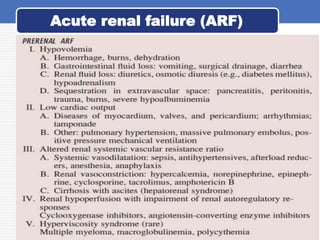
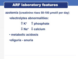
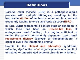
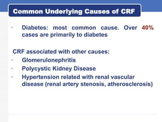
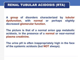
Common Renal
Disorders
Lecturer: Dr. Pablo R. Betancourt A.
Chemical Pathology I
University of the Gambia. SMAHS. 2013.
Contents
Acute renal failure
Chronic renal failure
Nephrotic syndrome
Nephrolithiasis
Renal Tubular Acidosis
Acute renal failure (ARF) is a syndrome
characterized by rapid decline in glomerular
filtration rate (hours to days), retention of
nitrogenous waste products, and perturbation
of extracellular fluid volume and electrolyte
and acid-base homeostasis.
Acute renal failure (ARF)
ARF complicates approximately 5% of hospital
admissions and up to 30% of admissions to intensive
care units.
Oliguria (urine output 400 mL/d) is a frequent but not
invariable clinical feature (50%).
ARF is usually asymptomatic and diagnosed when
biochemical monitoring of hospitalized patients
reveals a recent increase in blood urea and
creatinine concentrations.
Acute renal failure (ARF)
Both terms indicate an accumulation of nitrogenous
wastes in blood
Uremia - syndrome of renal failure
Represents numerous consequences related to renal
failure
Azotemia - increased serum urea and creatinine levels
Acute renal failure (ARF)
Uremia Vs Azotemia
Acute renal failure (ARF)
For purposes of diagnosis and management are
conveniently divided into three categories:
(1)diseases that cause renal hypoperfusion without
compromising the integrity of renal parenchyma
(prerenal ARF, prerenal azotemia) (55%)
(2) diseases that directly involve renal parenchyma
(intrinsic renal ARF, renal azotemia) (40%)
(3)diseases associated with urinary tract obstruction
(postrenal ARF, postrenal azotemia) (5%).
Acute renal failure (ARF)
(continuation)
Acute renal failure (ARF)
Clinical features
Rising blood urea and creatinine, trigger
uraemia.
Features of uraemia are:
 Anorexia
 Nausea & vomiting
 Pruritus
 Immune suppression (patients are often
septic)
 Reduced conscious level
 Haemorrhage (epistaxis and GI)
ARF diagnosis
• History and Physical examination
• Blood tests : CBC, BUN/creatinine, electrolytes,
uric acid
• Urinalysis
• Renal ultrasound (useful for obstructive forms)
• Doppler (to assess renal blood flow)
• CT (Computerized Tomography)
• MRI
azotemia (creatinine rises 50-100 μmol/l per day)
•electrolytes abnormalities:
 K+  phosphate
 Na+  calcium
• metabolic acidosis
•oliguria - anuria
ARF laboratory features
Chronic renal failure
Chronic renal disease (CRD) is a pathophysiologic
process with multiple etiologies, resulting in the
inexorable attrition of nephron number and function and
frequently leading to end-stage renal disease (ESRD).
In turn, ESRD represents a clinical state or condition in
which there has been an irreversible loss of
endogenous renal function, of a degree sufficient to
render the patient permanently dependent upon renal
replacement therapy (dialysis or transplantation) in
order to avoid life-threatening uremia.
Uremia is the clinical and laboratory syndrome,
reflecting dysfunction of all organ systems as a result of
untreated or undertreated acute or chronic renal failure.
Definitions
• Diabetes: most common cause. Over 40%
cases are primarily to diabetes
CRF associated with other causes:
• Glomerulonephritis
• Polycystic Kidney Disease
• Hypertension related with renal vascular
disease (renal artery stenosis, atherosclerosis)
Common Underlying Causes of CRF
Consequences
Metabolic features:
 Impairment in urinary concentration and dilution.
 Osmotic diuresis due to high solute concentration
for each functioning nephron
 Impairment of electrolytes and pH balance.
 Retention of waste products of metabolism.
 Decreased calcitriol synthesis.
 Decreased erythropoietin synthesis.
• Neurological: lethargy, peripheral
neuropathy.
• Musculoskeletal: growth failure, bone pain,
myopathy.
• Gastrointestinal: anorexia, nausea and
vomiting, GI bleeding.
• Cardiovascular: anaemia, hypertension,
pericarditis.
• Skin: pruritus, purpura, pallor.
• Genitourinary: nocturia, impotence.
Uremic Syndrome
Biochemical changes in plasma
Increased: potassium, urea, creatinine,
hydrogen ion, phosphate, magnesium.
Decreased: Sodium, bicarbonate, calcium.
Metabolic acidosis.
Hypocalcaemia and osteodystrophy
(secondary hyperparathyroidism= stimulates
PTH secretion).
Normochromic normocytic anaemia (ESRD)
due to decreased erythropoietin synthesis.
Nephrotic syndrome
The nephrotic syndrome is a clinical complex
characterized by a number of renal and
extrarenal features, the most prominent of
which are proteinuria of 3.5 g per 1.73 m2 per
24 h (in practice, 3.0 to 3.5 g per 24 h),
hypoalbuminemia, edema, hyperlipidemia and
hypercoagulation.
Definition
Pathophysiology
• Proteinuria: increased glomerular
permeability
• Hypoalbuminuria: decreased liver synthesis
• Oedema: primary salt and water retention
associated with reduced renal function, or
reduced plasma oncotic pressure.
• Hyperlipidaemia: increased apolipoprotein
liver synthesis
• Hypercoagulation: increased fibrinogen and
loss of antithrombin III
Nephrotic syndrome. Causes
Primary glomerular diseases
• glomerulosclerosis
• membranous glomerulonephritis
Systemic diseases:
• diabetes mellitus
• Connective tissue diseases (SLE)
• HIV/AIDS
• Nephrotoxins (nsaids, mercury poisoning,
etc)
Selectivity Index = UIgG/PIgG x 100
Ualb/Palb
Selectivity Index
This index will increase as the disease progresses.
Serum protein electrophoresis
Albumin α1 α2 β γ
Normal
Nephrotic
syndrome
RENAL STONES (Nephrolithiasis)
Causes
1. A high concentration of a substance in the urine due
to:
- low urine volume
- high excretion rate
2. pH changes
- alkaline urine predisposes to Ca deposition (e.g.
infection)
- acid urine predisposes to uric acid deposition.
3. Stagnation, usually due to obstruction.
RENAL STONES (Nephrolithiasis)
Types of stones (or calculi)
1. Calcium - oxalate
- phosphate
2. Uric acid - in about 10% of gouty cases. May be
associated with low urinary pH due to inadequate buffer
production.
3. Rare forms
- cystine: cystinuria, a transport defect of dibasic amino
acids and cysteine
- xanthine: xanthine oxidase deficiency
-2,8 dihydroxyadenine: Adenine Phosphoribosyl
Transferase (APRT - a purine salvage enzyme) deficiency.
Calculi are only partly mineral; up to 60%
may consist of protein, the rest being
varying proportions of calcium, magnesium,
ammonium, phosphate, etc
RENAL STONES (Nephrolithiasis)
RENAL TUBULAR ACIDOSIS (RTA)
A group of disorders characterized by tubular
dysfunction, with normal or perhaps slightly
decreased glomerular function.
The picture is that of a normal anion gap metabolic
acidosis, in the presence of a normal or near-normal
plasma creatinine.
The urine pH is often inappropriately high in the face
of the systemic acidosis (but NOT always).
Due to inability of distal nephron to excrete H+.
The urine pH is inappropriately high (pH > 5.5), but
does not contain significant bicarbonate.
Associated with hypokalemia, nephrocalcinosis and
rickets.
There are genetic and acquired forms (e.g. heavy
metal toxicity).
Type 1 (distal) RTA
Due to defective proximal bicarbonate reabsorption.
The renal threshold for bicarbonate is decreased
(normal value 24 mmol/l).
Whenever the plasma bicarbonate level exceeds the
(lowered) renal threshold (e.g. 16 mmol/l), the urine
contains large amounts of bicarbonate and the urine pH
is inappropriately high (called renal bicarbonate
wasting).
However, if the plasma bicarbonate drops to the level of
renal threshold, then all filtered bicarbonate can be
reabsorbed, and the urine pH can be appropriately
acidic (< 5.5) since distal tubular H+ excretion is normal
Type 2 (proximal) RTA

Renal diseases 2.ppt

  • 1.
    Common Renal Disorders Lecturer: Dr.Pablo R. Betancourt A. Chemical Pathology I University of the Gambia. SMAHS. 2013.
  • 2.
    Contents Acute renal failure Chronicrenal failure Nephrotic syndrome Nephrolithiasis Renal Tubular Acidosis
  • 3.
    Acute renal failure(ARF) is a syndrome characterized by rapid decline in glomerular filtration rate (hours to days), retention of nitrogenous waste products, and perturbation of extracellular fluid volume and electrolyte and acid-base homeostasis. Acute renal failure (ARF)
  • 4.
    ARF complicates approximately5% of hospital admissions and up to 30% of admissions to intensive care units. Oliguria (urine output 400 mL/d) is a frequent but not invariable clinical feature (50%). ARF is usually asymptomatic and diagnosed when biochemical monitoring of hospitalized patients reveals a recent increase in blood urea and creatinine concentrations. Acute renal failure (ARF)
  • 5.
    Both terms indicatean accumulation of nitrogenous wastes in blood Uremia - syndrome of renal failure Represents numerous consequences related to renal failure Azotemia - increased serum urea and creatinine levels Acute renal failure (ARF) Uremia Vs Azotemia
  • 6.
    Acute renal failure(ARF) For purposes of diagnosis and management are conveniently divided into three categories: (1)diseases that cause renal hypoperfusion without compromising the integrity of renal parenchyma (prerenal ARF, prerenal azotemia) (55%) (2) diseases that directly involve renal parenchyma (intrinsic renal ARF, renal azotemia) (40%) (3)diseases associated with urinary tract obstruction (postrenal ARF, postrenal azotemia) (5%).
  • 7.
  • 8.
  • 9.
    Acute renal failure(ARF) (continuation)
  • 10.
  • 11.
    Clinical features Rising bloodurea and creatinine, trigger uraemia. Features of uraemia are:  Anorexia  Nausea & vomiting  Pruritus  Immune suppression (patients are often septic)  Reduced conscious level  Haemorrhage (epistaxis and GI)
  • 12.
    ARF diagnosis • Historyand Physical examination • Blood tests : CBC, BUN/creatinine, electrolytes, uric acid • Urinalysis • Renal ultrasound (useful for obstructive forms) • Doppler (to assess renal blood flow) • CT (Computerized Tomography) • MRI
  • 13.
    azotemia (creatinine rises50-100 μmol/l per day) •electrolytes abnormalities:  K+  phosphate  Na+  calcium • metabolic acidosis •oliguria - anuria ARF laboratory features
  • 14.
  • 15.
    Chronic renal disease(CRD) is a pathophysiologic process with multiple etiologies, resulting in the inexorable attrition of nephron number and function and frequently leading to end-stage renal disease (ESRD). In turn, ESRD represents a clinical state or condition in which there has been an irreversible loss of endogenous renal function, of a degree sufficient to render the patient permanently dependent upon renal replacement therapy (dialysis or transplantation) in order to avoid life-threatening uremia. Uremia is the clinical and laboratory syndrome, reflecting dysfunction of all organ systems as a result of untreated or undertreated acute or chronic renal failure. Definitions
  • 16.
    • Diabetes: mostcommon cause. Over 40% cases are primarily to diabetes CRF associated with other causes: • Glomerulonephritis • Polycystic Kidney Disease • Hypertension related with renal vascular disease (renal artery stenosis, atherosclerosis) Common Underlying Causes of CRF
  • 17.
    Consequences Metabolic features:  Impairmentin urinary concentration and dilution.  Osmotic diuresis due to high solute concentration for each functioning nephron  Impairment of electrolytes and pH balance.  Retention of waste products of metabolism.  Decreased calcitriol synthesis.  Decreased erythropoietin synthesis.
  • 18.
    • Neurological: lethargy,peripheral neuropathy. • Musculoskeletal: growth failure, bone pain, myopathy. • Gastrointestinal: anorexia, nausea and vomiting, GI bleeding. • Cardiovascular: anaemia, hypertension, pericarditis. • Skin: pruritus, purpura, pallor. • Genitourinary: nocturia, impotence. Uremic Syndrome
  • 19.
    Biochemical changes inplasma Increased: potassium, urea, creatinine, hydrogen ion, phosphate, magnesium. Decreased: Sodium, bicarbonate, calcium. Metabolic acidosis. Hypocalcaemia and osteodystrophy (secondary hyperparathyroidism= stimulates PTH secretion). Normochromic normocytic anaemia (ESRD) due to decreased erythropoietin synthesis.
  • 20.
  • 21.
    The nephrotic syndromeis a clinical complex characterized by a number of renal and extrarenal features, the most prominent of which are proteinuria of 3.5 g per 1.73 m2 per 24 h (in practice, 3.0 to 3.5 g per 24 h), hypoalbuminemia, edema, hyperlipidemia and hypercoagulation. Definition
  • 22.
    Pathophysiology • Proteinuria: increasedglomerular permeability • Hypoalbuminuria: decreased liver synthesis • Oedema: primary salt and water retention associated with reduced renal function, or reduced plasma oncotic pressure. • Hyperlipidaemia: increased apolipoprotein liver synthesis • Hypercoagulation: increased fibrinogen and loss of antithrombin III
  • 23.
    Nephrotic syndrome. Causes Primaryglomerular diseases • glomerulosclerosis • membranous glomerulonephritis Systemic diseases: • diabetes mellitus • Connective tissue diseases (SLE) • HIV/AIDS • Nephrotoxins (nsaids, mercury poisoning, etc)
  • 24.
    Selectivity Index =UIgG/PIgG x 100 Ualb/Palb Selectivity Index This index will increase as the disease progresses.
  • 25.
    Serum protein electrophoresis Albuminα1 α2 β γ Normal Nephrotic syndrome
  • 26.
    RENAL STONES (Nephrolithiasis) Causes 1.A high concentration of a substance in the urine due to: - low urine volume - high excretion rate 2. pH changes - alkaline urine predisposes to Ca deposition (e.g. infection) - acid urine predisposes to uric acid deposition. 3. Stagnation, usually due to obstruction.
  • 27.
    RENAL STONES (Nephrolithiasis) Typesof stones (or calculi) 1. Calcium - oxalate - phosphate 2. Uric acid - in about 10% of gouty cases. May be associated with low urinary pH due to inadequate buffer production. 3. Rare forms - cystine: cystinuria, a transport defect of dibasic amino acids and cysteine - xanthine: xanthine oxidase deficiency -2,8 dihydroxyadenine: Adenine Phosphoribosyl Transferase (APRT - a purine salvage enzyme) deficiency.
  • 28.
    Calculi are onlypartly mineral; up to 60% may consist of protein, the rest being varying proportions of calcium, magnesium, ammonium, phosphate, etc RENAL STONES (Nephrolithiasis)
  • 29.
    RENAL TUBULAR ACIDOSIS(RTA) A group of disorders characterized by tubular dysfunction, with normal or perhaps slightly decreased glomerular function. The picture is that of a normal anion gap metabolic acidosis, in the presence of a normal or near-normal plasma creatinine. The urine pH is often inappropriately high in the face of the systemic acidosis (but NOT always).
  • 30.
    Due to inabilityof distal nephron to excrete H+. The urine pH is inappropriately high (pH > 5.5), but does not contain significant bicarbonate. Associated with hypokalemia, nephrocalcinosis and rickets. There are genetic and acquired forms (e.g. heavy metal toxicity). Type 1 (distal) RTA
  • 31.
    Due to defectiveproximal bicarbonate reabsorption. The renal threshold for bicarbonate is decreased (normal value 24 mmol/l). Whenever the plasma bicarbonate level exceeds the (lowered) renal threshold (e.g. 16 mmol/l), the urine contains large amounts of bicarbonate and the urine pH is inappropriately high (called renal bicarbonate wasting). However, if the plasma bicarbonate drops to the level of renal threshold, then all filtered bicarbonate can be reabsorbed, and the urine pH can be appropriately acidic (< 5.5) since distal tubular H+ excretion is normal Type 2 (proximal) RTA