Radiopharmaceuticals- Definition- structure- principle- diagnostic applications- therapeutic applications- Half life of radiopharmaceuticals-Criteria for an optimal radiopharmaceutical
DEFINITION:
Radiopharmaceuticals are theradioactive substances used for diagnostic
or therapeutic inventions
Radiopharmaceuticals
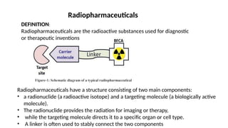
Radiopharmaceuticals have a structure consisting of two main components:
• a radionuclide (a radioactive isotope) and a targeting molecule (a biologically active
molecule).
• The radionuclide provides the radiation for imaging or therapy,
• while the targeting molecule directs it to a specific organ or cell type.
• A linker is often used to stably connect the two components
3.
PRINCILPLE
• radioactive isotopeshave a property to decompose or decay by emission of
nuclear particles.
• The three main types of radiation decay are α particles ,β particles and γ
photons.
• α particles have the largest mass. However, due to large charge, it does cause
a great deal of damage to the immediate area by breaking down DNA
• β particles are electrons. β particles are not as destructive as α particles but
can be used therapeutically.
• γ rays are electron magnetic vibrations comparable with light but of shorter
wavelength. Because of their shorter wavelength and high energy, they are
very penetrating
4.
Half life ofradiopharmaceuticals
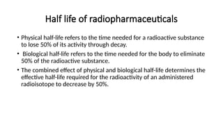
• Physical half-life refers to the time needed for a radioactive substance
to lose 50% of its activity through decay.
• Biological half-life refers to the time needed for the body to eliminate
50% of the radioactive substance.
• The combined effect of physical and biological half-life determines the
effective half-life required for the radioactivity of an administered
radioisotope to decrease by 50%.
5.
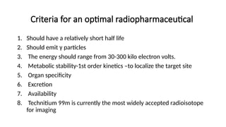
Criteria for anoptimal radiopharmaceutical
1. Should have a relatively short half life
2. Should emit γ particles
3. The energy should range from 30-300 kilo electron volts.
4. Metabolic stability-1st order kinetics –to localize the target site
5. Organ specificity
6. Excretion
7. Availability
8. Technitium 99m is currently the most widely accepted radioisotope
for imaging
6.
APPLICATIONS OF RADIOPHARMACEUTICALS
•Diagnostic uses
• Radiopharmaceuticals are used to diagnose the presence or
progression of disease following a specific therapy.
• Radiopharmaceuticals can also be used to evaluate drug induced
toxicity.
• to analyse thyroid function, a tracer dose of radioactive iodine is
administered orally; the agent concentrates in the thyroid gland. The
thyroid is then scanned to determine radioiodine concentration and
location. Greater than normal up-take by the thyroid indicates
hyperthyroidism
7.
• Therapeutic uses:
•Radioisotopes are used as internal or external radiation sources to
treat disorders such as hyperthyroidism and cancer.
• Internal radiation source
• Radioisotopes administered orally or intravenously or implanted in
the target tissue will reduce radiation that destroys disease, results in
prevention of new tissue growth.
• External radiation source
• Radiation used for therapy in cancer patients
8.
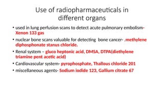
Use of radiopharmaceuticalsin
different organs
• used in lung perfusion scans to detect acute pulmonary embolism-
Xenon 133 gas
• nuclear bone scans valuable for detecting bone cancer- .methylene
diphosphonate stanus chloride.
• Renal system – gluco heptonic acid, DMSA, DTPA(diethylene
triamine pent acetic acid)
• Cardiovascular system- pyrophosphate, Thallous chloride 201
• miscellaneous agents- Sodium iodide 123, Gallium citrate 67