Radiology is a dynamic medical specialty that uses various imaging technologies to diagnose and treat diseases. While radiologists are often thought to work alone interpreting images in dark rooms, the reality is that radiology involves direct patient care, communication with other physicians, and teaching residents. The field also has many subspecialties and opportunities to specialize further in areas like interventional radiology that perform medical procedures. There are several pathways to become a radiologist, including integrated programs, with demand expected to remain steady due to advancing technologies.
Dr. Sudhir Kale is the head of radiology and lead consultant at Aster CMI hospital in Bangalore, India. He has over 14 years of experience in cross-sectional imaging and specializes in whole body CT/MRI. He is certified and trained in MR-guided HIFU surgeries and has authored chapters in textbooks. Dr. Kale has participated in various research projects involving neuroimaging and has reported on imaging for professional cricket and soccer players in India. He regularly presents at CME conferences on advanced applications like coronary, neuro, and sports injury imaging.
The document discusses ethical issues in clinical trials conducted in emerging regions. It summarizes that over 40% of US-regulated trials are now conducted overseas, driven by increasing trial numbers, large sample size demands, and availability of lower-cost patient recruitment abroad. However, ethical challenges arise from cultural differences, vulnerability of populations, and crisis opportunism in places with limited healthcare. Informed consent can be difficult given literacy and comprehension issues. Placebo controls remain controversial, though active controls are preferred from scientific, ethical and regulatory perspectives when proven treatments exist.
The document provides guidelines from the European Society of Radiology on producing high quality radiology reports. It recommends that reports should include the clinical context, examination details, objective findings described using standard terminology, an impression or diagnosis, and any advice for further evaluation. The goal is to concisely communicate relevant medical information to referring physicians in a clear, consistent format to aid patient care and management. Structured reporting is presented as a future direction that could standardize data capture and support additional uses of report information.
The Presentation will take the reader through various ethical issues in biomedical research. It covers topics like The Nuremberg Code, Declaration of Helsinki, Declaration of Geneva, selected code and regulations that guide research with human subjects, etc.
Ethical principles for conducting research with human participantsRaghad Abutair
This document discusses ethical principles for conducting research with human participants. It defines ethics, experiments on humans, and unethical human experimentation. It outlines controls and requirements for research, including obtaining consent from participants and oversight of the research process. Examples are given of some of the worst medical experiments in history, including those conducted by Nazi doctors and Unit 731 in Japan during World War II. The document stresses the importance of considering risks and benefits, protecting human dignity, and having proper scientific justification and oversight for any research with human subjects.
This document provides an overview of radiology reports and their structure. It explains that radiology reports are produced after imaging exams to communicate the radiologist's findings and impressions to referring physicians. The report typically includes sections identifying the exam type, patient history, comparison to prior exams, technical details, findings for each body area examined, and an overall impression or diagnosis. Electronic health records now allow patients direct access to their radiology reports online.
This document summarizes a review article on using deep learning techniques to detect and diagnose COVID-19 using radiology images. The review analyzed 37 studies published between November 2019 and July 2020. The studies used deep learning models on CT scans and X-rays to improve the detection and diagnosis of COVID-19 compared to traditional methods. Deep learning was shown to increase sensitivity and specificity by extracting hidden features from radiology images. The models helped address limitations of current diagnostic methods by providing fast, low-cost analysis of images to help identify COVID-19 cases.
The NIDIAG project aimed to develop diagnostic tools and guidance to improve clinical management of neglected infectious diseases (NIDs) in resource-limited settings. Researchers studied patients presenting with neurological disorders, persistent fever, or digestive disorders in multiple countries. They identified common infectious diseases causing these syndromes. New rapid diagnostic tests were developed for human African trypanosomiasis and visceral leishmaniasis. Diagnostic guidance tools incorporating useful clinical features and rapid tests were created to help health workers diagnose and treat patients. The project generated significant scientific output and new diagnostics that have the potential to improve NID control programs.
The document discusses diversity in the field of health physics, which deals with recognizing, evaluating, and controlling health hazards from ionizing radiation. It describes several areas of specialization within health physics including medical physics, radiology, radiotherapy, nuclear medicine, power reactors, defense, education, regulatory enforcement, occupational safety, and the environment. Medical physics focuses on using radiation for diagnosis and treatment, while health physicists ensure protection from radiation exposure. Radiotherapy uses radiation to treat cancer, radiology uses techniques like x-rays for imaging, and nuclear medicine uses radioactive tracers and gamma cameras. Other areas involve protection of workers and the public from radiation hazards.
This document describes a study that evaluated the safety and efficacy of TriPollar radiofrequency (RF) treatment for acne vulgaris. Twenty patients with active acne lesions received six weekly TriPollar RF treatments. Results found an average 42% reduction in active lesions after treatment, which was sustained at 4-week follow-up. Patients reported good to very good satisfaction with the results. No significant adverse events occurred. The findings confirm TriPollar RF is a safe and effective treatment for acne vulgaris that can provide comparable efficacy to other modalities in a more convenient treatment approach.
Change of neurosurgical planning during COVID-19 pandemic and endemic eraAmit Ghosh
- All patients undergoing surgery should be considered positive for COVID-19 unless proven otherwise, and appropriate PPE should be used.
- For elective surgeries, a chest CT scan and COVID test are recommended pre-operatively. Surgeries should be postponed for COVID-positive patients or those with unknown status.
- Special precautions are outlined for endoscopic or endonasal surgeries on COVID-positive patients due to risk of transmission, including use of PAPRs by all OR staff or choosing alternative surgical approaches if possible.
The study aimed to analyze patient demographics and disease patterns among those admitted to the intensive care unit (ICU) and coronary care unit (CCU) of BP Koirala Institute of Health Sciences, Nepal over one year. A total of 601 patients were admitted, with 269 in the ICU and 346 in the CCU. Most patients were aged 20-60 years and from surrounding districts. Common ICU admissions were for exploratory laparotomy complications, organophosphorus poisoning, and encephalitis. Acute coronary syndrome, myocardial infarction, and shock were leading CCU admissions. Outcomes could be improved as many patients were referred or left against medical advice, suggesting further investigation is needed.
This document provides an overview of ethics guidelines for biomedical research involving human participants in India. It discusses the evolution of ethics codes internationally and the role of the Indian Council of Medical Research (ICMR) in establishing ethics standards within India. The ICMR Code outlines 13 general principles for ethical research, including principles of voluntariness, non-exploitation, risk minimization, and institutional responsibility. The document provides background on the structure of ICMR and amendments made to India's ethics guidelines over time.
The document discusses criteria for admission and triage to the intensive care unit (ICU). It notes that demand often exceeds supply of ICU beds. It presents several models for prioritizing patients, including based on diagnosis and severity of illness. Factors considered for admission include prognosis, likelihood of benefit, and patient wishes. The document also discusses criteria for discharge and intermediate care units as alternatives to the ICU.
1) Back injuries are a major cause of missed work days for healthcare workers due to the physical demands of patient lifting and transfers. Proper lifting techniques are important for injury prevention.
2) A variety of patient transfer methods and equipment can help facilitate safe transfers while avoiding strain or injury. Factors like a patient's medical conditions and limitations must be considered.
3) Maintaining proper patient monitoring during transfers is important, as some patients can experience sudden changes in condition. Communication with the patient and awareness of any medical equipment or devices is also important for safety.
The document discusses critical care nursing and monitoring patients in the intensive care unit. It describes different levels of critical care from level 0 to level 3, with higher levels involving more advanced support and multi-organ failure. Various types of intensive care units are also outlined, including medical, surgical, neonatal and more specialized units. The document provides guidance on monitoring patients in the ICU, including assessing physiological parameters, vital signs, physical exams and laboratory reports. Close monitoring and documentation of the patient's condition is emphasized as important for patient care, legal protection and reimbursement.
The document summarizes The Belmont Report, which established ethical principles for protecting human subjects in research. It was created in 1979 by a commission in response to unethical human subject research studies. The Belmont Report outlines three basic ethical principles - respect for persons, beneficence, and justice. It also distinguishes between research and practice, and provides guidelines for informed consent, risk-benefit assessments, and subject selection to help apply these principles. While not formally adopted, The Belmont Report has become the primary framework for research ethics in the United States.
Adarsh paramedical institute in pune|best paramedical institute in pune.sara patil
Adarsh Paramedical Institute has offered paramedical courses since 2004. It provides diploma, graduation, and post-graduation courses in fields like medical laboratory technology, radiology, naturopathy, dialysis, ECG, and more. Course durations range from one to three years. The institute aims to give students requisite skills and knowledge to work in healthcare through innovative curriculum and hands-on learning. It is located in Pune, Maharashtra and accepts applications.
This presentation explores the origin of good Clinical Practice, Need of GCP. Principals of Good Clinical Practices. Its a ICH guideline E6.This presentation is very useful to prepare for interviews in the field of Clinical Research.
Elite Institute of Pharma Skills gives quality Education to Pharmacy and Life Science graduates in the field of Regulatory Affairs, Clinical Research, Pharmacovigilance and Medical Coding.
for more details log on to
www.elite-pharmaskills.com
7028042239
The document discusses preoperative assessment and perioperative management of obstructive sleep apnea. It notes that precise localization of airway obstruction sites is important for surgical success, but current diagnostic methods are imperfect. It also outlines considerations for patient selection, intraoperative care, and postoperative management to optimize surgical outcomes.
This document discusses office-based anesthesia. It notes that over 25% of surgeries are now performed in offices rather than hospitals. Office-based anesthesia, also called non-operating room anesthesia or ambulatory anesthesia, involves anesthesia performed in an outpatient setting like a doctor's office rather than an accredited surgery center or hospital. While standards of care are the same, office settings present unique challenges regarding safety, oversight, documentation, and experience level. The document outlines considerations for patient selection, appropriate procedures, personnel requirements, equipment needs, and pre-operative fasting guidelines for office-based anesthesia.
The document discusses improving safety standards for office-based surgery and anesthesia. It notes a lack of uniform regulation has led to increasing complexity of cases performed in office settings. It introduces the Institute for Safety in Office Based Surgery, a non-profit established in 2009 to promote patient safety, develop tools for risk detection, and encourage physician education and evidence-based standards of care. It also references the high-profile case of Joan Rivers' death from complications during an outpatient procedure.
This document provides guidance on developing a research proposal. It begins by welcoming participants to the first module on research and poses several questions about conducting research in Peru and Tumbes. The objectives are defined as helping participants develop a strong research question and proposal and publish their work. The final product will be a certification upon completion. Ideas are provided for research topics, such as conducting epidemiological studies on epilepsy in regions endemic for cysticercosis. Guidelines are outlined for structuring introductions, justifications, literature reviews and submitting documents to ethics boards. Sources of research ideas include personal experience, previous studies, consulting experts, and national and regional health priorities.
This document discusses ambulatory anesthesia and day-care surgery. It provides a brief history of ambulatory anesthesia, noting key developments from 1903 to present day. It describes the objectives and techniques of anesthesia for day-care surgery, including premedication, induction agents, maintenance techniques, regional versus general anesthesia, advantages and disadvantages, selection criteria for patients, and discharge criteria. The success of day-care surgery depends on appropriate patient selection, facility availability, and type of surgical procedure.
The document discusses the virtues and vices of teleradiology as well as potential malpractice issues. It notes that while teleradiology can provide timely interpretations from subspecialists, it also poses risks such as limited access to prior images and clinical data as well as less communication with ordering physicians. The document also examines issues radiologists may face with incidental findings, such as whether to report things like small cysts that likely have little clinical significance but could theoretically be early cancers. Overall it presents many complex factors around these areas of radiology practice.
RADIOTHERAPY FOR ENDOMETRIUM AND CERVICAL CANCERSKanhu Charan
1. The document discusses radiotherapy techniques for treating gynaecological malignancies such as cancer of the cervix, endometrium, vulva, and ovaries.
2. It describes the evolution of radiotherapy from conventional 2D techniques to newer approaches like 3D conformal radiation therapy and intensity-modulated radiation therapy.
3. Key factors in treatment planning and delivery discussed include patient immobilization, imaging techniques for treatment planning like CT and PET scanning, and methods of verifying accurate patient positioning and treatment delivery such as electronic portal imaging.
Nuclear medicine uses small amounts of radioactive tracers and imaging techniques to examine organ structure and function. It combines fields like chemistry, physics, and medicine. Nuclear imaging allows visualization of tissue structure and function by tracking how radiotracers are absorbed. Common nuclear medicine scans include thyroid scans to help diagnose thyroid abnormalities early. A thyroid scan involves administering a small amount of radioactive iodine and using a gamma camera to detect its distribution and uptake in the thyroid gland.
This document discusses radiation and its uses in dentistry. It begins with an overview of the electromagnetic spectrum and different types of radiation. Radiation has benefits in medical diagnosis and treatment through uses like x-rays, gamma rays for sterilization, and nuclear medicine. In dentistry, x-rays are commonly used for diagnostic purposes to detect issues like dental caries, periodontal disease, cysts and tumors. Radiation also has therapeutic uses in dentistry through procedures like radiotherapy for malignant diseases. The document outlines several specific diagnostic imaging techniques used in dentistry like bitewings, periapicals and panoramic x-rays.
This document discusses x-rays and medical imaging technology. It covers:
1. X-rays are a form of ionizing radiation that can potentially damage DNA and increase cancer risk, though risk from medical imaging is generally small depending on dose, age, sex, and body region exposed.
2. X-rays are used in medical imaging to non-invasively diagnose disease, monitor therapy, and guide procedures through techniques like radiography, fluoroscopy, and CT scans.
3. Principles of radiation protection include justifying exams based on medical need and optimizing techniques to use the lowest dose for adequate diagnosis. Risks include potential long-term cancer risk and rare tissue effects.
Adverse reactions and management of contrast reactions Ashim Budhathoki
The document provides information on contrast media used in medical imaging. It begins with acknowledging those who helped with the project. It then discusses the objectives of the study which are to define contrast media, explain types and reactions, and responsibilities during contrast studies. The document classifies contrast media as positive or negative. Positive contrast media make structures appear brighter on images while negative contrast media make structures appear darker. It provides details on various contrast agents used for different medical imaging modalities like CT, MRI, ultrasound and their routes of administration.
This document summarizes key points from a presentation on radiologic errors given at a conference. It discusses defining medical errors and differentiating them from complications. Error rates from retrospective studies of radiology are presented, ranging from 25-90% for missed cancers depending on the imaging modality and body area. Prospective "real-time" error rates are estimated to be 3-4%. Causes of errors including perceptual and cognitive factors are reviewed. Standards of care, hindsight bias, outcome bias and efforts to reduce errors are also discussed. An anecdote is shared about lawyers and doctors continuing adversarial relationship on flights.
Presentation slides from our first meeting, held on Tuesday 10th September 2013 at the Royal College of Surgeons.
Find us on
Twitter @STARSurgUK
Facebook.com/STARSurgUK
Email: STARSurgUK@gmail.com
This document provides an overview of the fourth edition of the textbook "Practical Radiotherapy Planning". The textbook is written by four authors who are professors and consultants in clinical oncology in the UK. It aims to provide guidance on radiotherapy treatment planning based on sound pathological and anatomical principles. The textbook covers topics such as radiobiology, organs at risk, brachytherapy, emergency radiotherapy, and treatment planning for many cancer sites. It emphasizes the underlying principles of treatment planning that can be applied to conventional, conformal and novel radiotherapy techniques. The textbook includes many clinical images to illustrate key planning concepts.
This survey of radiographers in Australian hospitals investigated their participation in abnormality detection systems for trauma radiographs, their perceptions of the benefits and barriers of radiographer commenting, and their views on radiographer image interpretation services. The results found that most radiographers participated in abnormality detection for less than 20% or more than 80% of examinations. Perceived benefits included assisting patient care, but barriers included limited access to image interpretation education and low confidence. Improving access to education was seen as an enabler for greater participation in detection and commenting systems.
The World Health Organization (WHO) is a specialized UN agency responsible for international health and public health. Through WHO, health professionals from 165 countries collaborate to help all people attain a level of health allowing a productive life by 2000. WHO promotes comprehensive health services, disease prevention and control, environmental improvement, health workforce development, research coordination, and health program planning and implementation. Progress requires international cooperation on standards, criteria, drug names, regulations, disease classification, and health statistics.
This document outlines a proposal for an Emergency Radiology Fellowship program. It notes the increasing subspecialization in medicine and complexity of technologies. An Emergency Radiology Fellowship would train radiologists in emergency settings through a competency-based curriculum involving practical skills. Selecting institutions with adequate emergency infrastructure and qualified faculty is important. While subspecialization provides expertise, the approach also needs to be multidisciplinary to avoid issues like fragmentation or conflicts of interest. The fellowship aims to address future imaging needs through specialized emergency training.
This study analyzed radiation doses received by patients from medical X-ray examinations in Nigeria between 2000-2014. The most common examinations were skull (32%), chest (22%), and lumbar spine (13%). Mean entrance surface doses for various examinations, such as chest and skull, were found to exceed internationally recommended diagnostic reference levels. To better protect patients and improve practices, the study concluded that Nigeria needs to develop comprehensive national diagnostic reference levels.
Nuclear medicine is a medical specialty that uses small amounts of radioactive tracers and imaging technologies to diagnose and treat diseases. It allows physicians to obtain medical information noninvasively that may otherwise require surgery or more invasive tests. Nuclear medicine procedures can identify abnormalities early in the progression of diseases and allow for earlier treatment when prognosis is better. Over 18 million nuclear medicine procedures are performed annually in the United States to diagnose and treat many different types of diseases.
This document provides an introduction to diagnostic radiography. It discusses what diagnostic radiography is, the early history of radiography dating back to Wilhelm Roentgen's discovery of x-rays in 1895. It then describes several common diagnostic imaging modalities used today including conventional x-ray, fluoroscopy, ultrasound, CT scanning, and MRI. Each modality is briefly explained in terms of its benefits, risks, and applications. The document concludes with a short section on the history and current state of diagnostic radiography in Ghana.
The document summarizes the Saudi neurology board, chaired by Dr. Hessa Alotaibi. It outlines the goals of establishing an excellent neurology training program and certifying competent neurologists. The 4-year residency involves rotations in internal medicine, clinical neurology, pediatrics, and electives. Residents are evaluated annually and must pass written and clinical board exams to become certified. The program aims to promote subspecialties and meet the growing need for neurologists in Saudi Arabia.
Radiography is a technique that uses radiation, such as x-rays, to view the internal structures of the body. It can be used for both medical and industrial purposes. Medically, radiography is used for diagnostic and therapeutic purposes to diagnose diseases and guide treatment. Some common medical radiography techniques include x-rays, CT scans, mammograms, and fluoroscopy. While radiography provides benefits for diagnosing and treating illnesses, it also carries some risks, such as a small increased risk of developing cancer from exposure to ionizing radiation. However, the benefits of radiography for improving health outcomes are considered to outweigh these potential risks.
Nuclear medicine uses radioactive substances to diagnose and treat disease. In diagnostic nuclear medicine, a radiopharmaceutical is administered to the patient and detected by a gamma camera to produce images of organ function. Positron emission tomography (PET) uses radiopharmaceuticals that emit positrons to produce highly accurate images of metabolic activity in the body, making it effective for cancer diagnosis, staging, assessing treatment response, and detecting recurrence. PET's most common radiopharmaceutical is fluorodeoxyglucose (FDG), which is taken up by metabolically active cells including many cancers.
MedicalResearch.com: Medical Research Exclusive Interviews December 14 2014Marie Benz MD FAAD
MedicalResearch.com features exclusive interviews with medical researchers from major and specialty medical research and health care journals and meetings.
How to Manage Reception Report in Odoo 17Celine George
A business may deal with both sales and purchases occasionally. They buy things from vendors and then sell them to their customers. Such dealings can be confusing at times. Because multiple clients may inquire about the same product at the same time, after purchasing those products, customers must be assigned to them. Odoo has a tool called Reception Report that can be used to complete this assignment. By enabling this, a reception report comes automatically after confirming a receipt, from which we can assign products to orders.
A Free 200-Page eBook ~ Brain and Mind Exercise.pptxOH TEIK BIN
(A Free eBook comprising 3 Sets of Presentation of a selection of Puzzles, Brain Teasers and Thinking Problems to exercise both the mind and the Right and Left Brain. To help keep the mind and brain fit and healthy. Good for both the young and old alike.
Answers are given for all the puzzles and problems.)
With Metta,
Bro. Oh Teik Bin 🙏🤓🤔🥰
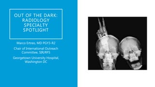
1. OUT OF THE DARK:
RADIOLOGY
SPECIALTY
SPOTLIGHT
Marco Ertreo, MD PGY3-R2
Chair of International Outreach
Committee, SIR/RFS
Georgetown University Hospital,
Washington DC
2. OVERVIEW
What a radiologist actually does
Common radiology myths and facts
Subspecialties in radiology and what they actually do
What is a radiology resident’s daily life like
Current training pathways
2
3. THE RADIOLOGIST
A physician specialized in radiology, the
branch of medicine that uses ionizing and
nonionizing radiation for the diagnosis and
treatment of disease.
3
9. MYTH: YOU WILL NEVER SEE OR
TALK TO A PATIENT EVER AGAIN…
• FACT: Radiologists see and talk to patients
all the time. Studying and evaluating x-ray
images is certainly a large part of being a
radiologist - and perhaps the most
recognizable part of the job.
• Departments are typically divided into
sections or subspecialty areas by body part
or imaging modality, and each section
performs a variety of diagnostic and
therapeutic procedures.
• In particular, fluoroscopy, interventional
radiology, ultrasound, and mammography
subspecialists are especially involved in
direct patient care, including counseling
patients, performing procedures, and post-
procedure management. 9
11. MYTH: RADIOLOGISTS ARE A
BUNCH OF ANTI-SOCIAL
INTROVERTS WHO ARE AFRAID OF
TALKING TO PEOPLE…
• FACT: Communication lies at the heart of
radiology. To be successful, radiologists are
in constant communication with patients,
referring physicians, as well as
multidisciplinary teams. While radiology
appears to the outsider as one person
examining one image, the reality goes
much deeper. The information gleaned by
examining the image is shared with others,
putting the radiologist in the role of
subject matter expert.
• Radiologist are, ultimately, consultants.
11
12. MYTH: YOU WILL BE ALONE IN A
DARK ROOM ALL DAY, EVERY DAY…
• FACT: Radiologists see the light of
day. While it is an undeniable truth
that radiologists spend time in a
dimly lit room, the radiology
reading room is filled with people
and activity throughout the day.
Unlike many other medical
specialties, attending physicians
work with residents throughout
the day, reviewing studies,
answering questions, performing
procedures, and teaching.
• Clinical teams routinely consult
with radiologists either directly by
visiting the reading room or
indirectly.
• Radiologists are prominent
members of all tumor boards and
most other multi-disciplinary
meetings that revolve around
medical imaging, often leading the
discussion. 12
14. MYTH: THE RADIOLOGY JOB
MARKET IS DISMAL, NO ONE CAN
GET A JOB…
• FACT: The radiology job market is
steady and reliable. Advancing
technology continues to expand the
field of radiology. The current
number of job openings remains
equal to the number of graduating
fellows, assuring a solid future for
individuals entering the field of
radiology.
14
15. MYTH: RADIOLOGY IS STATIC AND MUNDANE…
• FACT: The future of radiology is dynamic and innovative. Radiology is among the
most dynamic specialties in medicine with innovations and advancements
occurring on a regular basis. Commercial CT scanners were first introduced in
1972, the first MRI scan was performed in 1977. Imaging utilization continues to
increase as new applications and novel technologies in both diagnosis and
treatment of diseases are continually being researched. Few other medical
specialties can claim such dramatic advancements over the past few decades. In
fact, a textbook on radiology published in the 1990s would be considered
outdated given today’s technology.
15
16. MYTH: RADIATION FROM
MEDICAL IMAGING IS
DANGEROUS/RADIOLOGISTS
ARE EXPOSED TO LOTS OF
RADIATION…
Strict adherence to radiation safety
protocols
Most have little to no exposure -
except for IR most images are
obtained from technicians
IRs wear lead aprons, glasses and
even gloves
No risks for pregnant radiologists
16
17. MYTH: RADIOLOGISTS
AREN’T DOCTORS
“Saving lives one image at a time”
You actually see patients, how much is up
to you;
Perform procedures, how much is up to
you;
Provide consultations.
17
18. DIAGNOSTIC
• Body imaging
• Neuroradiology
• Cardiothoracic
imaging
• Musculoskeletal
imaging
• Pediatric imaging
• Breast imaging
VASCULAR AND INTERVENTIONAL
WHAT SUBSPECIALTIES IN RADIOLOGY?
NUCLEAR MEDICINE
18
• Neuro-interventional
• Hepatobiliary
• Interventional
oncology
• Arterial and venous
• Genitourinary
• Biopsies
• Pain management
19. BODY IMAGING
Diagnoses conditions affecting the
abdomen and pelvis, including the
colon, kidneys, pancreas, liver, lungs,
stomach, genitourinary system, etc.
MAIN MODALITIES: CT, MRI,
Ultrasound, XR, Fluoroscopy
19
27. BREAST IMAGING
Diagnoses conditions that specifically
affect women, particularly breast
disease.
Very involved with patient
management, guiding clinicians on
what to do next.
MAIN MODALITIES: US,
mammography, MRI
27
28. NUCLEAR MEDICINE
Diagnoses and treats diseases
utilizing small amounts of radioactive
material.
MAIN MODALITIES: PET/CT, Gamma
camera, SPECT
28
30. VASCULAR AND
INTERVENTIONAL RADIOLOGY
Diagnoses and treats benign
and malignant diseases using
different imaging technologies
and tools such as embolization,
angioplasty, stenting, biopsies,
chemoembolization, drainage
placement
MAIN MODALITIES: US,
fluoroscopy, CT.
30
42. A DAY IN THE LIFE OF A RADIOLOGY
RESIDENT
Report to service around
730-8 am depending on
specialty
CLINICAL SERVICE: Pick up
and read 5-10 cases, read
out with your attending and
repeat (Sprinkle with
intermittent email checking,
CNN reading and coffee run)
NOON CONFERENCE
CLINICAL SERVICE: Pick up
and read 5-10 cases, read
out with your attending and
repeat
Finish the day between 430-
6pm depending on service,
case load of the day and
institution
42
43. WHAT IS CALL
LIKE?
• Varies by institution
• SHORT CALL: covering stat cases
from close of business hours to when
the night float resident comes in
• NIGHT FLOAT: 10-12 hour shift at
night
• WEEKENDS: alternating day and
night residents
• Preliminary read all stat scan and
triage imaging appropriately.
Attendings on site or at home,
available via pager/cell phone
• Everything gets ready by the
attending in the morning
43
44. EXPECTATIONS FOR EACH YEAR
1st year:
just learn
not too many expectations,
usually no call
2nd year:
it’s call time
most call is usually in second
year. You’re learning what it’s
like to be a radiologist
3rd year: fellowship
and boards
you survived second year, now
you have to study for the Board
(yes, you take it as a PGY4-R3)
and apply and match for
fellowships
4th year:
sit back and relax
most rotations are electives
(except for 1 month of nuclear
medicine and 1 of
mammography).
44
45. AFTER FELLOWSHIP
• ACADEMIC SETTING
• Teaching residents and students
• Research
• Lower salaries
• PRIVATE PRACTICE
• Work in a group
• High volume of cases
• More cases – more money
45
46. FIRST STEPS IF YOU WANT TO BECOME
A RADIOLOGIST
Make a choice
• Choose between diagnostics
and interventional!
Can’t make a choice?
• If in doubt, do diagnostics and
then you can still get into
interventional – the other way
around is more difficult.
46
47. THE PATH TO
BECOME A
DIAGNOSTIC
RADIOLOGIST
Medical school
1
1 year internship
(general surgery,
internal medicine,
transitional year)
2
4 years
diagnostic
radiology
3
1 year fellowship
(subspecialty
training)
4
47
PGY DR NUCS IR
1 INTERNSHIP
2 11 1 1
3 11 1 1
4 11 1 1
5 ? 1 ?
6 FELLOWSHIP
48. THE “NEW” PATHS
TO BECOME AN
INTERVENTIONAL
RADIOLOGIST
48
1 2a 2b
Approved by the American Board of
Medical Specialties (ABMS) in 2012
IR/DR Certificate is npow one of 4
primary certificates offered by the
ABR (others are Diagnostic
Radiology, Radiation Oncology and
Medical Physics)
49. *IR or IR-related rotations - vascular surgery, medical
oncology or interventional procedural rotations housed
within diagnostic radiology sections 49
DIAGNOSTIC RESIDENCY
ESIR
DR/IR INTEGRATED
RESIDENCY
PGY DR IR* ICU DR IR* ICU DR IR* ICU
1 INTERSHIP INTERSHIP INTERNSHIP
2 12 1 0 12 1 0 12 1 0
3 12 1 0 12 1 0 12 1 0
4 12 1 0 12 1 0 12 1 0
5 13 0 0 0-4 8-12 1 0-4 8-12 1
6
IR RESIDENCY
“EX-FELLOWSHIP”
IR RESIDENCY
“EX-FELLOWSHIP”
13
7 NOTHING! NOTHING!
ESIR (Early specialization in IR) –
requires at least 500 IR procedures
and IR related rotations during the
DR residency
50. OPTIONS FOR
CURRENT 3RD YEAR
MEDICAL STUDENTS
Option 1 (6 years)
•1 year Internship +
•5 years Integrated IR residency
Option 2 (6 years)
•1 year Internship
•Match into DR residency program with an Integrated IR residency and hope to transfer within the same
program
Option 3a ESIR track (6 years)
•1 year internship +
•3 years DR residency +
•1 year ESIR +
•1 year advanced Independent IR residency
Option 3b (7 years)
•1 year internship +
•4 years DR residency +
•2 years Independent IR residency
50
5 years of training after a clinical internship year
3 DR, 2 IR under a IR Program Director
Match out of medical school or transfer in from DR residency (PGY 3-6) at the home institution
During PGY 3-5, could transfer out of IR and into DR residency
Qualifying residents may enter the PGY6 year if have adequate training experience including at least 12 IR or IR-related rotations and documentation of at least 500 procedures covering the broad domain of IR
During PGY 5 and 6, training in IR content can be achieved in the IR section or on IR-related rotations outside of the IR section proper. Examples include rotations in vascular surgery, medical oncology clinic or interventional procedural rotations housed within diagnostic radiology sections. However, residents must accrue a minimum number of designated IR procedures. Consequently, rotations outside of IR proper must be carefully tailored to meet the overall goals and requirements of the residency.
Independent format would allow continuation of programs that are not currently tied to a DR residency program (eg Miami Vascular). Residents transfer into the program from outside institutions but can also be from within the program. This might also allow blend of programs and residents at a single institution esp. during the transition period.
Residents complete 2 years of training after completing a 4-year DR residency
Candidates may enter the second year of the program provided they have adequate training experience including minimum of 11 IR or IR-related rotations and ICU rotation and documentation of at least 500 procedures during DR residency
During PGY 5 and 6, training in IR content can be achieved in the IR section or on IR-related rotations outside of the IR section proper. Examples include rotations in vascular surgery, medical oncology clinic or interventional procedural rotations housed within diagnostic radiology sections. However, residents must accrue a minimum number of designated IR procedures. Consequently, rotations outside of IR proper must be carefully tailored to meet the overall goals and requirements of the residency.