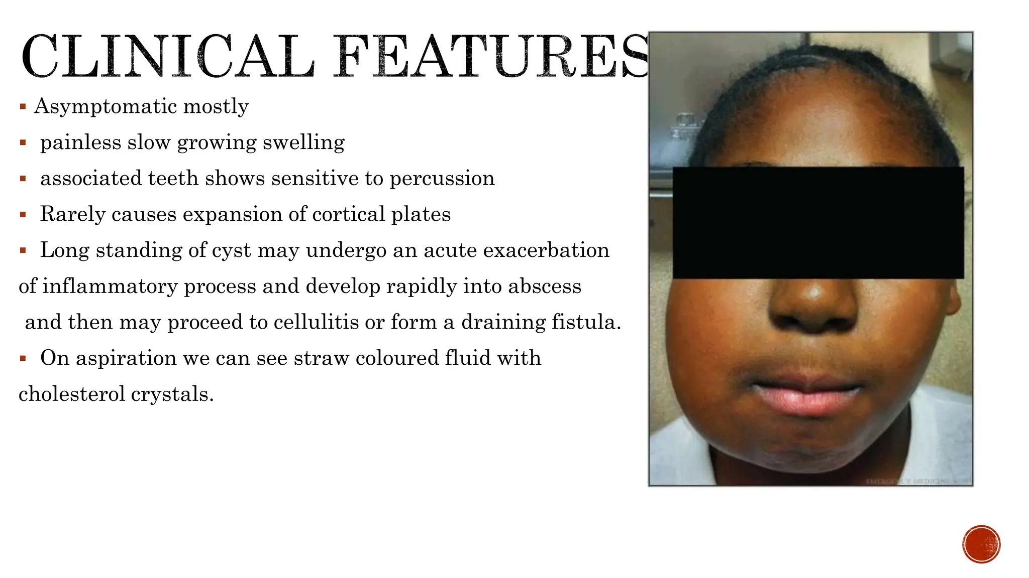
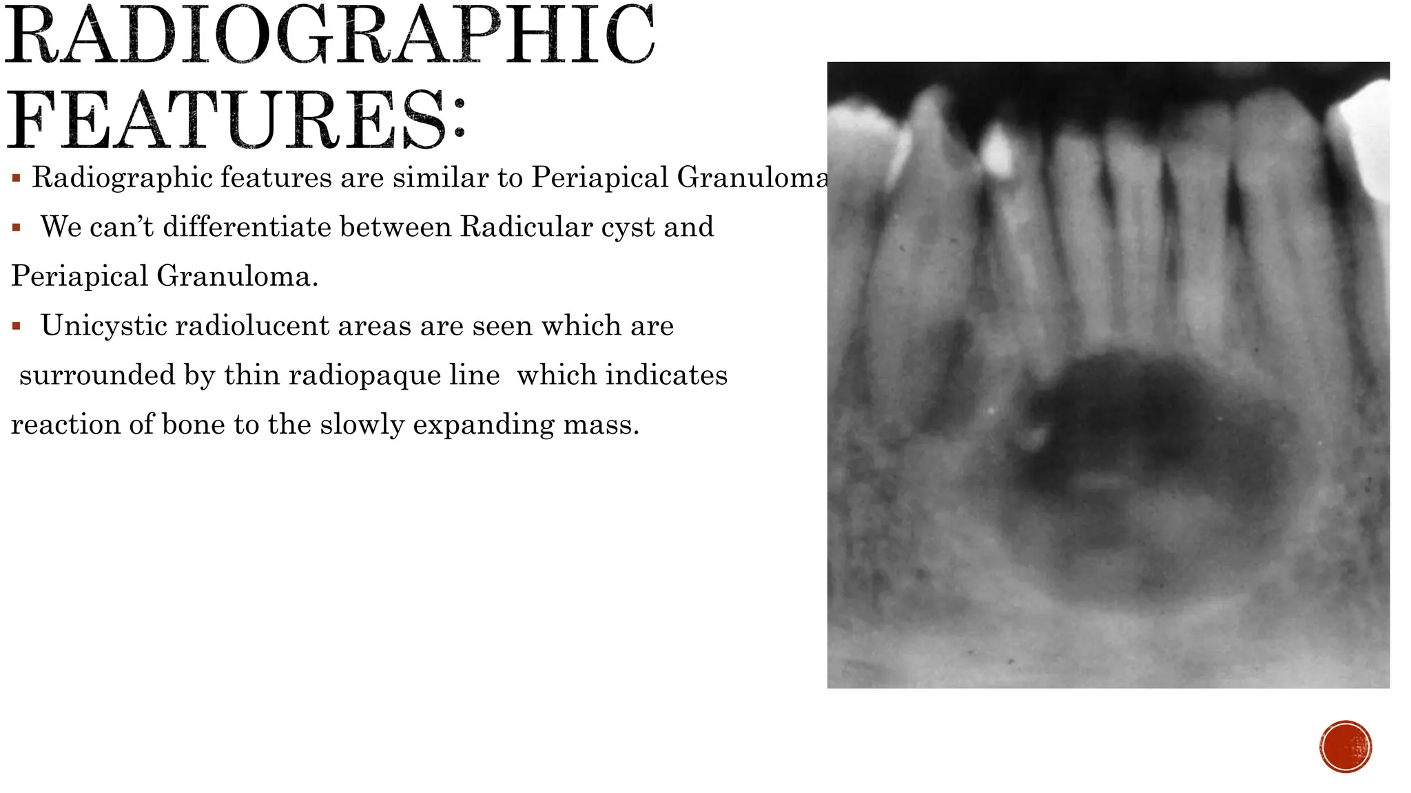
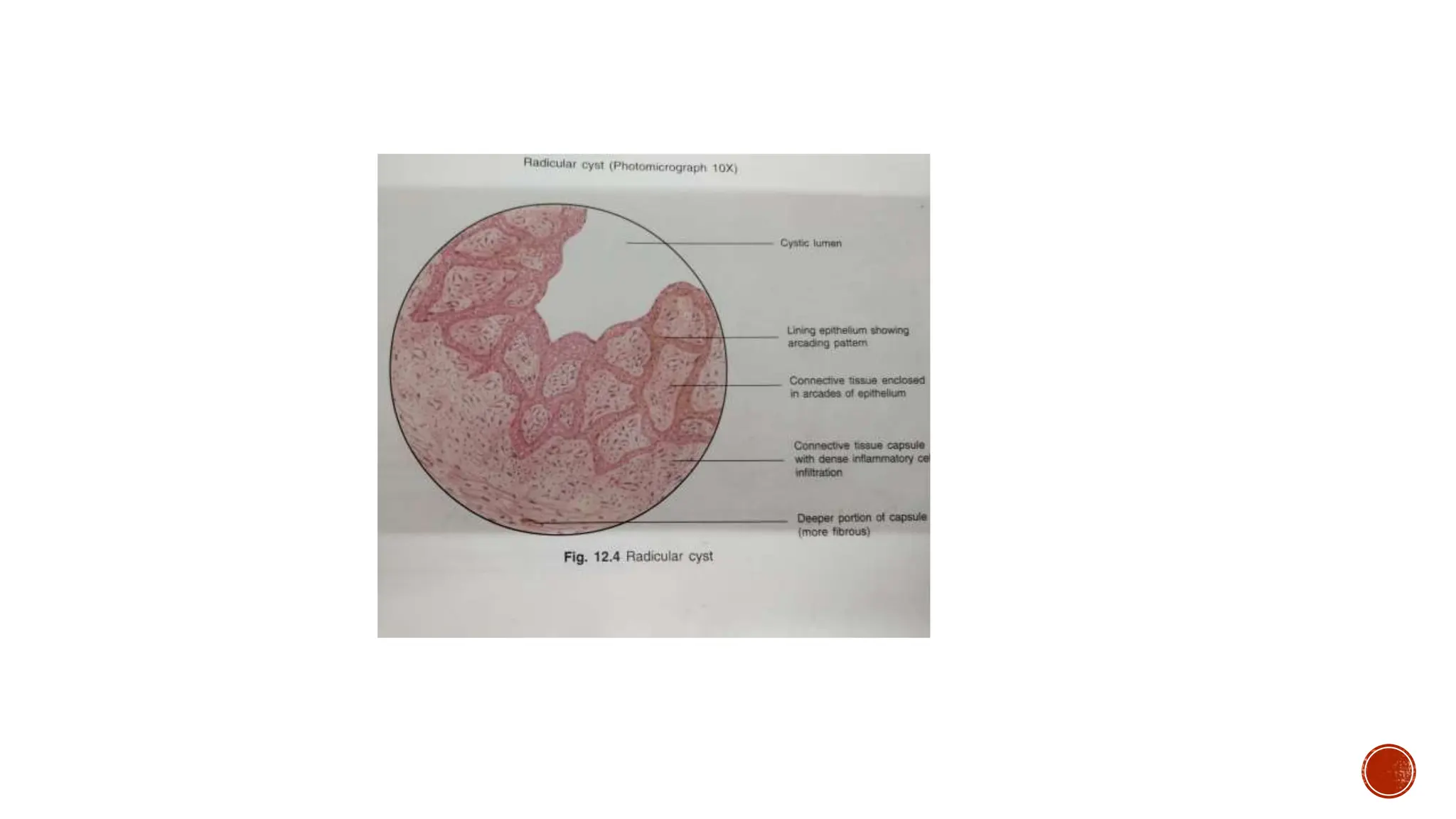
Sayyed Jani
BDS
 Most common odontogenic cyst
 Around 60% of all jaw cysts and 15% of all periapical lesions are Radicular cyst.
 It is an inflammatory odontogenic cyst surrounding the periapical region of tooth.
 It occurs as a result of
(a) Bacterial infections
(b) Necrosis of Dental pulp
(c) Dental caries
Gender prediction: Males > Females
Age prediction: 20 to 60 years
Site of occurance: Commonly maxillary anteriors followed by mandibular
premolars and molars .
Origin: odontogenic
 Asymptomatic mostly
 painless slow growing swelling
 associated teeth shows sensitive to percussion
 Rarely causes expansion of cortical plates
 Long standing of cyst may undergo an acute exacerbation
of inflammatory process and develop rapidly into abscess
and then may proceed to cellulitis or form a draining fistula.
 On aspiration we can see straw coloured fluid with
cholesterol crystals.
 Radiographic features are similar to Periapical Granuloma.
 We can’t differentiate between Radicular cyst and
Periapical Granuloma.
 Unicystic radiolucent areas are seen which are
surrounded by thin radiopaque line which indicates
reaction of bone to the slowly expanding mass.
 Lining epithelium is Non keratinized stratified squamous epithelium.
 Epithelium shows spongiosis and inflammatory cell infiltration.
 Epithelium shows arcading pattern
 Epithelium shows Rushton bodies .
 Rushton bodies - arc shaped ; eosinophilic ; hyaline bodies. They are refractile ;
amorphous in structure; Brittle in nature . There is no clinical significance and
their origin is unknown. They are also seen in residual cyst .
 Connective tissue – delicate with dense inflammatory cell infiltration.
 Deeper portion of connective tissue is more fibrous.
 Cystic lumen contains straw coloured fluid with cholesterol crystals.
 Root canal therapy of the involved teeth along with periapical surgery or
extraction of involved tooth followed by periapical curettage .
 If the cyst is incompletly removed then the remnant cells may develop into
Residual cyst.

Radicular cyst or periapical odontogenic cyst

  • 1.
  • 2.
     Most commonodontogenic cyst  Around 60% of all jaw cysts and 15% of all periapical lesions are Radicular cyst.  It is an inflammatory odontogenic cyst surrounding the periapical region of tooth.
  • 3.
     It occursas a result of (a) Bacterial infections (b) Necrosis of Dental pulp (c) Dental caries
  • 4.
    Gender prediction: Males> Females Age prediction: 20 to 60 years Site of occurance: Commonly maxillary anteriors followed by mandibular premolars and molars . Origin: odontogenic
  • 5.
     Asymptomatic mostly painless slow growing swelling  associated teeth shows sensitive to percussion  Rarely causes expansion of cortical plates  Long standing of cyst may undergo an acute exacerbation of inflammatory process and develop rapidly into abscess and then may proceed to cellulitis or form a draining fistula.  On aspiration we can see straw coloured fluid with cholesterol crystals.
  • 6.
     Radiographic featuresare similar to Periapical Granuloma.  We can’t differentiate between Radicular cyst and Periapical Granuloma.  Unicystic radiolucent areas are seen which are surrounded by thin radiopaque line which indicates reaction of bone to the slowly expanding mass.
  • 7.
     Lining epitheliumis Non keratinized stratified squamous epithelium.  Epithelium shows spongiosis and inflammatory cell infiltration.  Epithelium shows arcading pattern  Epithelium shows Rushton bodies .  Rushton bodies - arc shaped ; eosinophilic ; hyaline bodies. They are refractile ; amorphous in structure; Brittle in nature . There is no clinical significance and their origin is unknown. They are also seen in residual cyst .  Connective tissue – delicate with dense inflammatory cell infiltration.  Deeper portion of connective tissue is more fibrous.  Cystic lumen contains straw coloured fluid with cholesterol crystals.
  • 9.
     Root canaltherapy of the involved teeth along with periapical surgery or extraction of involved tooth followed by periapical curettage .  If the cyst is incompletly removed then the remnant cells may develop into Residual cyst.