SlideShare a Scribd company logo
1 of 32
PULMONARY INFECTIONS
• Pulmonary infections, in the form of pneumonia claims 1/6 of deaths in the USA.
• Pulmonary infections are common due to;
 the epithelial surfaces of the lung
are constantly exposed to air containing
various levels of microbial contaminants
 nasopharyngeal flora are regularly aspirated during sleep, even by
healthy persons and
 other common lung diseases render the lung parenchyma vulnerable to
virulent organisms.
• lung parenchyma remains sterile due to;
 Immune and non-immune defensive mechanisms in the airway tract.
• Any break in the immune mechanism (congenital or acquired) predispose to infection
• Acute and chronic pulmonary infections are generally caused by a wide variety of
microorganisms such as bacteria, viruses, fungi and mycoplasma.
• Common examples of acute pulmonary infectious ; pneumonias, lung abscess and
fungal infections, while pulmonary tuberculosis is generally regarded as an example
of chronic lung infections.
Pneumonia
• Pneumonia is defined as acute
inflammation of the lung parenchyma
distal to the terminal bronchioles.
• ‘pneumonia’ and ‘pneumonitis’; used
synonymously for inflammation of the
lungs.
• ‘consolidation’ (meaning solidification) is
the term used for gross and radiologic
appearance of the lungs in pneumonia.
PATHOGENESIS.
The microorganisms gain entry into the
lungs by one of the following four routes:
• Inhalation from the air
• Aspiration from the nasopharynx or oropharynx
• Haematogenous spread from a distant focus of infection
• Direct spread from an adjoining site of infection
The normal lung is free of bacteria due to lung defense
mechanisms at different levels as:
• Nasopharyngeal filtering,
• Mucociliary action of the lower respiratory airways, t
• Phagocytosing alveolar macrophages and
• immunoglobulins.
Failure of these mechanisms and presence of certain
predisposing factors result in pneumonias.
Predisposing conditions;
• Altered consciousness; aspiration of oropharyngeal
contents in cases of disturbed conscious level as in
coma, cranial trauma, seizures, cerebrovascular
accidents, drug overdose, alcoholism etc.
• Depressed cough and glottic reflexes in old age, pain
from trauma or thoracoabdominal surgery,
neuromuscular disease, weakness due to malnutrition,
kyphoscoliosis, severe obstructive pulmonary diseases,
endotracheal intubation and tracheostomy.
• Impaired mucociliary transport as in cigarette smoking,
viral respiratory infections, immotile cilia syndrome,
inhalation of hot or corrosive gases and old age.
Continue …
• Impaired alveolar macrophage function; by cigarette
smoke, hypoxia, starvation, anaemia, pulmonary
oedema and viral respiratory infections
• Endobronchial obstruction; tumour, foreign body, cystic
fibrosis and chronic bronchitis.
• Leucocyte dysfunctions; congenital and acquired
immunodeficiencies
CLASSIFICATION
Anatomical classification:
• Lobar pneumonia
• Bronchopneumonia (or Lobular pneumonia)
• Interstitial pneumonia
Etiologic classification:
• Bacterial pneumonia
• Viral pneumonia
• Pneumonias from other etiologies.
Usually a combined approach of
etiologic and morphologic classification is
followed.
BACTERIAL PNEUMONIA
• the most common cause of pneumonia or
consolidation of one or both the lungs.
 lobar pneumonia and
 broncho-(lobular-) pneumonia,
 confluent pneumonia; both lobar and lobular
involving larger (confluent) areas in both the
lungs irregularly – some consider it as a variant
of bronchopneumonia
Lobar Pneumonia
• an acute bacterial infection of a part of a
lobe, the entire lobe, or even two lobes of one or both the lungs.
• Based on the etiologic agent, lobar pneumonia can be classified:
 Pneumococcal pneumonia; more than 90% of lobar pneumonia is caused by
treptococcus pneumonia; majority of cases is community-acquired infection (3-S.
pneumoniae is more virulent).
 Staphylococcal pneumonia; by haematogenous spread of infection from another
focus or after viral infections.
 Streptococcal pneumonia; β-haemolytic streptococci
rarely cause pneumonia as in children, after measles or
influenza, in severely debilitated elderly patients and in
diabetics.
 Pneumonia by gram-negative aerobic bacteria; Less common, this group
includes Haemophilus influenzae, Klebsiella pneumonia, Pseudomonas
aeruginosa, Proteus and Escherichia coli.
common in children below 3 years of age after a preceding viral infection.
Pathologic phases:
• Stage of congestion (initial phase); early acute
inflammatory response to bacterial infection and lasts for
1 to 2 days, Grossly enlarged, heavy, dark red and congested
area, Cut surface exudes blood-stained frothy fluid.
Histologically, dilation/congestion of capillaries around the
alveolar wall, pale eosinophilic oedema in the alveolar space, few
RBCs and neutrophils in the intra-alveolar fluid and numerous
bacteria demonstrated in the alveolar fluid by Gram’s staining.
• Red hepatisation (early consolidation); lasts for 2 to 4 days,
Grossly red, firm and consolidated, the cut surface of the
involved lobe is airless, red-pink, dry, granular and has liver-like
consistency and accompanied by serofibrinous pleurisy.
Histologically The oedema is replaced by strands of fibrin,
marked cellular exudate of neutrophils and extravasation of red
cells, many neutrophils show ingested bacteria and the alveolar
septa are less prominent than in the first stage due to cellular
exudation.
• Grey hepatisation (late consolidation); lasts for 4 to 8 days, Grossly is a firm and heavy, the
cut surface is dry, granular and grey in appearance with liver-like consistency, from red to
grey change spreads from hilum to prephery, and fibrinous pleurisy is prominent.
Histologically the fibrin strands are dense and more numerous, the cellular exudate of
neutrophils is reduced due to disintegration of many inflammatory cells as evidenced by
their pyknotic nuclei, the red cells are also fewer, the macrophages begin to appear in the
exudate, The cellular exudate is often separated from the septal walls by a thin clear space,
the organisms are less numerous and appear as degenerated forms.
• Resolution; begins by 8th to 9th day, if not treated, and is completed in 1 to 3 weeks,
resolution proceeds in a progressive manner. Grossly the solid fibrinous constituent is
liquefied by enzymatic action eventually restoring the normal aeration, the process of
softening begins centrally to the periphery, the cut surface is grey-red or dirty brown, the
pleura may show resolution or undergo organisation fibrous obliteration of pleural cavity.
these stages seen in untreated cases and less common nowadays due to early antibiotic
therapy and improved medical care.
The sequence of pathologic changes described below represents the inflammatory response of
lungs in bacterial infection.
COMPLICATIONS
Because of antibiotics serious complications of lobar pneumonia are
uncommon, but in neglected cases and in those with impaired immunologic
defenses these complications can occur:
• Organisation; In about 3% ingrowth of fibroblasts from the alveolar septa
results in fibrosed, tough, airless leathery lung tissue –cornification.
• Pleural effusion; About 5% inflammation of pleura and effusion occurs
which may resolve or organize to fibrous adhesion.
• Empyema; in a small portion may develop encysted pus in the pleural
cavity termed empyema.
• Lung abscess; rarely, especially when there is secondary infection by
other organisms lung abscess is formed.
• Metastatic infection; Occasionally infection may extend into the
pericardium and the heart causing purulent pericarditis,
bacterial endocarditis and myocarditis.
other rare metastasis are otitis media, mastoiditis, meningitis, brain
abscess and purulent arthritis
CLINICAL FEATURES
Classically;
• the onset of lobar pneumonia is sudden.
• chills, fever, malaise, pleuritic chest pain, dyspnea,
productive cough which may be mucoid, purulent or
even bloody.
• Signs include fever, tachycardia, and tachypnoea, and
sometimes cyanosis and if severe, hypoxaemia.
• Lab test; marked neutrophilic leukocytosis, Blood culture
is +ve in about 30% of cases.
• Radiograph; reveal consolidation.
• The response to antibiotics is usually rapid with clinical
improvement in 48 to 72 hours.
Bronchopneumonia
• Bronchopneumonia is infection of the terminal
bronchioles that extends into the surrounding alveoli resulting
in patchy consolidation of the lung.
• It is is particularly frequent at the extremes of life as a
terminal event in chronic debilitating disease or secondary to
viral infection as influenza, measles
• ETIOLOGY; staphylococci, streptococci, pneumococci,
Klebsiella pneumoniae, Haemophilus influenzae, and gram-
negative bacilli like Pseudomonas and coliform bacteria.
Morphologically
• bronchopneumonia is identified by patchy areas of red or grey
consolidation affecting one or more lobes, usually bilateral,
involving lower zones,
• Histologically; Acute bronchiolitis, Suppurative exudate mainly
neutrophils, Thickening of the alveolar septa and Less involved
alveoli contain oedema fluid.
COMPLICATIONS:
• The complication of lobar pneumonia can also occur in
bronchopneumonia, However complete resolution of
latter is uncommon.
• Generally some degree of bronchiolar destruction
resulting in foci of fibrosis that may eventually cause
bronchiectasis.
CLINICAL FEATURES
• The patients are are generally infants or elderly individuals.
• There may be history of preceding bed-ridden illness, chronic
debility, aspiration of gastric contents or upper respiratory
infection.
• Initially there are features of acute bronchitis, but later signs and
symptoms similar to lobar pneumonia.
• Blood examination usually shows a neutrophilia.
• Chest radiograph shows mottled, focal opacities in both the lungs,
chiefly in the lower zones.
VIRAL AND MYCOPLASMAL PNEUMONIA
(interstitial pneumonitis )
• is characterised by patchy inflammatory changes,
mainly confined to interstitial tissue of the lungs,
without alveolar exudate.
• it is also called primary atypical pneumonia due to the
absence of alveolar exudate commonly present in other
pneumonias.
• It may occur in all ages and most of the cases are mild and
transient but in cases it may be severe and fulminant.
• In most cases, the infection is confined to the upper
respiratory tract presenting as common cold but
occasionally extends lower down and involve the
interstitium of the lungs.
• Malnutrition, chronic debilitating diseases and alcoholism
predisposes to atypical pneumonia.
ETIOLOGY
• Interstitial pneumonitis is caused by a wide variety of
agents, but commonly ;
 respiratory syncytial virus (RSV).
 Mycoplasma pneumoniae .
 Other viruses; influenza & parainfluenza viruses,
adenoviruses, rhinoviruses, coxsackieviruses and
cytomegaloviruses (CMV).
Pathologic features
• depending on severity, the involvement can be patchy or
massive and widespread consolidation of one lung or both.
• The lungs become heavy, congested and subcrepitant.
• The pleural reaction is usually infrequent and mild.
Histologically;
• Interstitial inflammation; alveolar wall thickening (oedema and
congestion), macrophages, lymphocytes and some plasma cells.
• Necrotising bronchiolitis; foci of epithelial necrosis, and mononuclear
infiltrate in the walls and lumina.
• Reactive changes; The lining epithelial cells proliferate in the presence
of virus forming multinucleate giant cells and syncytia in the
bronchiolar and alveolar walls, viral inclusions are found in CMV
pneumonitis.
• Alveolar change; in severe cases; oedema, fibrin scanty inflammatory
exudate, coating of alveolar walls by pink, hyaline membrane as RDS –
alveolar changes usually occurs with bacterial superinfection.
COMPLICATIONS
• superimposed bacterial infection and its
complications are the major complication of
interstitial pneumonitis.
• In severe cases, interstitial fibrosis and permanent
damage may occur.
• Otherwise, most cases recover completely.
CLINICAL FEATURES
• upper respiratory symptoms
• fever, headache and muscle-aches.
• Dry, hacking, non-productive cough with retrosternal
burning due to tracheitis and bronchitis appears few
days later.
• Blood film shows neutrophilia,
• Radiograph may show patchy or diffuse consolidation.
OTHER TYPES OF PNEUMONIAS
Pneumocystis carinii Pneumonia
• This pneumonia is caused by protozoan widespread in the
environment as an opportunistic in neonates and
immunosuppressed people (HIV, immunosuppressive therapy).
• Pathologically;
 the affected parts of the lung are consolidated, dry and grey.
 Interstitial pneumonitis with thickening and mononuclear
infiltration.
 Alveolar lumina contain pink frothy fluid containing the
organisms.
 No significant inflammatory exudate is seen in the air
spaces.
• Clinically;
 A rapid onset dyspnoea, tachycardia, cyanosis and non-
productive cough.
 If not treated, it causes death in 1 to 2 weeks.
 Radiograph shows diffuse alveolar and interstitial infiltrate.
Legionella Pneumonia
• An epidemic illness caused by gram-negative bacilli, Legionella
pneumophila which thrives in aquatic environment.
• The epidemic occurs in summer by pathogen spread through
contaminated drinking water or in air conditionings.
• impaired defense and smoking play important roles.
• Pathologically;
 Grossly, widespread bronchopneumonia or entire lung
consolidation and pleural effusion is frequently present.
 Histologically, Intra-alveolar exudate, initially neutrophils and
later macrophages dominate, alveolar septa hyperplasia and
vessel thrombosis, immunoflourocent technique may reveals
organism in the macrophage.
• Clinically;
 begins with malaise, headache and muscle-aches followed by
high fever, chills, cough and tachypnoea.
 due to bacteraemia and include abdominal pain, watery
diarrhoea, proteinuria and mild hepatic dysfunction.
Aspiration Pneumonia
• Results from inhalation of different agents into the lungs.
• Food, gastric contents, foreign body and infected material
from oral cavity cause AP.
• unconsciousness, drunkenness, bulbar palsy, in premature infants
and tracheo-oesophageal fistula predispose to AP.
• The patients may die immediately from asphyxiation or
laryngospasm without developing pneumonia.
• Pathologically;
 Sterile foreign matter can cause chemical pneumonitis.
 Chemical AP is characterised by hemorrhagic pulmonary
oedema with presence of particles in the bronchioles and
clinically develops; rapid cyanosis, dyspnea bloody sputum,
shock and likely dies due to cardiac failure.
 Non-sterile aspirate causing widespread bronchopneumonia
presents with areas of necrosis and suppuration.
 A granulomatous reaction with foreign body giant
cells may surround the aspirated vegetable matter.
Hypostatic Pneumonia
• When collection of fluid and secretions accumulate in
lung’s dependent parts –basal zone and posterior part,
of bed-ridden patient predisposes to bacterial
pneumonia.
• Hypostatic pneumonia is a common terminal event in
the old, feeble, comatose patients.
LUNG ABSCESS
• A localized area of necrosis of lung tissue
with suppuration.
Primary lung abscess; develops in an otherwise normal
lung and is usually due to aspiration of infected material.
Secondary lung abscess; develops as a complication of
other lung disease or from another site.
ETIOPATHOGENESIS
• streptococci, staphylococci and various gram-negatives are commonly isolated microorganisms.
• The pathogens are introduced into the lungs through;
 Aspiration of infected foreign material; food, decaying teeth, gastric contents, infected
gingivae in favorable circumstances.
 Preceding bacterial infection; abscess forms distal to an
obstructed bronchus as tumour, foreign body or impacted mucus.
 Septic embolism; from pyaemia, thrombophlebitis or from vegetative bacterial endocarditis
lodges in pulmonary circulation.
 Miscellaneous; from infected infarction, with Entamoeba
histolytica, trauma, direct extension from neighboring sites.
Continue..
• The abscess due to aspiration is more likely in right lung –lower part of the right
upper lobe or apex of right lower lobe, and frequently single.
• Abscesses of preceding pneumonia complication or septic and pyaemic origin are
often multiple and scattered throughout the lung.
• Grossly, the abscess is variable size of few millimeters to cavities of 5 to 6 cm in
diameter which contains exudate, acutely surrounded by pneumonia with ragged
edges then develops fibrous wall.
• Histologically, the cavity of destructed suppurative parenchyma is surrounded by
inflammatory cell infiltrates, and chronically a wall of firbocollagen is formed.
CLINICAL FEATURES
• The clinical manifestations are;
 Fever, malaise, loss of weight.
 Cough, purulent expectoration and haemoptysis in half the cases.
 Clubbing of the fingers and toes appears in about 20% of patients.
 Secondary amyloidosis may occur in chronic long-standing cases.
FUNGAL INFECTIONS OF LUNG
• Fungal infections in healthy people are rarely serious
but in immunosuppressed individuals it may prove fatal.
Aspergillosis;
• the most common infection caused by Aspergillus fumigatus which
grows best in cool and wet climate,
• It may result in allergic bronchopulmonary aspergillosis, Aspergilloma
and necrotising bronchitis.
• Immunocompromised persons develop more serious manifestations.
• Extensive haematogenous spread may result in widespread changes in
lung tissue due to arterial occlusion, thrombosis and infarction.
• pulmonary aspergillosis may occur within preexisting pulmonary cavities
or in bronchiectasis as fungal ball.
• Microscopically, the fungus may appear as a tangled mass within the
cavity, thin septate hyphae with dichotomous branching at acute angles
which stain positive for fungal stains such as PAS and silver
impregnation technique, the wall of the cavity shows chronic
inflammatory cells.
Mucormycosis/phycomycosis
• Caused by Mucor and Rhizopus, similar way as in aspergillosis,
• The pulmonary lesions are common in patients of DKA.
• Distinguished by its broad, non-parallel, nonseptate hyphae which branch at
an obtuse angle.
• More often angioinvasive, and disseminates; so more destructive than
aspergillosis.
Candidiasis
• Caused by Candida albicans which is a normal commensal in
oral cavity, gut and vagina but attains pathologic form in
immunocompromised host.
• Angioinvasive growth of the organism may occur in the
airways
• Caused by Blastomyces dermatitidis.
• The lesions result from inhalation of spores in the ground.
• may present as Ghon’s complex-like lesion, as a pneumonic
consolidation, and as multiple skin nodules.
• Uncommon condition.
Blastomycosis
Histoplasmosis
• Caused by oval organism, Histoplasma capsulatum, by
inhalation of infected dust or bird droppings.
• remain asymptomatic or may produce lesions similar to the
Ghon’s complex.
Cryptococcosis
• caused by Cryptococcus neoformans which is round yeast
having a halo around it due to shrinkage in tissue sections.
• The infection occurs from infection by inhalation of pigeon
droppings.
• The lesions in the body may range from a small parenchymal
granuloma in the lung to cryptococcal meningitis.
• caused by Coccidioides immitis which are spherical spores,
• The infection in human beings is acquired by close contact
with infected dogs.
• The lesions consist of peripheral parenchymal granuloma in
the lung.
Coccidioidomycosis

More Related Content

Similar to Pulmonary inections.pptx

BRONCHO PNEUMONIA PRESESNTATION.pptx
BRONCHO PNEUMONIA PRESESNTATION.pptxBRONCHO PNEUMONIA PRESESNTATION.pptx
BRONCHO PNEUMONIA PRESESNTATION.pptx
eguyujohn1
 
Pneumonia and case studies for medical students
Pneumonia and case studies for medical studentsPneumonia and case studies for medical students
Pneumonia and case studies for medical students
GokulnathMbbs
 
Pneumonia
PneumoniaPneumonia
Pneumonia
Kamal Bharathi
 

Similar to Pulmonary inections.pptx (20)

pneumonia.pptx
pneumonia.pptxpneumonia.pptx
pneumonia.pptx
 
Lung pathology 1
Lung pathology 1Lung pathology 1
Lung pathology 1
 
Tuberculosis.pptx
Tuberculosis.pptxTuberculosis.pptx
Tuberculosis.pptx
 
Pediatric Pneumonia.pptx
Pediatric Pneumonia.pptxPediatric Pneumonia.pptx
Pediatric Pneumonia.pptx
 
BRONCHO PNEUMONIA PRESESNTATION.pptx
BRONCHO PNEUMONIA PRESESNTATION.pptxBRONCHO PNEUMONIA PRESESNTATION.pptx
BRONCHO PNEUMONIA PRESESNTATION.pptx
 
BRONCHIECTASIS.pptx
BRONCHIECTASIS.pptxBRONCHIECTASIS.pptx
BRONCHIECTASIS.pptx
 
Pneumonia 5th year
Pneumonia 5th yearPneumonia 5th year
Pneumonia 5th year
 
Pneumonia and case studies for medical students
Pneumonia and case studies for medical studentsPneumonia and case studies for medical students
Pneumonia and case studies for medical students
 
Pulmonary Infection (2).pptx
Pulmonary Infection (2).pptxPulmonary Infection (2).pptx
Pulmonary Infection (2).pptx
 
Intrestitial lung disease 16 5-2016
Intrestitial  lung disease 16 5-2016Intrestitial  lung disease 16 5-2016
Intrestitial lung disease 16 5-2016
 
Intrestitial lung disease 9 5-2016
Intrestitial  lung disease 9 5-2016Intrestitial  lung disease 9 5-2016
Intrestitial lung disease 9 5-2016
 
Bronchiectasis
BronchiectasisBronchiectasis
Bronchiectasis
 
Pneumonia
PneumoniaPneumonia
Pneumonia
 
pneumonia00.pdf
pneumonia00.pdfpneumonia00.pdf
pneumonia00.pdf
 
pneumonia.pptx
pneumonia.pptxpneumonia.pptx
pneumonia.pptx
 
Pneumonia
PneumoniaPneumonia
Pneumonia
 
pneumonia for C-1.pptx
pneumonia for C-1.pptxpneumonia for C-1.pptx
pneumonia for C-1.pptx
 
Community acquired pneumonia
Community acquired pneumoniaCommunity acquired pneumonia
Community acquired pneumonia
 
Veterinary Pathology of Respiratory System
Veterinary Pathology of Respiratory SystemVeterinary Pathology of Respiratory System
Veterinary Pathology of Respiratory System
 
Pneu
PneuPneu
Pneu
 

More from yusufArashid

COMMUNICATION AND INTERPERSONAL RELATIONSHIPS IN NURSING
COMMUNICATION AND INTERPERSONAL RELATIONSHIPS IN NURSINGCOMMUNICATION AND INTERPERSONAL RELATIONSHIPS IN NURSING
COMMUNICATION AND INTERPERSONAL RELATIONSHIPS IN NURSING
yusufArashid
 
ETHICS IN CLINICAL NURSING CHAPTER FOUR..pptx
ETHICS IN CLINICAL NURSING  CHAPTER FOUR..pptxETHICS IN CLINICAL NURSING  CHAPTER FOUR..pptx
ETHICS IN CLINICAL NURSING CHAPTER FOUR..pptx
yusufArashid
 
Gastroesophageal Reflux Disease (1).pptx
Gastroesophageal Reflux Disease (1).pptxGastroesophageal Reflux Disease (1).pptx
Gastroesophageal Reflux Disease (1).pptx
yusufArashid
 
Nadia. sead clinical presentation in Nutrition..pptx
Nadia. sead   clinical presentation in Nutrition..pptxNadia. sead   clinical presentation in Nutrition..pptx
Nadia. sead clinical presentation in Nutrition..pptx
yusufArashid
 
Psychopathological terminology.pptx
Psychopathological terminology.pptxPsychopathological terminology.pptx
Psychopathological terminology.pptx
yusufArashid
 
Ethical issues related to patients rights.pptx
Ethical issues related to patients rights.pptxEthical issues related to patients rights.pptx
Ethical issues related to patients rights.pptx
yusufArashid
 

More from yusufArashid (16)

COMMUNICATION AND INTERPERSONAL RELATIONSHIPS IN NURSING
COMMUNICATION AND INTERPERSONAL RELATIONSHIPS IN NURSINGCOMMUNICATION AND INTERPERSONAL RELATIONSHIPS IN NURSING
COMMUNICATION AND INTERPERSONAL RELATIONSHIPS IN NURSING
 
ETHICS IN CLINICAL NURSING CHAPTER FOUR..pptx
ETHICS IN CLINICAL NURSING  CHAPTER FOUR..pptxETHICS IN CLINICAL NURSING  CHAPTER FOUR..pptx
ETHICS IN CLINICAL NURSING CHAPTER FOUR..pptx
 
Gastroesophageal Reflux Disease (1).pptx
Gastroesophageal Reflux Disease (1).pptxGastroesophageal Reflux Disease (1).pptx
Gastroesophageal Reflux Disease (1).pptx
 
Gastroesophageal Reflux Disease (1).pptx
Gastroesophageal Reflux Disease (1).pptxGastroesophageal Reflux Disease (1).pptx
Gastroesophageal Reflux Disease (1).pptx
 
Antenatal Care.pptx
Antenatal Care.pptxAntenatal Care.pptx
Antenatal Care.pptx
 
CHAPTER THREE.pptx
CHAPTER THREE.pptxCHAPTER THREE.pptx
CHAPTER THREE.pptx
 
Nadia. sead clinical presentation in Nutrition..pptx
Nadia. sead   clinical presentation in Nutrition..pptxNadia. sead   clinical presentation in Nutrition..pptx
Nadia. sead clinical presentation in Nutrition..pptx
 
anemia in pregnancy.pptx
anemia in pregnancy.pptxanemia in pregnancy.pptx
anemia in pregnancy.pptx
 
Psychopathological terminology.pptx
Psychopathological terminology.pptxPsychopathological terminology.pptx
Psychopathological terminology.pptx
 
ANATOMY OF FEMALE PELVIS.pptx
ANATOMY OF FEMALE PELVIS.pptxANATOMY OF FEMALE PELVIS.pptx
ANATOMY OF FEMALE PELVIS.pptx
 
introduction to midwifery.pptx
introduction to midwifery.pptxintroduction to midwifery.pptx
introduction to midwifery.pptx
 
FIRST AID CHAPTER SIX.pptx
FIRST AID CHAPTER SIX.pptxFIRST AID CHAPTER SIX.pptx
FIRST AID CHAPTER SIX.pptx
 
CHAPTER FIVE.pptx
CHAPTER FIVE.pptxCHAPTER FIVE.pptx
CHAPTER FIVE.pptx
 
CHAPTER EIGHT IN FUNDEMANTAL OF NURSING.pptx
CHAPTER EIGHT IN FUNDEMANTAL OF NURSING.pptxCHAPTER EIGHT IN FUNDEMANTAL OF NURSING.pptx
CHAPTER EIGHT IN FUNDEMANTAL OF NURSING.pptx
 
Ethical issues related to patients rights.pptx
Ethical issues related to patients rights.pptxEthical issues related to patients rights.pptx
Ethical issues related to patients rights.pptx
 
FIRST AID CONTINOUE CHAPTER FOUR.pptx
FIRST AID CONTINOUE CHAPTER FOUR.pptxFIRST AID CONTINOUE CHAPTER FOUR.pptx
FIRST AID CONTINOUE CHAPTER FOUR.pptx
 

Recently uploaded

Cara Menggugurkan Kandungan Dengan Cepat Selesai Dalam 24 Jam Secara Alami Bu...
Cara Menggugurkan Kandungan Dengan Cepat Selesai Dalam 24 Jam Secara Alami Bu...Cara Menggugurkan Kandungan Dengan Cepat Selesai Dalam 24 Jam Secara Alami Bu...
Cara Menggugurkan Kandungan Dengan Cepat Selesai Dalam 24 Jam Secara Alami Bu...
Cara Menggugurkan Kandungan 087776558899
 
👉 Chennai Sexy Aunty’s WhatsApp Number 👉📞 7427069034 👉📞 Just📲 Call Ruhi Colle...
👉 Chennai Sexy Aunty’s WhatsApp Number 👉📞 7427069034 👉📞 Just📲 Call Ruhi Colle...👉 Chennai Sexy Aunty’s WhatsApp Number 👉📞 7427069034 👉📞 Just📲 Call Ruhi Colle...
👉 Chennai Sexy Aunty’s WhatsApp Number 👉📞 7427069034 👉📞 Just📲 Call Ruhi Colle...
rajnisinghkjn
 
❤️ Chandigarh Call Girls☎️98151-579OO☎️ Call Girl service in Chandigarh ☎️ Ch...
❤️ Chandigarh Call Girls☎️98151-579OO☎️ Call Girl service in Chandigarh ☎️ Ch...❤️ Chandigarh Call Girls☎️98151-579OO☎️ Call Girl service in Chandigarh ☎️ Ch...
❤️ Chandigarh Call Girls☎️98151-579OO☎️ Call Girl service in Chandigarh ☎️ Ch...
Rashmi Entertainment
 
Call Girl in Chennai | Whatsapp No 📞 7427069034 📞 VIP Escorts Service Availab...
Call Girl in Chennai | Whatsapp No 📞 7427069034 📞 VIP Escorts Service Availab...Call Girl in Chennai | Whatsapp No 📞 7427069034 📞 VIP Escorts Service Availab...
Call Girl in Chennai | Whatsapp No 📞 7427069034 📞 VIP Escorts Service Availab...
amritaverma53
 
Difference Between Skeletal Smooth and Cardiac Muscles
Difference Between Skeletal Smooth and Cardiac MusclesDifference Between Skeletal Smooth and Cardiac Muscles
Difference Between Skeletal Smooth and Cardiac Muscles
MedicoseAcademics
 

Recently uploaded (20)

Call 8250092165 Patna Call Girls ₹4.5k Cash Payment With Room Delivery
Call 8250092165 Patna Call Girls ₹4.5k Cash Payment With Room DeliveryCall 8250092165 Patna Call Girls ₹4.5k Cash Payment With Room Delivery
Call 8250092165 Patna Call Girls ₹4.5k Cash Payment With Room Delivery
 
Cara Menggugurkan Kandungan Dengan Cepat Selesai Dalam 24 Jam Secara Alami Bu...
Cara Menggugurkan Kandungan Dengan Cepat Selesai Dalam 24 Jam Secara Alami Bu...Cara Menggugurkan Kandungan Dengan Cepat Selesai Dalam 24 Jam Secara Alami Bu...
Cara Menggugurkan Kandungan Dengan Cepat Selesai Dalam 24 Jam Secara Alami Bu...
 
👉 Chennai Sexy Aunty’s WhatsApp Number 👉📞 7427069034 👉📞 Just📲 Call Ruhi Colle...
👉 Chennai Sexy Aunty’s WhatsApp Number 👉📞 7427069034 👉📞 Just📲 Call Ruhi Colle...👉 Chennai Sexy Aunty’s WhatsApp Number 👉📞 7427069034 👉📞 Just📲 Call Ruhi Colle...
👉 Chennai Sexy Aunty’s WhatsApp Number 👉📞 7427069034 👉📞 Just📲 Call Ruhi Colle...
 
Call Girls Wayanad Just Call 8250077686 Top Class Call Girl Service Available
Call Girls Wayanad Just Call 8250077686 Top Class Call Girl Service AvailableCall Girls Wayanad Just Call 8250077686 Top Class Call Girl Service Available
Call Girls Wayanad Just Call 8250077686 Top Class Call Girl Service Available
 
ANATOMY AND PHYSIOLOGY OF RESPIRATORY SYSTEM.pptx
ANATOMY AND PHYSIOLOGY OF RESPIRATORY SYSTEM.pptxANATOMY AND PHYSIOLOGY OF RESPIRATORY SYSTEM.pptx
ANATOMY AND PHYSIOLOGY OF RESPIRATORY SYSTEM.pptx
 
💰Call Girl In Bangalore☎️63788-78445💰 Call Girl service in Bangalore☎️Bangalo...
💰Call Girl In Bangalore☎️63788-78445💰 Call Girl service in Bangalore☎️Bangalo...💰Call Girl In Bangalore☎️63788-78445💰 Call Girl service in Bangalore☎️Bangalo...
💰Call Girl In Bangalore☎️63788-78445💰 Call Girl service in Bangalore☎️Bangalo...
 
(RIYA)🎄Airhostess Call Girl Jaipur Call Now 8445551418 Premium Collection Of ...
(RIYA)🎄Airhostess Call Girl Jaipur Call Now 8445551418 Premium Collection Of ...(RIYA)🎄Airhostess Call Girl Jaipur Call Now 8445551418 Premium Collection Of ...
(RIYA)🎄Airhostess Call Girl Jaipur Call Now 8445551418 Premium Collection Of ...
 
❤️ Chandigarh Call Girls☎️98151-579OO☎️ Call Girl service in Chandigarh ☎️ Ch...
❤️ Chandigarh Call Girls☎️98151-579OO☎️ Call Girl service in Chandigarh ☎️ Ch...❤️ Chandigarh Call Girls☎️98151-579OO☎️ Call Girl service in Chandigarh ☎️ Ch...
❤️ Chandigarh Call Girls☎️98151-579OO☎️ Call Girl service in Chandigarh ☎️ Ch...
 
Call Girls in Lucknow Just Call 👉👉 8875999948 Top Class Call Girl Service Ava...
Call Girls in Lucknow Just Call 👉👉 8875999948 Top Class Call Girl Service Ava...Call Girls in Lucknow Just Call 👉👉 8875999948 Top Class Call Girl Service Ava...
Call Girls in Lucknow Just Call 👉👉 8875999948 Top Class Call Girl Service Ava...
 
Circulatory Shock, types and stages, compensatory mechanisms
Circulatory Shock, types and stages, compensatory mechanismsCirculatory Shock, types and stages, compensatory mechanisms
Circulatory Shock, types and stages, compensatory mechanisms
 
Call Girls Kathua Just Call 8250077686 Top Class Call Girl Service Available
Call Girls Kathua Just Call 8250077686 Top Class Call Girl Service AvailableCall Girls Kathua Just Call 8250077686 Top Class Call Girl Service Available
Call Girls Kathua Just Call 8250077686 Top Class Call Girl Service Available
 
💞 Safe And Secure Call Girls Coimbatore🧿 6378878445 🧿 High Class Coimbatore C...
💞 Safe And Secure Call Girls Coimbatore🧿 6378878445 🧿 High Class Coimbatore C...💞 Safe And Secure Call Girls Coimbatore🧿 6378878445 🧿 High Class Coimbatore C...
💞 Safe And Secure Call Girls Coimbatore🧿 6378878445 🧿 High Class Coimbatore C...
 
Call Girls Mussoorie Just Call 8854095900 Top Class Call Girl Service Available
Call Girls Mussoorie Just Call 8854095900 Top Class Call Girl Service AvailableCall Girls Mussoorie Just Call 8854095900 Top Class Call Girl Service Available
Call Girls Mussoorie Just Call 8854095900 Top Class Call Girl Service Available
 
Cardiac Output, Venous Return, and Their Regulation
Cardiac Output, Venous Return, and Their RegulationCardiac Output, Venous Return, and Their Regulation
Cardiac Output, Venous Return, and Their Regulation
 
Call Girls Service Jaipur {9521753030 } ❤️VVIP BHAWNA Call Girl in Jaipur Raj...
Call Girls Service Jaipur {9521753030 } ❤️VVIP BHAWNA Call Girl in Jaipur Raj...Call Girls Service Jaipur {9521753030 } ❤️VVIP BHAWNA Call Girl in Jaipur Raj...
Call Girls Service Jaipur {9521753030 } ❤️VVIP BHAWNA Call Girl in Jaipur Raj...
 
Bhawanipatna Call Girls 📞9332606886 Call Girls in Bhawanipatna Escorts servic...
Bhawanipatna Call Girls 📞9332606886 Call Girls in Bhawanipatna Escorts servic...Bhawanipatna Call Girls 📞9332606886 Call Girls in Bhawanipatna Escorts servic...
Bhawanipatna Call Girls 📞9332606886 Call Girls in Bhawanipatna Escorts servic...
 
Call Girl in Chennai | Whatsapp No 📞 7427069034 📞 VIP Escorts Service Availab...
Call Girl in Chennai | Whatsapp No 📞 7427069034 📞 VIP Escorts Service Availab...Call Girl in Chennai | Whatsapp No 📞 7427069034 📞 VIP Escorts Service Availab...
Call Girl in Chennai | Whatsapp No 📞 7427069034 📞 VIP Escorts Service Availab...
 
7 steps How to prevent Thalassemia : Dr Sharda Jain & Vandana Gupta
7 steps How to prevent Thalassemia : Dr Sharda Jain & Vandana Gupta7 steps How to prevent Thalassemia : Dr Sharda Jain & Vandana Gupta
7 steps How to prevent Thalassemia : Dr Sharda Jain & Vandana Gupta
 
Difference Between Skeletal Smooth and Cardiac Muscles
Difference Between Skeletal Smooth and Cardiac MusclesDifference Between Skeletal Smooth and Cardiac Muscles
Difference Between Skeletal Smooth and Cardiac Muscles
 
Call girls Service Phullen / 9332606886 Genuine Call girls with real Photos a...
Call girls Service Phullen / 9332606886 Genuine Call girls with real Photos a...Call girls Service Phullen / 9332606886 Genuine Call girls with real Photos a...
Call girls Service Phullen / 9332606886 Genuine Call girls with real Photos a...
 

Pulmonary inections.pptx

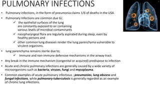
  • 1. PULMONARY INFECTIONS • Pulmonary infections, in the form of pneumonia claims 1/6 of deaths in the USA. • Pulmonary infections are common due to;  the epithelial surfaces of the lung are constantly exposed to air containing various levels of microbial contaminants  nasopharyngeal flora are regularly aspirated during sleep, even by healthy persons and  other common lung diseases render the lung parenchyma vulnerable to virulent organisms. • lung parenchyma remains sterile due to;  Immune and non-immune defensive mechanisms in the airway tract. • Any break in the immune mechanism (congenital or acquired) predispose to infection • Acute and chronic pulmonary infections are generally caused by a wide variety of microorganisms such as bacteria, viruses, fungi and mycoplasma. • Common examples of acute pulmonary infectious ; pneumonias, lung abscess and fungal infections, while pulmonary tuberculosis is generally regarded as an example of chronic lung infections.
  • 2. Pneumonia • Pneumonia is defined as acute inflammation of the lung parenchyma distal to the terminal bronchioles. • ‘pneumonia’ and ‘pneumonitis’; used synonymously for inflammation of the lungs. • ‘consolidation’ (meaning solidification) is the term used for gross and radiologic appearance of the lungs in pneumonia.
  • 3. PATHOGENESIS. The microorganisms gain entry into the lungs by one of the following four routes: • Inhalation from the air • Aspiration from the nasopharynx or oropharynx • Haematogenous spread from a distant focus of infection • Direct spread from an adjoining site of infection The normal lung is free of bacteria due to lung defense mechanisms at different levels as: • Nasopharyngeal filtering, • Mucociliary action of the lower respiratory airways, t • Phagocytosing alveolar macrophages and • immunoglobulins. Failure of these mechanisms and presence of certain predisposing factors result in pneumonias.
  • 4. Predisposing conditions; • Altered consciousness; aspiration of oropharyngeal contents in cases of disturbed conscious level as in coma, cranial trauma, seizures, cerebrovascular accidents, drug overdose, alcoholism etc. • Depressed cough and glottic reflexes in old age, pain from trauma or thoracoabdominal surgery, neuromuscular disease, weakness due to malnutrition, kyphoscoliosis, severe obstructive pulmonary diseases, endotracheal intubation and tracheostomy. • Impaired mucociliary transport as in cigarette smoking, viral respiratory infections, immotile cilia syndrome, inhalation of hot or corrosive gases and old age.
  • 5. Continue … • Impaired alveolar macrophage function; by cigarette smoke, hypoxia, starvation, anaemia, pulmonary oedema and viral respiratory infections • Endobronchial obstruction; tumour, foreign body, cystic fibrosis and chronic bronchitis. • Leucocyte dysfunctions; congenital and acquired immunodeficiencies
  • 6. CLASSIFICATION Anatomical classification: • Lobar pneumonia • Bronchopneumonia (or Lobular pneumonia) • Interstitial pneumonia Etiologic classification: • Bacterial pneumonia • Viral pneumonia • Pneumonias from other etiologies. Usually a combined approach of etiologic and morphologic classification is followed.
  • 7. BACTERIAL PNEUMONIA • the most common cause of pneumonia or consolidation of one or both the lungs.  lobar pneumonia and  broncho-(lobular-) pneumonia,  confluent pneumonia; both lobar and lobular involving larger (confluent) areas in both the lungs irregularly – some consider it as a variant of bronchopneumonia
  • 8. Lobar Pneumonia • an acute bacterial infection of a part of a lobe, the entire lobe, or even two lobes of one or both the lungs. • Based on the etiologic agent, lobar pneumonia can be classified:  Pneumococcal pneumonia; more than 90% of lobar pneumonia is caused by treptococcus pneumonia; majority of cases is community-acquired infection (3-S. pneumoniae is more virulent).  Staphylococcal pneumonia; by haematogenous spread of infection from another focus or after viral infections.  Streptococcal pneumonia; β-haemolytic streptococci rarely cause pneumonia as in children, after measles or influenza, in severely debilitated elderly patients and in diabetics.  Pneumonia by gram-negative aerobic bacteria; Less common, this group includes Haemophilus influenzae, Klebsiella pneumonia, Pseudomonas aeruginosa, Proteus and Escherichia coli. common in children below 3 years of age after a preceding viral infection.
  • 9. Pathologic phases: • Stage of congestion (initial phase); early acute inflammatory response to bacterial infection and lasts for 1 to 2 days, Grossly enlarged, heavy, dark red and congested area, Cut surface exudes blood-stained frothy fluid. Histologically, dilation/congestion of capillaries around the alveolar wall, pale eosinophilic oedema in the alveolar space, few RBCs and neutrophils in the intra-alveolar fluid and numerous bacteria demonstrated in the alveolar fluid by Gram’s staining. • Red hepatisation (early consolidation); lasts for 2 to 4 days, Grossly red, firm and consolidated, the cut surface of the involved lobe is airless, red-pink, dry, granular and has liver-like consistency and accompanied by serofibrinous pleurisy. Histologically The oedema is replaced by strands of fibrin, marked cellular exudate of neutrophils and extravasation of red cells, many neutrophils show ingested bacteria and the alveolar septa are less prominent than in the first stage due to cellular exudation.
  • 10.
  • 11. • Grey hepatisation (late consolidation); lasts for 4 to 8 days, Grossly is a firm and heavy, the cut surface is dry, granular and grey in appearance with liver-like consistency, from red to grey change spreads from hilum to prephery, and fibrinous pleurisy is prominent. Histologically the fibrin strands are dense and more numerous, the cellular exudate of neutrophils is reduced due to disintegration of many inflammatory cells as evidenced by their pyknotic nuclei, the red cells are also fewer, the macrophages begin to appear in the exudate, The cellular exudate is often separated from the septal walls by a thin clear space, the organisms are less numerous and appear as degenerated forms. • Resolution; begins by 8th to 9th day, if not treated, and is completed in 1 to 3 weeks, resolution proceeds in a progressive manner. Grossly the solid fibrinous constituent is liquefied by enzymatic action eventually restoring the normal aeration, the process of softening begins centrally to the periphery, the cut surface is grey-red or dirty brown, the pleura may show resolution or undergo organisation fibrous obliteration of pleural cavity. these stages seen in untreated cases and less common nowadays due to early antibiotic therapy and improved medical care. The sequence of pathologic changes described below represents the inflammatory response of lungs in bacterial infection.
  • 12. COMPLICATIONS Because of antibiotics serious complications of lobar pneumonia are uncommon, but in neglected cases and in those with impaired immunologic defenses these complications can occur: • Organisation; In about 3% ingrowth of fibroblasts from the alveolar septa results in fibrosed, tough, airless leathery lung tissue –cornification. • Pleural effusion; About 5% inflammation of pleura and effusion occurs which may resolve or organize to fibrous adhesion. • Empyema; in a small portion may develop encysted pus in the pleural cavity termed empyema. • Lung abscess; rarely, especially when there is secondary infection by other organisms lung abscess is formed. • Metastatic infection; Occasionally infection may extend into the pericardium and the heart causing purulent pericarditis, bacterial endocarditis and myocarditis. other rare metastasis are otitis media, mastoiditis, meningitis, brain abscess and purulent arthritis
  • 13. CLINICAL FEATURES Classically; • the onset of lobar pneumonia is sudden. • chills, fever, malaise, pleuritic chest pain, dyspnea, productive cough which may be mucoid, purulent or even bloody. • Signs include fever, tachycardia, and tachypnoea, and sometimes cyanosis and if severe, hypoxaemia. • Lab test; marked neutrophilic leukocytosis, Blood culture is +ve in about 30% of cases. • Radiograph; reveal consolidation. • The response to antibiotics is usually rapid with clinical improvement in 48 to 72 hours.
  • 14. Bronchopneumonia • Bronchopneumonia is infection of the terminal bronchioles that extends into the surrounding alveoli resulting in patchy consolidation of the lung. • It is is particularly frequent at the extremes of life as a terminal event in chronic debilitating disease or secondary to viral infection as influenza, measles • ETIOLOGY; staphylococci, streptococci, pneumococci, Klebsiella pneumoniae, Haemophilus influenzae, and gram- negative bacilli like Pseudomonas and coliform bacteria.
  • 15. Morphologically • bronchopneumonia is identified by patchy areas of red or grey consolidation affecting one or more lobes, usually bilateral, involving lower zones, • Histologically; Acute bronchiolitis, Suppurative exudate mainly neutrophils, Thickening of the alveolar septa and Less involved alveoli contain oedema fluid. COMPLICATIONS: • The complication of lobar pneumonia can also occur in bronchopneumonia, However complete resolution of latter is uncommon. • Generally some degree of bronchiolar destruction resulting in foci of fibrosis that may eventually cause bronchiectasis.
  • 16. CLINICAL FEATURES • The patients are are generally infants or elderly individuals. • There may be history of preceding bed-ridden illness, chronic debility, aspiration of gastric contents or upper respiratory infection. • Initially there are features of acute bronchitis, but later signs and symptoms similar to lobar pneumonia. • Blood examination usually shows a neutrophilia. • Chest radiograph shows mottled, focal opacities in both the lungs, chiefly in the lower zones.
  • 17.
  • 18. VIRAL AND MYCOPLASMAL PNEUMONIA (interstitial pneumonitis ) • is characterised by patchy inflammatory changes, mainly confined to interstitial tissue of the lungs, without alveolar exudate. • it is also called primary atypical pneumonia due to the absence of alveolar exudate commonly present in other pneumonias. • It may occur in all ages and most of the cases are mild and transient but in cases it may be severe and fulminant. • In most cases, the infection is confined to the upper respiratory tract presenting as common cold but occasionally extends lower down and involve the interstitium of the lungs. • Malnutrition, chronic debilitating diseases and alcoholism predisposes to atypical pneumonia.
  • 19. ETIOLOGY • Interstitial pneumonitis is caused by a wide variety of agents, but commonly ;  respiratory syncytial virus (RSV).  Mycoplasma pneumoniae .  Other viruses; influenza & parainfluenza viruses, adenoviruses, rhinoviruses, coxsackieviruses and cytomegaloviruses (CMV).
  • 20. Pathologic features • depending on severity, the involvement can be patchy or massive and widespread consolidation of one lung or both. • The lungs become heavy, congested and subcrepitant. • The pleural reaction is usually infrequent and mild. Histologically; • Interstitial inflammation; alveolar wall thickening (oedema and congestion), macrophages, lymphocytes and some plasma cells. • Necrotising bronchiolitis; foci of epithelial necrosis, and mononuclear infiltrate in the walls and lumina. • Reactive changes; The lining epithelial cells proliferate in the presence of virus forming multinucleate giant cells and syncytia in the bronchiolar and alveolar walls, viral inclusions are found in CMV pneumonitis. • Alveolar change; in severe cases; oedema, fibrin scanty inflammatory exudate, coating of alveolar walls by pink, hyaline membrane as RDS – alveolar changes usually occurs with bacterial superinfection.
  • 21. COMPLICATIONS • superimposed bacterial infection and its complications are the major complication of interstitial pneumonitis. • In severe cases, interstitial fibrosis and permanent damage may occur. • Otherwise, most cases recover completely. CLINICAL FEATURES • upper respiratory symptoms • fever, headache and muscle-aches. • Dry, hacking, non-productive cough with retrosternal burning due to tracheitis and bronchitis appears few days later. • Blood film shows neutrophilia, • Radiograph may show patchy or diffuse consolidation.
  • 22. OTHER TYPES OF PNEUMONIAS Pneumocystis carinii Pneumonia • This pneumonia is caused by protozoan widespread in the environment as an opportunistic in neonates and immunosuppressed people (HIV, immunosuppressive therapy). • Pathologically;  the affected parts of the lung are consolidated, dry and grey.  Interstitial pneumonitis with thickening and mononuclear infiltration.  Alveolar lumina contain pink frothy fluid containing the organisms.  No significant inflammatory exudate is seen in the air spaces. • Clinically;  A rapid onset dyspnoea, tachycardia, cyanosis and non- productive cough.  If not treated, it causes death in 1 to 2 weeks.  Radiograph shows diffuse alveolar and interstitial infiltrate.
  • 23. Legionella Pneumonia • An epidemic illness caused by gram-negative bacilli, Legionella pneumophila which thrives in aquatic environment. • The epidemic occurs in summer by pathogen spread through contaminated drinking water or in air conditionings. • impaired defense and smoking play important roles. • Pathologically;  Grossly, widespread bronchopneumonia or entire lung consolidation and pleural effusion is frequently present.  Histologically, Intra-alveolar exudate, initially neutrophils and later macrophages dominate, alveolar septa hyperplasia and vessel thrombosis, immunoflourocent technique may reveals organism in the macrophage. • Clinically;  begins with malaise, headache and muscle-aches followed by high fever, chills, cough and tachypnoea.  due to bacteraemia and include abdominal pain, watery diarrhoea, proteinuria and mild hepatic dysfunction.
  • 24. Aspiration Pneumonia • Results from inhalation of different agents into the lungs. • Food, gastric contents, foreign body and infected material from oral cavity cause AP. • unconsciousness, drunkenness, bulbar palsy, in premature infants and tracheo-oesophageal fistula predispose to AP. • The patients may die immediately from asphyxiation or laryngospasm without developing pneumonia. • Pathologically;  Sterile foreign matter can cause chemical pneumonitis.  Chemical AP is characterised by hemorrhagic pulmonary oedema with presence of particles in the bronchioles and clinically develops; rapid cyanosis, dyspnea bloody sputum, shock and likely dies due to cardiac failure.  Non-sterile aspirate causing widespread bronchopneumonia presents with areas of necrosis and suppuration.  A granulomatous reaction with foreign body giant cells may surround the aspirated vegetable matter.
  • 25. Hypostatic Pneumonia • When collection of fluid and secretions accumulate in lung’s dependent parts –basal zone and posterior part, of bed-ridden patient predisposes to bacterial pneumonia. • Hypostatic pneumonia is a common terminal event in the old, feeble, comatose patients.
  • 26. LUNG ABSCESS • A localized area of necrosis of lung tissue with suppuration. Primary lung abscess; develops in an otherwise normal lung and is usually due to aspiration of infected material. Secondary lung abscess; develops as a complication of other lung disease or from another site. ETIOPATHOGENESIS • streptococci, staphylococci and various gram-negatives are commonly isolated microorganisms. • The pathogens are introduced into the lungs through;  Aspiration of infected foreign material; food, decaying teeth, gastric contents, infected gingivae in favorable circumstances.  Preceding bacterial infection; abscess forms distal to an obstructed bronchus as tumour, foreign body or impacted mucus.  Septic embolism; from pyaemia, thrombophlebitis or from vegetative bacterial endocarditis lodges in pulmonary circulation.  Miscellaneous; from infected infarction, with Entamoeba histolytica, trauma, direct extension from neighboring sites.
  • 27.
  • 28. Continue.. • The abscess due to aspiration is more likely in right lung –lower part of the right upper lobe or apex of right lower lobe, and frequently single. • Abscesses of preceding pneumonia complication or septic and pyaemic origin are often multiple and scattered throughout the lung. • Grossly, the abscess is variable size of few millimeters to cavities of 5 to 6 cm in diameter which contains exudate, acutely surrounded by pneumonia with ragged edges then develops fibrous wall. • Histologically, the cavity of destructed suppurative parenchyma is surrounded by inflammatory cell infiltrates, and chronically a wall of firbocollagen is formed. CLINICAL FEATURES • The clinical manifestations are;  Fever, malaise, loss of weight.  Cough, purulent expectoration and haemoptysis in half the cases.  Clubbing of the fingers and toes appears in about 20% of patients.  Secondary amyloidosis may occur in chronic long-standing cases.
  • 29. FUNGAL INFECTIONS OF LUNG • Fungal infections in healthy people are rarely serious but in immunosuppressed individuals it may prove fatal. Aspergillosis; • the most common infection caused by Aspergillus fumigatus which grows best in cool and wet climate, • It may result in allergic bronchopulmonary aspergillosis, Aspergilloma and necrotising bronchitis. • Immunocompromised persons develop more serious manifestations. • Extensive haematogenous spread may result in widespread changes in lung tissue due to arterial occlusion, thrombosis and infarction. • pulmonary aspergillosis may occur within preexisting pulmonary cavities or in bronchiectasis as fungal ball. • Microscopically, the fungus may appear as a tangled mass within the cavity, thin septate hyphae with dichotomous branching at acute angles which stain positive for fungal stains such as PAS and silver impregnation technique, the wall of the cavity shows chronic inflammatory cells.
  • 30.
  • 31. Mucormycosis/phycomycosis • Caused by Mucor and Rhizopus, similar way as in aspergillosis, • The pulmonary lesions are common in patients of DKA. • Distinguished by its broad, non-parallel, nonseptate hyphae which branch at an obtuse angle. • More often angioinvasive, and disseminates; so more destructive than aspergillosis. Candidiasis • Caused by Candida albicans which is a normal commensal in oral cavity, gut and vagina but attains pathologic form in immunocompromised host. • Angioinvasive growth of the organism may occur in the airways • Caused by Blastomyces dermatitidis. • The lesions result from inhalation of spores in the ground. • may present as Ghon’s complex-like lesion, as a pneumonic consolidation, and as multiple skin nodules. • Uncommon condition. Blastomycosis
  • 32. Histoplasmosis • Caused by oval organism, Histoplasma capsulatum, by inhalation of infected dust or bird droppings. • remain asymptomatic or may produce lesions similar to the Ghon’s complex. Cryptococcosis • caused by Cryptococcus neoformans which is round yeast having a halo around it due to shrinkage in tissue sections. • The infection occurs from infection by inhalation of pigeon droppings. • The lesions in the body may range from a small parenchymal granuloma in the lung to cryptococcal meningitis. • caused by Coccidioides immitis which are spherical spores, • The infection in human beings is acquired by close contact with infected dogs. • The lesions consist of peripheral parenchymal granuloma in the lung. Coccidioidomycosis