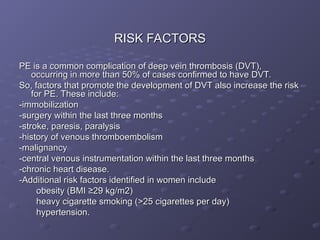
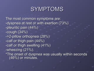
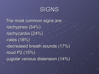
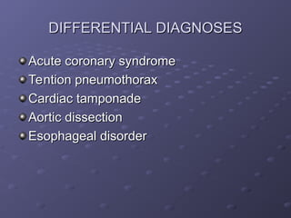
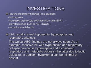
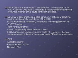
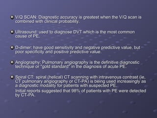
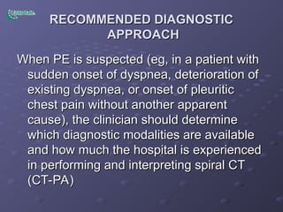
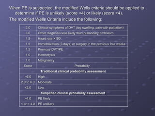
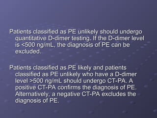
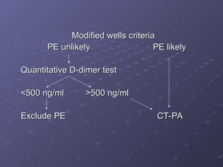
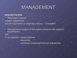
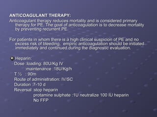
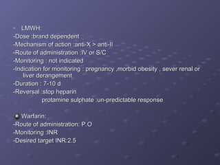
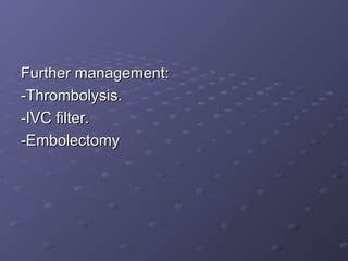
PULMONARY EMBOLISM
PULMONARY EMBOLISM
PREPARED BY:
PREPARED BY:
DR. IBRAHIM AYOUB
DR. IBRAHIM AYOUB
DR. SUHAIL KHOJAH
DR. SUHAIL KHOJAH
INTRODUCTION
INTRODUCTION
Acute pulmonary embolism (PE) is a common
and often fatal disease. Mortality can be reduced
from 30% to up to 2-8% by prompt diagnosis
and therapy.
and therapy.
Unfortunately, the clinical presentation of PE is
variable and nonspecific, making accurate
diagnosis difficult.
CLASSIFICATION
CLASSIFICATION
PE can be classified as
PE can be classified as acute
acute or
or chronic
chronic
-Patients with acute PE typically develop
symptoms and signs immediately after
obstruction of pulmonary vessels.
-In contrast, patients with chronic PE tend to
develop slowly progressive dyspnea over a
period of years due to pulmonary hypertension.
Acute PE can be further classified as
Acute PE can be further classified as massive
massive or
or submassive
submassive:
:
-Massive PE causes hypotension, defined as:
a systolic blood pressure <90 mmHg
or
or
a drop in systolic blood pressure of ≥40 mmHg from
baseline for a period >15 minutes.
It should be suspected anytime there is hypotension
accompanied by an elevated central venous pressure (or
neck vein distension), which is not otherwise explained by
acute myocardial infarction, tension pneumothorax,
pericardial tamponade, or a new arrhythmia.
-All acute PE not meeting the definition of massive PE are
considered submassive PE.
RISK FACTORS
RISK FACTORS
PE is a common complication of deep vein thrombosis (DVT),
occurring in more than 50% of cases confirmed to have DVT.
So, factors that promote the development of DVT also increase the risk
for PE. These include:
-immobilization
-immobilization
-surgery within the last three months
-stroke, paresis, paralysis
-history of venous thromboembolism
-malignancy
-malignancy
-central venous instrumentation within the last three months
-chronic heart disease.
-Additional risk factors identified in women include
obesity (BMI ≥29 kg/m2)
heavy cigarette smoking (>25 cigarettes per day)
hypertension.
hypertension.
SYMPTOMS
SYMPTOMS
The most common symptoms are:
-dyspnea at rest or with exertion (73%)
-pleuritic pain (44%)
-cough (34%)
-cough (34%)
->2-pillow orthopnea (28%)
-calf or thigh pain (44%)
-calf or thigh swelling (41%)
-wheezing (21%).
-wheezing (21%).
*The onset of dyspnea was usually within seconds
(46%) or minutes.
(46%) or minutes.
SIGNS
SIGNS
The most common signs are:
-tachypnea (54%)
-tachypnea (54%)
-tachycardia (24%)
-tachycardia (24%)
-rales (18%)
-rales (18%)
-decreased breath sounds (17%)
-loud P2 (15%)
-loud P2 (15%)
-jugular venous distension (14%)
DIFFERENTIAL DIAGNOSES
Acute coronary syndrome
Tention pneumothorax
Cardiac tamponade
Cardiac tamponade
Aortic dissection
Aortic dissection
Esophageal disorder
Esophageal disorder
INVESTIGATIONS
INVESTIGATIONS
Routine laboratory findings (non-specific):
-leukocytosis
-leukocytosis
-increased erythrocyte sedimentation rate (ESR)
-elevated serum LDH or AST (SGOT)
-normal serum bilirubin
ABG:
ABG: usually reveal hypoxemia, hypocapnia, and
usually reveal hypoxemia, hypocapnia, and
respiratory alkalosis.
The typical ABG findings are not always seen. As an
example, massive PE with hypotension and respiratory
collapse can cause hypercapnia and a combined
respiratory and metabolic acidosis (the latter due to lactic
acidosis). In addition, hypoxemia can be minimal or
absent.
absent.
TROPONIN: Serum troponin I and troponin T are elevated in 30-
50% of patients who have a moderate to large pulmonary embolism.
presumed mechanism is acute right heart overload.
ECG: ECG abnormalities are also common in patients without PE,
limiting the diagnostic usefulness of the ECG.
ECG abnormalities historically considered to be suggestive of PE
-S1Q3T3 pattern
-S1Q3T3 pattern
-right ventricular strain
-new incomplete right bundle branch block
ECG changes are infrequent during acute PE. However, they are
common among patients with massive acute PE and cor pulmonale.
CXR:
CXR:
-Atelectasis (69%)
-Atelectasis (69%)
-Pleural effusion (47%)
-Normal (12%)
-Normal (12%)
V/Q SCAN: Diagnostic accuracy is greatest when the V/Q scan is
combined with clinical probability.
Ultrasound: used to diagnose DVT which is the most common
cause of PE.
cause of PE.
D-dimer: have good sensitivity and negative predictive value, but
poor specificity and positive predictive value.
Angiography: Pulmonary angiography is the definitive diagnostic
technique or "gold standard" in the diagnosis of acute PE.
Spiral CT: spiral (helical) CT scanning with intravenous contrast (ie,
CT pulmonary angiography or CT-PA) is being used increasingly as
a diagnostic modality for patients with suspected PE.
Initial reports suggested that 98% of patients with PE were detected
by CT-PA.
by CT-PA.
RECOMMENDED DIAGNOSTIC
APPROACH
APPROACH
When PE is suspected (eg, in a patient with
sudden onset of dyspnea, deterioration of
existing dyspnea, or onset of pleuritic
chest pain without another apparent
cause), the clinician should determine
which diagnostic modalities are available
and how much the hospital is experienced
in performing and interpreting spiral CT
(CT-PA)
(CT-PA)
When PE is suspected, the modified Wells criteria should be applied to
determine if PE is unlikely (score <4) or likely (score >4).
The modified Wells Criteria include the following:
Clinical symptoms of DVT (leg swelling, pain with palpation)
3.0
3.0
Other diagnosis less likely than pulmonary embolism
3.0
3.0
Heart rate >100
Heart rate >100
1.5
1.5
Immobilization (3 days) or surgery in the previous four weeks
1.5
1.5
Previous DVT/PE
Previous DVT/PE
1.5
1.5
Hemoptysis
Hemoptysis
1.0
1.0
Malignancy
Malignancy
1.0
1.0
Probability
Probability
Score
Score
Traditional clinical probability assessment
High
High
>6.0
>6.0
Moderate
Moderate
2.0 to 6.0
2.0 to 6.0
Low
Low
<2.0
<2.0
Simplified clinical probability assessment
PE likely
PE likely
>4.0
>4.0
PE unlikely
PE unlikely
< or = 4.0
< or = 4.0
Patients classified as PE unlikely should undergo
quantitative D-dimer testing. If the D-dimer level
is <500 ng/mL, the diagnosis of PE can be
excluded.
excluded.
Patients classified as PE likely and patients
classified as PE unlikely who have a D-dimer
level >500 ng/mL should undergo CT-PA. A
positive CT-PA confirms the diagnosis of PE.
Alternatively, a negative CT-PA excludes the
diagnosis of PE.
diagnosis of PE.
Modified wells criteria
PE unlikely PE likely
Quantitative D-dimer test
<500 ng/ml >500 ng/ml
Exclude PE CT-PA
Exclude PE CT-PA
In case you are working in CT inexperienced hospital or the
patieng can’t undergo CP-PA (e.g. renal insufficiency or
morbid obecity), a ventilation-perfusion (V/Q) scan is then
performed, with the following combinations of outcomes
possible:
possible:
-Normal V/Q scan plus any clinical probability excludes PE.
-Low probability V/Q scan plus low clinical probability
excludes PE.
excludes PE.
-High probability V/Q scan plus high clinical probability
confirms PE.
confirms PE.
Any other combination of V/Q scan result plus clinical
probability should prompt either a pulmonary angiogram
or serial lower extremity venous ultrasound exams.
A reasonable alternative approach for patients with a low or
intermediate clinical probability of PE is to obtain a D-
dimer. A negative D-Dimer by the SimpliRed assay
excludes PE.
excludes PE.
MANAGEMENT
MANAGEMENT
RESUSCITATION
RESUSCITATION:
:
Respiratory support:
-oxygen supplement
-oxygen supplement
-severe hypoxemia or respiratory failure Intubation
Hemodynamic support (If the patient presents with systemic
hypotension):
hypotension):
-IVF
-IVF
-if not resolved: -norepinephrine
-dopamine
-dopamine
-combined norepinephrine and dobutamine
ANTICOAGULANT THERAPY:
Anticoagulant therapy reduces mortality and is considered primary
therapy for PE. The goal of anticoagulation is to decrease mortality
by preventing recurrent PE.
For patients in whom there is a high clinical suspicion of PE and no
excess risk of bleeding, empiric anticoagulation should be initiated
immediately and continued during the diagnostic evaluation.
Heparin:
Heparin:
Dose :loading: 80U/Kg IV
:maintenance :18U/Kg/h
T ½ : 90m
T ½ : 90m
Route of administration: IV/SC
Duration :7-10 d
Duration :7-10 d
Reversal :stop heparin
protamine sulphate :1U neutralize 100 IU heparin
No FFP
No FFP
• LMWH:
LMWH:
-Dose :brand dependent
-Mechanism of action :anti-X > anti-II
-Route of administration :IV or S/C
-Monitoring : not indicated
-Indication for monitoring : pregnancy ,morbid obesity , sever renal or
liver derangement
liver derangement
-Duration : 7-10 d
-Duration : 7-10 d
-Reversal :stop heparin
protamine sulphate :un-predictable response
Warfarin:
Warfarin:
-Route of administration: P.O
-Monitoring :INR
-Monitoring :INR
-Desired target INR:2.5
Further management:
Further management:
-Thrombolysis.
-Thrombolysis.
-IVC filter.
-IVC filter.
-Embolectomy
-Embolectomy
Pulmonary embolism for tb chest students

Pulmonary embolism for tb chest students

  • 1.
    PULMONARY EMBOLISM PULMONARY EMBOLISM PREPAREDBY: PREPARED BY: DR. IBRAHIM AYOUB DR. IBRAHIM AYOUB DR. SUHAIL KHOJAH DR. SUHAIL KHOJAH
  • 2.
    INTRODUCTION INTRODUCTION Acute pulmonary embolism(PE) is a common Acute pulmonary embolism (PE) is a common and often fatal disease. Mortality can be reduced and often fatal disease. Mortality can be reduced from 30% to up to 2-8% by prompt diagnosis from 30% to up to 2-8% by prompt diagnosis and therapy. and therapy. Unfortunately, the clinical presentation of PE is Unfortunately, the clinical presentation of PE is variable and nonspecific, making accurate variable and nonspecific, making accurate diagnosis difficult. diagnosis difficult.
  • 3.
    CLASSIFICATION CLASSIFICATION PE can beclassified as PE can be classified as acute acute or or chronic chronic -Patients with acute PE typically develop -Patients with acute PE typically develop symptoms and signs immediately after symptoms and signs immediately after obstruction of pulmonary vessels. obstruction of pulmonary vessels. -In contrast, patients with chronic PE tend to -In contrast, patients with chronic PE tend to develop slowly progressive dyspnea over a develop slowly progressive dyspnea over a period of years due to pulmonary hypertension. period of years due to pulmonary hypertension.
  • 4.
    Acute PE canbe further classified as Acute PE can be further classified as massive massive or or submassive submassive: : -Massive PE causes hypotension, defined as: -Massive PE causes hypotension, defined as: a systolic blood pressure <90 mmHg a systolic blood pressure <90 mmHg or or a drop in systolic blood pressure of ≥40 mmHg from a drop in systolic blood pressure of ≥40 mmHg from baseline for a period >15 minutes. baseline for a period >15 minutes. It should be suspected anytime there is hypotension It should be suspected anytime there is hypotension accompanied by an elevated central venous pressure (or accompanied by an elevated central venous pressure (or neck vein distension), which is not otherwise explained by neck vein distension), which is not otherwise explained by acute myocardial infarction, tension pneumothorax, acute myocardial infarction, tension pneumothorax, pericardial tamponade, or a new arrhythmia. pericardial tamponade, or a new arrhythmia. -All acute PE not meeting the definition of massive PE are -All acute PE not meeting the definition of massive PE are considered submassive PE. considered submassive PE.
  • 5.
    RISK FACTORS RISK FACTORS PEis a common complication of deep vein thrombosis (DVT), PE is a common complication of deep vein thrombosis (DVT), occurring in more than 50% of cases confirmed to have DVT. occurring in more than 50% of cases confirmed to have DVT. So, factors that promote the development of DVT also increase the risk So, factors that promote the development of DVT also increase the risk for PE. These include: for PE. These include: -immobilization -immobilization -surgery within the last three months -surgery within the last three months -stroke, paresis, paralysis -stroke, paresis, paralysis -history of venous thromboembolism -history of venous thromboembolism -malignancy -malignancy -central venous instrumentation within the last three months -central venous instrumentation within the last three months -chronic heart disease. -chronic heart disease. -Additional risk factors identified in women include -Additional risk factors identified in women include obesity (BMI ≥29 kg/m2) obesity (BMI ≥29 kg/m2) heavy cigarette smoking (>25 cigarettes per day) heavy cigarette smoking (>25 cigarettes per day) hypertension. hypertension.
  • 6.
    SYMPTOMS SYMPTOMS The most commonsymptoms are: The most common symptoms are: -dyspnea at rest or with exertion (73%) -dyspnea at rest or with exertion (73%) -pleuritic pain (44%) -pleuritic pain (44%) -cough (34%) -cough (34%) ->2-pillow orthopnea (28%) ->2-pillow orthopnea (28%) -calf or thigh pain (44%) -calf or thigh pain (44%) -calf or thigh swelling (41%) -calf or thigh swelling (41%) -wheezing (21%). -wheezing (21%). *The onset of dyspnea was usually within seconds *The onset of dyspnea was usually within seconds (46%) or minutes. (46%) or minutes.
  • 7.
    SIGNS SIGNS The most commonsigns are: The most common signs are: -tachypnea (54%) -tachypnea (54%) -tachycardia (24%) -tachycardia (24%) -rales (18%) -rales (18%) -decreased breath sounds (17%) -decreased breath sounds (17%) -loud P2 (15%) -loud P2 (15%) -jugular venous distension (14%) -jugular venous distension (14%)
  • 8.
    DIFFERENTIAL DIAGNOSES DIFFERENTIAL DIAGNOSES Acutecoronary syndrome Acute coronary syndrome Tention pneumothorax Tention pneumothorax Cardiac tamponade Cardiac tamponade Aortic dissection Aortic dissection Esophageal disorder Esophageal disorder
  • 9.
    INVESTIGATIONS INVESTIGATIONS Routine laboratory findings(non-specific): Routine laboratory findings (non-specific): -leukocytosis -leukocytosis -increased erythrocyte sedimentation rate (ESR) -increased erythrocyte sedimentation rate (ESR) -elevated serum LDH or AST (SGOT) -elevated serum LDH or AST (SGOT) -normal serum bilirubin -normal serum bilirubin ABG: ABG: usually reveal hypoxemia, hypocapnia, and usually reveal hypoxemia, hypocapnia, and respiratory alkalosis. respiratory alkalosis. The typical ABG findings are not always seen. As an The typical ABG findings are not always seen. As an example, massive PE with hypotension and respiratory example, massive PE with hypotension and respiratory collapse can cause hypercapnia and a combined collapse can cause hypercapnia and a combined respiratory and metabolic acidosis (the latter due to lactic respiratory and metabolic acidosis (the latter due to lactic acidosis). In addition, hypoxemia can be minimal or acidosis). In addition, hypoxemia can be minimal or absent. absent.
  • 10.
    TROPONIN: Serum troponinI and troponin T are elevated in 30- TROPONIN: Serum troponin I and troponin T are elevated in 30- 50% of patients who have a moderate to large pulmonary embolism. 50% of patients who have a moderate to large pulmonary embolism. presumed mechanism is acute right heart overload. presumed mechanism is acute right heart overload. ECG: ECG abnormalities are also common in patients without PE, ECG: ECG abnormalities are also common in patients without PE, limiting the diagnostic usefulness of the ECG. limiting the diagnostic usefulness of the ECG. ECG abnormalities historically considered to be suggestive of PE ECG abnormalities historically considered to be suggestive of PE -S1Q3T3 pattern -S1Q3T3 pattern -right ventricular strain -right ventricular strain -new incomplete right bundle branch block -new incomplete right bundle branch block ECG changes are infrequent during acute PE. However, they are ECG changes are infrequent during acute PE. However, they are common among patients with massive acute PE and cor pulmonale. common among patients with massive acute PE and cor pulmonale. CXR: CXR: -Atelectasis (69%) -Atelectasis (69%) -Pleural effusion (47%) -Pleural effusion (47%) -Normal (12%) -Normal (12%)
  • 11.
    V/Q SCAN: Diagnosticaccuracy is greatest when the V/Q scan is V/Q SCAN: Diagnostic accuracy is greatest when the V/Q scan is combined with clinical probability. combined with clinical probability. Ultrasound: used to diagnose DVT which is the most common Ultrasound: used to diagnose DVT which is the most common cause of PE. cause of PE. D-dimer: have good sensitivity and negative predictive value, but D-dimer: have good sensitivity and negative predictive value, but poor specificity and positive predictive value. poor specificity and positive predictive value. Angiography: Pulmonary angiography is the definitive diagnostic Angiography: Pulmonary angiography is the definitive diagnostic technique or "gold standard" in the diagnosis of acute PE. technique or "gold standard" in the diagnosis of acute PE. Spiral CT: spiral (helical) CT scanning with intravenous contrast (ie, Spiral CT: spiral (helical) CT scanning with intravenous contrast (ie, CT pulmonary angiography or CT-PA) is being used increasingly as CT pulmonary angiography or CT-PA) is being used increasingly as a diagnostic modality for patients with suspected PE. a diagnostic modality for patients with suspected PE. Initial reports suggested that 98% of patients with PE were detected Initial reports suggested that 98% of patients with PE were detected by CT-PA. by CT-PA.
  • 12.
    RECOMMENDED DIAGNOSTIC RECOMMENDED DIAGNOSTIC APPROACH APPROACH WhenPE is suspected (eg, in a patient with When PE is suspected (eg, in a patient with sudden onset of dyspnea, deterioration of sudden onset of dyspnea, deterioration of existing dyspnea, or onset of pleuritic existing dyspnea, or onset of pleuritic chest pain without another apparent chest pain without another apparent cause), the clinician should determine cause), the clinician should determine which diagnostic modalities are available which diagnostic modalities are available and how much the hospital is experienced and how much the hospital is experienced in performing and interpreting spiral CT in performing and interpreting spiral CT (CT-PA) (CT-PA)
  • 13.
    When PE issuspected, the modified Wells criteria should be applied to When PE is suspected, the modified Wells criteria should be applied to determine if PE is unlikely (score <4) or likely (score >4). determine if PE is unlikely (score <4) or likely (score >4). The modified Wells Criteria include the following: The modified Wells Criteria include the following: Clinical symptoms of DVT (leg swelling, pain with palpation) Clinical symptoms of DVT (leg swelling, pain with palpation) 3.0 3.0 Other diagnosis less likely than pulmonary embolism Other diagnosis less likely than pulmonary embolism 3.0 3.0 Heart rate >100 Heart rate >100 1.5 1.5 Immobilization (3 days) or surgery in the previous four weeks Immobilization (3 days) or surgery in the previous four weeks 1.5 1.5 Previous DVT/PE Previous DVT/PE 1.5 1.5 Hemoptysis Hemoptysis 1.0 1.0 Malignancy Malignancy 1.0 1.0 Probability Probability Score Score Traditional clinical probability assessment Traditional clinical probability assessment High High >6.0 >6.0 Moderate Moderate 2.0 to 6.0 2.0 to 6.0 Low Low <2.0 <2.0 Simplified clinical probability assessment Simplified clinical probability assessment PE likely PE likely >4.0 >4.0 PE unlikely PE unlikely < or = 4.0 < or = 4.0
  • 14.
    Patients classified asPE unlikely should undergo Patients classified as PE unlikely should undergo quantitative D-dimer testing. If the D-dimer level quantitative D-dimer testing. If the D-dimer level is <500 ng/mL, the diagnosis of PE can be is <500 ng/mL, the diagnosis of PE can be excluded. excluded. Patients classified as PE likely and patients Patients classified as PE likely and patients classified as PE unlikely who have a D-dimer classified as PE unlikely who have a D-dimer level >500 ng/mL should undergo CT-PA. A level >500 ng/mL should undergo CT-PA. A positive CT-PA confirms the diagnosis of PE. positive CT-PA confirms the diagnosis of PE. Alternatively, a negative CT-PA excludes the Alternatively, a negative CT-PA excludes the diagnosis of PE. diagnosis of PE.
  • 15.
    Modified wells criteria Modifiedwells criteria PE unlikely PE likely PE unlikely PE likely Quantitative D-dimer test Quantitative D-dimer test <500 ng/ml >500 ng/ml <500 ng/ml >500 ng/ml Exclude PE CT-PA Exclude PE CT-PA
  • 16.
    In case youare working in CT inexperienced hospital or the In case you are working in CT inexperienced hospital or the patieng can’t undergo CP-PA (e.g. renal insufficiency or patieng can’t undergo CP-PA (e.g. renal insufficiency or morbid obecity), a ventilation-perfusion (V/Q) scan is then morbid obecity), a ventilation-perfusion (V/Q) scan is then performed, with the following combinations of outcomes performed, with the following combinations of outcomes possible: possible: -Normal V/Q scan plus any clinical probability excludes PE. -Normal V/Q scan plus any clinical probability excludes PE. -Low probability V/Q scan plus low clinical probability -Low probability V/Q scan plus low clinical probability excludes PE. excludes PE. -High probability V/Q scan plus high clinical probability -High probability V/Q scan plus high clinical probability confirms PE. confirms PE. Any other combination of V/Q scan result plus clinical Any other combination of V/Q scan result plus clinical probability should prompt either a pulmonary angiogram probability should prompt either a pulmonary angiogram or serial lower extremity venous ultrasound exams. or serial lower extremity venous ultrasound exams. A reasonable alternative approach for patients with a low or A reasonable alternative approach for patients with a low or intermediate clinical probability of PE is to obtain a D- intermediate clinical probability of PE is to obtain a D- dimer. A negative D-Dimer by the SimpliRed assay dimer. A negative D-Dimer by the SimpliRed assay excludes PE. excludes PE.
  • 17.
    MANAGEMENT MANAGEMENT RESUSCITATION RESUSCITATION: : Respiratory support: Respiratory support: -oxygensupplement -oxygen supplement -severe hypoxemia or respiratory failure Intubation -severe hypoxemia or respiratory failure Intubation Hemodynamic support (If the patient presents with systemic Hemodynamic support (If the patient presents with systemic hypotension): hypotension): -IVF -IVF -if not resolved: -norepinephrine -if not resolved: -norepinephrine -dopamine -dopamine -combined norepinephrine and dobutamine -combined norepinephrine and dobutamine
  • 18.
    ANTICOAGULANT THERAPY: ANTICOAGULANT THERAPY: Anticoagulanttherapy reduces mortality and is considered primary Anticoagulant therapy reduces mortality and is considered primary therapy for PE. The goal of anticoagulation is to decrease mortality therapy for PE. The goal of anticoagulation is to decrease mortality by preventing recurrent PE. by preventing recurrent PE. For patients in whom there is a high clinical suspicion of PE and no For patients in whom there is a high clinical suspicion of PE and no excess risk of bleeding, empiric anticoagulation should be initiated excess risk of bleeding, empiric anticoagulation should be initiated immediately and continued during the diagnostic evaluation. immediately and continued during the diagnostic evaluation. Heparin: Heparin: Dose :loading: 80U/Kg IV Dose :loading: 80U/Kg IV :maintenance :18U/Kg/h :maintenance :18U/Kg/h T ½ : 90m T ½ : 90m Route of administration: IV/SC Route of administration: IV/SC Duration :7-10 d Duration :7-10 d Reversal :stop heparin Reversal :stop heparin protamine sulphate :1U neutralize 100 IU heparin protamine sulphate :1U neutralize 100 IU heparin No FFP No FFP
  • 19.
    • LMWH: LMWH: -Dose :branddependent -Dose :brand dependent -Mechanism of action :anti-X > anti-II -Mechanism of action :anti-X > anti-II -Route of administration :IV or S/C -Route of administration :IV or S/C -Monitoring : not indicated -Monitoring : not indicated -Indication for monitoring : pregnancy ,morbid obesity , sever renal or -Indication for monitoring : pregnancy ,morbid obesity , sever renal or liver derangement liver derangement -Duration : 7-10 d -Duration : 7-10 d -Reversal :stop heparin -Reversal :stop heparin protamine sulphate :un-predictable response protamine sulphate :un-predictable response Warfarin: Warfarin: -Route of administration: P.O -Route of administration: P.O -Monitoring :INR -Monitoring :INR -Desired target INR:2.5 -Desired target INR:2.5
  • 20.