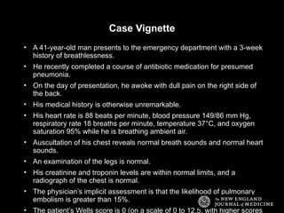
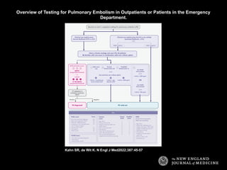
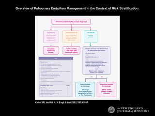
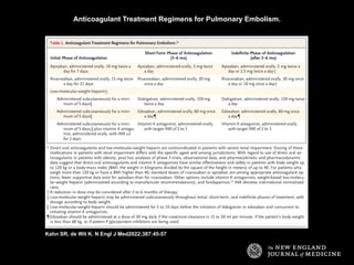
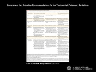
Clinical Practice
Pulmonary Embolism
Susan R. Kahn, M.D., and Kerstin de Wit, M.B., Ch.B., M.D.
N Engl J Med
Volume 387(1):45-57
July 7, 2022
Case Vignette
• A 41-year-old man presents to the emergency department with a 3-week
history of breathlessness.
• He recently completed a course of antibiotic medication for presumed
pneumonia.
• On the day of presentation, he awoke with dull pain on the right side of
the back.
• His medical history is otherwise unremarkable.
• His heart rate is 88 beats per minute, blood pressure 149/86 mm Hg,
respiratory rate 18 breaths per minute, temperature 37°C, and oxygen
saturation 95% while he is breathing ambient air.
• Auscultation of his chest reveals normal breath sounds and normal heart
sounds.
• An examination of the legs is normal.
• His creatinine and troponin levels are within normal limits, and a
radiograph of the chest is normal.
• The physician’s implicit assessment is that the likelihood of pulmonary
embolism is greater than 15%.
• The patient’s Wells score is 0 (on a scale of 0 to 12.5, with higher scores
Overview of Testing for Pulmonary Embolism in Outpatients or Patients in the Emergency
Department.
Kahn SR, de Wit K. N Engl J Med2022;387:45-57
Overview of Pulmonary Embolism Management in the Context of Risk Stratification.
Kahn SR, de Wit K. N Engl J Med2022;387:45-57
Anticoagulant Treatment Regimens for Pulmonary Embolism.
Kahn SR, de Wit K. N Engl J Med2022;387:45-57
Summary of Key Guideline Recommendations for the Treatment of Pulmonary Embolism.
Kahn SR, de Wit K. N Engl J Med2022;387:45-57
Conclusions and Recommendations
• The patient with breathlessness described in the vignette was estimated
to have greater than a 15% likelihood of pulmonary embolism.
• In the context of the patient’s low Wells score for pulmonary embolism,
<sc xmlns:xlink="http://www.w3.org/1999/xlink"
xmlns:mml="http://www.w3.org/1998/Math/MathML"
xmlns:xsi="http://www.w3.org/2001/XMLSchema-instance"
xmlns:oasis="http://www.niso.org/standards/z39-96/ns/oasis-exchange/
table">d</sc> -dimer testing was warranted to guide the need for
imaging; CT is indicated, given the <sc
xmlns:xlink="http://www.w3.org/1999/xlink"
xmlns:mml="http://www.w3.org/1998/Math/MathML"
xmlns:xsi="http://www.w3.org/2001/XMLSchema-instance"
xmlns:oasis="http://www.niso.org/standards/z39-96/ns/oasis-exchange/
table">d</sc> -dimer level of more than 1000 ng per milliliter.
• Under the presumption that the patient’s CT scan confirms pulmonary
embolism and shows normal right-ventricle dimensions, he would be
classified as having low-risk pulmonary embolism, given his normal
troponin level.
• Treatment with a direct oral anticoagulant should be started promptly,
and the patient should be given information about the pulmonary
Conclusions and Recommendations
• We would recommend that he undergo cancer screening appropriate for
his age and personal risk.
• After the patient receives 3 to 6 months of therapy with a direct oral
anticoagulant administered at a treatment-level dose, in the absence of
an increased bleeding risk and considering his preferences, we would
recommend switching to a low-dose direct oral anticoagulant on a long-
term basis for secondary prevention.

Pulmonary Embolism, classification and management.

  • 1.
    Clinical Practice Pulmonary Embolism SusanR. Kahn, M.D., and Kerstin de Wit, M.B., Ch.B., M.D. N Engl J Med Volume 387(1):45-57 July 7, 2022
  • 2.
    Case Vignette • A41-year-old man presents to the emergency department with a 3-week history of breathlessness. • He recently completed a course of antibiotic medication for presumed pneumonia. • On the day of presentation, he awoke with dull pain on the right side of the back. • His medical history is otherwise unremarkable. • His heart rate is 88 beats per minute, blood pressure 149/86 mm Hg, respiratory rate 18 breaths per minute, temperature 37°C, and oxygen saturation 95% while he is breathing ambient air. • Auscultation of his chest reveals normal breath sounds and normal heart sounds. • An examination of the legs is normal. • His creatinine and troponin levels are within normal limits, and a radiograph of the chest is normal. • The physician’s implicit assessment is that the likelihood of pulmonary embolism is greater than 15%. • The patient’s Wells score is 0 (on a scale of 0 to 12.5, with higher scores
  • 3.
    Overview of Testingfor Pulmonary Embolism in Outpatients or Patients in the Emergency Department. Kahn SR, de Wit K. N Engl J Med2022;387:45-57
  • 4.
    Overview of PulmonaryEmbolism Management in the Context of Risk Stratification. Kahn SR, de Wit K. N Engl J Med2022;387:45-57
  • 5.
    Anticoagulant Treatment Regimensfor Pulmonary Embolism. Kahn SR, de Wit K. N Engl J Med2022;387:45-57
  • 6.
    Summary of KeyGuideline Recommendations for the Treatment of Pulmonary Embolism. Kahn SR, de Wit K. N Engl J Med2022;387:45-57
  • 7.
    Conclusions and Recommendations •The patient with breathlessness described in the vignette was estimated to have greater than a 15% likelihood of pulmonary embolism. • In the context of the patient’s low Wells score for pulmonary embolism, <sc xmlns:xlink="http://www.w3.org/1999/xlink" xmlns:mml="http://www.w3.org/1998/Math/MathML" xmlns:xsi="http://www.w3.org/2001/XMLSchema-instance" xmlns:oasis="http://www.niso.org/standards/z39-96/ns/oasis-exchange/ table">d</sc> -dimer testing was warranted to guide the need for imaging; CT is indicated, given the <sc xmlns:xlink="http://www.w3.org/1999/xlink" xmlns:mml="http://www.w3.org/1998/Math/MathML" xmlns:xsi="http://www.w3.org/2001/XMLSchema-instance" xmlns:oasis="http://www.niso.org/standards/z39-96/ns/oasis-exchange/ table">d</sc> -dimer level of more than 1000 ng per milliliter. • Under the presumption that the patient’s CT scan confirms pulmonary embolism and shows normal right-ventricle dimensions, he would be classified as having low-risk pulmonary embolism, given his normal troponin level. • Treatment with a direct oral anticoagulant should be started promptly, and the patient should be given information about the pulmonary
  • 8.
    Conclusions and Recommendations •We would recommend that he undergo cancer screening appropriate for his age and personal risk. • After the patient receives 3 to 6 months of therapy with a direct oral anticoagulant administered at a treatment-level dose, in the absence of an increased bleeding risk and considering his preferences, we would recommend switching to a low-dose direct oral anticoagulant on a long- term basis for secondary prevention.

Editor's Notes

  • #3 Figure 1 Overview of Testing for Pulmonary Embolism in Outpatients or Patients in the Emergency Department. Physicians may use Pulmonary Embolism Rule-out Criteria (PERC) to rule out pulmonary embolism if their implicit sense suggests there is less than 15% probability that the patient has pulmonary embolism. Otherwise, physicians should use a D-dimer assay to rule out pulmonary embolism in patients who have a low structured clinical probability score (a Wells score of ≤4.0 on a scale of 0 to 12.5, a revised Geneva score of ≤10 on a scale ranging from 0 to 22, or a simplified Geneva score of ≤4 on a scale of 0 to 9; on all three scales, higher scores indicate a greater probability of pulmonary embolism) or should use the YEARS algorithm. Each circled number refers to a different clinical decision rule, and each circled letter to a distinct D-dimer strategy. Imaging can be avoided in patients with clinical probability scores at or below the given cutoff and D-dimer level below the given cutoff. Computed tomography (CT) and ventilation–perfusion single-photon-emission computed tomography (SPECT) are reserved for patients with a clinical probability score above the preset cutoff for the chosen score or a D-dimer at or above the preset cutoff for the chosen D-dimer option. The adjusted D-dimer thresholds have been validated for assays with a manufacturer-recommended cutoff of 500 ng per milliliter. DVT denotes deep-vein thrombosis.
  • #4 Figure 2 Overview of Pulmonary Embolism Management in the Context of Risk Stratification. At the time of diagnosis, pulmonary embolism should be stratified as low risk, intermediate risk, or high risk. Patients with high-risk pulmonary embolism should be assessed for immediate reperfusion interventions such as systemic thrombolysis. Patients with intermediate-risk pulmonary embolism should be carefully monitored and assessed for initiation of treatment with low-molecular-weight heparin (LMWH). Most patients have low-risk pulmonary embolism and can be assessed for outpatient anticoagulant therapy according to their Hestia score, score on the simplified Pulmonary Embolism Severity Index (PESI), or the physician’s implicit judgment. All patients discharged home would benefit from rapid, reliable outpatient follow-up. BNP denotes brain natriuretic peptide, DOAC direct oral anticoagulant, IV intravenous, and VKA vitamin K antagonist.
  • #5 Table 1 Anticoagulant Treatment Regimens for Pulmonary Embolism.
  • #6 Table 2 Summary of Key Guideline Recommendations for the Treatment of Pulmonary Embolism.