The document provides protocols and guidelines for the Department of Obstetrics including definitions, classifications, investigations, and management guidelines for various obstetric conditions. It covers protocols for pre-eclampsia and eclampsia, liver diseases in pregnancy, deep venous thrombosis in pregnancy, preterm labour, preterm PROM, breech presentation, APH, induction of labour, normal labour and delivery, PPH, umbilical cord prolapse, Rh prophylaxis, and GDM. The department aims to provide high quality, empathetic and research-based care through comprehensive training and by reviewing and creating protocols according to population needs.
A comprehensive guide to the management of hyperglycaemia in pregnancy aimed at the primary care physician and based on latest evidenced based criteria. Includes information from latest studies such as HAPO study and ACHOIS, and involves guidelines from the IADPSG, ADA, WHO and Malaysia.
Slide 1: Title Slide
Extrachromosomal Inheritance
Slide 2: Introduction to Extrachromosomal Inheritance
Definition: Extrachromosomal inheritance refers to the transmission of genetic material that is not found within the nucleus.
Key Components: Involves genes located in mitochondria, chloroplasts, and plasmids.
Slide 3: Mitochondrial Inheritance
Mitochondria: Organelles responsible for energy production.
Mitochondrial DNA (mtDNA): Circular DNA molecule found in mitochondria.
Inheritance Pattern: Maternally inherited, meaning it is passed from mothers to all their offspring.
Diseases: Examples include Leber’s hereditary optic neuropathy (LHON) and mitochondrial myopathy.
Slide 4: Chloroplast Inheritance
Chloroplasts: Organelles responsible for photosynthesis in plants.
Chloroplast DNA (cpDNA): Circular DNA molecule found in chloroplasts.
Inheritance Pattern: Often maternally inherited in most plants, but can vary in some species.
Examples: Variegation in plants, where leaf color patterns are determined by chloroplast DNA.
Slide 5: Plasmid Inheritance
Plasmids: Small, circular DNA molecules found in bacteria and some eukaryotes.
Features: Can carry antibiotic resistance genes and can be transferred between cells through processes like conjugation.
Significance: Important in biotechnology for gene cloning and genetic engineering.
Slide 6: Mechanisms of Extrachromosomal Inheritance
Non-Mendelian Patterns: Do not follow Mendel’s laws of inheritance.
Cytoplasmic Segregation: During cell division, organelles like mitochondria and chloroplasts are randomly distributed to daughter cells.
Heteroplasmy: Presence of more than one type of organellar genome within a cell, leading to variation in expression.
Slide 7: Examples of Extrachromosomal Inheritance
Four O’clock Plant (Mirabilis jalapa): Shows variegated leaves due to different cpDNA in leaf cells.
Petite Mutants in Yeast: Result from mutations in mitochondrial DNA affecting respiration.
Slide 8: Importance of Extrachromosomal Inheritance
Evolution: Provides insight into the evolution of eukaryotic cells.
Medicine: Understanding mitochondrial inheritance helps in diagnosing and treating mitochondrial diseases.
Agriculture: Chloroplast inheritance can be used in plant breeding and genetic modification.
Slide 9: Recent Research and Advances
Gene Editing: Techniques like CRISPR-Cas9 are being used to edit mitochondrial and chloroplast DNA.
Therapies: Development of mitochondrial replacement therapy (MRT) for preventing mitochondrial diseases.
Slide 10: Conclusion
Summary: Extrachromosomal inheritance involves the transmission of genetic material outside the nucleus and plays a crucial role in genetics, medicine, and biotechnology.
Future Directions: Continued research and technological advancements hold promise for new treatments and applications.
Slide 11: Questions and Discussion
Invite Audience: Open the floor for any questions or further discussion on the topic.
Nutraceutical market, scope and growth: Herbal drug technologyLokesh Patil
As consumer awareness of health and wellness rises, the nutraceutical market—which includes goods like functional meals, drinks, and dietary supplements that provide health advantages beyond basic nutrition—is growing significantly. As healthcare expenses rise, the population ages, and people want natural and preventative health solutions more and more, this industry is increasing quickly. Further driving market expansion are product formulation innovations and the use of cutting-edge technology for customized nutrition. With its worldwide reach, the nutraceutical industry is expected to keep growing and provide significant chances for research and investment in a number of categories, including vitamins, minerals, probiotics, and herbal supplements.
Multi-source connectivity as the driver of solar wind variability in the heli...Sérgio Sacani
The ambient solar wind that flls the heliosphere originates from multiple
sources in the solar corona and is highly structured. It is often described
as high-speed, relatively homogeneous, plasma streams from coronal
holes and slow-speed, highly variable, streams whose source regions are
under debate. A key goal of ESA/NASA’s Solar Orbiter mission is to identify
solar wind sources and understand what drives the complexity seen in the
heliosphere. By combining magnetic feld modelling and spectroscopic
techniques with high-resolution observations and measurements, we show
that the solar wind variability detected in situ by Solar Orbiter in March
2022 is driven by spatio-temporal changes in the magnetic connectivity to
multiple sources in the solar atmosphere. The magnetic feld footpoints
connected to the spacecraft moved from the boundaries of a coronal hole
to one active region (12961) and then across to another region (12957). This
is refected in the in situ measurements, which show the transition from fast
to highly Alfvénic then to slow solar wind that is disrupted by the arrival of
a coronal mass ejection. Our results describe solar wind variability at 0.5 au
but are applicable to near-Earth observatories.
Richard's aventures in two entangled wonderlandsRichard Gill
Since the loophole-free Bell experiments of 2020 and the Nobel prizes in physics of 2022, critics of Bell's work have retreated to the fortress of super-determinism. Now, super-determinism is a derogatory word - it just means "determinism". Palmer, Hance and Hossenfelder argue that quantum mechanics and determinism are not incompatible, using a sophisticated mathematical construction based on a subtle thinning of allowed states and measurements in quantum mechanics, such that what is left appears to make Bell's argument fail, without altering the empirical predictions of quantum mechanics. I think however that it is a smoke screen, and the slogan "lost in math" comes to my mind. I will discuss some other recent disproofs of Bell's theorem using the language of causality based on causal graphs. Causal thinking is also central to law and justice. I will mention surprising connections to my work on serial killer nurse cases, in particular the Dutch case of Lucia de Berk and the current UK case of Lucy Letby.
4. 4 Procedure/ Protocol/SOP’s Obstetrics & Gynecology
1. PURPOSE
The eminent faculty of Dr. Ziauddin University /Hospitals Department of Obstetrics and Gynecology provide
women with empathetic, research-based care.
To department/ faculty review international guidelines and create ones accordingly to our local population.
The teams of highly trained experts are advancing the same field through remarkable patient care, exploring
new ways and methods in order to gear towards continuous improvement, whereas providing
comprehensive training of the next generation of women’s health care leaders.
2. SCOPE
This procedures / protocols mentioned are on Ziauddin Website as well as in the form of guidebook. It is
applicable to the functioning of all the campuses. The Quality Managements Services and approaches of
obstetrics and Gynecology provide one of the best cares in maintaining the mother and child’s best health.
3. RESPONSIBILITIES
We accept all emergencies from the primary and second level tertiary hospitals and provide quality treatment
and care.
Our academic mission is to train and coach as well as develops medical students (under graduates) and
trainees as safe clinicians and surgeons. To advance the field of Obstetrics and Gynaecology at DZH, we work
towards making the best of the given relevant and related assignments and also by integrating education and
research with clinical services.
5. 5 Procedure/ Protocol/SOP’s Obstetrics & Gynecology
1. PRE-ECLAMPSIA& ECLAMPSIA
DEFINITION:
It is pregnancy-induced hypertension i.e. diastolic BP >90 mm Hg/ systolic>140mm Hg on two separate
occasions, 4 to 6 hours apart, along with proteinuria (> 0.3 g in 24 hours) after 20 weeks of gestation in a
woman who is previously normotensive and settles after 6 weeks of delivery
HISTORY:
Headache, visual disturbance, epigastric pain, vomiting, oliguria, abdominal pain,may have previous history /
family history of pre-eclampsia (mother or sister).
EXAMINATION:
GPE
Pulse, Temp, R.R.,B.P. :If B.P. is raised greater than 140/90 mm Hg
then repeat after 4 hours, edema,deep tendon reflexes,& signs of
clonus
P/A HOF , lie, Presenting Part , Fetal Heart Sounds, liver tenderness
Fundoscopy (if available) Papilledema
Urinary proteins Proteinuria (> 0.3 g in 24 hours)
ASSESSMENT AND DIAGNOSIS
Senior obstetric and anaesthetic staff and experienced midwives should be involved in the assessment and
management of women with severe pre-eclampsia and eclampsia.
HOW SHOULD THE BLOOD PRESSURE BE TAKEN?
When taking blood pressure, the woman should be rested and sitting at a 45-degree angle. The BP cuff
should be of the appropriate size and should be placed at the level of the heart. Multiple readings should be
used to confirm the diagnosis. Korotkoff phase 5 is the appropriate measurement of diastolic blood pressure.
The method used should be consistent and documented.
6. 6 Procedure/ Protocol/SOP’s Obstetrics & Gynecology
HOW SHOULD PROTEINURIA BE MEASURED?
The usual screening test is visual dipstick assessment.
A two plus dipstick measurement can be taken as evidence of proteinuria
Ideally a more accurate test (either a spot protein creatinine ratio or ideally a 24-hour urine collection) is
required to confirm this.
Diagnosed significant proteinuria if the urine protein: creatinine ratio is greater than
30mgmmol or validated 24 hr urine collection result shows greater than 300mg
protein.
HOW SHOULD THE WOMAN BE MONITORED?
The blood pressure should be checked every 15 minutes until the woman is stabilized Every 30 minutes in the
initial phase of assessment.
The blood pressure should be checked 4-hourly if a conservative management plan is in place and the woman
is stable and asymptomatic
INVESTIGATIONS
Blood group and save
Complete blood count ( Alert if Platelet count <100000 per cubic millimeter)
Renal function test
Serum Uric Acid.
LFTs (ALT or AST rising to above 70 iu/l)
Coagulation Screen ( if Platelet count <100000 per cubic millimeter )
Urine: urine for albumin / protein.
MANAGEMENT FOR MILD PRE-ECLAMPSIA
GENERAL:
Expectant treatment of mild preeclampsia may be used with patients at <37 weeks gestation.
Patients may be treated at home.
TREATMENT:
Tab: Labetalol 100 mg x B.D or Methyldopa ( 250 / 500 mg ) can be considered
Bi weekly fetal surveillance. May be done more often if indicated.
Blood pressure checks at least twice per week.
Baseline coagulation, renal, and liver function tests - to be repeated on clinical indication
Daily urine protein checks with dipstick. Do 24-hour collection if 1+ or more.
Anti platelet agent:
Advise women at high risk for preeclampsia to take 75 mg of aspirin daily from 12 weeks until the birth of
the baby.
Women at high risk are those with any of the following
7. 7 Procedure/ Protocol/SOP’s Obstetrics & Gynecology
Hypertensive disease during a previous pregnancy
Chronic kidney disease
Autoimmune disease such as systemic lupus erythematosis or anti phospholipid syndrome
Type 1 or Type 2 diabetes
Chronic hypertension
Advise women with more than one moderate risk factor for pre-eclampsia to take 75 mg of aspirin* daily from
12 weeks until the birth of the baby. Factors indicating moderate risk are:
First pregnancy
Age 40 years or older
Pregnancy interval of more than 10 years
Body mass index (BMI) of 35 kg/m2 or more at first visit
Family history of pre-eclampsia
Multiple pregnancy.
INDICATIONS FOR TERMINATION OF PREGNANCY
Gestational age >37 weeks
Offer birth to women who have preeclampsia with mild or moderate hypertension at 34+0
to 36+6
weeks
depending on maternal and fetal condition
Fetal deterioration
Maternal deterioration
Any associated condition dictating delivery (post dates, IUGR, etc.)
In general, in the presence of an immature fetus, overriding considerations must be present to terminate
pregnancy in patients with mild pre-eclampsia.
If BP more than 160/110 mm hg OR with Sign and Symptoms treat as Severe Pre Eclampsia
CLASSIFICATION:
The classical distinction between mild and severe preeclampsia is still useful from the therapeutic point of view.
Preeclampsia is classified as severe if any one of the following signs or symptoms is present:
1. Blood pressure of 160 or more systolic or 110 or more diastolic
2. Proteinuria of 5 gm or more/24hours
3. Oliguria (400 ml or less/24 hours)
4. Cerebral or visual disturbances
5. Pulmonary edema or cyanosis
6. HELLP syndrome
8. 8 Procedure/ Protocol/SOP’s Obstetrics & Gynecology
HELLP SYNDROME WILL BE DIAGNOSED IN THE PRESENCE OF:
Haemolysis, defined by abnormal peripheral smear, increased bilirubin, >1.2 mg per decilitre, and
increased lactic dehydrogenase, > 900 units per litre.
Elevated liver enzymes, defined as increased SGPT >70 units per litre.
Low platelets, defined as platelet count < 150 x 103
per mm3
MANAGEMENT OF SEVERE PRE - ECLAMPSIA
Consider admission when
Diastolic BP greater than or equal to 110 mmHg, or if
Hypertension and Proteinuria ++
Presence of symptoms, e.g., epigastric pain, with hypertension +/- proteinuria
1-Counsel the patient & husband regarding:
Outcome and Mode of delivery
Prognosis of the Baby and NICU admission need for ventilator support
Patient may need ICU care
2- I / V antihypertensive
● Inform and Alert Consultant Obstetrician, Consultant Pediatrician, Consultant Anesthetist
Monitor BP 1/2 hourly till patient stabilizes and then 4 hourly
Reflexes +/- clonus
Test urine for albumin
Urinary output I/O charting urine output should be >30ml/hr
IV fluids not exceeding 80 ml/hr.
Fetal cardiotocograph & ultrasound scan on admission
Doppler Ultrasound studies to rule out placental insufficiency.
3-Antihypertensives
AGENT DOSAGE
Hydralazine
(preferred)
5 mg iv bolus, then 5 - 10 mg every 20 to 30
minutes to a maximum of 25 mg,repeat as
necessary
Labetalol
20 mg iv bolus, then 40 mg 10 minutes later, 80
mg every 10 minutes for 2additional doses to a
maximum of 220 mg
9. 9 Procedure/ Protocol/SOP’s Obstetrics & Gynecology
Nifedipine 10 mg p.o repeat every 20 minutes to a maximum
of 30 mg
Antihypertensive treatment should be started in women with a systolic blood pressure over 160 mmHg or
a diastolic blood pressure over 110 mmHg.
In women with other markers of potentially severe disease, treatment can be considered at lower degrees
of hypertension.
Labetalol, given orally or intravenously,
Nifedipine given orally or
Intravenous hydralazine can be used for the acute management of severe hypertension.
In moderate hypertension, treatment may assist prolongation of the pregnancy.
Atenolol, angiotensin converting enzyme (ACE) inhibitors, angiotensin receptor-blocking drugs (ARB) and
diuretics should be avoided.
Nifedipine should be given orally not sublingually.
Labetalol should be avoided in women with known asthma.
MAGNESIUM SULFATE FOR PREVENTING SEIZURES DURING PREGNANCY:
Loading Dose: Give 4 g of magnesium sulphate (20 ml of 20% MgSo4 Solution) IV SLOWLY over 5 to 10
min (patient may feel warmth during injection)
Maintenance Dose: infusion of 1 g/hour maintained for 24 hours after the last seizure.
Recurrent seizures should be treated with either a further bolus of 2 g magnesium sulphate or an
increase in the infusion rate to 1.5 g or 2.0 g/hour.
Watch for Magnesium toxicity:
Urine output <30 ml/hr
Loss of deep tendon reflexes (prolonged relaxation phase)
Respiratory rate < 16 breaths per minute.
Antidote Calcium gluconate 1 gm. / (10 ml) I/V over 10 mints should be given.
6-Betamethasone If gestation <34 weeks 12 mg I/M 24 hours apart 2 doses.
7 -Labour The third stage of labour, should be managed with 5 units intramuscular Syntocinon or 5 units
intravenous Syntocinon
8 -Anti-hypertensive should be continued after delivery according to blood pressure.
10. 10 Procedure/ Protocol/SOP’s Obstetrics & Gynecology
9-Fetal Monitoring
Severe gestational hypertension or preeclampsia
Carry out cardiotocography and diagnosis of severe gestational hypertension and
preeclampsia
If conservative management of gestational hypertension or preeclampsia is planned,
carry out all the following test at diagnosis :
Ultrasound fetal growth and amniotic fluid assessment
Umbilical artery Doppler velocimetry
If the results of all the fetal monitoring are normal in women with severe gestational
hypertension or preeclampsia, donot routinely repeat cardiotocography more than
weekly.
:
11. 11 Procedure/ Protocol/SOP’s Obstetrics & Gynecology
ECLAMPSIA It is defined as Tonic Colonic convulsions in the absence of any neurological disease like
epilepsy.
INCIDENCE: 1%
DIAGNOSIS: History, Examination, Investigation
HISTORY: History of blurring of vision, severe headache and epigastric pain, seizures
EXAMINATION
Pulse, B.P., Temp, R.R. and Edema.
Vitals
Check patients‟ consciousness level. GCS,
P/A HOF, Presenting Part, Fetal Heart Sounds.
12. 12 Procedure/ Protocol/SOP’s Obstetrics & Gynecology
P/V Examination To assess bishop’s score
Fetal Assessment CTG, U/S pelvis for FWB + Doppler
INVESTIGATIONS:
MATERNAL
Complete blood count
Urea
Creatinine.
LFT‟s (SGPT)
PT, APTT,INR
Blood group and Rh Factor
Uric Acid
FETAL
CTG
U/S pelvis for fetal wellbeing ( Doppler U/S if required)
MANAGEMENT
Call for help senior obstetrician and Anesthetist.
Do not leave the woman alone.
Maintain circulation and breathing; check BP, R/R
Secure and maintain airway
Maintain 100 % oxygenation.
Maintain I/V line with 2 wide bore cannulae draw blood and send for above mentioned investigations
Catheterize the patient and maintain intake output chart.
Keep patient left lateral
FLUID MANAGEMENT:
Fluid restriction in the intrapartum and postpartum period..Give total of 80 ml/hr or 1 ml/kg/hr or previous
hours urine output + 30 ml, because excess fluid may cause pulmonary edema and ARDS.
13. 13 Procedure/ Protocol/SOP’s Obstetrics & Gynecology
ANTICONVULSANT
MgSO4 is the drug of choice.
Mode of action of MgSO4
vasodilator
Membrane stabilizer
LOADING DOSE:
Give 4 g of magnesium sulphate (20 ml of 20% MgSo4 Solution) IV SLOWLY over 5 to 10 min (Patient may feel
warmth during injection)
MAINTENANCE DOSE:
Give 1gm i/v infusion/hour for at least 24 hours or from the last fit.
WATCH FOR MAGNESIUM TOXICITY:
Urine output <30 ml/hr
Loss of deep tendon reflexes
Respiratory rate <16 breaths per minute.
ANTIDOTE:
Calcium gluconate 1 gm (10 ml) I/V over 10 mins should be given.
RECURRENT SEIZURE:
Should be treated with a bolus of 2 gm MgSO4 and increase in the infusion rate to 1.5 g or 2.0 g/hour.
ANTIHYPERTENSIVES:
As in severe preeclampsia
DELIVERY IS THE ULTIMATE MANAGEMENT
14. 14 Procedure/ Protocol/SOP’s Obstetrics & Gynecology
< 34 weeks >34 weeks
Give 2 doses of betamethasone
Defer delivery for 24
hours if possible
Assess bishop
Poor Favourable
C/section vaginal delivery
DONOT GIVE ERGOMETRINE
POST DELIVERY CARE
HDU care
TED stockings
BP monitoring and start oral Antihypertensive’s like beta blockers in postnatal/postoperative period.
MgSO4 continued till 24 hours after the last fit or delivery.
Close monitoring for postpartum eclampsia.
Breastfeeding : Avoid diuretics
Drugs given safely- Labetolol, Nifidipine, Metaprolol,
REFERENCES
1. National Institute For Health And Clinical Experience Hypertension in pregnancy (CG107) Clinical
guidelines, CG107 - Issued: August 2011
2. Obstetrics and Gynaecology ; An Evidence – Based Text for MRCOG(2nd edition) 2010
15. 15 Procedure/ Protocol/SOP’s Obstetrics & Gynecology
2. LIVER DISEASES IN PREGNANCY
Viral Hepatitis is the commonest cause of liver dysfunction in pregnancy, worldwide.
Hepatitis may be caused by numerous viruses, drugs, or toxic chemicals .The most common viral agents
causing
Hepatitis in pregnancy are Hepatitis A virus, Hepatitis B virus, Hepatitis C virus & Hepatitis E virus
Pregnancy does not affect the course of hepatitis unless a woman has hepatitis E, which can worsen severely
in some cases.
HEPATITIS A IN PREGNANCY:
Mode of transmission is the fecal/oral route.
SYMPTOMS: fever, chills, anorexia, nausea and vomiting, dark urine, pale stools,
SIGNS: jaundice & hepatomegaly.
DIAGNOSIS: Serum IgM anti-HAV anti-bodies (persist for months after the infection.)
Mother to Child Transmission (MTCT) of HAV is very rare.
If the new born is exposed, the infection is usually mild and will have a lifelong immunity to the disease.
If a pregnant woman comes in contact with chronic carriers or during travelling, immune gamma globulin
(IgG) should be given to protect her from getting the disease.
At present Vaccination in pregnancy is not recommended
HEPATITIS B IN PREGNANCY
Mode of transmission: contact with infected blood and bodily fluids. (Saliva, semen, vaginal fluid and
breast milk)or mucous membranes (e.g. in the nose, mouth, throat and genitals)
One of the highly transmitted forms of hepatitis from mother to child
The risk to the new born for developing hepatitis B is10 - 20% if the mother is HBsAg positive and is 90%
if she is also positive for HBeAg.
ANTENATAL SCREENING
All pregnant women should be screened for hepatitis B (HBsAg) during first antenatal visit & those who are
found to be positive, should be given verbal and written information about:
Their chronic hepatitis B infection& course of the illness,
the associated risk to baby & therefore preventing transmission
16. 16 Procedure/ Protocol/SOP’s Obstetrics & Gynecology
Need for further serology and monitoring throughout pregnancy and lifelong monitoring of liver health.
They all should be referred to an appropriate specialist for a comprehensive assessment with expertise
in the management of hepatitis B
A positive diagnosis gives psychological trauma so reassure them about confidentiality.
Oral antiviral agents lamivudine (safe in pregnancy) given from 32 to 34 weeks gestation have been shown to
reduce the viral load (if >200,000 IU/ml) and reduce risk of mother-to-child transmission at delivery.
LIVER FUNCTION TEST (REPEAT AT 28 WEEKS)
Routine vaccination for HBV to those pregnant women who are negative for HBsAg and who have not
received any immunization in their life. 1st dose at 20weeks, 2nd dose at 24 weeks followed by 3rd
dose at 6 weeks postpartum.
Standard precautions with blood and body secretions when giving injections, taking blood or performing
vaginal examinations.
Hepatitis B immunoglobulin (HBIgG) should be administered to women who developed acute hepatitis B
during pregnancy.
SYMPTOMS:
Fever, chills, anorexia, nausea and vomiting, dark urine, pale stools, jaundice and hepatomegaly.
DIAGNOSIS:
Serum HBsAg and HBc IgM
EXPOSURE TO HBV DURING PREGNANCY
If previously known to be hepatitis B immune (anti HBs positive) no intervention is required
If maternal anti HBs titre < 10 IU / mL, give mother:
Hepatitis B immunoglobulin (HBIG) (400 IU, IM) as soon as possible but within 72 hours of
HB vaccine as soon as possible but within 7 days(percutaneous, ocular or mucous membrane
exposures) or 14 days (sexual exposures) of exposure,
repeat at 1 and 6 months post initial dose
INTRAPARTUM MANAGEMENT
Vaginal delivery should be the aim, as there is limited research on the best mode of delivery
Routine caesarean section is not recommended
17. 17 Procedure/ Protocol/SOP’s Obstetrics & Gynecology
Immunoprophylaxis of new-borns born to hepatitis B carriers should be done
with hepatitis B immune globulin & hepatitis B vaccine within 12hrs of birth.
Avoid procedures that may inoculate the baby, for example:
Fetal scalp electrodes
Fetal scalp blood sampling
Avoid where possible:
Vigorous nasopharyngeal aspiration or oral suctioning of the baby
Instrumental modes of birth
Ventouse delivery
At birth
Standard precautions - protective eyewear, gown / apron and gloves should be worn by the
attending obstetrician
CARE OF THE NEWBORN BABY
Standard precautions should be utilized when handling the baby Before administering hepatitis B
vaccine, immunoglobulin and Konakion (vitamin K), the skin at the injection site should be cleaned
with soap and water (if visible blood) OR with an alcohol swab
Breast feeding does not appear to transmit the hepatitis B virus (World Health
Organization).
However avoid breastfeeding if her nipples are cracked and bleeding
HEPATITIS C IN PREGNANCY
Hepatitis C virus can develop into chronic infection leading to cirrhosis and hepatocellular carcinoma.
The incidence of hepatitis C infection is rising most rapidly between 20 to 40years during which most
women become pregnant
MODE OF TRANSMISSION: Blood transfusions, contaminated needles or injected drug, medical
procedures in poor settings, sexual behavior or tattooing.
Among adults it is regarded as a blood borne infection but among infants and children, mother to child
transmission is the leading cause of hepatitis C infection.
The risk of transmission of hepatitis C virus from mother to her unborn child depends on quantitative
levels of RNA i.e. viral load in her blood & presence of HIV infection.
Interferon & ribavirin are contraindicated in pregnancy
● Breast feeding is not contraindicated
All infants of the women positive for anti HCV antibodies will have detectable levels of maternal HCV
18. 18 Procedure/ Protocol/SOP’s Obstetrics & Gynecology
antibodies for the first few months of life. Diagnosis of MTCT should be considered
when HCV RNA is detected in at least 2 serum samples in at least 3months apart during first year of life or
when HCV antibody is detected after 18 months of life
There are no vaccines for HCV infection
SYMPTOMS:
Fever, chills, anorexia, nausea and vomiting, dark urine, pale stools, jaundice and hepatomegaly.
DIAGNOSIS: serum Anti HCV antibody.
HEPATITIS E IN PREGNANCY
Hepatitis E virus (HEV) causes an acute, usually self-limiting hepatitis & may lead to fulminant hepatic
failure.
Pregnant women are more susceptible to HEV infection especially during second and third trimesters &
HEV infection occurring in the third trimester with genotype-1 increases maternal morbidity and mortality
HEV infection does not cause chronic hepatitis, cirrhosis or hepatocellular carcinoma.
Mode of transmission: Fecal-oral route via drinking water contaminated with feces 50% rate of vertical
transmission
Liver injury coincides with marked ALT elevation and appearance of anti-HEV IgM
Neonatal death may result from massive hepatic necrosis secondary to trans-placental transmission of
HEV as shown by specific HEV IgM in cord blood and virus isolation by PCR.
However infants generally recover from in Utero infection.
SYMPTOMS: fever, chills, anorexia, nausea and vomiting, dark urine, pale stools
SIGNS: Jaundice and hepatomegaly.
DIAGNOSIS: serum of HEV IgM, IgG.
EXAMINATION:
VITALS B.P., Pulse, Temperature, R.R, jaundice
P/A
Abdominal Tenderness, Hepatomegaly, Height of fundus, P/p,
lie
Fetal head engagement
INVESTIGATIONS FOR PREGNANT WOMEN WITH ACUTE HEPATITIS
19. 19 Procedure/ Protocol/SOP’s Obstetrics & Gynecology
Complete blood count
LFTs
PT/APTT/INR
Diagnosis of Hepatitis A----- IgM anti-HAV anti-bodies (which can persist for months after the infection)
Hepatitis B ----HBsAg and HBc IgM. HBeAg (the e antigen identifies a high infective status)
HBV viral load (HBV DNA) provides an accurate reflection of infectivity (high risk carriers have high viral
loads)
Anti-HBe ( HBeAb positive status indicates the woman is at lower risk of Spreading HBV infection than
HBeAg positive women)
Hepatitis C Anti HCV (In case of hepatitis C)
Hepatitis E HEVIgM, IgG
U/S whole abdomen
U/S pelvis for FWB + G/A
CTG
GENERAL MANAGEMENT:
● Advice admission, consent, proper counseling of family about Risk of preterm birth. Betamethasone
cover.
● Bed rest
● General supportive measures
● Intravenous hydration if nausea, vomiting, or anorexia is prominent.
● Gastroenterologist / liver expert review.
OBSTETRIC CHOLESTASIS:
It is multi factorial condition of pregnancy characterized by pruritus in absence of a skin rash with
abnormal liver function test, neither of which has an alternative cause and both of which resolves after
birth.
INCIDENCE:
1.2–1.5% Pakistani–Asian origin
DIAGNOSIS:
20. 20 Procedure/ Protocol/SOP’s Obstetrics & Gynecology
ICP is a diagnosis of exclusion based on unexplained Pruritus & abnormal LFTs (elevated Transaminases /
raised bile salts) in the pregnant women and both of which resolve after delivery.
Use pregnancy-specific reference ranges for LFTs.
Exclude other causes of itching and of liver dysfunction
LFTs should be repeated every 1–2 weeks in women with persistent pruritus but normal biochemistry.
Confirm postnatal resolution of pruritus and abnormal LFTs
Once obstetrics Cholestasis is confirmed, repeat LFTs weekly.
HISTORY:
Itching worse at night, involving palms and soles, disturbed sleep, dark colour urine, pale stool, jaundice,
presence of other risk factors such as a personal or family history of obstetric cholestasis, multiple pregnancy,
hepatitis C carrier and presence of gallstones, drug history
EXAMINATION
General physical examination Pulse, B.P, Temp, and R.R, scratch marks on body sp extremities. Presence
of rash, jaundice
Abdominal examination Height of fundus, lie, Presenting Part, Fetal Heart Sounds.
Fetal Assessment CTG,
U/S pelvis for FWB
Other causes of pruritus and abnormal LFTs should be sought. This may include screening for viral hepatitis A,
B, and C, Epstein Barr and cytomegalovirus, a liver autoimmune screen for chronic active hepatitis and primary
biliary cirrhosis (for example, anti-smooth muscle and anti-mitochondrial antibodies)
INVESTIGATIONS:
ICP is a diagnosis of exclusion therefore other causes of pruritus & hepatobiliary pathology should be excluded, most
notably: cholecystitis, cholelithiasis, viral hepatitis, liver autoimmune disease HELLP syndrome and acute fatty liver of
pregnancy are pregnancy-specific causes of abnormal LFTs that might form part of the differential diagnosis in
atypical or early cases. Also review patient by dermatologist
Complete blood count
Random blood sugar
LFT‟s
PT, APTT
a viral screen for hepatitis A, B, and C, Epstein Barr and cytomegalovirus
a liver autoimmune screen primary biliary cirrhosis(for example ,anti-smooth muscle and anti-
21. 21 Procedure/ Protocol/SOP’s Obstetrics & Gynecology
mitochondrial antibodies)
U/S whole abdomen
U/S FWB
MONITORING OF OBSTETRIC CHOLESTASIS:
MATERNAL
Blood pressure monitoring,
LFTs weekly till delivery, postnatal after 10 days
Coagulation profile weekly
FETAL:
C.T.G Daily
Continuous monitoring during labour is advised.
US Pelvis Weekly
RISKS TO MOTHER & FETUS
An increased incidence of passage of meconium, premature delivery, fetal distress, delivery by caesarean
section and postpartum haemorrhage
MANAGEMENT
There is no evidence that any specific treatment improves fetal or neonatal outcome
● Topical Emollients e.g. Calamine lotion
● Ursodeoxycholic acid
Cap. Ursofalk 500 mg BD up to 1500mg can be given till sign &symptoms
disappear or till delivery.
Vitamin K (menadiol sodium phosphate) 10 mg daily orally if
prothrombin time prolong.
● Antihistamines (diphenhydramine, hydroxyzine) Relief of pruritic symptoms mainly through sedative
side effects
MODE AND TIME OF DELIVERY
Induction of labour after 37 completed weeks of gestation, if more severe biochemical abnormality
(transaminases and bile acids).Women should be informed about risk of perinatal morbidity and
maternal morbidity.
No test of fetal surveillance is reliable
Continuous fetal monitoring in labour should be advised.
Close monitoring for PPH after delivery and haematoma formation.
22. 22 Procedure/ Protocol/SOP’s Obstetrics & Gynecology
C-section according to obstetrical indication
FOLLOW UP
Check for LFTs at least after 10 days of delivery/after 6 weeks
Counseling about recurrence (45-90%) of Obstetric cholestasis in next pregnancy& risk of increased
incidence in family members.
OCP should be avoided.
After delivery, combined oral contraceptives are not absolutely contraindicated but should be
administered with caution as they may exacerbate cholestasis.
Review by Gastroenterologist if symptoms persist.
ACUTE FATTY LIVER OF PREGNANCY
AFLP is the serious condition that is unique to pregnancy & disease of primiparous women in their third
trimester between the 30th and 38th weeks of gestation but may occur in the second trimester
Association with male fetus (ratio 3:1) & twin pregnancy is found (20%) of cases Incidences a rare 1 in
7000 to 1in 20,000 deliveries, outcomes can be grave with significant maternal and fetal morbidity and
mortality
The UK Obstetric surveillance system study of AFLP found maternal mortality of 2% and perinatal mortality
rate of 11%
PATHOGENESIS
The exact pathophysiology of AFLP is not known.
Women who develop AFLP are found to have deficiency of LCHAD enzyme (long-chain hydroxyacyl-coenzyme
A dehydrogenase). LCHAD is involved in the beta oxidation of long chain fatty acids & is found on the
mitochondrial membrane. This gene mutation is recessive & women are heterozygous; therefore, under
normal physiological conditions when women are not pregnant, they have normal fatty acid oxidation. Fetus
will be unable to oxidize fatty acids, if it is homozygous for this mutation. These acids are passed to the
mother, who cannot metabolize the additional fatty acids because of decreased enzyme function. The result is
hepatic insufficiency leading to the development of AFLP, the only treatment of which is the delivery of fetus.
CLINICAL PRESENTATION
The initial presentation include severe nausea &vomiting(60%), headache, and fatigue.
Abdominal pain (60%) i.e. right upper quadrant pain or epigastric pain.
Jaundice common, and early jaundice (within 2 weeks of the onset of symptoms) may indicate severe disease
Other reported features include hypoglycaemia, renal failure, coagulopathy, and ascites
23. 23 Procedure/ Protocol/SOP’s Obstetrics & Gynecology
Hypertension can be present, severe hypertension is secondary to the reduction in peripheral vascular
resistance associated with liver failure.
Metabolic acidosis due to renal failure
Infection
Pancreatitis
Fulminant liver failure with hepatic encephalopathy
Postpartum haemorrhage due to DIC secondary to coagulopathy
PROPOSED (SWANSEA) DIAGNOSTIC CRITERIA FOR ACUTE FATTY LIVEROF PREGNANCY
Vomiting Abdominal pain
Polydipsia/polyuria Encephalopathy
Elevated bilirubin Hypoglycaemia
Elevated uric acid Leucocytosis
Ascites or bright liver on US Elevated transaminases
Elevated ammonia Renal impairment
Coagulopathy Microvesicular steatosis on liver biopsy
To meet the criteria the patient should have 6 or more of these clinical findings.
Swansea criteria had a sensitivity of 100% and specificity of 57%, with positive and negative predictive values
of 85% and 100% in one report
In early and mild cases of AFLP, especially if diagnosis is not clear Liver biopsy can be helpful
In severe cases Liver biopsy is not needed and should be avoided due to high risk of bleeding and need of
prompt therapeutic intervention.
Distinctive Features of AFLP from HELLP syndrome
Profound hypoglycaemia (70%)
Marked hyper uricaemia (out of proportion to the other features of preeclampsia ---90%)
Coagulopathy (95%) in the absence of thrombocytopenia
INVESTIGATION:
CBC Leukocytosis, thrombocytopenia
LFT Elevated bilirubin levels
Elevated aminotransferases, .
24. 24 Procedure/ Protocol/SOP’s Obstetrics & Gynecology
PT/APTT INR prolonged leading to DIC(disseminated intravascular coagulopathy) due to
decreased production of clotting factors by the damaged liver.
Low Serum fibrinogen
Urine D/R for Ketonuria and proteinuria.
Elevated Blood urea nitrogen and creatinine.
Decreased serum albumin and
RBS hypoglycaemia due to decreased gluconeogenesis by damaged hepatocytes
Increased serum Uric acid and ammonia levels due to in effective liver detoxification.
Hyperuricaemia an early indicator and develop before hyperbilirubinemia.
Liver biopsy usually displays microvesicular steatosis Electron microscopy shows mitochondrial disruption.
IMAGING STUDIES
Ultrasonographic examination of Liver may reveal increased echogenicity in severe cases.
A computed tomography (CT) scan may show decreased or diffuse attenuation in the liver
HISTOLOGIC FINDINGS
The standard criterion for diagnosis of AFLP is liver biopsy, but rarely done due to the risk of haemorrhage.
AFLP can easily be differentiated from viral or drug-induced hepatitis by viral serologies and measuring
acetaminophen levels in serum.
In case of liver biopsy, the histological findings include pericentral microvesicular fat infiltration with minimal
inflammation or necrosis
MANAGEMENT
AFLP is a medical and obstetric emergency and therefore prompt diagnosis should be made relying on clinical
and laboratory findings
Stabilization of the mother, early recognition and expeditious delivery are the mainstay for successful
management.
SUPPORTIVE TREATMENT
To improve outcomes close monitoring and management of associated complications is necessary
Intensive care with multidisciplinary approach involving the hepatologist, is required for severely ill women in
intensive care setting
Women with abnormal coagulation profile i.e. prolonged P.T/APTT INR should be treated with fresh frozen
plasma (FFPs) & vit K (10mg) before delivery.
25. 25 Procedure/ Protocol/SOP’s Obstetrics & Gynecology
Large amounts of 10% or 50% glucose is given for aggressive treatment of
hypoglycaemia which may lead to coma /death
Because of the risk of sepsis in AFLP, antibiotics should be given Tazocin4.5 gm. I/V t.d.s (adjusted for
impaired renal function) to decolonize the gut, Gentamicin is contraindicated b/c of high incidence of renal
injury.
For impaired intrinsic liver function empirical antifungal therapy fluconazole may be given
Renal function may be affected by several factors, including maternal hemorrhage, which can lead to acute
tubular necrosis and hepatorenal syndrome. Fluid balance should be closely monitored, as patients may
develop pulmonary edema due to low plasma oncotic pressures
If urine volume is excessive Desmopressin may be given intranasally or 1 µg subcutaneously for the treatment
of Diabetes insipidus (DI).Repeat dosing is given
If urine O/P exceeds 400ml/hr.
Na >140mmol/l &
plasma osmolality >290 mOsmol /L
Women with fulminant hepatic failure & encephalopathy should be referred to special liver unit
DEFINITE TREATMENT
Once the diagnosis has been made, the only treatment for acute fatty liver of pregnancy (AFLP) isthe delivery
of the fetus, irrespective of gestational age
Mode of delivery is decided on the basis of following several factors:
Fetal status: risk of asphyxia and hypoxia requires close monitoring with CTG, along with the ability to
expedite delivery, should fetal compromise be evident.
Maternal coagulation status: If coagulation abnormalities are present & caesarean delivery is found to be
necessary then coagulopathy should be corrected before the surgery
Regional anaesthesia may be preferred in the absence of coagulopathy.
If a coagulopathy is present, it should be corrected prior to regional anaesthesia due to risk of bleeding at the
puncture site
Some of the anaesthetic agents are hepatotoxic so these shouldn’t’ be used if general anaesthesia is to be
given e.g. halothane
Isoflurane may be used as it has no hepatotoxicity and may improve hepatic blood flow.
Labor may be induced if vaginal delivery seems to accomplish safely within 24 hours from the time of
diagnosis& maternal & fetal condition are stable otherwise caesarean delivery may be optimal.
PROGNOSIS
26. 26 Procedure/ Protocol/SOP’s Obstetrics & Gynecology
The prognosis for women who have survived the acute event of AFLP is excellent.
Rate of recurrence in subsequent pregnancies is increased if the women are heterozygous for disorders for β-
fatty acid oxidation so screening for LCHAD deficiency should be done
References
1. American College of Obstetricians and Gynecologists. Viral hepatitis in pregnancy. Practice Bulletin
86; Obs. Gynecology 2007 Oct; 110(4):941-56.
2. British Viral Hepatitis Group. UK guidelines for the initial management of hepatitis B infection, London
27 June 2008. BVHG Consensus Statement – UK guidelines for the management of babies born to
women who are HbsAg Positive.
3. British Viral Hepatitis Group. UK guidelines for the initial management of hepatitis B infection, London
27 June 2008. BVHG Consensus Statement – Initial Testing and Referral of Individuals who are HBsAg
Positive
4. Clinical Effectiveness Group British Association of Sexual Health and HIV /United Kingdom
National Guideline on the Management of the Viral Hepatitis A, B & C 2008
5. European Association for the Study of the Liver. EASL Clinical Practice Guidelines: Management of
chronic hepatitis B. Journal of Hepatology 50 (2009) 227–242.
6. ACIP: Guidance for Vaccine Recommendations in Pregnant and Breastfeeding Women March 2014
7. Hepatitis B in pregnancy (query bank) rcog guideline Published: 25/01/2012
8. Policy Clinical Guideline: Hepatitis B in pregnancy developed by: SA Maternal & Neonatal Clinical
Network&
9. Approved SA Health Safety & Quality Strategic Governance Committee on:19 December 2014
10. Forthcoming Cochrane reviews: Eke Ahizechukwu C, Eke Uzoamaka A, Uchenna Eleje. Hepatitis B
immunoglobulin during pregnancy for the prevention of mother to child transmission of hepatitis B
virus. Cochrane Database of Systematic Reviews: Protocols 2010 Issue 6 John Wiley & Sons, Ltd
Chichester, UK DOI: 10.1002/14651858.CD008545
11. Mumtaz Khalid, Ahmed Umair Syed, Zuberi Nadeem F, Salamat Sumaira, Jafri Wasim. Lamivudine during
pregnancy for preventing hepatitis B virus infection in newborns. Cochrane Database of Systematic
Reviews: Protocols 2010 Issue 9 John Wiley & Sons, Ltd Chichester, UK DOI:
10.1002/14651858.CD008718
27. 27 Procedure/ Protocol/SOP’s Obstetrics & Gynecology
12. Sangkomkamhang Ussanee S, Lumbiganon Pisake, Laopaiboon Malinee. Hepatitis B vaccination during
pregnancy for preventing infant infection. Cochrane Database of Systematic Reviews: Protocols 2009
Issue 3 John Wiley & Sons, Ltd Chichester, UK DOI: 10.1002/14651858.CD007879
13. Royal College of Obstetrics and Gynecologists. Obstetric Cholestasis Green-top Guideline No. 43 April
2011
14. Obstetrics and Gynaecology; An Evidence – Based Text for MRCOG(2nd
Edition 2010)
15. Rajasri AG, Srestha R, Mitchell J. Acutefatty liver of pregnancy (AFLP)--an overview. J Obstet Gynaecol.
2007 Apr. 27(3):237-4. [Medline].
16. Ahmed KT, Almashhrawi AA, Rahman RN, Hammoud GM, Ibdah JA. Liver diseases in pregnancy: diseases
unique to pregnancy. World J Gastroenterol. 2013 Nov 21. 19(43):7639-46. [Medline]. [Full Text].
17. Bellig LL. Maternal acute fatty liver of pregnancy and the associated risk for long-chain 3-hydroxyacyl-
coenzyme a dehydrogsenase (LCHAD) deficiency in infants. AdvNeonatal Care. 2004 Feb. 4(1):26-32.
[Medline].
18. Vigil-de Gracia P, Montufar-Rueda C. Acute fatty liver of pregnancy: diagnosis, treatment, and outcome
based on 35 consecutive cases. J Matern Fetal Neonatal Med. 2011 Sep. 24(9):1143-6. [Medline].
19. Hand book of Obstetric Medicine; Catherine Nelson-Piercy fifth edition chapter 11 Liver disease 207-227
20. Kaplan MM. Acute fatty liver of pregnancy. N Engl J Med. 1985 Aug 8. 313(6):367-70. [Medline].
21. Rajasri AG, Srestha R, Mitchell J. Acute fatty liver of pregnancy (AFLP)--an overview. J Obstet Gynaecol.
2007 Apr. 27(3):237-40. [Medline].
22. Hepburn IS, Schade RR. Pregnancy-associated liver disorders. Dig Dis Sci. 2008 Sep. 53(9):2334-58.
[Medline].
23. Ockner SA, Brunt EM, Cohn SM, Krul ES, Hanto DW, Peters MG. Fulminant hepatic failure caused by acute
fatty liver of pregnancy treated by orthotopic liver transplantation. Hepatology. 1990 Jan. 11(1):59-64.
[Medline].
24. Ibdah JA. Acute fatty liver of pregnancy: an update on pathogenesis and clinical implications. World J
Gastroenterol. 2006 Dec 14. 12(46):7397-404. [Medline].
28. 28 Procedure/ Protocol/SOP’s Obstetrics & Gynecology
25. Khulood T Ahmed, Ashraf A Almashhrawi, Rubayat N Rahman, Ghassan M
Hammoud, and Jamal A Ibdah Liver diseases in pregnancy: Diseases unique to pregnancy. World J
Gastroenterol. 2013 Nov 21; 19(43):
3. DEEP VENOUS THROMBOSIS IN PREGNANCY & PUERPERIUM
INTRODUCTION:
Pregnancy is a hypercoagulable state with a 4-5 times increase in DVTs in pregnancy. When one adds
additional risks for deep venous thrombosis such as surgery, infection, immobilization this risk can increase
dramatically. Although it appears that 2/3 of DVT occur antepartum (with 50% being silent), pulmonary
embolism more commonly (up to 60%) occurs postpartum. This VTE risk is up to 20 fold higher after a c-
section.
RISK ASSESSMENT FOR VENOUS THROMBOEMBOLISM (VTE)
Risk assessment should be repeated if the women is admitted to hospital for any reason or develops other
inter problems.
Pre-existing risk factors
Previous recurrent VTE
Previous VTE – unprovoked or estrogen related
Previous VTE – provoked
Family history of VTE
Known thrombophilia
Medical comorbidities
Age (35 years)
29. 29 Procedure/ Protocol/SOP’s Obstetrics & Gynecology
Parity (more than 3)
Smoker
Gross varicose veins
Obstetric risk factors
Pre-eclampsia
Dehydration/hyperemesis/OHSS
Multiple pregnancy or ART
Caesarean section in labour
Elective caesarean section
Mid-cavity or rotational forceps
Prolonged labour( 24 hours)
PPH (1 litre or transfusion)
Anti-phospholipid antibodies
Transient risk factors
Current systemic infection
Immobility
Obesity
Surgical procedure in pregnancy or 6 weeks postpartum
Cesarean Section
DIAGNOSIS:
History, examination and investigations, it is recommended to involve an obstetrician, Physician, radiologist
and hematologist.
HISTORY:
Majority of symptoms occurring in pregnancy are in the left leg. Pain in affected calf/ leg, swelling, fever,
erythema and increased heat of the affected leg
EXAMINATION
GPE Pulse, B.P., Temp. R.R.
Edema, tenderness, warmth & redness over the
affected limb
30. 30 Procedure/ Protocol/SOP’s Obstetrics & Gynecology
Homan’s sign is unreliable.
P/A HOF, Presenting Part, Fetal Heart Sounds.
Fetal Assessment CTG
U/S Pelvis for FWB
INVESTIGATIONS
● Complete blood count
● Urea, creatinine, electrolytes
● LFTs
● D-dimer (not needed in pregnancy), increased levels of blood D-dimer. However, elevations in the D-
dimer level are found in uncomplicated pregnancy.
● PT, APTT, INR
● Compression Duplex Ultrasound; high sensitivity and non-invasive. If there is a high index Of suspicion,
then patient should be started on the treatment and U/S should be repeated on day 3 &7 or
venography performed.
● CXR shielded
● Compression Duplex Doppler
● Oxygen saturation (femoral artery)
● ECG (in case of sign & symptoms of PE)
● Ventilation – Perfusion (V/Q) lung scan OR CT Pulmonary Angiogram (in case of Pulmonary) embolism.
● Venography; visualizes calf and deep veins (inform consent should be undertaken before V/Q scan,
CTPA).
MANAGEMENT
1 TED (thromboembolic deterrans) stockings should be worn in acute period. And it is recommended
that they should be worn on the affected leg for two years after the event to reduce the incidence of
post thrombotic syndrome.
2. LMWH is the treatment of choice, given daily in two divided doses subcutaneously. Dosage titrated
against the women’s booking or most recent weight. Treatment for VTE occurring in relation to
pregnancy should continue at least 6-12 weeks after delivery or 6 months after the initial episode.
31. 31 Procedure/ Protocol/SOP’s Obstetrics & Gynecology
Anti – Xa level not measured routinely except in women at extremes of body weight.
Antidote; protamine sulphate can be used to reverse the effects of Heparin, but usually only receives 60%
reversal.
Calculation of initial doses of drugs by early pregnancy weight
Initial Dose early pregnancy weight(kg)
<50 50-69 70-89 >90
Enoxaprin 40 mg bd 60 mg bd 80 mg bd 100 mg bd
Dalteparin 5000 IU bd 6000 IU bd 8000 IU bd 10,000 IU bd
Tinzaparin 175 units/ kg once daily (all weights)
Bd= twice daily
MANAGEMENT SHOULD INVOLVE A MULTIDISCIPLINARY RESUSCITATION TEAM
INCLUDING SENIOR PHYSICIANS, OBSTETRICIANS AND RADIOLOGISTS.
o In the initial management of DVT, the leg should be elevated and a graduated elastic compression
stocking applied to reduce edema. Mobilization with graduated elastic compression stockings
should be encouraged.
o Consideration should be given to the use of a temporary inferior vena caval filter in the perinatal
period for women with iliac vein VTE, to reduce the risk of PTE or in women with
●
proven DVT and who have continuing PTE despite adequate anticoagulation.
o Treatment with therapeutic doses of subcutaneous LMWH should be employed during the
remainder of the pregnancy and postnatal.
o Outpatient follow up should include clinical assessment and advice with assessment of platelets
o If LMWH require monitoring (e.g. extreme body wt< 50 kg > 90 kg or renal impairment) anti X-a
level, 3 hrs post injection, 0.5 – 1.2 litre / ml.
o Women receiving therapeutic dose unfractionated heparin should have their platelet count
monitored at least every other day until day 14 or until the unfractionated heparin is stopped,
whichever occurs first.
32. 32 Procedure/ Protocol/SOP’s Obstetrics & Gynecology
o Pregnant women who develop HIT or have heparin allergy and require continuing
anticoagulant therapy should be managed with the heparinoid, danaparoid sodium or
fondaparinux, under specialist advice if available.
o In cases of EL-LSCS or IOL, LMWH discontinued 24 hrs prior to delivery.
o A thromboprophylactic dose of LMWH should be given by 3 hrs after a caesarian section (more
than 4 hrs after removal of the epidural catheter, if appropriate)
o Regional techniques should be avoided if possible until at least 12 hours after the previous
prophylactic dose of LMWH.
o LMWH should not be given for 4 hours after use of spinal anaesthesia or after the epidural
catheter has been removed and the catheter should not be removed within 12 hours of the most
recent injection.
o When a woman presents while on a therapeutic regimen of LMWH, regional techniques should
be avoided if possible for at least 24 hours after the last dose of LMWH.
o In women receiving therapeutic doses of LMWH, wound drains should be considered at C/S and
the skin incision should be closed with staples or interrupted sutures to allow drainage of any
hematoma.
o Any woman who is considered to be at a high risk of hemorrhage and in whom continued heparin
treatment is considered essential should be managed with intravenous, unfractionated heparin until
the risk factors for hemorrhage have resolved.
o Therapeutic anticoagulant therapy should be continued for the duration of the pregnancy and
for at least 6 weeks postnatal and until at least 3 months of treatment has been given in total,
when discontinue risk should be assessed again.
o Women should be offered a choice of LMWH or oral anticoagulant for postnatal therapy after
●
Discussion about the need for regular blood tests for monitoring or warfarin, particularly during the
first 10 days of treatment.
o Women should be advised that neither heparin nor warfarin is contraindicated in breastfeeding.
●
Postpartum warfarin should be avoided until at least the 5th
day and for longer in women at
33. 33 Procedure/ Protocol/SOP’s Obstetrics & Gynecology
increased risk of postpartum hemorrhage
●
Graduated elastic compression stockings should be worn on the affected for leg for 2 yrs after
the acute event, if swelling persists, to reduce the risk of post thrombotic syndrome.
●
Postnatal review for women who develop VTE during pregnancy or the puerperium should,
whenever possible, be at an obstetric medicine clinic or a joint obstetric-hematology clinic.
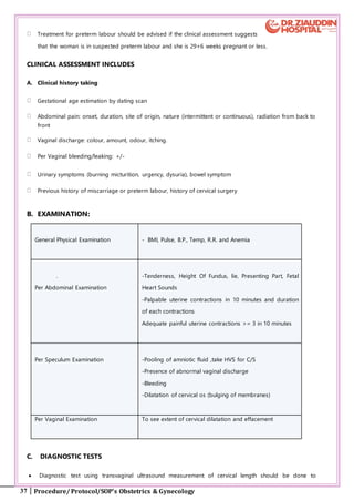
CONTRAINDICATIONS/CAUTIONS TO LMWH USE
Known bleeding disorder (e.g. haemophilia, von Willebrand’s disease or
acquired coagulopathy)
Active antenatal or postpartum bleeding
Women considered at increased risk of major haemorrhage (e.g. placenta praevia)
Thrombocytopenia (platelet count < 75 × 109/l)
Acute stroke in previous 4 weeks (haemorrhagic or ischaemic)
Severe renal disease (glomerular filtration rate [GFR] < 30 ml/minute/1.73m2)
Severe liver disease (prothrombin time above normal range or known varices)
Uncontrolled hypertension (blood pressure > 200 mmHg systolic or > 120 mmHg diastolic)
SUMMARY OF GUIDELINE FOR THROMBOPROPHYLAXIS IN WOMEN WITH
PREVIOUS VTE AND/OR THROMBOPHILIA
Very high risk Previous VTE on long-term
oral anticoagulant therapy
Antithrombin deficiency
Antiphospholipid syndrome
with previous VTE
Recommend antenatal
high-dose LMWH and at
least 6 weeks’ postnatal
LMWH or until switched
back to oral anticoagulant
therapy
These women require
specialist management by
experts in haemostasis and
pregnancy
34. 34 Procedure/ Protocol/SOP’s Obstetrics & Gynecology
High risk Any previous VTE (except a
single VTE related to major
surgery)
Recommend antenatal and
6 weeks’ postnatal
prophylactic LMWH
Intermediate risk Asymptomatic high-risk
thrombophilia homozygous
factor V Leiden/compound
heterozygote Protein C or S
deficiency
Single previous VTE
associated with major surgery
without thrombophilia, family
history or other risk factors
Low risk Asymptomatic low-risk
thrombophilia (prothrombin
gene mutation or factor V
Leiden)
Consider as a risk factor and
score appropriately (see
Appendix III) Recommend
10 days’ if other risk factor
postpartum (or 6 weeks’ if
significant family history)
postnatal prophylactic
LMWH
References
1. Royal College of Obstetrics and Gynecologists. Reducing The Risk Of Thrombosis And Embolism
During Pregnancy And The Puerperium Green-top Guideline No.37a April 2015
2. Royal College of Obstetrics and Gynecologists. The Acute Management Of Thrombosis And
Embolism During Pregnancy And The Puerperium Green-top Guideline No.37b April2015
3. Obstetrics and Gynecology ;An Evidence – Based Text for MRCOG
35. 35 Procedure/ Protocol/SOP’s Obstetrics & Gynecology
4. PRETERM LABOR
DEFINITION:
Preterm labour is defined as regular uterine contractions, every 5-10 minutes or 6-10 in one hour,
before 37+6 weeks gestation associated with:
1. Progressive cervical effacement or dilatation or
36. 36 Procedure/ Protocol/SOP’s Obstetrics & Gynecology
2. > 2cm dilation or >80% effacement
All deliveries occurring between 24+0 and 36+6 weeks
INCIDENCE: - 7-12%
TERMS USED IN THIS PROTOCOL
Symptoms of preterm labour
● A woman presenting with symptoms (such as abdominal pain), might be indicative of preterm labour
before 37+6 weeks of pregnancy but without any clinical assessment (including speculum or digital vaginal
examination)
SUSPECTED PRETERM LABOUR
● A woman with symptoms of preterm labour &a clinical assessment (including a speculum or digital vaginal
examination) that confirms the possibility of preterm labour & exclude established labour
DIAGNOSED PRETERM LABOUR
● A woman is in diagnosed preterm labour if she is in suspected preterm labour along with a positive
diagnostic test for preterm labour.
ESTABLISHED PRETERM LABOUR
● A woman is in established preterm labour if she has progressive cervical dilatation from 4 cm with regular
contractions
RESCUE' CERVICAL CERCLAGE
● Cervical cerclage performed as an emergency procedure in a woman with premature cervical dilatation and
often with exposed fetal membranes
DIAGNOSIS: - Diagnosing preterm labour for women with intact membranes
Women presenting with symptoms of preterm labour & have intact membranes should undergo clinical
assessment &should be told about
the clinical assessment and diagnostic tests that are available
how the clinical assessment and diagnostic tests are carried out
Risks and possible consequences of the false positive and false negative test results taking into account
gestational age.
37. 37 Procedure/ Protocol/SOP’s Obstetrics & Gynecology
Treatment for preterm labour should be advised if the clinical assessment suggests
that the woman is in suspected preterm labour and she is 29+6 weeks pregnant or less.
CLINICAL ASSESSMENT INCLUDES
A. Clinical history taking
Gestational age estimation by dating scan
Abdominal pain: onset, duration, site of origin, nature (intermittent or continuous), radiation from back to
front
Vaginal discharge: colour, amount, odour, itching.
Per Vaginal bleeding/leaking: +/-
Urinary symptoms (burning micturition, urgency, dysuria), bowel symptom
Previous history of miscarriage or preterm labour, history of cervical surgery
B. EXAMINATION:
General Physical Examination - BMI, Pulse, B.P., Temp, R.R. and Anemia
.
Per Abdominal Examination
-Tenderness, Height Of Fundus, lie, Presenting Part, Fetal
Heart Sounds
-Palpable uterine contractions in 10 minutes and duration
of each contractions
Adequate painful uterine contractions >= 3 in 10 minutes
Per Speculum Examination -Pooling of amniotic fluid ,take HVS for C/S
-Presence of abnormal vaginal discharge
-Bleeding
-Dilatation of cervical os (bulging of membranes)
Per Vaginal Examination To see extent of cervical dilatation and effacement
C. DIAGNOSTIC TESTS
Diagnostic test using transvaginal ultrasound measurement of cervical length should be done to
38. 38 Procedure/ Protocol/SOP’s Obstetrics & Gynecology
determine likelihood of birth within 48 hours.
Act on the results as follows:
If cervical length is more than 15 mm,
Tell the woman that it is unlikely, that she is in preterm labour
Discuss with her the advantages & disadvantages of going home compared with continued monitoring
and treatment in hospital
Tell her that if she does decide to go home, she should return if symptoms suggestive of preterm labour
recur
If cervical length is 15 mm or less,
Diagnose the woman as being in preterm labour
Offer treatment
If transvaginal ultrasound measurement of cervical length to exclude preterm labour is not available & a
woman who is 30+0 weeks pregnant or more, is in suspected preterm labour, treat her as being in
diagnosed preterm labour
Ultrasound scans should be performed by trained healthcare professionals, and experience of,
transvaginal ultrasound measurement of cervical length
MATERNAL CORTICOSTEROIDS
The use of maternal corticosteroids should be discussed with women between 23+0 and 23+6 weeks of
pregnancy who are in suspected or established preterm labour, are having a planned preterm birth or
have P-PROM in the context of her individual circumstances
Consider maternal corticosteroids for women between 24+0 and 26+0 weeks of pregnancy who are in
suspected or established preterm labour, are having a planned preterm birth or have P-PROM
Maternal corticosteroids should be given to women between 26+1 and 35+6 weeks of pregnancy who
are in suspected, diagnosed or established preterm labour, are having a planned preterm birth or have P-
PROM.
When offering or considering maternal corticosteroids, discuss with the woman (and her family members)
How corticosteroids may help the potential risks associated with them. Do not routinely offer repeat
courses of maternal corticosteroids, but take into account:
Interval since the end of last course more than 10 weeks
Gestational age
39. 39 Procedure/ Protocol/SOP’s Obstetrics & Gynecology
The likelihood of birth within 48 hours
METHOD
Betamethasone 12mg IM x 2 doses 24 hours apart
dexamethasone 6 mg given intramuscularly in four doses
A single course of antenatal corticosteroid treatment should be given to women with multiple pregnancies at
risk of imminent iatrogenic or spontaneous preterm delivery between 24+0 and 34+6 weeks of gestation.
INVESTIGATION:
Complete blood count
Urine D/R and C/S
High Vaginal Swab(HVS), for C/S in case of discharge
U/S pelvis for Amniotic Fluid Index (AFI) and fetal weight
Transvaginal ultrasound for cervical length
MANAGEMENT:
COUNSELING
The couple should be provided with information& support & this should be done as early as possible
depending on the status of labour & likelihood of preterm birth
Information should include
Both oral and written information
The symptoms and signs of preterm labour Chances of neonatal survival& long term
consequences of premature birth, giving values as natural frequencies (for example, 1 in 100)
The neonatal care of preterm babies, including location of care e.g. nursery
The immediate problems that can arise when a baby is born preterm e.g. need of incubator &ventilator
The possible long-term consequences of prematurity for the baby (how premature babies grow and
develop)
Ongoing opportunities to talk about their wishes regarding resuscitation of the baby
An opportunity to speak to a neonatologist or paediatrician.
Admit the patient
In utero transfer
40. 40 Procedure/ Protocol/SOP’s Obstetrics & Gynecology
C.T.G daily depending on gestational age
Prophylactic vaginal progesterone should be offered to women with
● No history of spontaneous preterm birth or mid-trimester loss
● A cervical length of less than 25 mm on transvaginal ultrasound scan done between 16+0 &
24+0 weeks of pregnancy
Prophylactic cervical cerclage should be offered to women who have
History of preterm prelabour rupture of membranes (P-PROM) in a previous pregnancy or
History of cervical trauma.
Cervical length of less than 25 mm on transvaginal ultrasound scan, carried out between 16+0
and 24+0 weeks of pregnancy
History of spontaneous preterm birth or mid-trimester loss between 16+0 and 34+0 weeks of
pregnancy and
Benefits and risks of prophylactic progesterone and cervical cerclage should be discussed with the woman
and her preferences should be taken into account.
RESCUE’ CERVICAL CERCLAGE
INDICATION women with a dilated cervix and exposed, unruptured fetal membranes between 16+0 and
27+6 weeks of pregnancy
CONTRAINDICATION women with
Signs of infection,
Active vaginal bleeding
Uterine contractions.
When deciding whether to offer ‘rescue’ cervical cerclage
take into account gestational age and
the extent of cervical dilatation
discuss with a consultant obstetrician and consultant paediatrician.
While counseling regarding ‘rescue’ cervical cerclage, women and their family members should be told
About the risks of the procedure
Aim to delay the birth, and thus increasing the likelihood of the neonatal survival and reducing serious
neonatal morbidity.
41. 41 Procedure/ Protocol/SOP’s Obstetrics & Gynecology
Offer maternal corticosteroids to women between 26+1 and 35+6 weeks of
pregnancy who are in suspected, diagnosed or established preterm labour, are having a planned preterm
birth or have P-PROM.
Counsel the women & their attendant about the benefits & potential risk associated with corticosteroid
Repeat courses of maternal corticosteroids should be avoided, but take into account:
The interval since the end of last course is more than10 weeks
Gestational age
The likelihood of birth within 48 hours.
Magnesium sulfate for neuro protection
Intravenous magnesium sulphate should be given to women between 24+0 and 34+0 weeks of pregnancy for
neuroprotection of the baby in case of
Established preterm labour or
Planned preterm birth within 24 hours.
METHOD:
Intravenous bolus of a 4 g magnesium sulfate over15 minutes,
Followed by an intravenous infusion of 1 g per hour until the birth or for 24 hours (whichever is sooner).
MONITORING
Monitor for clinical signs of magnesium toxicity, for women on magnesium sulphate at least every 4 hours
by recording
Pulse
Blood pressure,
Respiratory rate and
Deep tendon (for example, patellar) reflexes.
More frequent monitoring for magnesium toxicities required, or the dose of magnesium sulphate need to be
reduced, if a woman has or develops oliguria or other signs of renal failure
TOCOLYSIS
Decide to start tocolysis after taking following factors into account
Whether the woman is in suspected or diagnosed preterm labour
Gestational age at presentation
Likely benefit of maternal corticosteroids
42. 42 Procedure/ Protocol/SOP’s Obstetrics & Gynecology
Availability of neonatal care (need for transfer to another unit)
The preference of the woman.
TOCOLYTIC DRUGS:
Calcium channel blockers to women between 24+1 and 34+0 weeks of pregnancy who have intact
membranes and are in suspected or diagnosed preterm labour.
Initial oral dose of nifedipine (Tab. Adalat Retard) is 20 mg followed by 10–20 mg three to four times daily,
adjusted according to uterine activity for up to 48 hours.
A total dose above 60 mg appears to be associated with a three- to four-fold increase in adverse events.
Side effect: Headache, flushing and hypotension
If calcium channel blockers are contraindicated, offer oxytocin receptor antagonists atosiban for tocolysis
Initial bolus dose of an atosiban of 6.75 mg over 1 minute, followed by an infusion of 18 mg/hour for 3 hours,
then 6 mg/hour for up to 45 hours (to a maximum of 330 mg)
There is an absence of evidence about all tocolytic medicines before 26+0 weeks of pregnancy.
Avoid betamimetics for tocolysis.
Absolute Contraindications for Tocolysis
Conditions in which prolongation of pregnancy is contraindicated per se, e.g.
● Intrauterine infection,
● Fetus with lethal congenital malformation,
● Fulminating proteinuric preeclampsia
● and any other urgent feto maternal indication for delivery.
Relative contraindications Conditions in which the risks and benefits of intervention have to be weighed
● Antepartum haemorrhage due to abruption placenta
● Ruptured membranes,
● No reassuring fetal heart rate pattern on cardiotocography,
● Intrauterine growth restriction,
● Insulin-dependent diabetes
FETAL MONITORING
Monitoring options: cardiotocography and intermittent auscultation
Discuss with women in suspected, diagnosed or established preterm labour (and their family members or
careers as appropriate):
43. 43 Procedure/ Protocol/SOP’s Obstetrics & Gynecology
the purpose of fetal monitoring
the clinical decisions it informs at different gestational ages a normal cardiotocography trace is
reassuring and indicates that the baby is coping well with labour,
an abnormal trace does not necessarily indicate that fetal hypoxia or acidosis is present.
The woman's preferences should be considered while deciding on choice of monitoring option.
MODE OF BIRTH
Counsel the women in suspected or diagnosed preterm labour & women with PROM (and their family
members) regarding
The general benefits and risks of caesarean section and vaginal birth
Benefits and risks of caesarean section that are specific to gestational age.
The difficulties associated with performing a caesarean section for a preterm birth, especially the
increased likelihood of a vertical uterine incision and the implications of this for future pregnancies .
There are no known benefits or harms for the baby from caesarean section, but the evidence is very
limited.
Caesarean section should be done for women presenting in suspected or diagnosed preterm labour between
26+0 and 36+6 weeks of pregnancy with breech presentation, and explain to the woman that:
Caesarean section for breech presentation for preterm babies is common but not universal practice
Other indication for caesarean section is indicated if;
Breech presentation
FBW < 1.5 kg
Fetal Distress / abnormal CTG and multiple pregnancies
Timing of cord clamping for preterm babies (born vaginally or by caesarean section)
If a preterm baby needs to be moved away from the mother for resuscitation, or there is significant maternal
bleeding:
consider milking the cord and
Clamp the cord as soon as possible.
Wait at least 30 seconds, but no longer than 3 minutes, before clamping the cord of preterm babies if the
mother and baby are stable.
Position the baby at or below the level of the placenta before clamping the cord.
44. 44 Procedure/ Protocol/SOP’s Obstetrics & Gynecology
If Preterm labour subsides: patient can be discharged home with advice of:
● Bed rest
● Avoidance of Coitus
● Tablet Nifedipine 10 mg 1+0+1
● Follow up in OPD after one week
● If labour progresses then accomplish delivery safely (Vaginal route if safe)
REFERENCES
1. Royal College of Obstetrics and Gynaecologists. Tocolysis for Women in Preterm
Labour Green top Guideline No. 1bFebruary 2011
2. Preterm labour and birth NICE guideline: short version Draft for consultation, June
2015
3. Obstetrics and Gynaecology; An Evidence – Based Text for MRCOG
4. Antenatal Corticosteroids to Reduce Neonatal Morbidity and Mortality Green–top
Guideline No. 7October 2010
5. PRETERM PRE LABOR RUPTURE OF MEMBRANES (PPROM):
DEFINITION
Rupture of membrane with leakage of amniotic fluid in absence of uterine activity before 37th
week of
gestation
INCIDENCE
2-3%( associated with approximately one third of all preterm deliveries)
DIAGNOSIS
45. 45 Procedure/ Protocol/SOP’s Obstetrics & Gynecology
History, Examination and Investigation
HISTORY:
Sudden gush of fluid or excessive discharge from vagina.
Gestational age
EXAMINATION
Vitals: BP, Pulse, Temp
P/A: HOF, lie, presentation, uterine tenderness, palpable uterine contractions, and fetal heart sounds
Sterile Speculum examination: To see pooling of amniotic fluid in posterior fornix of vagina, HVS for C/S to
be taken simultaneously and to see cord
P/V Examination: Should be avoided (only done if uterine contractions are present)
If pooling of amniotic fluid is not observed, consider performing an insulin-like growth factor binding protein-
1 test or placenta alpha-microglobulin-1 test of vaginal fluid.
If the results of the above mentioned test are positive, do not use the test results alone to decide what care to
offer the women, but also take into account her clinical condition, her medical and pregnancy history and
gestational age and either:
Offer her antenatal prophylactic antibiotics
or
Re-evaluate the women’s diagnostic status at a later time point.
If the results of the above tests are negative and no amniotic fluid is observed:
Do not offer antenatal prophylactic antibiotics
Explain to the women, it is unlikely that she has P-PROM, but she should return if she has any further
symptoms suggestive of P-PROM of preterm labour
If pooling of amniotic fluid is observed or if labour becomes established in a women reporting symptoms
suggestive of P-PROM do not perform diagnostic tests for P-PROM.
Do not use nitrazine to diagnose P-PROM
INVESTIGATIONS
Complete blood count (C.B.C) to see leukocytosis
C reactive protein (C.R.P)
Urine detail report
46. 46 Procedure/ Protocol/SOP’s Obstetrics & Gynecology
high vaginal swab for culture and sensitivity
Cardiotocography (C.T.G) to see fetal Tachycardia.
Ultra Sound fetal wellbeing & assessment of liquor volume
MONITORING
TLC - Daily
C.R.P - Alternate days
C.T.G - Twice daily
US Pelvis for AFI - Twice Weekly
MANAGEMENT
All patients with P-PROM must be admitted.
a. EXPECTANT TREATMENT:
If labor ensues do not use tocolytic agents.
Steroids should be used if patient is between 24 and 34 weeks
Admit the patient
Provide Autoclave Pad
Counsel the patient & husband regarding:
Outcome and mode of delivery
Prognosis of baby
Need of N.I.C.U / Ventilator
Meeting with the pediatrician & arrangement for surfactant before delivery.
CONSERVATIVE MANAGEMENT:
If no signs of Chorioamnionitis ( tachycardia , pyrexia , uterine tenderness )
Pulse, temperature, Fetal Heart Rate, uterine tenderness 8 hourly
Injection Betamethasone (12 mg ) x 2 doses 24 hours apart
47. 47 Procedure/ Protocol/SOP’s Obstetrics & Gynecology
Tab Erythromycin (250 mg) x 6 hourly for 10 days
For women with P-PROM who cannot tolerate erythromycin or in whom erythromycin is contraindicated,
consider oral penicillin for a maximum of 10 days or until the woman is in established labour (Whichever
is sooner)
Do not offer women with P-PROM Co-Amoxiclav as prophylaxis for intrauterine infection
,
OUT PATIENT MANAGEMENT
Woman should be educated about sign and symptoms of chorioamnionitis.
Woman should check her temperature twice daily
Weekly visit
Delivery by 34-36 weeks
Do not use any of the following in isolation to confirm or exclude intrauterine infection in women with
P-PROM:
a single test of C-reactive protein
white blood cell count
measurement of fetal heart rate using Cardiotocography
If the results of the clinical assessment for any of the test are not consistent with each other, continue to
observe the women and consider repeating the tests.
CHORIOAMNIONITIS
• Deliver baby if signs & symptoms of chorioamnionitis develop i.e.
• maternal pyrexia > 100.2 F
• Uterine tenderness
• Offensive vaginal discharge
• Fetal tachycardia > 160 bpm
• Mode of delivery abdominal or vaginal according to Bishop’s Scoring
48. 48 Procedure/ Protocol/SOP’s Obstetrics & Gynecology
REFERENCES
1. Obstetrics and Gynaecology An Evidence – Based Text for MRCOG
2. Preterm labour and birth NICE guideline: Published 20th
Nov 2015
6. THE MANAGEMENT OF BREECH PRESENTATION & EXTERNAL
49. 49 Procedure/ Protocol/SOP’s Obstetrics & Gynecology
CEPHALIC VERSION
INCIDENCE
3–4% at term
INTRODUCTION
The term breech presents a challenge for delivery. The publication of the Term Breech Trial showed babies
born by planned vaginal delivery had a three-fold higher rate of mortality or serious morbidity than those
born by planned elective caesarean section.
In 2001, the American College of Obstetricians and Gynecologists issued an opinion that, “Patients with
persistent breech presentation at term in a singleton gestation should undergo a planned cesarean delivery.”
Since some patient and may want a planned vaginal delivery of a term singleton breech fetus or come in labor
So it may be reasonable under hospital specific protocol guidelines for both eligibility and labor management.
MANAGEMENT OF THE BREECH FETUS AT TERM
External cephalic version should be offered to all women at term with a fetus known to be breech unless there
is an absolute contraindication. If unsuccessful, however, or if the patient declines the attempt, the plan for
delivery must be carefully thought out.
The patient should be counseled as to the relative risks of alternate modes of delivery. Counseling should be
well documented & the risk of perinatal mortality in planned delivery is 2/1000 as compared to Caesarean
Section 0.5/ 1000.
Women should be informed that planned vaginal delivery increases the risk of low Apgar score & serious
short term complications, but has not been shown to increase the risk of long term morbidity.
The cesarean section increases the risk of complication in future pregnancies, including the risk of opting for
vaginal birth after cesarean section, risk of complication at repeat Cesarean Section, risk of an abnormal
placement of placenta.
FACTORS THAT FAVOURS / INDICATES THE SAFETY OF VAGINAL BREECH DELIVERY
1. Extended breech or flexed breech with buttock presentation.
2. Clinically adequate pelvis.
3. EFW not more than 3.8 kg.
4. Multiparty
5. No other obstetrics or medical risk factor.
50. 50 Procedure/ Protocol/SOP’s Obstetrics & Gynecology
6. A practitioner skilled in the conduct of labour with breech presentation and vaginal
breech birth should be present at all vaginal breech births. Trainees should perform vaginal breech
deliveries under supervision.
7. Unscarred uterus
8. Flexed attitude
9. Avoid breech delivery after 39th
week.
FACTORS UNFAVORABLEFOR VAGINAL BREECH BIRTH INCLUDE THE FOLLOWING
Placenta praevia, compromised fetal condition.
Clinically inadequate pelvis
Footling or kneeling breech presentation
Large baby (usually defined as larger than 3800 g)
Growth-restricted baby (usually defined as smaller than 2000 g)
Hyperextended fetal neck in labour
Lack of presence of a clinician trained in vaginal breech delivery
Previous caesarean section.
Women who do not meet these criteria should be advised to undergo caesarean section, and further
counseled that the risk of perinatal morbidity is likely to be higher than the figures in the Term Breech Trial.
ROLE OF PELVIMETRY is unclear
ANTENATAL ASSESSMENT:
Breech should be delivered at maternity unit where skills level and experience are great. Birth attendant
should be experienced. The position and weight of fetus should be confirmed by Ultrasound and the mode
of delivery is assessed during labour. Induction not recommended.
INTRAPARTUM MANAGEMENT
● Fetal monitoring by CTG
● Maintain partogram
● Spontaneous progress
● Delayed amniotomy
● Epidural analgesia
● Low threshold for c/ section
● Senior should be informed
51. 51 Procedure/ Protocol/SOP’s Obstetrics & Gynecology
FIRST STAGE
Adequate descent of the breech in the passive 2nd
stage is the prerequisite for the active 2nd
stage.
SECOND STAGE
Consider episiotomy when breech seems climbing up the perineum
Baby allows delivering spontaneously up to the umbilicus i-e Hands off Policy.
The arms should be delivered by sweeping them across the baby’s face and downwards or by the
Lovset‟s Manoeuvre (rotation of the baby to facilitate delivery of the arms).
Suprapubic pressure by an assistant should be used to assist flexion of the head.
The after coming head may be delivered with forceps, by the Mariceau- Smellie- Veit manoeuvre,
avoid Burns- Marshall method.
Role of epidural analgesia in vaginal breech delivery in unclear.
Baby should be seen by pediatrician after delivery.
Consider inj. Rhogam if woman blood group negative.
MANAGEMENT OF THE PRETERM BREECH AND TWIN BREECH
ANTEPARTUM
Woman with threaten preterm should be admitted in hospital for observation and intramuscular betamethasone
should be given for fetal lung maturity.
Caesarean Section is recommended for pre term breech presentation where delivery is planned due to
maternal and/ or fetal compromise.
The mode of delivery of pre term breech should be discussed according to the maternal and fetal condition,
stage of labour, type of breech.
Semi recumbent or an all four position may be adopted for delivery and should depend on maternal choice.
INTRAPARTUM
The mode of delivery of the preterm breech presentation should be discussed according to maternal and fetal
condition an individual basis with woman and her husband stage of labor type of breech.
In case of preterm breech delivery should be managed as term breech.
Routine caesarean section for twin pregnancy with breech presentation of the second twin should not be
performed.
C/S is recommended for preterm breech presentation where delivery is planned due to maternal and/ or fetal
compromise.
52. 52 Procedure/ Protocol/SOP’s Obstetrics & Gynecology
EXTERNAL CEPHALIC VERSION (2017):
OBJECTIVE:
3-4% of term births are breech with most resulting in a c-section, successful external version may allow a
woman to avoid an operative delivery. External cephalic version (ECV) is the manipulation of the fetus, through
the maternal abdomen, to a cephalic presentation.
● ECV should be offered to all women with breech presentation at 37 week onward till 42 week or pt in early
labor
PRE - REQUISITES:
Counsel Women that ECV reduces the chance of breech presentation at delivery, and
Lowers their chances of having a Caesarean section.
Explain the procedure
Explain small risk of need of EMLSCS 1/ 200 .
Informed Consent
Ultrasound scan to check fetal well being, type of breech, position of head and placental localization
The immediate presenting part of the fetus is unengaged
Reactive CTG
Tocolytic may be administered before the procedure. i..e ,Inj. salbutamol 0.5mg s/c 30 min before the ECV
PROCEDURE
Place both hand on patient abdomen. Perform forward roll by keeping the breech in flexed position
breech upwards while pressure downwards towards pelvis avoid jerky movement.
Check fetal position on ultrasound and do CTG again after the procedure.
Anti D for Rh negative mothers
If unsuccessful try again Maximum attempt three. If fetal distress, maternal discomfort,
abandon the procedure.
Keep the patient for few hours in recovery room then send her home if no complain and fetal heart rate
tracing reassuring.
ABSOLUTE CONTRAINDICATIONS TO ECV
53. 53 Procedure/ Protocol/SOP’s Obstetrics & Gynecology
Ante partum hemorrhage
Abnormal CTG
Major uterine anomaly/ fetal anomalies
Ruptured membranes
Multiple pregnancy (except delivery of second twin)
RELATIVE CONTRAINDICATIONS WHERE ECV MIGHT BE MORE COMPLICATED:
Gestational Hypertension especially protienuric preeclampsia
Oligohydramnios
Major fetal anomalies
scarred uterus
Unstable lie
Women should be informed that planned C/S for breech presentation at term causes a small increase in
immediate complication for the mother as compared with planned vaginal birth.
REFERENCES
1. RCOG Green Top Guidelines 2017
2. NICE clinical guideline 55 – Intrapartum care 30 September 2007
3. The Society of Obstetrics & Gynecologists Pakistan in accordance with Clinical Governance Guideline 1d
of Royal College of Obstetricians & Gynecologists has adapted Green Top guideline 20b 2006 on
“Management Of Breech presentation”
4. The Society of Obstetrics & Gynecologists Pakistan in accordance with Clinical Governance Guideline
1d of Royal College of Obstetricians & Gynecologists has adapted Green Top guideline Number 20a
on “External Cephalic Version and reducing the incidence of breech presentation” in January 2011.
54. 54 Procedure/ Protocol/SOP’s Obstetrics & Gynecology
7. ANTEPARTUM HEMORRHAGE (APH)
DEFINITION:
Antepartum haemorrhage is described as bleeding from genital tract in pregnancy after 24 weeks &
before the onset of labour.
INCIDENCE:
3-5% of all pregnancies
The causes of APH can be divided into 3 main groups:
Placental Abruption (5%)
Placenta Previa (0.4 – 0.8%)
Others; uterine rupture, cervical ectropion, vaginal infection, vulval varices, marginal placental bleeding,
genital tract tumors
HISTORY:
Amount of blood loss, abdominal pain, provoking factor, previous LSCS, myomectomy. history of D&E.
EXAMINATION
VITALS B.P., Pulse, Temperature, R.R
P/A
H.O.F., Abdominal Tenderness(woody hard), Lie, Presenting Part, Fetal
Hearts
P/V Examination Contraindicated unless placenta previa excluded
INVESTIGATIONS:
CTG
Complete blood count
55. 55 Procedure/ Protocol/SOP’s Obstetrics & Gynecology
Coagulation profile
Blood Sugar
Electrolytes/LFTS
Renal Function Tests
Blood group and Rh Factor
Hepatitis HBsAg
Anti HCV
U/S Pelvis for F.W.B and placental localization
MANAGEMENT:
Call for help, senior staff, the consultant, the pediatrician, the hematologist, anesthetist
Inform the O.T.
ABC, if patient is in shock
Access----- Airway
Maintain -----100 % O2
Breathing
Circulation
15 degree tilt
2 large bore IV Cannula 18G
Cross match 4 unit of packed cells and 6 FFP initially and later according to amount of blood loss.
Start infusion with Normal Saline followed by colloids
Counseling of husband and immediate relative about the present condition and need of emergency
LSCS, need of ICU,NICU, further need of blood
If bleeding not stopped, need of uterine artery embolization
As a last resort when all the procedures fail, need of obstetric hysterectomy
57. 57 Procedure/ Protocol/SOP’s Obstetrics & Gynecology
PLACENTA PRAEVIA
PLACENTA PREVIA
Mild bleeding: (< 200ml)
- Wait and watch & keep under observation with fetal
monitoring.
Moderate bleeding: (200- 1000ml)
-Need of injection betamethasone 12mg, 2 doses IM
12hours apart
-Transfuse blood and monitor vigilantly.
-EL LSCS at 38 wks for Previa only.
-EL LSCS at 36-37 wks for suspected Placenta Accreta.
-Placental edge < 2cm from internal os in 3rd
trimester is likely to deliver by LSCS
Massive bleeding: > 1000ml
-Systolic B.P <100mmHg, pulse >120/min,
altered consciousness
-Manage shock.
-Deliver by LSCS irrespective of gestation.
-Prior to delivery patient & partner should be
counseled about all risks including hysterectomy.
-Anesthesia choice would be decided by
anesthetist.
-Consent form should include :
Blood transfusion,
Hysterectomy.
Admission in ICU
-Anterior placenta overlying previous scar
manage as accreta.
-Before starting C/S misoprostol per rectally
And/or
-After C/S, Insert 800 microgram misoprostol per
rectally to prevent PPH
PLACENTAL ABRUPTION
PLACENTA ABRUPTION
Mild to Moderate bleeding:
-Transfuse blood.
If Fetal distress:
-Delivery imminent: Vaginal delivery
-Delivery not imminent: LSCS
If No fetal distress or fetus is dead:
-Cervix favorable:
deliver vaginally
-Cervix not favorable:
Induce/Augment accordingly, if no progress in 2-4 hrs/fetal
distress/heavy bleeding :LSCS
-Active management of 3rd
stage of labour
Massive bleeding: >1000ml
Manage shock.
Transfuse Blood, FFPs & Cryoprecipitate
Fully dilated & imminent delivery: Deliver vaginally. Delivery not
imminent & FHS present: LSCS
Counsel about NICU care and outcome of baby according to gestatio
age.
58. 58 Procedure/ Protocol/SOP’s Obstetrics & Gynecology
COMPLICATIONS OF APH
Maternal complications Fetal complications
Anemia Fetal hypoxia
Infection Small for gestational age and fetal growth restriction
Maternal shock Prematurity (iatrogenic and spontaneous)
Renal tubular necrosis Fetal death
Consumption Coagulopathy
Postpartum Hemorrhage
Prolonged hospital stay
Psychological sequelae
Complications of blood transfusion
REFERENCES
1. Royal College of Obstetrics and Gynecologists. Antepartum Hemorrhage Green-top Guideline No. 63
1st edition November 2011
2. Royal College of Obstetricians and Gynaecologists. Placenta Praevia, Placenta Praevia Accreta
and Vasa Praevia:Diagnosis and Management Green-top Guideline No. 27.London: RCOG; 2011.
59. 59 Procedure/ Protocol/SOP’s Obstetrics & Gynecology
8. INDUCTION OF LABOUR:
DEFINITION
Induction of labour is defined as an intervention designed to artificially initiate uterine contractions leading to
progressive dilatation and effacement of the cervix and birth of the baby
INCIDENCE: 15-20%
INDICATION FOR INDUCTION
Pregnancy after 41 completed weeks gestation
Prelabour spontaneous rupture of membranes
Maternal diseases
Diabetes
Hypertension
Any other medical disorders
Pregnancy related conditions
Pre-eclampsia / Eclampsia
Obstetric cholestasis
Abruptio placenta
Fetal Intra uterine growth restriction
chorioamnionitis
Intra uterine fetal demise
Maternal request
CONTRAINDICATION FOR INDUCTION
Major placenta praevia(Type 3 & 4)
Vasa praevia
Transverse lie
Previous classical caesarean section
Cord prolapse
Previous one or more caesarean sections
Previous myomectomy entering endometrial cavity.
60. 60 Procedure/ Protocol/SOP’s Obstetrics & Gynecology
PRE INDUCTION ASSESSMENT
Ensure that indication of the induction persists
Counsel the patient and Take written consent
Per abdominal examination to confirm lie and presentation
INFORMATION AND DECISION-MAKING
Women should be informed that most women will go into labour spontaneously by 42 weeks at the 38 week
antenatal visit; all women should be offered information about the risks associated with pregnancies that last
longer than42 weeks, and their options
The information should cover membrane sweeping that membrane sweeping makes spontaneous labour
more likely, and so reduces the need for formal induction of labour to prevent prolonged pregnancy.
membrane sweep can cause discomfort and vaginal bleeding
Healthcare professionals should explain the following points to women being Offered
induction of labour:
Reasons for induction
When, where and how induction could be carried out
Arrangements for support and pain relief
Alternative options if the woman chooses not to have induction of labour
The risks and benefits of induction of labour in specific circumstances and the proposed induction
methods
Inform induction may not be successful and what the woman's options would be.
TIME OF INDUCTION OF LABOR
Women with uncomplicated pregnancies should usually be offered induction of
labour between 41+0and 42+0 women who decline induction of labour should be offered
increased antenatal monitoring consisting of at least twice-weekly
cardiotocography and ultrasound estimation of maximum amniotic pool depth
PRETERM PRE LABOUR RUPTUREOF MEMBRANE
If a woman has preterm pre labour rupture of membranes, induction of labour should not be carried out
before 34 weeks unless there are additional obstetric indications (for example, infection or fetal compromise).
If a woman has preterm prelabour rupture of membranes after 34 weeks, the maternity team should discuss
the following factors with her before a decision is made about whether to induce labour, using vaginal PGE2
61. 61 Procedure/ Protocol/SOP’s Obstetrics & Gynecology
PRE LABOUR RUPTURE OF MEMBRANES AT TERM
Women with prelabour rupture of membranes at term (at or over 37 weeks) should be offered a choice of
induction of labour with vaginal PGE or expectant management. Induction of labour is appropriate
approximately 24 hours after prelabour rupture of the membranes at term.
PREVIOUS CAESAREAN SECTION
If delivery is indicated, women who have had a previous caesarean section may be offered induction of labour
with vaginal PGE2, caesarean section or expectant management on an individual basis, taking into account the
woman's circumstances and wishes. Women should be informed of the following risks with induction of
labour increased risk of need for emergency caesarean section during induced labour increased risk of uterine
rupture.
MATERNAL REQUEST
Induction of labour should not routinely be offered on maternal request alone. However, under exceptional
circumstances (for example, if the woman's partner is soon to be posted abroad with the armed forces),
induction may be considered at or after 40 weeks.
BREECH PRESENTATION
Induction of labour is not generally recommended if a woman's baby is in the breech presentation. If external
cephalic version is unsuccessful, declined or contraindicated, and the woman chooses not to have an elective
caesarean section, induction of labour should be offered, if delivery is indicated, after discussing the
associated risks with the woman.
FETAL GROWTH RESTRICTION
If there is severe fetal growth restriction with confirmed fetal compromise, induction of labour is not
recommended.
HISTORY OF PRECIPITATE LABOUR
Induction of labour to avoid a birth unattended by healthcare professionals should not be routinely offered
to women with a history of precipitate labour.
INTRAUTERINEFETAL DEATH
In the event of an intrauterine fetal death, healthcare professionals should offer support to help women and
their partners and/or family cope with the emotional and physical consequences of the death. This should
include offering information about specialist support.
62. 62 Procedure/ Protocol/SOP’s Obstetrics & Gynecology
In the event of an intrauterine fetal death, if the woman appears to be physically well,
her membranes are intact and there is no evidence of infection or bleeding, she should be offered a choice of
immediate induction of labour or expectant management.
In the event of an intrauterine fetal death, if there is evidence of ruptured membranes, infection or bleeding,
immediate induction of labour is the preferred management option.
If a woman who has had an intrauterine fetal death chooses to proceed with induction of labour, oral
mifepristone, followed by vaginal PGE or vaginal misoprostol, should be offered. The choice and dose of
vaginal prostaglandin should take into account the clinical circumstances, availability of preparations and local
protocol. For women who have intrauterine fetal death and who have had a previous caesarean s ection, the
risk of uterine rupture is increased. The dose of vaginal prostaglandin should be reduced accordingly,
particularly in the third trimester.
SUSPECTED FETAL MACROSOMIA
In the absence of any other indications, induction of labour should not be carried out simply because a
healthcare professional suspects a baby is large for gestational age (macrosomic).
RECOMMENDED METHODS FOR INDUCTION OF LABOUR
Membrane sweeping involves the examining finger passing through the cervix to rotate against the wall of the
uterus, to separate the chorionic membrane from the decidua. If the cervix will not admit a finger, massaging
around the cervix in the vaginal fornices may achieve a similar effect. For the purpose of this guideline,
membrane sweeping is regarded as an adjunct to induction of labour rather than an actual method of
induction.
The Bishop score is a group of measurements made by doing a vaginal examination, and is based on the
station, dilation, effacement (or length), position and consistency of the cervix. A score of eight or more
generally indicates that the cervix is ripe, or 'favourable' – when there is a high chance of spontaneous labour,
or response to interventions made to induce labour.
PHARMACOLOGICAL METHODS
Vaginal PGE is the preferred method of induction of labour, unless there are specific clinical reasons for not
using it (in particular the risk of uterine hyper stimulation). The recommended regimens are:
One cycle of vaginal PGE2, 2 tablets in total: one dose, followed by a second dose after 6 hours if labour is not
established (up to a maximum of two doses)
one cycle of vaginal PGE2: one dose over 24 hours. for induction of labour, healthcare professionals should
inform women about the associated risks of uterine hyper stimulation.
63. 63 Procedure/ Protocol/SOP’s Obstetrics & Gynecology
MISOPROSTOL
Should only be offered as a method of induction of labour to women who have intrauterine fetal death
Mifepristone should only be offered as a method of induction of labour to women who have intrauterine fetal
death
Methods that are not recommended for induction of labour
oral PGE2
intravenous PGE
extra-amniotic PGE
intracervical PGE
intravenous oxytocin alone
Surgical methods Amniotomy, alone or with oxytocin, should not be used as a primary method of
induction of labour unless there are specific clinical reasons for not using vaginalPGE2
MONITORING AND PAIN RELIEF
Wherever induction of labour is carried out, facilities should be available for continuous electronic fetal heart
rate and uterine contraction monitoring.
Before induction of labour is carried out, Bishop score should be assessed and recorded, and a normal fetal
heart rate pattern should be confirmed using electronic fetal monitoring.
After administration of vaginal PGE, when contractions begin, fetal wellbeing should be assessed with
continuous electronic fetal monitoring. Once the cardiotocogram is confirmed as normal, intermittent
auscultation should be used unless there are clear indications for continuous electronic fetal monitoring
If the fetal heart rate is abnormal after administration of vaginal PGE Bishop score should be reassessed 6
hours after vaginal PGE tablet insertion, or 24 hours after vaginal PGE controlled-release pessary insertion, to
monitor progress
During induction of labour, healthcare professionals should provide women with the pain relief appropriate
for them and their pain.
This can range from simple analgesics to epidural analgesia.
Birth attendants (carers and healthcare professionals) should offer women support and analgesia as required,
and should encourage women to use their own coping strategies for pain relief.
Prevention and management of complication
64. 64 Procedure/ Protocol/SOP’s Obstetrics & Gynecology
Uterine hyperstimulation: Tocolysis should be considered if uterine hyperstimulation occurs during induction
of labour.
Failed induction
Per vaginal examination to assess bishop score for cervical ripening.
CALDER MODIFIED BISHOP SCORE
Score 0 1 2 3
Dilatation (cm) 0 1-2 3-4 >5
Length of cervix(cm) >2 2-1 1-0.5 <0.5
Position of cervix Posterior Central Anterior
Consistency of cervix Firm Medium Soft
Station -3 -2 -1 or 0 Below spine
On p/v examination assess the position of suture and mal position
METHODS FOR INDUCTION
MECHANICAL INDUCTION
Membrane sweeping
Intra cervical balloon of foleys
Amniotomy
MEDICAL METHOD
Prostaglandin E2
Can be given by various routes but the intra vaginal route is the preferred method of induction
Recommended regimen is shown in the given table
Type Interval Dose regimen Total dose
65. 65 Procedure/ Protocol/SOP’s Obstetrics & Gynecology
Tablets 6-hourly 3mg 6mg
PROSTAGLANDIN E1 (MISOPROSTOL)
50 μg administered 4- hourly per vaginum to a maximum of five doses
100 μg as single or repeat doses
PLACE FOR INDUCTION
Labour should be induced in a setting in which, there are adequate staffing levels to monitor both
the fetus and mother
TIME FOR INDUCTION
NICE recommends that the induction should be started in the morning at 6am
DIET
Liquid diet
FETAL SURVEILLANCE FOLLOWING INDUCTION OF LABOUR
CTG should be performed for a minimum of 20 minutes before its administration and for 60 minutes
thereafter CTG should be recommenced once the uterine contractions begin.
On p/v examination feel for sutures, presence of cord , station of head , caput and moulding also watch for
color of liquor.
REFERENCES
1. Obstetrics and Gynaecology An evidence –based text for MRCOG second edition Edited by David M
Luesley and Philip N Baker
2. Dewhurst‟s Text Book of Obstetrics & Gynaecology Edited By D.Keith Edmonds
3. Inducing labour nicc.org.uk/guidance / cg 70.
66. 66 Procedure/ Protocol/SOP’s Obstetrics & Gynecology
9. NORMAL LABOUR AND DELIVERY
LABOUR:
It is defined as an onset of regular, painful and effective uterine contractions leading to progressive dilatation
and effacement of the cervix.
DURATION OF LABOR:
Primigravida 12-14 hours
Multigravida 6-8 hours
STAGES OF LABOUR: Three stages:
FIRST STAGE:
Onset of labour till full dilatation of cervix Consists of two phases
Latent Phase: a period of time not necessarily continuous when
There are painful contractions,
There is some cervical change including cervical effacement and dilatation up to 4 cm.
Active Phase: when increase regular painful contraction there is progressive cervical dilatation 4cm to
full dilatation of cervix i.e. 10cm.
The length of established first stage of labor varies between women:
Established first step of labor when there are regular painful contraction there is same progressive cervical
dilatation from 4 cm
first labours last on average 8 hours and are unlikely to last over 18 hours
67. 67 Procedure/ Protocol/SOP’s Obstetrics & Gynecology
second and subsequent labours last on average 5 hours and are unlikely to last over 12 hours
SECOND STAGE:
From full dilatation of cervix till delivery of fetus
Duration in Multilgravida: 1hour
With epidural analgesia: 2 hours
Duration in Primigravida: 2hours
With epidural analgesia 3hours
THIRD STAGE:
From delivery of fetus to expulsion of placenta and membranes.
Duration 10-30 minutes
MANAGEMENT
VERTEX
POSTERIOR FONTANELLE FRONTAL SUTURE
ANTERIOR FONTANELLE
SAGITTAL SUTURE
Anatomy of fetal head and sutures
OBSERVATIONS DURING THE ESTABLISHED FIRST STAGE
• half-hourly documentation of frequency of contractions
• Auscultate the fetal heart rate for a minimum of 1 minute immediately after a contraction every 15
minutes and record it as a single rate
• hourly pulse
68. 68 Procedure/ Protocol/SOP’s Obstetrics & Gynecology
• 4-hourly temperature and blood pressure
• frequency of passing urine
• offer a vaginal examination 4-hourly or if there is concern about progress or in response to the
woman's wishes
FIRST STAGE
Diagnose labour with history and examination
P/A-- Height of fundus, lie, presentation, palpable uterine contractions i.e. frequency of contractions
in 10 minutes and duration of each contraction
P/V- dilatation of os, effacement of cervix, presenting part, presence of membranes, station and
position of presenting part in active phase, show)
Maintain partogram
Watch for progress of labour and timely intervention if it becomes abnormal (normal progress is at a
rate of 1cm/hour)
Provide proper care and emotional support to mother
Monitor fetal wellbeing with CTG
Clear liquid diet
Adequate and appropriate pain relief e.g. epidural analgesia If a woman is contemplating regional
analgesia, talk with her about the risks and benefits and the implications for her labour, including the
arrangements and time .
Provide information about epidural analgesia, including the following:
It provides more effective pain relief than opioids.
It is not associated with long-term backache.
It is not associated with a longer first stage of labour or an increased chance of caesarean birth.
It is associated with a longer second stage of labour and an increased chance of vaginal instrumental
birth.
It will be accompanied by a more intensive level of monitoring and intravenous access, and so mobility
may be reduced.
DELAY IN FIRST STAGE OF LABOUR:
If delay in the established first stage is suspected, take the following into account:
Parity
Cervical dilatation and rate of change
Uterine contractions