هذا التقرير يشير إلى حالة مريض بتلاسيميا بيتا يبلغ من العمر 25 عامًا من كراتشي، باكستان، ويستعرض تأثير المرض على الفم والوجه. تشدد النتائج على أهمية بدء نقل الدم في وقت مبكر والتحكم في التحميل الزائد للحديد لتقليل شدة الأعراض الفموية والوجهية، مما قد يزيد من احتمالية حياة المريض. كما يقدم التقرير صورة شاملة للتشوهات الفموية والعظام المرتبطة بحالة المريض.
![Professional Med J 2017;24(2):352-356. www.theprofesional.com
β THALASSEMIA
353
2
mandible apparently prevent the expansion. The
bony changes may occur early in life and tend
to persist, particularly in skull.16,25,32,36
Furthermore
tint of lemon color is observed in oral mucosa.
CASE REPORT
A registered 25year old male β thalassemic
patient coming at Husaini blood bank and
Institute of Hematological diseases Karachi for
blood transfusion and iron therapy was randomly
selected for Clinical and Radiological Studies
of Oral and Maxillofacial manifestation as case
report. He is habitual of pan and betel nut chewing
since childhood. His mother tongue is Sindhi. He
belongs to a low income family and is not able
to afford thalassemic treatment expenses. His
family consist of 9 members. Mother and father
are normal (carrier). One sister of 12 years and
one brother of 11 years died in β - thalassemia.
Rest of four members are phenotypically healthy
(Carrier). Patient was regularly blood transfused
from the age of 5years. From the last three years,
he has been coming to Husaini blood bank and
InstituteofHematologicaldiseasesKarachiforfree
blood transfusion and iron over loaded treatment
and his blood transfusion motivation is internal.
He has enlarged spleen but Spleenectomy was
not carried out. He has complain of pain in his
joints for last three months and also complain
of bleeding gum during brushing and sensitive
teeth to hot and cold.
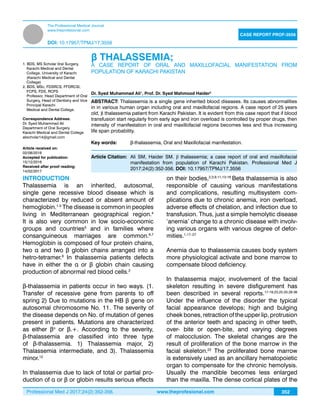
On general examination, he looks under-built,
under-nourished and short stature, with evident
icterus, and yellow tinged finger nails. Intra oral
examination showed intra oral pigmentation on
hard palate and buccal mucosa. Spacing are not
evident. Proclination of upper anterior teeth are
prominent. Multiple decayed teeth with heavy
deposition of calculus and plaque on upper
and lower teeth with dark stains are prominent.
Mamelons are not present and intra oral color of
mucosa is pale yellow.
Extra oral examination findings, disclosed that
color of skin is light muddy black. Frontal and
parietal bossing are present. Depressed nasal
bridge is evident. Maxillary pragmatism is present.
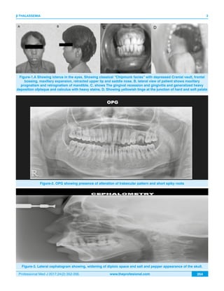
Radiographic findings(X- ray cephlomatric lateral
view) illustrated that class 2 skeletal patterns,
Mandibular retrognatism, high angle in dental
analysis, bimaxillary proclination in soft tissue
analysis, in competent lips, deep mento labial
sulcus skeletal high angle case and increase
lower facial height, widening of diploic space and
salt and pepper appearance of skull are present.
X ray OPG findings disclose that all permanent
teeth are presents. All 3rd
molar are present and
erupted. Periodontal recession of upper and low
anterior teeth vertical bone lose. Short spiky roots
and alteration of trabecular pattern is evident.
DISCUSSION
Generally the findings recorded in this case
confirm the work carried out by different worker
throughout the world19,23,25,30,32
Elhametal., (2002)19
and Hashemipour etal. (2007)22
, Bassimiti et al.
(1996)29
described mandible less enlarged than
maxilla and it is also found in the present case
report. Class 2 skeletal pattern was reported by
Aminiet al. (2007)24
Hattab &Patterns. (2013)27
and
in the present study the skeletal pattern class 2.
Depressednasalbridge,yellowtingeoralmucusa,
bimaxillary proclination, maxillary prognatism are
reported by Amini et al. (2007)20
Hashemipouri et
al. (2007)22
,Babu & Amitha (2014).31
Nagaraj et al.
(2011).42
And these all features are also present in
present study confirm their results. Malnourished,
underweight and short stature are very common
in thalassemic patient Anonym (2012)37
and it is
also true and found in present study. Generally
the skin color in thalassemic patient were reported
pale yellow.1,10,12,16
However skin muddy black is
also reported [41]. In our case the skin color of
patient is muddy black. Mamelons and space
between teeth are generally found in thalassemic
patient40,41
, but these features are not found in
this case. Short spiky roots and diploic space
reported by Patil [2006)17
, Hazza. etal. (2006)22
,
Rashin etal. (2010)23
, Babu & Amitha (2014)40
and
Nagaraj et al. (2011).41](https://image.slidesharecdn.com/prof-3556-170322194915/85/Prof-3556-2-320.jpg)
![Professional Med J 2017;24(2):352-356. www.theprofesional.com
β THALASSEMIA
355
4
CONCLUSION
Findings found In this case report are generally
agree with the previous work done in other
parts of world. However in the present study
teeth spaces among the teeth are not present.
Manifestation intensity in oral and maxillofacial
organs are comparatively low and this seems to
be due to starting of early age blood transfusion
and control on iron over load and thus the
abnormalities are not so prominent, where as
in the same family where blood transfusion was
not started early and properly two members of
the family died in thalassemia. In present study
patient with minimum abnormalities and reaching
the 25 years age is an achievement with the hope
that if patient will continue blood transfusion
regularly and continued treatment of over loading
of iron then, he will have a long spin of life.
Copyright© 15 Dec, 2016.
REFERENCES
1. Rund, D. and E. Rachmilewitz.2005. Medical progress
β-Thalassemia. NEJM353 (11): 1135-1146.
2. Verma, I. C., R. Saxena, and S. Kohli. 2011. Past,
present & future scenario of thalassaemic care &
control in India. Indian J Med Res134: 507-521.
3. Flint, J., F.M. Harding, A. J., Boyce and J. B., Clegg.1998.
The population genetics of haemoglobinopathies.
Bailliere;s Clin. Haematol.11 (1):1-15.
4. Hussain, R. 2000. Socio-demorphic correlates of in
Muslim population of Ind. J. BiosocialSci.32:433-444.
5. Jaber, L., G. J. Halpern, and M. Shohat.1998. The
impact of consanguinity worldwide. Community
Genetics 1: 12-17
6. Naidu L.D., S. M. Raju, and G, sumit. 2010. Effect of
consanguineous marriages on oral and cariofacial
structure: A study on dental patients in north India.
Annals Essences of Dentistry 2(4): 199-203
7. Thein, S. L., (2005). Genetic modifiers of β
-thalassamia. Haematologia. 90: 649-660.
8. S. Riazuddin and R. Galanello. 2000. Identification of
three rare β-thalassemia mutations in the Pakistani
population. Hemoglobin24(1):15-22.
9. Raihan, S., G. Farooq, A., Salman and K., Mohammad.
2009. Thalassemia Major. Journal of the Pakistan
Medical Association 59(6):388-90.
10. Galanello, R., and R. Origa. 2010. β-thalassemia
Orphanet Journal of Rare Diseases 2010, 5:11.
11. Khan, S. A., S.A., Khattak, A. Jaleel, N.A. Anwar, H.S.
Kashif. 2012. β-thalassemia and its association with
haematological parameters. J Pak Med Assoc. 62
(1):40-3.
12. Bejaoui, M and N. Guirat. 2013. β-Thalassemia Major
in a Developing Country: Epidemiological, Clinical
and Evolutionary Aspects Mediterr. J.Hemato. Infect.
Dis. 5(1): e2013002.
13. Kang, J. H. B. R. Park, K. S. Kim, D. Y. Kim, H. J. Huh,
S. L. Chae, S. J. Shin. 2013. β -Thalassemia Minor
Is Associated with IgA Nephropathy. Ann Lab Med
33(2): 153 – 155.
14. Bunn.H.F.F.B.1984. Hamoglobin Moleculare Genetics
and clinical assessment. W. B. Saunders Copampany.
15. Bridge, K., 1998. How do people get thalassemia?
Information center for sickle cell and thalassemia
disorder [cited 16/11/2007, available from http//
sickle bwh. Harvared. Edu / tha inheritance.html,].
16. Patil, S. 2006, Clinical and Radiological study of Oro-
facial manifestation in Thalassemia. Thesis of Master
of Dental surgery in oral Medicine and surgery. Deptt. of
Oral.
17. VandisM.L.andR.P.,Langlias.1986.Thethalassaemia:
Oral manifestations and complications. Oral surgery,
Oral Medicine, Oral Pathology62:229-223.
18. Higgs, D. R., S. L, Thein and W.G.Wood., 2001.The
molecular pathology of thalassemias.
19. Old, J.M., N.I.Oliveri and S.L., Thein. 2001. Diagnosis
and management of Thalassemia in: Weather all, D.
J., B., Clegg, eds. The thalassemia syndromes 4th ed.
Oxford, England: Blackwell Sciene. 30-685.
20. Weatherall, D. J., B., Clegg, eds 2005. The thalassemia
syndromes 4th ed. Oxford, England: Blackwell Sciene.
30-685.
21. Chakraborty and S.P. Basu. 1971. Observations
on radiological changes of bones in thalassemia
syndrome. J. Indian Medical Association 57:90-95.
22. Logothetis, J., J. Economidou, M. Constantoulakis, O.
Augoustaki, R.B. Lowe and M.
23. Bilek. 1971. Cephalo facial deformities in thalassemia
major (Cooley’s anemia): A corrective study among
138 cases. American J. Diseases in children 121:300-
306.](https://image.slidesharecdn.com/prof-3556-170322194915/85/Prof-3556-4-320.jpg)