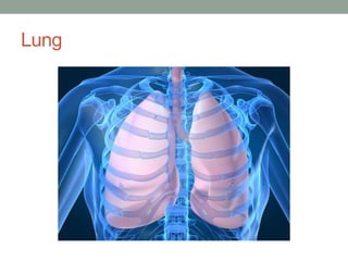
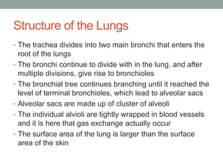
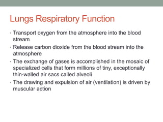
PRESENTATION 2
By
Kidane Hailemichael
Lung
Gross Anatomy of Lungs
• Each cone shaped lung is surrounded by pleurae and
    connected to the mediastinum by vascular and bronchial
    attachment , called the lung root
•   The anterior, lateral, and posterior lung surface lie in close
    contact with the ribs and form the continuously curving
    costal surface
•   The narrow superior tip of the lung is called the apex
•   The concave, inferior surface that rests on the diaphragm
    is the base
•   On the mediastinal surface of each lung, there is an
    indentation called the hilum
Gross anatomy of lungs
• The two lungs differ in size and shape because the apex
    of the heart is slightly to the left of the median plane
•   The left lung is smaller than the right lung
•   Cardiac notch is a concavity in it’s medial aspect that is
    modeled to and accommodates the heart
•   The left lung is subdivided into two superior and inferior
    lobes by the oblique fissure
•   The right lung is subdivided into superior, middle and
    inferior lobes by the oblique and horizontal fissure
•   The smallest subdivisions of the lung visible with the
    naked eye are the lobules
Structure of the Lungs
• The trachea divides into two main bronchi that enters the
    root of the lungs
•   The bronchi continue to divide with in the lung, and after
    multiple divisions, give rise to bronchioles
•   The bronchial tree continues branching until it reached the
    level of terminal bronchioles, which lead to alveolar sacs
•   Alveolar sacs are made up of cluster of alveoli
•   The individual alvioli are tightly wrapped in blood vessels
    and it is here that gas exchange actually occur
•   The surface area of the lung is larger than the surface
    area of the skin
Lungs Respiratory Function
• Transport oxygen from the atmosphere into the blood
  stream
• Release carbon dioxide from the blood stream into the
  atmosphere
• The exchange of gases is accomplished in the mosaic of
  specialized cells that form millions of tiny, exceptionally
  thin-walled air sacs called alveoli
• The drawing and expulsion of air (ventilation) is driven by
  muscular action
Lungs Non Respiratory Function
• Filter out small blood clots formed in veins
• May serve as a layer of soft, shock- absorbent for the
    heart
•   Maintains sterility by producing mucus containing
    antimicrobial compounds
•   Celiar escalator action is an important defense system
    against air-born infections
•   Provide airflow for the creation of vocal sound
•   Immunoglobin-A is secreted in the bronchial secretion and
    protects against respiratory infection
Lung Conditions
• Chronic Bronchitis: Repeated, frequent episodes of
    productive cough
•   Pneumonia: Infection in one or both lugs
•   Asthma: The lungs airways (bronchi) becomes inflamed
    and can spasm, causing shortness of breath and
    wheezing
•   Acute Bronchitis: An infection of the lungs large airways,
    usually caused by virus
•   Pulmonary Fibrosis: The interstitum becomes scarred,
    making the lungs stiff and causing shortness of breath
•   Pleurisy: Inflammation of the lining of the lung (pleura),
    which often causes pain when breathing in
Lung Conditions
• Lung Cancer: Cancer can affect almost any part of the
  lung. Most lung cancer is caused by smoking
• Tuberculosis: A slowly progressive pneumonia caused by
  a bacteria Mycobacterium Tuberculosis
• Emphysema: The fragile walls between the lungs air sacs
  are damaged, trapping air in the lungs and making
  breathing difficult
Lung Tests
• Chest X-ray: X-ray is most common first test for lung
    problem
•   Computer Tomography (CT Scan): A CT scan uses x-rays
    and a computer to make detailed picture of the lung
•   Spirometer: Measures how fast and how much air a
    person can breath out
•   Pulmonary Function Test (PFTs): Serious of tests to
    evaluate how well the lungs work
•   Sputum Culture: Culturing mucus can sometimes identify
    the organism responsible for a Pneumonia or Bronchitis
•   Lung Biopsy: A small piece of tissue is taken from the
    lung. Examining the biopsied tissue under microscope
    can help diagnose lung conditions
Lung Treatments
• Thoracotomy: A surgery that enters the chest wall (thorax)
• Video Assisted Thorascopic surgery (VATS): Less-
    invasive chest wall surgery using an endoscope
•   Plurocentesis: A needle is placed into the chest cavity to
    drain fluid that’s around the lung
•   Bronchodilators: Inhaled medicines can help expand the
    airways (bronchi)
•   Lung Transplant: Surgical removal of diseased lung and
    replacement with organ donor lungs
•   Lung Resection: A diseased portion of the lung is
    removed through surgery
Medical Terminology
• Pulmon/o – Lung
• Pulmonary – Pertaining to the lung
• Pulmonology – Study of lungs
• Pulmonologist – A physician who specializes in lungs
• Pneumonitis – Inflammation of the lungs
References
• Book – Human Anatomy and Physiology
•         By Elain N. Marieb, Katja Hoehn
• Www. WebMD.com
• en.wikipenda.org

Presentation 2 kidane

  • 1.
  • 2.
  • 3.
    Gross Anatomy ofLungs • Each cone shaped lung is surrounded by pleurae and connected to the mediastinum by vascular and bronchial attachment , called the lung root • The anterior, lateral, and posterior lung surface lie in close contact with the ribs and form the continuously curving costal surface • The narrow superior tip of the lung is called the apex • The concave, inferior surface that rests on the diaphragm is the base • On the mediastinal surface of each lung, there is an indentation called the hilum
  • 4.
    Gross anatomy oflungs • The two lungs differ in size and shape because the apex of the heart is slightly to the left of the median plane • The left lung is smaller than the right lung • Cardiac notch is a concavity in it’s medial aspect that is modeled to and accommodates the heart • The left lung is subdivided into two superior and inferior lobes by the oblique fissure • The right lung is subdivided into superior, middle and inferior lobes by the oblique and horizontal fissure • The smallest subdivisions of the lung visible with the naked eye are the lobules
  • 5.
    Structure of theLungs • The trachea divides into two main bronchi that enters the root of the lungs • The bronchi continue to divide with in the lung, and after multiple divisions, give rise to bronchioles • The bronchial tree continues branching until it reached the level of terminal bronchioles, which lead to alveolar sacs • Alveolar sacs are made up of cluster of alveoli • The individual alvioli are tightly wrapped in blood vessels and it is here that gas exchange actually occur • The surface area of the lung is larger than the surface area of the skin
  • 6.
    Lungs Respiratory Function •Transport oxygen from the atmosphere into the blood stream • Release carbon dioxide from the blood stream into the atmosphere • The exchange of gases is accomplished in the mosaic of specialized cells that form millions of tiny, exceptionally thin-walled air sacs called alveoli • The drawing and expulsion of air (ventilation) is driven by muscular action
  • 7.
    Lungs Non RespiratoryFunction • Filter out small blood clots formed in veins • May serve as a layer of soft, shock- absorbent for the heart • Maintains sterility by producing mucus containing antimicrobial compounds • Celiar escalator action is an important defense system against air-born infections • Provide airflow for the creation of vocal sound • Immunoglobin-A is secreted in the bronchial secretion and protects against respiratory infection
  • 8.
    Lung Conditions • ChronicBronchitis: Repeated, frequent episodes of productive cough • Pneumonia: Infection in one or both lugs • Asthma: The lungs airways (bronchi) becomes inflamed and can spasm, causing shortness of breath and wheezing • Acute Bronchitis: An infection of the lungs large airways, usually caused by virus • Pulmonary Fibrosis: The interstitum becomes scarred, making the lungs stiff and causing shortness of breath • Pleurisy: Inflammation of the lining of the lung (pleura), which often causes pain when breathing in
  • 9.
    Lung Conditions • LungCancer: Cancer can affect almost any part of the lung. Most lung cancer is caused by smoking • Tuberculosis: A slowly progressive pneumonia caused by a bacteria Mycobacterium Tuberculosis • Emphysema: The fragile walls between the lungs air sacs are damaged, trapping air in the lungs and making breathing difficult
  • 10.
    Lung Tests • ChestX-ray: X-ray is most common first test for lung problem • Computer Tomography (CT Scan): A CT scan uses x-rays and a computer to make detailed picture of the lung • Spirometer: Measures how fast and how much air a person can breath out • Pulmonary Function Test (PFTs): Serious of tests to evaluate how well the lungs work • Sputum Culture: Culturing mucus can sometimes identify the organism responsible for a Pneumonia or Bronchitis • Lung Biopsy: A small piece of tissue is taken from the lung. Examining the biopsied tissue under microscope can help diagnose lung conditions
  • 11.
    Lung Treatments • Thoracotomy:A surgery that enters the chest wall (thorax) • Video Assisted Thorascopic surgery (VATS): Less- invasive chest wall surgery using an endoscope • Plurocentesis: A needle is placed into the chest cavity to drain fluid that’s around the lung • Bronchodilators: Inhaled medicines can help expand the airways (bronchi) • Lung Transplant: Surgical removal of diseased lung and replacement with organ donor lungs • Lung Resection: A diseased portion of the lung is removed through surgery
  • 12.
    Medical Terminology • Pulmon/o– Lung • Pulmonary – Pertaining to the lung • Pulmonology – Study of lungs • Pulmonologist – A physician who specializes in lungs • Pneumonitis – Inflammation of the lungs
  • 13.
    References • Book –Human Anatomy and Physiology • By Elain N. Marieb, Katja Hoehn • Www. WebMD.com • en.wikipenda.org