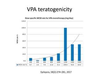
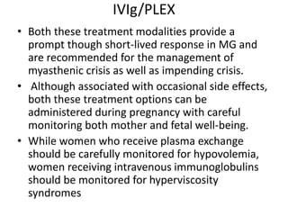
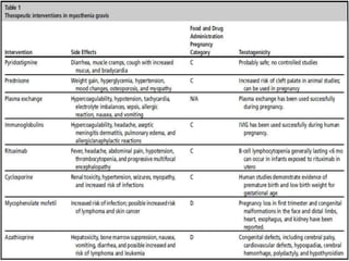
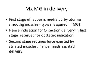
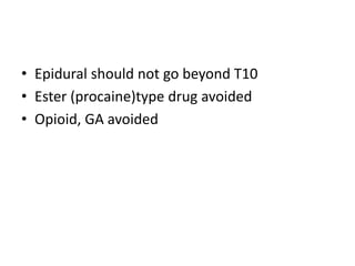
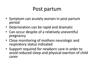
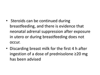
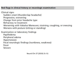
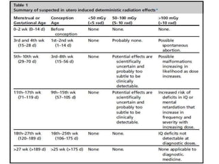
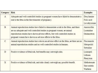
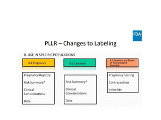
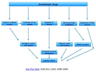
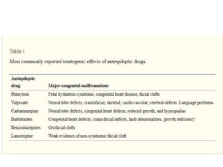
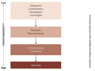
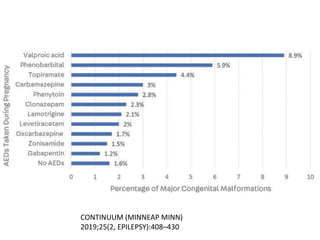
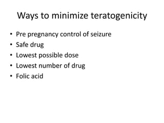
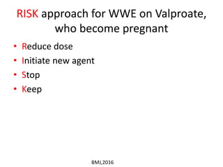
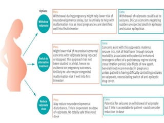
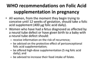
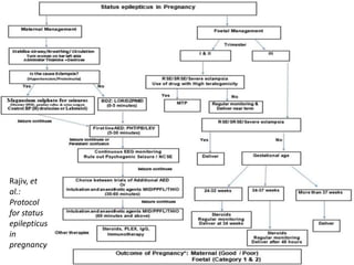
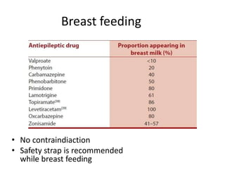
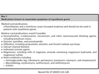
Selected Topics in
“Neurology of Pregnancy”
Dr Srimant Pattnaik
DM PDT , BIN
29.04.2019
Outline
• Diagnostic Imaging in pregnancy
• Pre existing diseases
– Epilepsy
– Myasthenia Gravis
– Headache
• What pregnancy does to the disease
• What disease does to mother
(ante/intra/postpartum/lactation)
• What disease/medication does to baby
Diagnostic Imaging
• Situation include stroke, venous thrombosis ,
Eclampsia , Pituitary issues , multiple sclerosis
, optic neuritis
• Case – by –case approach best
• Benefit should outweigh the risk
• Acceptable maximum accummulated fetal
dose is 5 rad
Safety of Radiographic Imaging During Pregnancy
Am Fam Physician. 1999 Apr 1;59(7):1813-1818
MRI
• Usually no increased clinical side effects
• heating of the tissue, teratogenicity risk
theoratical
• ACR recommends MRI if it leads to benefit to
mother and fetus
Contrast agents
• Iodinated contrast was shown to be mutagenic in vitro, can
cause neonatal hypothyroidism
• currently most studies use nonionic contrast that has no
effect on the thyroid gland.
• MR contrast agents categorized as a Class C drug by the FDA
• Intravenous gadolinium chelate was shown to be teratogenic
in animal studies (with high and repeated dose), although was
not observed in a small number of human studies.
• Gadolinium crosses the placenta and is excreted by fetal
kidneys
Lactation after contrast use
• In the past, cessation of breastfeeding was
recommended for 24 hours after administration
of contrast in a lactating mother.
• However, studies found that less than 0.01% of
CT and 0.04% of MR contrast gets into breast milk
and only a tiny fraction of ingested contrast is
absorbed by the infant’s gastrointestinal tract.
• It was thus determined that the tiny risk is not
sufficient to advocate cessation of breastfeeding
To conclude
• Deterministic effects are negligible at any gestational
age for low risk examinations such as Head CT, Chest X
ray
• The risks and benefits of MR are the same as without
pregnancy.
• CT contrast is safe to use, but MR contrast be used
only if necessary
• There is no need to stop breastfeeding, except with the
use of nuclear medicine agents.
• Although many imaging examinations are considered
safe in pregnancy, elective imaging should be delayed if
possible
PLLR
(Pregnancy And Lactation Labelling Rule)
Women With Epilepsy(WWE)
Funct Neurol. 2016 Jul-Sep; 31(3): 127–134
Infertility
• Direct relationship between epilepsy and
infertlity remain unclear , though birth rate
among WWE is low .
• Low marriage rates , less desire to have
children
• Phenobarbital, polytherapy and EIAED use
may contribute to infertility
Contraception
• Each visit gives an oppurtunity for
preconceptional counselling esp regarding a
planned pregnancy.
• Special indication for contraception is to delay
pregnancy till seizure control is achieved
• Interaction between AED and hormonal
contraceptive is a major issue , which
manifests as 1. contraceptive failure
2. reduced AED efficacy
EIAED
CYP3A4
ACTIVITY
INCREASED
ESTROGEN ,
PROGESTIN LEVEL
DECREASE
EIAED
1. CARBAMAZEPINE
2. OXCARBAZEPINE
3. FELBAMATE
4. PHENYTOIN
5. PRIMIDONE
6. TOPIRAMATE
7. PAREMPANEL
8. PHENOBARBITAL
AEDs with less
interaction with
hormonal contraceptive
1.Valproic acid
2. Vigabartin
3. Gabapentin
4. Tiagabine
5. Levetiracetam
6.Zonisamide
7.Ethosuximide
8. Clobazam
9.Clonazepam
Neurol Clin 37(2019) 53-62
Estrogen
Increased
UGT
Decreased level of
Lamotrigine, Valproate ,
Eslicarbazepine
Estrogenic component of
COCs can lower lamotrigine levels by 40% to
60%, increasing the risk of breakthrough
Seizures
Lamotrigine levels should be carefully
monitored before and after starting a COC,
and doses adjusted accordingly, potentially by
up to 50%
UGT = uridine diphospho glucoronosyl transferase
• Lamotrigine levels are not affected by the
progestins of COCs.
• lamotrigine induces the clearance of progestins
with a resultant decrease in progestin level of
approximately 20%.
• although the progestin-only OC may seem to be
reasonable for women taking lamotrigine, the
risk of contraceptive failure would predictably be
increased with this combination as well.
Physiologic changes in pregnancy
affecting drug level
• Decreased absorption of drug due to gastric
stasis and vomiting
• Increased blood volume
• Increased renal clearance of drugs
• AED level monitoring at baseline and monthly
once in pregnancy has been recommended by
AAN Practice guidelines
Risk of adverse outcomes in mother
• Pregnancy usually does not alter the seizure
frequency in WWE
• If a patient was seizure free for 9 months -1 year
before pregnancy , 80% chance that she will
remain seiezure free (Vajda et al , Epilepsia 2008 )
• However, this predictor is dependent upon AED
drug serum concentrations staying within 35% of
the preconception value throughout the
pregnancy (Epilepsy Behav. 2013)
• Rate of status epilepticus is similar to general
pregnancy
Pregnancy related factors contributing to
worsening seizure control
• Lack of sleep during late pregnancy, labour, and after
delivery
• Stress and anxiety
• Hormonal changes
• Pharmacokinetic changes with decreased plasma
antiepileptic drug levels, particularly lamotrigine
• Lack of adherence to medication due to concerns
about fetal development
• Change to less effective drug because of perceived
lower risk of teratogenicity
BMJ 2016;353:i2880
• Risk of Eclampsia increases slightly
• Risk of SUDEP is 10 times higher than general
population
• In a study , half of epilepsy related death in
pregnancy ( 79 % from SUDEP) occured in
patients who were on lamotrigine
Risk of adverse outcome in fetus
• SGA, low head circumference
• No immediate complication to baby due to
seizure
• Fetal bradycardia may be there due to
maternal hypoxia and acidosis
AED teratogenicity
Exp Ther Med. 2016 Oct; 12(4): 2400–2404.
AustralianPregnancyRegisterdataandotherliterature
CONTINUUM (MINNEAP MINN)
2019;25(2, EPILEPSY):408–430
VPA teratogenicity
Epilepsia, 58(2):274–281, 2017
More chance of teratogenicity at
following doses
• Valproic acid (>700 mg/d)
• Carbamazepine (>400mg/d)
• Phenobarbital (>150 mg/d)
• Lamotrigine (>300 mg/d).
EURAP
This dataset from South India confirms the increased risk of
MCM with exposure to AEDs, particularly polytherapy.
A dose-dependent increased risk was observed with
valproate.
The increased risk associated with clobazam monotherapy
is an important signal that needs to be confirmed in a larger
sample
The newer generation AEDs, lamotrigine and levetiracetam, were not associated with
significant increased risks of CMs compared to control, and were significantly less likely
to be associated with children experiencing cardiac malformations than control.
However, this does not mean that these agents are not harmful to infants/children
exposed in utero. Counselling is advised concerning teratogenic risks when the
prescription is written for a woman of childbearing age and before women continue
with these agents when considering pregnancy, such as switching from polytherapy to
monotherapy with evidence of lower risk and avoiding AEDs, such as valproate, that
are consistently associated with CMs. These decisions must be balanced against the
need for seizure control
Valproate alone or combined with another AED is
associated with the greatest odds of adverse
neurodevelopmental outcomes compared with control.
Oxcarbazepine and lamotrigine were associated with
increased occurrence of autism. Counselling is advised for
women considering pregnancy to tailor the safest regimen
Continuum (Minneap Minn) 2014;20(1):60–79
Ways to minimize teratogenicity
• Pre pregnancy control of seizure
• Safe drug
• Lowest possible dose
• Lowest number of drug
• Folic acid
RISK approach for WWE on Valproate,
who become pregnant
• Reduce dose
• Initiate new agent
• Stop
• Keep
BMJ,2016
WHO recommendations on Folic Acid
supplementation in pregnancy
• All women, from the moment they begin trying to
conceive until 12 weeks of gestation, should take a folic
acid supplement (400 μg folic acid daily).
• Women who have had a fetus diagnosed as affected by
a neural tube defect or have given birth to a baby with
a neural tube defect should:
– receive information on the risk of recurrence;
– be advised on the protective effect of periconceptional
folic acid supplementation;
– be offered high-dose supplementation (5 mg folic acid
daily); and
– be advised to increase their food intake of folate.
RCOG guidelines regarding Folic Acid
supplemenation in pregnancy
• All WWE should be advised to take 5 mg/day
of folic acid prior to conception and to
continue the intake until at least the end of
the first trimester to reduce the incidence of
major congenital malformation.
• Pre-pregnancy folic acid 5 mg/day may be
helpful in reducing the risk of AED-related
cognitive deficits
Labour and delivery
• Risk of seizure during delivery 2 %
• Associated with subtherapeutic AED level
• Epilepsy is not an indicaction for C- section
delivery
• AED to be continued through labour
• Maternal mortality is ten times higher in WWE
than in those without the disorder
(MacDonald et al , 2015)
Status epilepticus in pregnancy
• Status epilepticus (SE) is a single epileptic seizure
lasting more than 5 min or two or more seizures
occurring within a 5-minute period without the person
returning to normal between them
• Nonconvulsive status epilepticus (NCSE) is either a de
novo change in behavior and mental processes from
baseline, or prolonged postictal confusion following a
generalized tonic–clonic seizure, lasting for at least 30
min associated with continuous epileptiform EEG
changes but without major motor signs
K.R. Rajiv, A.
Radhakrishnan / Epilepsy
& Behavior (2017)
Rajiv, et
al.:
Protocol
for status
epilepticus
in
pregnancy
Breast feeding
• No contraindiaction
• Safety strap is recommended
while breast feeding
Myasthenia Gravis
Effect of Pregnancy on Myasthenia
• Variable from woman to woman and
pergnancy to pregnancy in same woman
• Around 40% worsen, 30 % improve and 30 %
remain unchanged
• Disease exacerbation more in first trimester
and postpartum period ; 20 % require
ventillatory support
• Increased dosage requirement
• Pre pregnancy control of disease activity has poor
correlation with pregnancy related exacerbations
• Pregnancy termination is not an option for
controlloing the disease
• Myasthenic exacerbations occur in initial 1-2
years after diagnosis , hence pregnancy may be
delayed to that point after diagnosis
• The above discussion also part of preconception
counselling
Contraception
• Continuous hormonal contraceptive in form of
implant, ring or OC pills are preferred
• cyclic withdrawal of OC pills precipitate
weakness
• IUD is effective alternative
Antenatal Care
• When a patient with MG becomes pregnant,
care is required to avoid exacerbation.
• Starting glucocorticoid therapy or
withdrawing immunosuppressant therapy
might cause transient worsening of MG.
• Infections should be treated promptly ( no
aminoglycoside, macrolide or colistin)
• An initial evaluation of pregnant patients with
MG includes
– Assessment of baseline motor strength
– Pulmonary function
– Electrocardiographs
– Thyroid profile test
Neurol Clin 37 (2019) 113–120
Issue of thymectomy
• During pregnancy, patients with thymoma who
have not undergone thymectomy present with a
higher incidence of exacerbation than those who
have undergone thymectomy.
• Infants born to MG patients who have undergone
thymectomy have less risk of developing neonatal
MG.
• Thus, thymectomy could be considered before
conception or after delivery if a patient with MG
plans to become pregnant in the future, but not
during pregnancy.
Pyridostigmine (category B)
• Not expected to cross the placenta in significant
amounts
• No reports of fetal malformations with
pyridostigmine
• Dose may have to be increased in view of
increased volume of distribution, increased renal
clearance , decreased absorption hyperemesis
gravidarum
• Higher doses may induce premature contraction
of uterus
Steroid (FDA category B)
• Maintenance steroids at the lowest possible dose
are usually continued in pregnancy, because they
cause minimal adverse effects in the mother and
fetus.
• Women with MG who are prescribed corticosteroids
must be informed before conception of the
increased risk of oral clefts.
• As withdrawal of steroid may lead to exacerbation of
MG, pregnant women who are on steroids should
continue steroids during pregnancy.
• However, for women who are not on steroids, one
can withhold temptation to start steroids during 1st
trimester as palate is completely formed by 12
weeks
• Prednisolone is preferred, because it is
metabolized by the placenta, and just 10%
crosses into the fetal circulation at maternal
doses of <20 mg.
• A typical regimen is to start with 5 mg
prednisolone once daily and then increase by 5
mg every 5–7 days till target dose is achieved
(usually 0.75–1 mg/kg body weight).
• Mothers receiving more than 7.5 mg
prednisolone per day or 15 mg on alternate days
for more than 2 weeks before delivery should
receive parenteral steroids to cover the stress of
delivery.
Azathioprine (FDA category D)
• Azathioprine has not been associated with
increased rates of congenital abnormalities.
• This is likely related to the absence of enzyme
in fetal liver which converts azathioprine to its
active metabolite
• However, there is evidence of possible
intrauterine growth retardation and infants
with low birthweight, as well as concern about
immunological changes
• Currently, azathioprine is considered as
non-steroidal drug of choice in the
management of pregnant myasthenic women
in Europe given in a dose of 2–3 mg/Kg body
weight
Cyclosporine A
• Although reported to cross placenta and associated with
transient neutropenia, lymphopenia, and
thrombocytopenia as well as prematurity, intrauterine
growth retardation and abortions, use of this drug is not
associated with any serious harm to the fetus
• Most of the data regarding the use of cyclosporine in
pregnancy are obtained from transplant recipients and
other autoimmune diseases rather than MG per se.
• However, the current literature suggests that this drug can
be used in pregnancy provided the perceived benefits to
mother outweigh the perceived risks to fetus.
• The usual starting dose is 1.25 mg/kg body weight twice
daily which can be increased by 0.5 mg/kg at 4–8 weeks
interval up to a maximal dose of 4 mg/kg/day
MMF
• The current evidence suggests teratogenic effects
(short fingers and toenails, cleft palate and lip,
and corpus callosum agenesis) as well high rates
of spontaneous abortions following the use of
mycophenolate during pregnancy.
• Thus, the use of this drug is contraindicated
during pregnancy and recipients of
mycophenolate should practice contraception
during and for 6 weeks after discontinuation of
this drug
Methotrexate
• The use of methotrexate during pregnancy is
associated with high risk of neural tube
defects including anencephaly as well as high
rate of abortions.
• Thus, the use of methotrexate during
pregnancy is contraindicated, and women
who are on methotrexate should practice
contraception
cyclophosphamide
• Similarly, the use of during pregnancy is
associated with major congenital
malformations
• this drug should be avoided in pregnant
women
Rituximab
• This drug is being used with increasing frequency
in the management of MG especially severe MG
and MuSK antibody positive MG.
• Although reported to cross placenta, use of this
drug has not been reported to have major fetal
adverse effects other than transiently decreased
B-cell counts.
• However, experience regarding the use of this
drug in pregnancy is still limited, and it should be
used only when perceived benefits to mother
outweigh perceived risks to fetus.
• Frequent USG for fetal well-being may be
recommended whenever this drug is used in
pregnant women with MG.
IVIg/PLEX
• Both these treatment modalities provide a
prompt though short-lived response in MG and
are recommended for the management of
myasthenic crisis as well as impending crisis.
• Although associated with occasional side effects,
both these treatment options can be
administered during pregnancy with careful
monitoring both mother and fetal well-being.
• While women who receive plasma exchange
should be carefully monitored for hypovolemia,
women receiving intravenous immunoglobulins
should be monitored for hyperviscosity
syndromes
Mx MG in delivery
• First stage of labour is mediated by uterine
smoothg muscles ( typically spared in MG)
• Hence indication for C- section delivery in first
stage reserved for obstetric indication
• Second stage requires force exerted by
striated muscles , hence needs assisted
delivery
• Epidural should not go beyond T10
• Ester (procaine)type drug avoided
• Opioid, GA avoided
Treatment of Eclampsia in MG
• No Mg for MG
• Phenytoin increases muscle weakness
• IV Levetiractam recommended
Effect of MG on fetus
• 10–30% of infants develop TNMG
• placental transfer of immunoglobulin G (IgG)
anti-AChR antibodies in the second and third
trimesters
• respiratory problems, muscle weakness,
feeble cry, poor sucking or ptosis, within 12 h
to 4 days that persist from 2 weeks to several
months after birth.
• All infants delivered from MG mothers should
be observed particularly carefully through the
first week.
• These conditions recover spontaneously
within 3–4 weeks after birth as the antibodies
transported from the mother are degraded.
• The durations of maternal disease and
medication for MG are not associated with the
occurrence of neonatal MG.
Lactation
• Prednisone , prednisolone , pyridostigmine
safe
• Cyclosporine , methotrexate , azathioprine
contraindicated
• MMF, retuximab need more study in lactation
Post partum
• Symptom can acutely worsen in post partum
period
• Deterioration can be rapid and dramatic
• Can occur despite of a relatively uneventful
pregnancy
• Close monitoring of mothers neurologic and
respiratory status indicated
• Support required for newborn care in order to
avoid reduced sleep and physical exertion of child
carev
• Steroids can be continued during
breastfeeding, and there is evidence that
neonatal adrenal suppression after exposure
in utero or during breastfeeding does not
occur.
• Discarding breast milk for the first 4 h after
ingestion of a dose of prednisolone ≥20 mg
has been advised
Headache in pregnancy
Neurol Clin 37 (2019) 31–51
Migraine in Pregnancy
Effect of pregnancy on migraine
• Migraine without aura typically improves or
remits altogether in most women when pregnant
• Improvement or remission observed in nearly
– 47% of women during the first trimester
– 83% of women during the second trimester
– 87% of women during the third trimester
• Migraine improvement during pregnancy is
related to increasing levels of estradiol during the
gestational period, as well as the lack of cycling
related to the menstrual cycle where estrogen
withdrawal can serve as a major migraine trigger
• Migraine with aura is less likely to improve
during pregnancy than migraine without aura.
• New-onset migraine with aura and even aura
without headache may occur in the later
stages of pregnancy
Effect of migraine on pregnancy
• Maternal disability may lead to less calori
intake , dehydration
• No acute effect on fetus
General advice regarding pregnancy
• Non-pharmacologic therapies should always be
emphasized as an important aspect of migraine
management
• Recognizing and avoiding commonly reported
migraine attack triggers such as diet and
especially sleep
• Proactively addressing risk factors for migraine
progression and co-morbidities is also crucial
since many of these factors are modifiable.
– Medication overuse
– excessive caffeine intake,
– psychiatric and pain comorbidities,
– obesity,
– sleep disturbance
Nonpharmacologic treatment
• many of which are evidence-based and safe
during pregnancy, are highly recommended
and include
– Ice pack
– Relaxation strategies
– Biofeedback
– Cognitive-behavioral therapy
• Emphasizing the good prognosis for migraine
in pregnancy is also helpful to relieve some of
the anxiety for women planning to conceive,
particularly for those who have episodic
migraine without aura
Acute treatment of migraine in
pregnancy
 Acetaminophen and metoclopramide are
traditional first-line therapies
 Ibuprofen helpful in 2nd trimester
Triptans can be considered 2nd line
Rescue therapy : iv metoclopramide,
diphenhydramine +/- opioids
Occipital nerve blocks using lidocaine alone
are helpful in the office setting and are
considered compatible with pregnancy
Prophylactic treatment of migraine
• Prevention should be considered in women who
have frequent, moderate to severe attacks that
do not respond well to acute treatment
• Propranolol is the preventive with the best
evidence for safe use during pregnancy and is
preferred as the first-line preventive treatment
• Amitriptyline and verapamil have a lower risk
profile than other preventive options and are
second-line preventive treatments
Lactation

Pregnancy neurology

  • 1.
    Selected Topics in “Neurologyof Pregnancy” Dr Srimant Pattnaik DM PDT , BIN 29.04.2019
  • 2.
    Outline • Diagnostic Imagingin pregnancy • Pre existing diseases – Epilepsy – Myasthenia Gravis – Headache • What pregnancy does to the disease • What disease does to mother (ante/intra/postpartum/lactation) • What disease/medication does to baby
  • 3.
    Diagnostic Imaging • Situationinclude stroke, venous thrombosis , Eclampsia , Pituitary issues , multiple sclerosis , optic neuritis • Case – by –case approach best • Benefit should outweigh the risk • Acceptable maximum accummulated fetal dose is 5 rad
  • 8.
    Safety of RadiographicImaging During Pregnancy Am Fam Physician. 1999 Apr 1;59(7):1813-1818
  • 9.
    MRI • Usually noincreased clinical side effects • heating of the tissue, teratogenicity risk theoratical • ACR recommends MRI if it leads to benefit to mother and fetus
  • 10.
    Contrast agents • Iodinatedcontrast was shown to be mutagenic in vitro, can cause neonatal hypothyroidism • currently most studies use nonionic contrast that has no effect on the thyroid gland. • MR contrast agents categorized as a Class C drug by the FDA • Intravenous gadolinium chelate was shown to be teratogenic in animal studies (with high and repeated dose), although was not observed in a small number of human studies. • Gadolinium crosses the placenta and is excreted by fetal kidneys
  • 11.
    Lactation after contrastuse • In the past, cessation of breastfeeding was recommended for 24 hours after administration of contrast in a lactating mother. • However, studies found that less than 0.01% of CT and 0.04% of MR contrast gets into breast milk and only a tiny fraction of ingested contrast is absorbed by the infant’s gastrointestinal tract. • It was thus determined that the tiny risk is not sufficient to advocate cessation of breastfeeding
  • 12.
    To conclude • Deterministiceffects are negligible at any gestational age for low risk examinations such as Head CT, Chest X ray • The risks and benefits of MR are the same as without pregnancy. • CT contrast is safe to use, but MR contrast be used only if necessary • There is no need to stop breastfeeding, except with the use of nuclear medicine agents. • Although many imaging examinations are considered safe in pregnancy, elective imaging should be delayed if possible
  • 14.
  • 16.
  • 17.
    Funct Neurol. 2016Jul-Sep; 31(3): 127–134
  • 18.
    Infertility • Direct relationshipbetween epilepsy and infertlity remain unclear , though birth rate among WWE is low . • Low marriage rates , less desire to have children • Phenobarbital, polytherapy and EIAED use may contribute to infertility
  • 19.
    Contraception • Each visitgives an oppurtunity for preconceptional counselling esp regarding a planned pregnancy. • Special indication for contraception is to delay pregnancy till seizure control is achieved • Interaction between AED and hormonal contraceptive is a major issue , which manifests as 1. contraceptive failure 2. reduced AED efficacy
  • 20.
    EIAED CYP3A4 ACTIVITY INCREASED ESTROGEN , PROGESTIN LEVEL DECREASE EIAED 1.CARBAMAZEPINE 2. OXCARBAZEPINE 3. FELBAMATE 4. PHENYTOIN 5. PRIMIDONE 6. TOPIRAMATE 7. PAREMPANEL 8. PHENOBARBITAL AEDs with less interaction with hormonal contraceptive 1.Valproic acid 2. Vigabartin 3. Gabapentin 4. Tiagabine 5. Levetiracetam 6.Zonisamide 7.Ethosuximide 8. Clobazam 9.Clonazepam
  • 21.
  • 22.
    Estrogen Increased UGT Decreased level of Lamotrigine,Valproate , Eslicarbazepine Estrogenic component of COCs can lower lamotrigine levels by 40% to 60%, increasing the risk of breakthrough Seizures Lamotrigine levels should be carefully monitored before and after starting a COC, and doses adjusted accordingly, potentially by up to 50% UGT = uridine diphospho glucoronosyl transferase
  • 23.
    • Lamotrigine levelsare not affected by the progestins of COCs. • lamotrigine induces the clearance of progestins with a resultant decrease in progestin level of approximately 20%. • although the progestin-only OC may seem to be reasonable for women taking lamotrigine, the risk of contraceptive failure would predictably be increased with this combination as well.
  • 24.
    Physiologic changes inpregnancy affecting drug level • Decreased absorption of drug due to gastric stasis and vomiting • Increased blood volume • Increased renal clearance of drugs • AED level monitoring at baseline and monthly once in pregnancy has been recommended by AAN Practice guidelines
  • 25.
    Risk of adverseoutcomes in mother • Pregnancy usually does not alter the seizure frequency in WWE • If a patient was seizure free for 9 months -1 year before pregnancy , 80% chance that she will remain seiezure free (Vajda et al , Epilepsia 2008 ) • However, this predictor is dependent upon AED drug serum concentrations staying within 35% of the preconception value throughout the pregnancy (Epilepsy Behav. 2013) • Rate of status epilepticus is similar to general pregnancy
  • 26.
    Pregnancy related factorscontributing to worsening seizure control • Lack of sleep during late pregnancy, labour, and after delivery • Stress and anxiety • Hormonal changes • Pharmacokinetic changes with decreased plasma antiepileptic drug levels, particularly lamotrigine • Lack of adherence to medication due to concerns about fetal development • Change to less effective drug because of perceived lower risk of teratogenicity BMJ 2016;353:i2880
  • 27.
    • Risk ofEclampsia increases slightly • Risk of SUDEP is 10 times higher than general population • In a study , half of epilepsy related death in pregnancy ( 79 % from SUDEP) occured in patients who were on lamotrigine
  • 28.
    Risk of adverseoutcome in fetus • SGA, low head circumference • No immediate complication to baby due to seizure • Fetal bradycardia may be there due to maternal hypoxia and acidosis
  • 29.
  • 30.
    Exp Ther Med.2016 Oct; 12(4): 2400–2404.
  • 32.
  • 33.
  • 34.
  • 35.
    More chance ofteratogenicity at following doses • Valproic acid (>700 mg/d) • Carbamazepine (>400mg/d) • Phenobarbital (>150 mg/d) • Lamotrigine (>300 mg/d). EURAP
  • 36.
    This dataset fromSouth India confirms the increased risk of MCM with exposure to AEDs, particularly polytherapy. A dose-dependent increased risk was observed with valproate. The increased risk associated with clobazam monotherapy is an important signal that needs to be confirmed in a larger sample
  • 37.
    The newer generationAEDs, lamotrigine and levetiracetam, were not associated with significant increased risks of CMs compared to control, and were significantly less likely to be associated with children experiencing cardiac malformations than control. However, this does not mean that these agents are not harmful to infants/children exposed in utero. Counselling is advised concerning teratogenic risks when the prescription is written for a woman of childbearing age and before women continue with these agents when considering pregnancy, such as switching from polytherapy to monotherapy with evidence of lower risk and avoiding AEDs, such as valproate, that are consistently associated with CMs. These decisions must be balanced against the need for seizure control
  • 38.
    Valproate alone orcombined with another AED is associated with the greatest odds of adverse neurodevelopmental outcomes compared with control. Oxcarbazepine and lamotrigine were associated with increased occurrence of autism. Counselling is advised for women considering pregnancy to tailor the safest regimen
  • 39.
    Continuum (Minneap Minn)2014;20(1):60–79
  • 40.
    Ways to minimizeteratogenicity • Pre pregnancy control of seizure • Safe drug • Lowest possible dose • Lowest number of drug • Folic acid
  • 41.
    RISK approach forWWE on Valproate, who become pregnant • Reduce dose • Initiate new agent • Stop • Keep BMJ,2016
  • 43.
    WHO recommendations onFolic Acid supplementation in pregnancy • All women, from the moment they begin trying to conceive until 12 weeks of gestation, should take a folic acid supplement (400 μg folic acid daily). • Women who have had a fetus diagnosed as affected by a neural tube defect or have given birth to a baby with a neural tube defect should: – receive information on the risk of recurrence; – be advised on the protective effect of periconceptional folic acid supplementation; – be offered high-dose supplementation (5 mg folic acid daily); and – be advised to increase their food intake of folate.
  • 44.
    RCOG guidelines regardingFolic Acid supplemenation in pregnancy • All WWE should be advised to take 5 mg/day of folic acid prior to conception and to continue the intake until at least the end of the first trimester to reduce the incidence of major congenital malformation. • Pre-pregnancy folic acid 5 mg/day may be helpful in reducing the risk of AED-related cognitive deficits
  • 45.
    Labour and delivery •Risk of seizure during delivery 2 % • Associated with subtherapeutic AED level • Epilepsy is not an indicaction for C- section delivery • AED to be continued through labour • Maternal mortality is ten times higher in WWE than in those without the disorder (MacDonald et al , 2015)
  • 46.
    Status epilepticus inpregnancy • Status epilepticus (SE) is a single epileptic seizure lasting more than 5 min or two or more seizures occurring within a 5-minute period without the person returning to normal between them • Nonconvulsive status epilepticus (NCSE) is either a de novo change in behavior and mental processes from baseline, or prolonged postictal confusion following a generalized tonic–clonic seizure, lasting for at least 30 min associated with continuous epileptiform EEG changes but without major motor signs
  • 47.
    K.R. Rajiv, A. Radhakrishnan/ Epilepsy & Behavior (2017)
  • 48.
  • 49.
    Breast feeding • Nocontraindiaction • Safety strap is recommended while breast feeding
  • 50.
  • 51.
    Effect of Pregnancyon Myasthenia • Variable from woman to woman and pergnancy to pregnancy in same woman • Around 40% worsen, 30 % improve and 30 % remain unchanged • Disease exacerbation more in first trimester and postpartum period ; 20 % require ventillatory support • Increased dosage requirement
  • 52.
    • Pre pregnancycontrol of disease activity has poor correlation with pregnancy related exacerbations • Pregnancy termination is not an option for controlloing the disease • Myasthenic exacerbations occur in initial 1-2 years after diagnosis , hence pregnancy may be delayed to that point after diagnosis • The above discussion also part of preconception counselling
  • 53.
    Contraception • Continuous hormonalcontraceptive in form of implant, ring or OC pills are preferred • cyclic withdrawal of OC pills precipitate weakness • IUD is effective alternative
  • 54.
    Antenatal Care • Whena patient with MG becomes pregnant, care is required to avoid exacerbation. • Starting glucocorticoid therapy or withdrawing immunosuppressant therapy might cause transient worsening of MG. • Infections should be treated promptly ( no aminoglycoside, macrolide or colistin)
  • 55.
    • An initialevaluation of pregnant patients with MG includes – Assessment of baseline motor strength – Pulmonary function – Electrocardiographs – Thyroid profile test
  • 56.
    Neurol Clin 37(2019) 113–120
  • 57.
    Issue of thymectomy •During pregnancy, patients with thymoma who have not undergone thymectomy present with a higher incidence of exacerbation than those who have undergone thymectomy. • Infants born to MG patients who have undergone thymectomy have less risk of developing neonatal MG. • Thus, thymectomy could be considered before conception or after delivery if a patient with MG plans to become pregnant in the future, but not during pregnancy.
  • 58.
    Pyridostigmine (category B) •Not expected to cross the placenta in significant amounts • No reports of fetal malformations with pyridostigmine • Dose may have to be increased in view of increased volume of distribution, increased renal clearance , decreased absorption hyperemesis gravidarum • Higher doses may induce premature contraction of uterus
  • 59.
    Steroid (FDA categoryB) • Maintenance steroids at the lowest possible dose are usually continued in pregnancy, because they cause minimal adverse effects in the mother and fetus. • Women with MG who are prescribed corticosteroids must be informed before conception of the increased risk of oral clefts. • As withdrawal of steroid may lead to exacerbation of MG, pregnant women who are on steroids should continue steroids during pregnancy. • However, for women who are not on steroids, one can withhold temptation to start steroids during 1st trimester as palate is completely formed by 12 weeks
  • 60.
    • Prednisolone ispreferred, because it is metabolized by the placenta, and just 10% crosses into the fetal circulation at maternal doses of <20 mg. • A typical regimen is to start with 5 mg prednisolone once daily and then increase by 5 mg every 5–7 days till target dose is achieved (usually 0.75–1 mg/kg body weight). • Mothers receiving more than 7.5 mg prednisolone per day or 15 mg on alternate days for more than 2 weeks before delivery should receive parenteral steroids to cover the stress of delivery.
  • 61.
    Azathioprine (FDA categoryD) • Azathioprine has not been associated with increased rates of congenital abnormalities. • This is likely related to the absence of enzyme in fetal liver which converts azathioprine to its active metabolite • However, there is evidence of possible intrauterine growth retardation and infants with low birthweight, as well as concern about immunological changes
  • 62.
    • Currently, azathioprineis considered as non-steroidal drug of choice in the management of pregnant myasthenic women in Europe given in a dose of 2–3 mg/Kg body weight
  • 63.
    Cyclosporine A • Althoughreported to cross placenta and associated with transient neutropenia, lymphopenia, and thrombocytopenia as well as prematurity, intrauterine growth retardation and abortions, use of this drug is not associated with any serious harm to the fetus • Most of the data regarding the use of cyclosporine in pregnancy are obtained from transplant recipients and other autoimmune diseases rather than MG per se. • However, the current literature suggests that this drug can be used in pregnancy provided the perceived benefits to mother outweigh the perceived risks to fetus. • The usual starting dose is 1.25 mg/kg body weight twice daily which can be increased by 0.5 mg/kg at 4–8 weeks interval up to a maximal dose of 4 mg/kg/day
  • 64.
    MMF • The currentevidence suggests teratogenic effects (short fingers and toenails, cleft palate and lip, and corpus callosum agenesis) as well high rates of spontaneous abortions following the use of mycophenolate during pregnancy. • Thus, the use of this drug is contraindicated during pregnancy and recipients of mycophenolate should practice contraception during and for 6 weeks after discontinuation of this drug
  • 65.
    Methotrexate • The useof methotrexate during pregnancy is associated with high risk of neural tube defects including anencephaly as well as high rate of abortions. • Thus, the use of methotrexate during pregnancy is contraindicated, and women who are on methotrexate should practice contraception
  • 66.
    cyclophosphamide • Similarly, theuse of during pregnancy is associated with major congenital malformations • this drug should be avoided in pregnant women
  • 67.
    Rituximab • This drugis being used with increasing frequency in the management of MG especially severe MG and MuSK antibody positive MG. • Although reported to cross placenta, use of this drug has not been reported to have major fetal adverse effects other than transiently decreased B-cell counts. • However, experience regarding the use of this drug in pregnancy is still limited, and it should be used only when perceived benefits to mother outweigh perceived risks to fetus. • Frequent USG for fetal well-being may be recommended whenever this drug is used in pregnant women with MG.
  • 68.
    IVIg/PLEX • Both thesetreatment modalities provide a prompt though short-lived response in MG and are recommended for the management of myasthenic crisis as well as impending crisis. • Although associated with occasional side effects, both these treatment options can be administered during pregnancy with careful monitoring both mother and fetal well-being. • While women who receive plasma exchange should be carefully monitored for hypovolemia, women receiving intravenous immunoglobulins should be monitored for hyperviscosity syndromes
  • 70.
    Mx MG indelivery • First stage of labour is mediated by uterine smoothg muscles ( typically spared in MG) • Hence indication for C- section delivery in first stage reserved for obstetric indication • Second stage requires force exerted by striated muscles , hence needs assisted delivery
  • 71.
    • Epidural shouldnot go beyond T10 • Ester (procaine)type drug avoided • Opioid, GA avoided
  • 72.
    Treatment of Eclampsiain MG • No Mg for MG • Phenytoin increases muscle weakness • IV Levetiractam recommended
  • 73.
    Effect of MGon fetus • 10–30% of infants develop TNMG • placental transfer of immunoglobulin G (IgG) anti-AChR antibodies in the second and third trimesters • respiratory problems, muscle weakness, feeble cry, poor sucking or ptosis, within 12 h to 4 days that persist from 2 weeks to several months after birth. • All infants delivered from MG mothers should be observed particularly carefully through the first week.
  • 74.
    • These conditionsrecover spontaneously within 3–4 weeks after birth as the antibodies transported from the mother are degraded. • The durations of maternal disease and medication for MG are not associated with the occurrence of neonatal MG.
  • 75.
    Lactation • Prednisone ,prednisolone , pyridostigmine safe • Cyclosporine , methotrexate , azathioprine contraindicated • MMF, retuximab need more study in lactation
  • 76.
    Post partum • Symptomcan acutely worsen in post partum period • Deterioration can be rapid and dramatic • Can occur despite of a relatively uneventful pregnancy • Close monitoring of mothers neurologic and respiratory status indicated • Support required for newborn care in order to avoid reduced sleep and physical exertion of child carev
  • 77.
    • Steroids canbe continued during breastfeeding, and there is evidence that neonatal adrenal suppression after exposure in utero or during breastfeeding does not occur. • Discarding breast milk for the first 4 h after ingestion of a dose of prednisolone ≥20 mg has been advised
  • 78.
  • 79.
    Neurol Clin 37(2019) 31–51
  • 82.
  • 83.
    Effect of pregnancyon migraine • Migraine without aura typically improves or remits altogether in most women when pregnant • Improvement or remission observed in nearly – 47% of women during the first trimester – 83% of women during the second trimester – 87% of women during the third trimester • Migraine improvement during pregnancy is related to increasing levels of estradiol during the gestational period, as well as the lack of cycling related to the menstrual cycle where estrogen withdrawal can serve as a major migraine trigger
  • 84.
    • Migraine withaura is less likely to improve during pregnancy than migraine without aura. • New-onset migraine with aura and even aura without headache may occur in the later stages of pregnancy
  • 85.
    Effect of migraineon pregnancy • Maternal disability may lead to less calori intake , dehydration • No acute effect on fetus
  • 86.
    General advice regardingpregnancy • Non-pharmacologic therapies should always be emphasized as an important aspect of migraine management • Recognizing and avoiding commonly reported migraine attack triggers such as diet and especially sleep • Proactively addressing risk factors for migraine progression and co-morbidities is also crucial since many of these factors are modifiable. – Medication overuse – excessive caffeine intake, – psychiatric and pain comorbidities, – obesity, – sleep disturbance
  • 87.
    Nonpharmacologic treatment • manyof which are evidence-based and safe during pregnancy, are highly recommended and include – Ice pack – Relaxation strategies – Biofeedback – Cognitive-behavioral therapy • Emphasizing the good prognosis for migraine in pregnancy is also helpful to relieve some of the anxiety for women planning to conceive, particularly for those who have episodic migraine without aura
  • 88.
    Acute treatment ofmigraine in pregnancy  Acetaminophen and metoclopramide are traditional first-line therapies  Ibuprofen helpful in 2nd trimester Triptans can be considered 2nd line Rescue therapy : iv metoclopramide, diphenhydramine +/- opioids Occipital nerve blocks using lidocaine alone are helpful in the office setting and are considered compatible with pregnancy
  • 90.
    Prophylactic treatment ofmigraine • Prevention should be considered in women who have frequent, moderate to severe attacks that do not respond well to acute treatment • Propranolol is the preventive with the best evidence for safe use during pregnancy and is preferred as the first-line preventive treatment • Amitriptyline and verapamil have a lower risk profile than other preventive options and are second-line preventive treatments
  • 92.