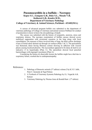
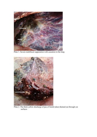
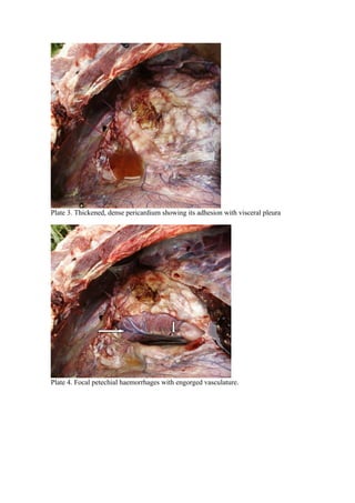
Pneumocarditis in a buffalo – Necropsy
           Kapse S.U., Gangane G.R., Dake S.L., Musale V.B,
                     Kulkarni G.B., Kondre B.M.,
                  Department of Veterinary Pathology
   College of Veterinary & Animal Sciences, Parbhani - 431402(M.S.)

         A carcass of advanced pregnant buffalo was submitted to the department of
   veterinary pathology of College of veterinary and animal sciences Parbhani for conduct
   of postmortem to know to know the etio-pathology of death.
         The carcass was submitted with the history of tympanitis, anorexia, stress and
   respiratory distress. The necropsy examination of buffalo carcass showed severe
   multifocal suppuration with occasional caseation in the lung along with focal
   consolidation. On exposure of these involved lobes of lung, the thick yellow discharge
   of pus of foetid odour drained out through cut surfaces (Plate 1 & 2.). The pericardium
   was thickened, dense having fibrinous content showing its adhesion with visceral
   pleura causing localized pleuritis. The myocardium appeared to be dense & hard to cut
   indicative if its hypertrophy (Plate 3.). The epicardium showed focal petechial
   haemorrhages with engorged vasculature (Plate 4.).
         Considering the history & observable lesions the buffalo might have died due to
   respiratory failure, resulted due to cardiopneumopathy.


References:
              1. Pathology of Domestic animals (3rd edition) volume-2 by K.V.F. Jubb,
                 Peter C. Kennedy & Nigel Palmer.
              2. A Textbook of Veterinary Systemic Pathology by J.L. Vegad & A.K.
                 Katiyar
              3. Veterinary Pathology by Thomas Jones & Ronald Hunt ( 5th edition)
Plate 1. Severe multifocal suppuration with caseation in the lung


Plate 2. The thick yellow discharge of pus of foetid odour drained out through cut
         surfaces
Plate 3. Thickened, dense pericardium showing its adhesion with visceral pleura


Plate 4. Focal petechial haemorrhages with engorged vasculature.

Pneumocarditis

  • 1.
    Pneumocarditis in abuffalo – Necropsy Kapse S.U., Gangane G.R., Dake S.L., Musale V.B, Kulkarni G.B., Kondre B.M., Department of Veterinary Pathology College of Veterinary & Animal Sciences, Parbhani - 431402(M.S.) A carcass of advanced pregnant buffalo was submitted to the department of veterinary pathology of College of veterinary and animal sciences Parbhani for conduct of postmortem to know to know the etio-pathology of death. The carcass was submitted with the history of tympanitis, anorexia, stress and respiratory distress. The necropsy examination of buffalo carcass showed severe multifocal suppuration with occasional caseation in the lung along with focal consolidation. On exposure of these involved lobes of lung, the thick yellow discharge of pus of foetid odour drained out through cut surfaces (Plate 1 & 2.). The pericardium was thickened, dense having fibrinous content showing its adhesion with visceral pleura causing localized pleuritis. The myocardium appeared to be dense & hard to cut indicative if its hypertrophy (Plate 3.). The epicardium showed focal petechial haemorrhages with engorged vasculature (Plate 4.). Considering the history & observable lesions the buffalo might have died due to respiratory failure, resulted due to cardiopneumopathy. References: 1. Pathology of Domestic animals (3rd edition) volume-2 by K.V.F. Jubb, Peter C. Kennedy & Nigel Palmer. 2. A Textbook of Veterinary Systemic Pathology by J.L. Vegad & A.K. Katiyar 3. Veterinary Pathology by Thomas Jones & Ronald Hunt ( 5th edition)
  • 2.
    Plate 1. Severemultifocal suppuration with caseation in the lung Plate 2. The thick yellow discharge of pus of foetid odour drained out through cut surfaces
  • 3.
    Plate 3. Thickened,dense pericardium showing its adhesion with visceral pleura Plate 4. Focal petechial haemorrhages with engorged vasculature.