KESSY JULIUS, P., MD.
Obstetrics & Gynecology Department,
Njombe Regional Referral Hospital.
18th August, 2020.
PLACENTA
ABRUPTION
1. INTRODUCTION
2. Epidemiology
3. Placentalanatomy
4. PATHOPHYSIOLOGY
5. RISKFACTORS
6. CLASSIFICATION
7. SIGNSANDSYMPTOMS
8. COMPLICATIONS
9. DIAGNOSIS
10. MANAGEMENT
OUTLINE
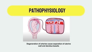
Placenta abruption is the premature
separation of all or part of placenta
from the uterine wall resulting in
bleeding (hemorrhage).
Introduction
Most common after 20 weeks of
gestation.
Significant cause of perinatal
morbidity and mortality.
Introduction
20 weeks
Third trimester
Affects 1% of all pregnancies
worldwide.
epidemiology
PLACENTALANATOMY
The embryo
attaches to the
uterine wall.
Placenta forms
from the maternal
and fetal.
Permits gas and
nutrient
exchange.
Pathophysiology
Degeneration of arteries cause separation of uterine
wall and decidua basalis
RISKFACTORS
1. Blunt trauma (fall, domestic violence, car crush, etc.
2. Drug use e.g. Cocaine and methamphetamine.
3. Multiparity
4. Maternal age (< 20 and > 35 years)
5. Previous abruptions
6. Chronic hypertension with pre-eclampsia
7. Cigarette smoking
8. Chorioamnionitis
9. Oligohydramnios
CLASSIFICATION
1. CONCEALED
• Intact placental edges with
hemorrhage within the decidua
basalis beneath the placenta.
• Spotting or limited bleeding.
• Dark red or clotted blood.
• No bleeding.
• Unexplained hypotension or
unexplained anemia.
CLASSIFICATION
2. REVEALED
•Placental edges fully removed
from maternal tissue, blood leaks
into the uterine space, and exits
through the vagina.
•Bright red bleeding.
•More bleeding than in concealed.
•The amount of bleeding doesn’t
correlate to severity of
hemorrhage.
SIGNS&SYMPTOMS
1. Vaginal bleeding or spotting
2. Abdominal pain or cramping
3. ↑Frequency ↓Amplitude contractions
4. Tender and hard uterus on palpation
5. Abnormalities in FHR
6. Pre-mature labour
COMPLICATIONS
MATERNAL
Hypovolemic shock
Sheehan syndrome
Acute Kidney Injury
DIC
FETAL
Intrauterine hypoxia and
asphyxia
Premature birth
IUGR
Fetal death
DIAGNOSIS
CLINICALLY IMAGING
Ultrasound – Retro placental
collection of blood
MANAGEMENT
Placenta abruption is an emergency
Management depends on the maternal and fetal physiological status
IV fluids to increase circulating volume
Blood products to prevent coagulation
Emergency cesarean section if there is severe hemorrhage and fetal compromise

Placenta abruption

  • 1.
    KESSY JULIUS, P.,MD. Obstetrics & Gynecology Department, Njombe Regional Referral Hospital. 18th August, 2020. PLACENTA ABRUPTION
  • 2.
    1. INTRODUCTION 2. Epidemiology 3.Placentalanatomy 4. PATHOPHYSIOLOGY 5. RISKFACTORS 6. CLASSIFICATION 7. SIGNSANDSYMPTOMS 8. COMPLICATIONS 9. DIAGNOSIS 10. MANAGEMENT OUTLINE
  • 3.
    Placenta abruption isthe premature separation of all or part of placenta from the uterine wall resulting in bleeding (hemorrhage). Introduction
  • 4.
    Most common after20 weeks of gestation. Significant cause of perinatal morbidity and mortality. Introduction 20 weeks Third trimester
  • 5.
    Affects 1% ofall pregnancies worldwide. epidemiology
  • 6.
    PLACENTALANATOMY The embryo attaches tothe uterine wall. Placenta forms from the maternal and fetal. Permits gas and nutrient exchange.
  • 7.
    Pathophysiology Degeneration of arteriescause separation of uterine wall and decidua basalis
  • 8.
    RISKFACTORS 1. Blunt trauma(fall, domestic violence, car crush, etc. 2. Drug use e.g. Cocaine and methamphetamine. 3. Multiparity 4. Maternal age (< 20 and > 35 years) 5. Previous abruptions 6. Chronic hypertension with pre-eclampsia 7. Cigarette smoking 8. Chorioamnionitis 9. Oligohydramnios
  • 9.
    CLASSIFICATION 1. CONCEALED • Intactplacental edges with hemorrhage within the decidua basalis beneath the placenta. • Spotting or limited bleeding. • Dark red or clotted blood. • No bleeding. • Unexplained hypotension or unexplained anemia.
  • 10.
    CLASSIFICATION 2. REVEALED •Placental edgesfully removed from maternal tissue, blood leaks into the uterine space, and exits through the vagina. •Bright red bleeding. •More bleeding than in concealed. •The amount of bleeding doesn’t correlate to severity of hemorrhage.
  • 11.
    SIGNS&SYMPTOMS 1. Vaginal bleedingor spotting 2. Abdominal pain or cramping 3. ↑Frequency ↓Amplitude contractions 4. Tender and hard uterus on palpation 5. Abnormalities in FHR 6. Pre-mature labour
  • 12.
    COMPLICATIONS MATERNAL Hypovolemic shock Sheehan syndrome AcuteKidney Injury DIC FETAL Intrauterine hypoxia and asphyxia Premature birth IUGR Fetal death
  • 13.
    DIAGNOSIS CLINICALLY IMAGING Ultrasound –Retro placental collection of blood
  • 14.
    MANAGEMENT Placenta abruption isan emergency Management depends on the maternal and fetal physiological status IV fluids to increase circulating volume Blood products to prevent coagulation Emergency cesarean section if there is severe hemorrhage and fetal compromise