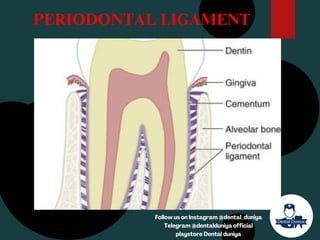
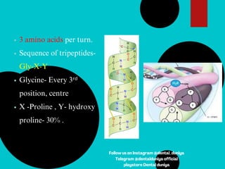
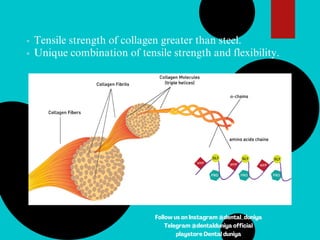
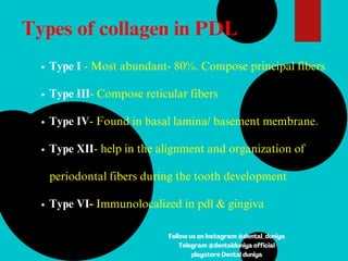
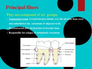
Detail explantion about periodontal ligament .
The periodontal ligament (PDL) is a crucial component in the field of periodontics, playing a pivotal role in the support and maintenance of tooth structure. This connective tissue is situated between the cementum of the tooth and the alveolar bone of the jaw, creating a dynamic and complex environment within the oral cavity.
3. Introduction
Periodontium
It is a dynamic structure composed
of the tissues supporting and investing
the teeth.
Peri – around
Odontos - tooth
Four principal components
Gingiva.
1.
Periodontal ligament.
2.
Cementum.
3.
Alveolar bone.
4.
Follow us on Instagram @dental_duniya
Telegram @dentalduniya official
playstore Dental duniya
4. Periodontal ligament
It is composed of complex vascular and highly cellular connective
tissue that surrounds the tooth root and connects it to the inner wall
of the alveolar bone.
Shape : Hourglass.
Other names:
Periodontal membrane
Pericemental membrane
Alveolo-dental ligament
Desmodent
Gomphosis
Follow us on Instagram @dental_duniya
Telegram @dentalduniya official
playstore Dental duniya
5. Periodontal ligament space = 0.2mm.
Decreased in teeth which are not in
function/unerupted teeth.
Increased in teeth subjected to
hyperfunction .
Follow us on Instagram @dental_duniya
Telegram @dentalduniya official
playstore Dental duniya
7. Periodontal fibers
Principal fibers collagenous.
1.
Non- collagenous fibres/elastic fibers.
2.
Indifferent fiber plexus.
3.
Follow us on Instagram @dental_duniya
Telegram @dentalduniya official
playstore Dental duniya
8. Collagen
Collagen molecule –
Tropocollagen -3 alpha chains.
Three helical alpha chains- coiled
around each other triple helix
rigid structure like rope.
Follow us on Instagram @dental_duniya
Telegram @dentalduniya official
playstore Dental duniya
9. 3 amino acids per turn.
Sequence of tripeptides-
Gly-X-Y
Glycine- Every 3ʳᵈ
position, centre
X -Proline , Y- hydroxy
proline- 30% .
Follow us on Instagram @dental_duniya
Telegram @dentalduniya official
playstore Dental duniya
10. Most abundant amino acids present in
collagen:
Glycine
Proline
Hydroxylysine and
Hydroxyproline
Follow us on Instagram @dental_duniya
Telegram @dentalduniya official
playstore Dental duniya
11. Tensile strength of collagen greater than steel.
Unique combination of tensile strength and flexibility.
Follow us on Instagram @dental_duniya
Telegram @dentalduniya official
playstore Dental duniya
12. Types of collagen in PDL
Type I - Most abundant- 80%. Compose principal fibers
Type III- Compose reticular fibers
Type IV- Found in basal lamina/ basement membrane.
Type XII- help in the alignment and organization of
periodontal fibers during the tooth development
Type VI- Immunolocalized in pdl & gingiva
Follow us on Instagram @dental_duniya
Telegram @dentalduniya official
playstore Dental duniya
13. Principal fibers
They are composed of six groups
Transseptal group: Extend interproximally over the alveolar bone crest
and embedded in the cementum of adjacent teeth.
Reconstructed after destruction of alveolar bone.
Responsible for relapse of orthodontic treatment.
14. Alveolar crest group
From alv crest to cementum just beneath the junctional ep.
prevents extrusion of tooth)
incision of these fibres during periodontal sugery doesnt
lead to increase in mobility.
19. Follow us on Instagram @dental_duniya
Telegram @dentalduniya official
playstore Dental duniya
20. Sharpey’s fibres
Terminal portions of the
principal fibers
Calcify to a significant
degree
Associated with proteins :
osteopontin and
bone sialoprotein
Follow us on Instagram @dental_duniya
Telegram @dentalduniya official
playstore Dental duniya
21. Non collagenous fibers/elastic fibres:
Two immature forms found
are:-
oxytalan fibers
Eluanin fibers
Functions
Regulate vascular flow
Role in tooth support
Facilitate fibroblast attachment
and migration
22. Indifferent fiber plexus
Small collagen fibers along with large principal
fibers
Randomly oriented forming plexus.
Follow us on Instagram @dental_duniya
Telegram @dentalduniya official
playstore Dental duniya
23. Cellular elements
Connective tissue cells
Epithelial cells
Immune system cells
Cells associated with neuro- vascular
elements
Follow us on Instagram @dental_duniya
Telegram @dentalduniya official
playstore Dental duniya
24. Fibroblasts:
Most common cells
Ovoid, elongated or stellate str.
Oriented along principal fibers
Synthesize collagen
Connective tissue cells
Follow us on Instagram @dental_duniya
Telegram @dentalduniya official
playstore Dental duniya
25. Possess capacity to phagocytose
old collagen & degrade them – enzyme hydrolysis
Regulates collagen turnover
Follow us on Instagram @dental_duniya
Telegram @dentalduniya official
playstore Dental duniya
26. Epithelial rests of Malassez
Considered as remnants of hertwig’s
epithelial root sheath
Distributed close to cementum and
throughout the periodontal ligament of
most teeth.
Clinical considertaions
Periapical cyst
Lateral root cyst
28. Ground substance:
Contains 70% water
Proteoglycans participates in various biological functions
like cell adhesion, cell-cell matrix interaction.
Glycosaminoglycans
Glycoprotein
Hyaluronic acid
Proteoglycans
Fibronectin
Osteopontin
29. CEMENTICLES
Calcified masses found adherent to or detached from the root
surfaces.
Develop from
Calcified epithelial rests
Small spicules of cementum
Alveolar bone traumatically displaced into pdl
Calcified sharpey’s fibers
Thrombosed vessels with in pdl
31. Physical Function
Protection- provide sof tissue casing for nerves
and vessels.
1.
Transmission of occlusal forces to the bone.
2.
Shock absorption: resistance to the impact of
occlusal forces.
3.
Attachment of the teeth to the bone.
4.
Maintenance of the gingival tissues in the
5.
33. Tensional theory
Forces are applied
principal fibers unfold & straighten
transmission of forces to alveolar bone
elastic deformation of the bony socket
transmitted to the basal bone
34. Viscoelastic theory
Extracellular fluid passes from pdl to marrow spaces of
bone
depletion of tissue
fluid
Fibers slacken & tighten
Blood vessels
stenosis
Forces are
applied
Arterial back pressure causes ballooning of vessels
& passage of blood into tissues, replenishing tissue
35. TRANSMISSION OF OCCLUSAL
FORCES TO THE BONE
When there is axial force-
oblique fibres become taut-
assume their full length -
sustain the force
When there is horizontal
force- the tooth rotates
around centre of rotation-
Area of compression- fibres
are compressed
Area of tension- fibres are
tensed
36. Helps in formation & resorption of
cementum and bone during physiologic tooth
movement.
Constantly undergoes remodeling.
Formative and Remodeling
Function
37. Supplies nutrients to cementum ,bone & gingiva
Abundantly supplied with sensory nerve fibers capable
of transmitting tactile ,pressure and pain sensation by
trigeminal pathway.
Nutritional and sensory function
38. Free nerve endings-pain sensation
Ruffini like mechano receptors-apical region
Meissner’s corpuscles(mechano receptors)-mid
root region
Spindle like(pressure,vibration nerve endings)-
apical region
4 TYPES OF NEURAL
TERMINATIONS :
40. BLOOD SUPPLY
Inferior and superior alveolar
arteries
Branches from
Apical vessels
1.
Penetrating vessels from the
alveolar bone
2.
Anastomosing vessels from the
gingiva.
3.
42. Follow @dental_duniya for new updates of
medical and dental jobs
,check stories for recent job opportunities,
check out our website for question papers ,e
books ,dentalassistance for dental students
share our instgram page with friends and
colleagues