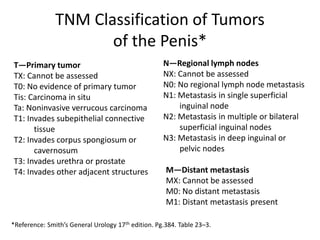
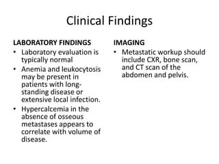
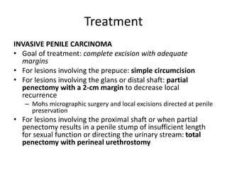
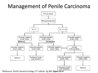
TUMORS OF THE PENIS
Tumors of the penis
• less than 1% of cancers among males
• The one etiologic factor most commonly
associated with penile carcinoma is poor
hygiene
• The disease is virtually unheard of in males
circumcised near birth.
Tumors of the penis
• One theory postulates that smegma
accumulation under the phimotic foreskin
results in chronic inflammation leading to
carcinoma.
• A viral cause has also been suggested as a
result of the association of this tumor with
cervical carcinoma.
CARCINOMA IN SITU
BOWEN DISEASE
• squamous cell carcinoma in situ typically involving the
penile shaft.
• The lesion appears as a red plaque with encrustations
ERYTHROPLASIA OF QUEYRAT
• a velvety, red lesion with ulcerations
• involve the glans
• Microscopic examination shows typical, hyperplastic
cells in a disordered array with vacuolated cytoplasm
and mitotic figures.
INVASIVE CARCINOMA OF THE PENIS
Squamous cell carcinoma
• composes most penile cancers.
• most commonly originates on the glans
• Other common sites: prepuce and shaft
• The appearance may be papillary or ulcerative.
Verrucous carcinoma
• a variant of squamous cell carcinoma composing 5–16% of
penile carcinomas
• papillary in appearance
• have a well-demarcated deep margin unlike the infiltrating
margin of the typical squamous cell carcinoma on histology
TNM Classification of Tumors
of the Penis*
T—Primary tumor
TX: Cannot be assessed
T0: No evidence of primary tumor
Tis: Carcinoma in situ
Ta: Noninvasive verrucous carcinoma
T1: Invades subepithelial connective
tissue
T2: Invades corpus spongiosum or
cavernosum
T3: Invades urethra or prostate
T4: Invades other adjacent structures
N—Regional lymph nodes
NX: Cannot be assessed
N0: No regional lymph node metastasis
N1: Metastasis in single superficial
inguinal node
N2: Metastasis in multiple or bilateral
superficial inguinal nodes
N3: Metastasis in deep inguinal or
pelvic nodes
M—Distant metastasis
MX: Cannot be assessed
M0: No distant metastasis
M1: Distant metastasis present
*Reference: Smith’s General Urology 17th edition. Pg.384. Table 23–3.
Clinical Findings
SYMPTOMS
• may appear as an area of
induration or erythema,
• an ulceration, a small
nodule, or an exophytic
growth
• Phimosis may obscure the
lesion and result in a delay
in seeking medical attention
• pain, discharge, irritative
voiding symptoms, and
bleeding
SIGNS
• Lesions are typically
confined to the penis at
presentation
• more than 50% of
patients present with
enlarged inguinal
nodes.
Clinical Findings
LABORATORY FINDINGS
• Laboratory evaluation is
typically normal
• Anemia and leukocytosis
may be present in
patients with long-
standing disease or
extensive local infection.
• Hypercalcemia in the
absence of osseous
metastases appears to
correlate with volume of
disease.
IMAGING
• Metastatic workup should
include CXR, bone scan,
and CT scan of the
abdomen and pelvis.
Treatment
PRIMARY LESION
• Biopsy of the primary lesion - to establish the
diagnosis of malignancy
CARCINOMA IN SITU
• treated conservatively in reliable patients
• Fluorouracil cream application or
neodymium:YAG laser treatment
Treatment
INVASIVE PENILE CARCINOMA
• Goal of treatment: complete excision with adequate
margins
• For lesions involving the prepuce: simple circumcision
• For lesions involving the glans or distal shaft: partial
penectomy with a 2-cm margin to decrease local
recurrence
– Mohs micrographic surgery and local excisions directed at penile
preservation
• For lesions involving the proximal shaft or when partial
penectomy results in a penile stump of insufficient length
for sexual function or directing the urinary stream: total
penectomy with perineal urethrostomy
Treatment: Lymph Nodes
• Enlarged node commonly due to inflammation
• Should undergo treatment of the primary lesion followed by a 4- to 6-week course of
oral broad-spectrum antibiotics
• sequential bilateral ilioinguinal node dissections
– For persistent adenopathy following antibiotic treatment
• observation in low-stage primary tumors (Tis, T1)
– For Resolved lymphadenopathy with antibiotics
• sentinel node biopsy or a modified (limited) dissection
– If lymphadenopathy resolves in higher-stage tumors, more limited lymph node
samplings should be considered
• bilateral ilioinguinal node dissection
– If positive nodes are encountered
• unilateral ilioinguinal node dissection
– Patients who initially have clinically negative nodes but in whom clinically palpable
nodes later develop
• chemotherapy (cisplatin and 5-fluorouracil)
– Patients who have inoperable disease and bulky inguinal metastases
• Regional radiotherapy
– For palliation by delaying ulceration and infectious complications and alleviating
pain.
Management of Penile Carcinoma
*Reference: Smith’s General Urology 17th edition. Pg.386. Figure 23–4
TUMORS OF THE SCROTUM
Tumors of the Scrotum
• Tumors of the scrotal skin are rare.
• The most common benign lesion is a sebaceous cyst
• Most common malignant tumor of the scrotum is
Squamous cell carcinoma
• Rare cases: melanoma, basal cell carcinoma, and
Kaposi sarcoma
• Etiology of SCC of the Scrotum: poor hygiene and
chronic inflammation
Tumors of the Scrotum: Management
• Biopsy
• Wide excision with a 2-cm margin should be
performed for malignant tumors
• Surrounding subcutaneous tissue should be
excised with the primary tumor
• Primary closure using the redundant scrotal skin
is usually possible.
• The management of inguinal nodes should be
similar to that of penile cancer.
Tumors of the Scrotum: Prognosis
Prognosis correlates with the presence or
absence of nodal involvement.
In the presence of inguinal node metastasis, the
5-year survival rate is approximately 25%
There are virtually no survivors if iliac nodes are
involved.

PENILE CARCINOMA

  • 1.
  • 2.
    Tumors of thepenis • less than 1% of cancers among males • The one etiologic factor most commonly associated with penile carcinoma is poor hygiene • The disease is virtually unheard of in males circumcised near birth.
  • 3.
    Tumors of thepenis • One theory postulates that smegma accumulation under the phimotic foreskin results in chronic inflammation leading to carcinoma. • A viral cause has also been suggested as a result of the association of this tumor with cervical carcinoma.
  • 4.
    CARCINOMA IN SITU BOWENDISEASE • squamous cell carcinoma in situ typically involving the penile shaft. • The lesion appears as a red plaque with encrustations ERYTHROPLASIA OF QUEYRAT • a velvety, red lesion with ulcerations • involve the glans • Microscopic examination shows typical, hyperplastic cells in a disordered array with vacuolated cytoplasm and mitotic figures.
  • 5.
    INVASIVE CARCINOMA OFTHE PENIS Squamous cell carcinoma • composes most penile cancers. • most commonly originates on the glans • Other common sites: prepuce and shaft • The appearance may be papillary or ulcerative. Verrucous carcinoma • a variant of squamous cell carcinoma composing 5–16% of penile carcinomas • papillary in appearance • have a well-demarcated deep margin unlike the infiltrating margin of the typical squamous cell carcinoma on histology
  • 6.
    TNM Classification ofTumors of the Penis* T—Primary tumor TX: Cannot be assessed T0: No evidence of primary tumor Tis: Carcinoma in situ Ta: Noninvasive verrucous carcinoma T1: Invades subepithelial connective tissue T2: Invades corpus spongiosum or cavernosum T3: Invades urethra or prostate T4: Invades other adjacent structures N—Regional lymph nodes NX: Cannot be assessed N0: No regional lymph node metastasis N1: Metastasis in single superficial inguinal node N2: Metastasis in multiple or bilateral superficial inguinal nodes N3: Metastasis in deep inguinal or pelvic nodes M—Distant metastasis MX: Cannot be assessed M0: No distant metastasis M1: Distant metastasis present *Reference: Smith’s General Urology 17th edition. Pg.384. Table 23–3.
  • 7.
    Clinical Findings SYMPTOMS • mayappear as an area of induration or erythema, • an ulceration, a small nodule, or an exophytic growth • Phimosis may obscure the lesion and result in a delay in seeking medical attention • pain, discharge, irritative voiding symptoms, and bleeding SIGNS • Lesions are typically confined to the penis at presentation • more than 50% of patients present with enlarged inguinal nodes.
  • 8.
    Clinical Findings LABORATORY FINDINGS •Laboratory evaluation is typically normal • Anemia and leukocytosis may be present in patients with long- standing disease or extensive local infection. • Hypercalcemia in the absence of osseous metastases appears to correlate with volume of disease. IMAGING • Metastatic workup should include CXR, bone scan, and CT scan of the abdomen and pelvis.
  • 9.
    Treatment PRIMARY LESION • Biopsyof the primary lesion - to establish the diagnosis of malignancy CARCINOMA IN SITU • treated conservatively in reliable patients • Fluorouracil cream application or neodymium:YAG laser treatment
  • 10.
    Treatment INVASIVE PENILE CARCINOMA •Goal of treatment: complete excision with adequate margins • For lesions involving the prepuce: simple circumcision • For lesions involving the glans or distal shaft: partial penectomy with a 2-cm margin to decrease local recurrence – Mohs micrographic surgery and local excisions directed at penile preservation • For lesions involving the proximal shaft or when partial penectomy results in a penile stump of insufficient length for sexual function or directing the urinary stream: total penectomy with perineal urethrostomy
  • 11.
    Treatment: Lymph Nodes •Enlarged node commonly due to inflammation • Should undergo treatment of the primary lesion followed by a 4- to 6-week course of oral broad-spectrum antibiotics • sequential bilateral ilioinguinal node dissections – For persistent adenopathy following antibiotic treatment • observation in low-stage primary tumors (Tis, T1) – For Resolved lymphadenopathy with antibiotics • sentinel node biopsy or a modified (limited) dissection – If lymphadenopathy resolves in higher-stage tumors, more limited lymph node samplings should be considered • bilateral ilioinguinal node dissection – If positive nodes are encountered • unilateral ilioinguinal node dissection – Patients who initially have clinically negative nodes but in whom clinically palpable nodes later develop • chemotherapy (cisplatin and 5-fluorouracil) – Patients who have inoperable disease and bulky inguinal metastases • Regional radiotherapy – For palliation by delaying ulceration and infectious complications and alleviating pain.
  • 12.
    Management of PenileCarcinoma *Reference: Smith’s General Urology 17th edition. Pg.386. Figure 23–4
  • 13.
  • 14.
    Tumors of theScrotum • Tumors of the scrotal skin are rare. • The most common benign lesion is a sebaceous cyst • Most common malignant tumor of the scrotum is Squamous cell carcinoma • Rare cases: melanoma, basal cell carcinoma, and Kaposi sarcoma • Etiology of SCC of the Scrotum: poor hygiene and chronic inflammation
  • 15.
    Tumors of theScrotum: Management • Biopsy • Wide excision with a 2-cm margin should be performed for malignant tumors • Surrounding subcutaneous tissue should be excised with the primary tumor • Primary closure using the redundant scrotal skin is usually possible. • The management of inguinal nodes should be similar to that of penile cancer.
  • 16.
    Tumors of theScrotum: Prognosis Prognosis correlates with the presence or absence of nodal involvement. In the presence of inguinal node metastasis, the 5-year survival rate is approximately 25% There are virtually no survivors if iliac nodes are involved.

Editor's Notes

  • #7 Tumor Staging The staging system used most commonly in the United States was proposed by Jackson (1966), as follows: In stage I, the tumor is confined to the glans or prepuce. Stage II involves the penile shaft. Stage III has operable inguinal node metastasis. In stage IV, the tumor extends beyond the penile shaft, with inoperable inguinal or distant metastases. The TNM classification of the American Joint Committee (1996) is given in Table 23–3.