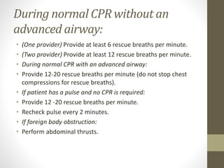
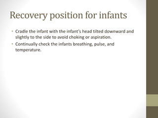
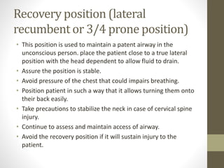
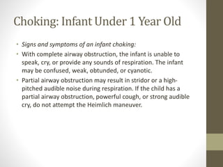
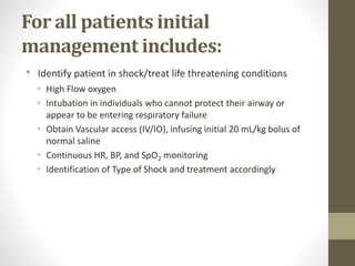
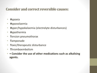
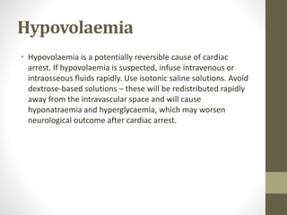
2. Objectives
• Be able to define PALS
• Be familiar with common sings
• Be able to identify
• Take promote resuscitative actions when necessary
• Know drugs being in use in PALS
• Identify reversible causes
3. Introduction
• Pediatric Life Support describes protocols to
resuscitate critically ill pediatric patients.
• Basic Life Support (BLS) is intended for use by
both healthcare professionals and laypersons to
maintain individuals in nonhospital or limited-
resource settings until they can be stabilized in a
healthcare facility.
4. Pediatric Advanced Life Support
(PALS)
• Is a training to healthcare providers to enforce a
systematic approach to evaluation, management, and
resuscitation of critically ill patients.
• Survival to discharge from in hospital cardiac arrest in
pediatric patients has improved since 2001, with return
of spontaneous circulation (ROSC) rates from 39% to
77% and survival to discharge from 24% to 43%.
• Prolonged CPR also has been shown to be favorable, with
12% of patients surviving. 60% of those survivors had a
favorable neurological outcome at discharge.
• American Heart Association (AHA) in 2015
5. Definition
• PALS is a specialized training for emergency care of children
from the AHA training guidelines. The care includes infants,
children and adolescents that require life support following
injury or SCA.
6. CPR: for infants 0-1 in age
• CABD (Circulation, Airway, Breathing, Defibrillate)
• An infant is found lying on the ground.
• Assess to make sure the scene is safe for you to respond to
the down patient.
• Assess Unresponsiveness: Lightly shake or tap the infant’s foot
and say their name. Look at the chest and torso for movement
and normal breathing.
7. If the infant is unresponsive:
• (One provider) If alone and collapse is un-witnessed: First
perform 2 minutes of CPR then call the emergency response
team and bring an AED to the patient.
• (One provider) If alone and collapse is witnessed: First call the
emergency response team and bring an AED, then start CPR.
• (Two providers) Have someone near call the emergency
response team and bring the AED and you start CPR.
• Place patient supine on a hard flat surface.
8. Circulation
• Feel for either the brachial or femoral pulse (Do not check for
more than 10 seconds).
• If the infant has a pulse:
• Move to the airway and rescue breathing portion of the
algorithm.
• Give 12-20 breaths per minute.
• Recheck the pulse every 2 minutes.
• If the infant doesn’t have a pulse:
• Begin 5 cycles of CPR (lasts approximately 2 minutes).
9.
10. Start with Chest Compressions:
• Provide 100 to 120 compressions per minute. This is 30
compressions every 15 to 18 seconds.
• (One provider) Place two fingers on the sternum of the lower
chest. One between the nipple line and the other 1cm below.
• (Two providers) Encircle the infant’s torso with both hands
with both thumbs pointing cephalic positioned 1cm below the
nipples over the sternum.
• Chest Compressions should be at least 1.5 inches or 1/3 the
depth of infant’s chest.
• Press hard and fast.
• Allow for full chest recoil.
11.
12. • Only allow minimal interruptions to the chest compressions.
One Provider: 1 cycle is 30 chest compressions to 2 rescue
breaths) (Two Providers: 1 cycle is 15 chest compressions to 2
rescue breaths)
• If you have two providers: switch rolls between compressor
and rescue breather every 2 minutes or 5 cycles of CPR.
13. Airway
• In the event of an unwitnessed collapse, drowning, or trauma:
• Use the Jaw-Thrust maneuver. (This maneuver is used when
cervical spine injury cannot be ruled out.):
• Place your thumbs on the upper cheek bones of the infant.
• Place your fingers on the lower rami of the jaw.
• Provide anterior pressure to advance the jaw forward.
14. • In the event of a witnessed collapse and there’s no reason to
assume C-spine injury:
• Use the Head Tilt-Chin Lift maneuver:
• place your palm on the patient’s forehead and apply pressure
to tilt the head backward.
• place the fingers of your other hand under the mental
protuberance of the chin and pull the chin forward and
cephalic.
15. Breathing
• Scan the patients chest and torso for possible movement
during the “assess unresponsiveness” portion of the
algorithm. Watch for abnormal breathing or gasping.
• If the infant has adequate breathing:
• Continue to assess and maintain a patent airway and place the
infant in the infant recovery position. (Only use the recovery
position if its unlikely to worsen patient injury)
16. • If the infant is not breathing or is inadequately breathing:
• If the infant has a pulse:
• commence rescue breaths immediately.
• If the infant doesn’t have a pulse:
• begin CPR (go to Circulation portion of the algorithm).
17. • Use a barrier device if available.
• Make a seal using your mouth over the mouth and nose of the
patient.
• Each rescue breath should be small and last approximately 1
second.
• Watch for chest rise.
• Allow time for the air to expel from the patient.
18. During normal CPR with an
advanced airway:
• Provide 12-20 rescue breaths per minute (do not stop chest
compressions for rescue breaths).
• If the patient has a pulse and no CPR is required:
• Provide 12-20 rescue breaths per minute.
• Recheck pulse every 2 minutes.
19. Defibrillate
• Arrival of AED Power:
• Turn AED On NOW! (early defibrillation is the single most
important therapy for survival of cardiac arrest. Begin use on
patient as soon as it arrives).
• Follow verbal AED prompts.
• Attachment:
• Firmly place appropriate pads (adult/pediatric) to patient’s
skin to the indicated locations (pad image).
20.
21. Analyze:
• A short pause in CPR is required to allow the AED to analyze
the rhythm.
• If the rhythm is not shockable:
• Initiate 5 cycles of CPR.
• Recheck the rhythm at the end of the 5 cycles of CPR.
• If shock is indicated:
• Assure no one is touching the patient or in mutual contact of a
good conductor of electricity by yelling “Clear, I’m Clear,
you’re Clear!” prior to delivering a shock.
• Press the shock button when the providers are clear of the
patient.
• Resume 5 cycles of CPR.
22. • Manual defibrillators are preferred for infant use. If the
manuals defibrillator is not available the next best option is an
AED with a pediatric attenuator. An AED without a pediatric
attenuator can also be used.
• CPR: for a child older than 1 year of age to puberty
• CABD (Circulation, Airway, Breathing, Defibrillate)
23. If not or inadequate breathing:
• has a pulse: Commence rescue breaths immediately.
• no pulse: Begin CPR (go to Circulation portion of the
algorithm).
• Use a barrier device if available.
• Pinch the patient’s nose closed.
• Make a seal using your mouth over the mouth of the patient.
• Each rescue breath should last approximately 1 second.
• Watch for chest rise.
• Allow time for the air to expel from patient.
24. During normal CPR without an
advanced airway:
• (One provider) Provide at least 6 rescue breaths per minute.
• (Two provider) Provide at least 12 rescue breaths per minute.
• During normal CPR with an advanced airway:
• Provide 12-20 rescue breaths per minute (do not stop chest
compressions for rescue breaths).
• If patient has a pulse and no CPR is required:
• Provide 12 -20 rescue breaths per minute.
• Recheck pulse every 2 minutes.
• If foreign body obstruction:
• Perform abdominal thrusts.
25. Recovery position for infants
• Cradle the infant with the infant’s head tilted downward and
slightly to the side to avoid choking or aspiration.
• Continually check the infants breathing, pulse, and
temperature.
26. Recovery position (lateral
recumbent or 3/4 prone position)
• This position is used to maintain a patent airway in the
unconscious person. place the patient close to a true lateral
position with the head dependent to allow fluid to drain.
• Assure the position is stable.
• Avoid pressure of the chest that could impairs breathing.
• Position patient in such a way that it allows turning them onto
their back easily.
• Take precautions to stabilize the neck in case of cervical spine
injury.
• Continue to assess and maintain access of airway.
• Avoid the recovery position if it will sustain injury to the
patient.
27. Choking: Infant Under 1 Year Old
• Signs and symptoms of an infant choking:
• With complete airway obstruction, the infant is unable to
speak, cry, or provide any sounds of respiration. The infant
may be confused, weak, obtunded, or cyanotic.
• Partial airway obstruction may result in stridor or a high-
pitched audible noise during respiration. If the child has a
partial airway obstruction, powerful cough, or strong audible
cry, do not attempt the Heimlich maneuver.
28. If signs andsymptoms of chokingare present
and infantis conscious:
• (one provider) immediately call the emergency response
team.
• (one provider) Assess the airway for any visually present
obstruction and manually remove it if possible.
• (two provider) Send someone to call the emergency response
team while you assess the airway.
• Never use a blind finger sweep.
29. Position the patient:
• Lay infant’s face and torso down on forearm (prone) with
chest being supported by your palm and their head and neck
by your fingers.
• Tilt the infant’s body at a 30 degree angle, head downward
(trandelenburg).
• Use your thigh or other object for support.
• Interventional Back Blows:
• Provide 5 rapid forceful blows using a flat palm on the infant’s
back between the two scapula.
• Reposition the patient:
• Rotate the infant face up (supine), head downward
(trandelenburg) by switching the infant to the opposite arm.
30.
31. Interventional Chest Thrusts:
• Place your two fingers on the center of the infant’s sternum
immediately below the nipple line.
• Provide 5 rapid compressions, with thrusts equaling 1/3 to 1/2
the total depth of the chest.
• Continue cycling back and forth between interventional back
blows and chest thrusts until the obstruction is removed or
until consciousness is lost.
• If becomes unconscious:
• Initiate CPR.
• Before attempting rescue breaths during normal CPR, assess
the airway, removing any visually present obstruction.
• Do not use a blind finger sweep in an attempt to remove an
obstruction.
32.
33. Stabilization of Patients in Shock
• Critical to care for patients in shock is early
identification, signs and symptoms include
• Diminished pulses
• Cool, pale, mottled skin
• Increased capillary refill time
• Altered Mental Status
• Vital Sign Abnormalities (importantly, individuals in
shock may not display vital sign abnormalities but can
display tachypnea, tachycardia, hypotension, pulse
pressure abnormalities)
• ALS protocols aim to identify the type of shock to target
the therapeutic interventions.
34. For all patients initial
management includes:
• Identify patient in shock/treat life threatening conditions
• High Flow oxygen
• Intubation in individuals who cannot protect their airway or
appear to be entering respiratory failure
• Obtain Vascular access (IV/IO), infusing initial 20 mL/kg bolus of
normal saline
• Continuous HR, BP, and SpO2 monitoring
• Identification of Type of Shock and treatment accordingly
35. Considerandcorrectreversiblecauses:
• Hypoxia
• Hypovolaemia
• Hyper/hypokalaemia (electrolyte disturbances)
• Hypothermia
• Tension pneumothorax
• Tamponade
• Toxic/therapeutic disturbance
• Thromboembolism
• • Consider the use of other medications such as alkalising
agents.
36. Hypovolaemia
• Hypovolaemia is a potentially reversible cause of cardiac
arrest. If hypovolaemia is suspected, infuse intravenous or
intraosseous fluids rapidly. Use isotonic saline solutions. Avoid
dextrose-based solutions – these will be redistributed rapidly
away from the intravascular space and will cause
hyponatraemia and hyperglycaemia, which may worsen
neurological outcome after cardiac arrest.
37. Therapeutic hypothermia
• Mild hypothermia is thought to suppress many of the
chemical reactions associated with reperfusion injury. Two
randomised clinical trials showed improved outcome in adults
remaining comatose after initial resuscitation from out-of-
hospital VF cardiac arrest. There are insufficient data for a firm
recommendation for children.
• Current guidance is that post-arrest infants and children with
core temperatures less than 37.5°C should not be actively
rewarmed, unless the core temperature is less than 33°C
when they should be rewarmed to 34°C.
38. Parental Presence
• Many parents would like to be present during a resuscitation
attempt; they can see that everything possible is being done
for their child. Reports show that being at the side of the child
is comforting to the parents or carers, and helps them to gain
a realistic view of attempted resuscitation and death. Families
who have been present in the resuscitation room show less
anxiety and depression several months after the death.
39.
40. Drugs used in CPR
• Adrenaline
• This drug still plays a major role in the treatment algorithms
both for non-shockable and shockable cardiac arrest rhythms.
This is supported by experimental studies and its known effect
of improving relative coronary and cerebral perfusion.
41. Amiodarone
• Amoiodarone is a membrane-stabilising anti-arrhythmic drug
that increases the duration of the action potential and
refractory period in atrial and ventricular myocardium.
Atrioventricular conduction is slowed, and a similar effect is
seen with accessory pathways.
42. Lidocaine
• Until the publication of Guidelines 2000, lidocaine was the
anti-arrhythmic drug of choice. Comparative studies with
amiodarone have displaced it from this position and lidocaine
is now recommended only for use when amiodarone is
unavailable.
43. Atropine
• When bradycardia is unresponsive to improved ventilation and
circulatory support, atropine may be used. The dose of
atropine is 20 microgram kg-1, with a maximum dose of 600
microgram, and a minimum dose of 100 microgram to avoid a
paradoxical effect at low doses.
44. Magnesium
• This is a major intracellular cation and serves as a cofactor in a
number of enzymatic reactions. Magnesium treatment is
indicated in children with documented hypomagnesemia or
with polymorphic VT (’torsade de pointes’), regardless of
cause.
45. Sodium bicarbonate
• Cardiac arrest results in combined respiratory and metabolic
acidosis, caused by cessation of pulmonary gas exchange, and
the development of anaerobic cellular metabolism
respectively. The best treatment for acidaemia in cardiac
arrest is a combination of chest compression and ventilation.
Furthermore, giving bicarbonate causes generation of carbon
dioxide which diffuses rapidly into the cells.
46. Questions
• What is PALS
• What are reversible causes of SCA
• Which drugs can be used in PALS
• How can we handle choking
• What are signs of choking
• What are the signs of shock