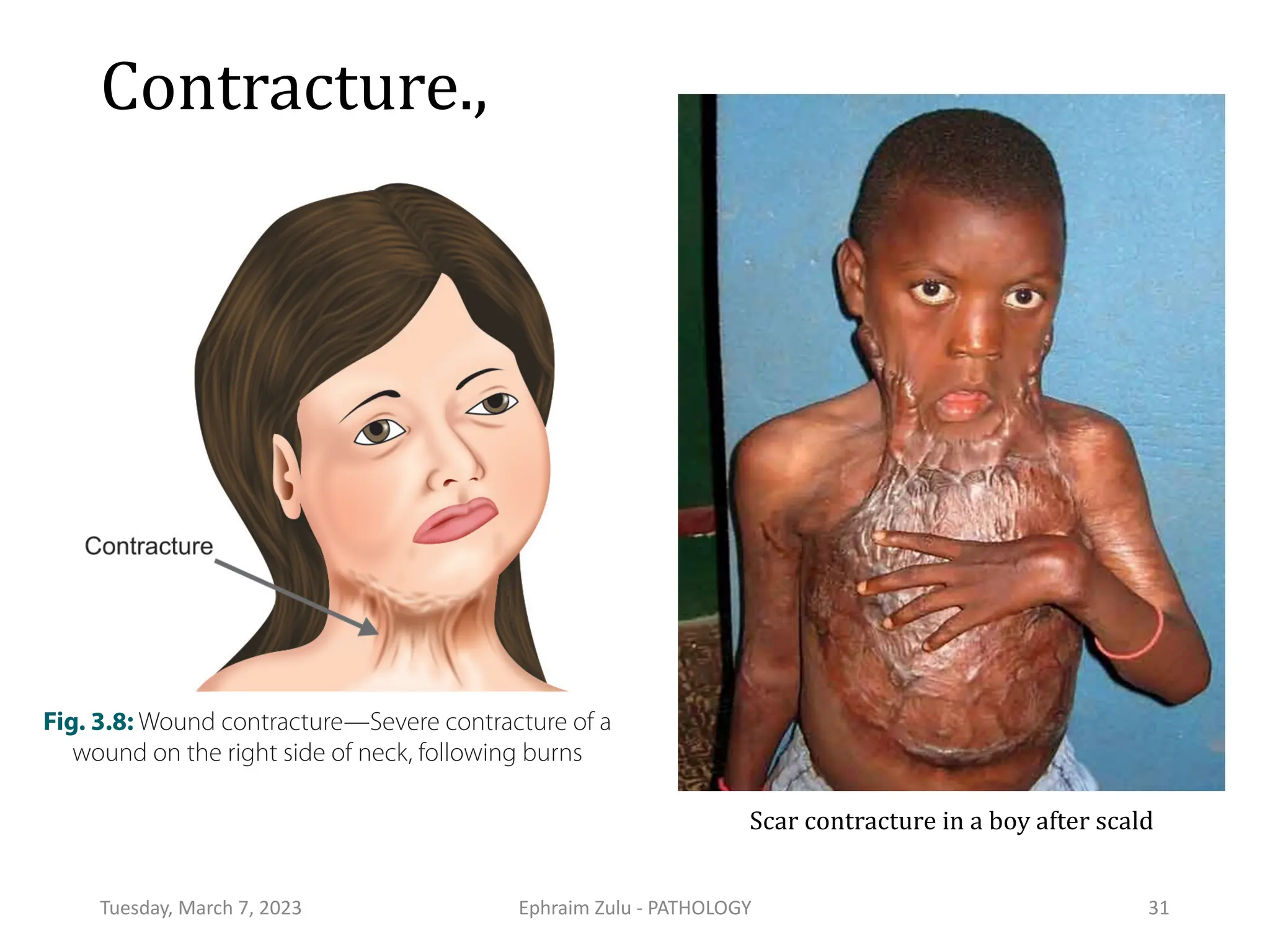
Ephraim Imhotep Zulu, BSc BMS, MSc Path
University of Zambia
School of Health Sciences,
Dept. of Biomedical Sciences,
Pathology
Lecture #4
Tissue Healing & Repair
Procedural document:
Rare disease nomenclature in English
www.orpha.net www.orphadata.org
Tuesday, March 7, 2023 Ephraim Zulu - PATHOLOGY 2
Lecture Outline
• Proliferative Capacities of Tissues
• Tissue Repair
• Patterns of Wound Healing
• Factors that Influence Wound Healing
• Complications of Wound Healing
• Fracture Healing
Tuesday, March 7, 2023 Ephraim Zulu - PATHOLOGY 3
Learning Objectives:
At the end of this lecture, the student is expected to
• Know the different types and Patterns of Wound Healing processes
• Understand the process of Tissue Repair
• Appreciate the Proliferative Capacities of Tissues
• Differentiate/Compare and Contrast the following pairs of terms:
• Primary union and Secondary union
• Keloids and Hypertrophic scars
• Understand and Appreciate the different Factors that Influence Wound Healing
• Know the Complications of Wound Healing
• Understand how Fracture Healing occurs
Tuesday, March 7, 2023 Ephraim Zulu - PATHOLOGY 4
Synopsis
• Healing, as used in a pathological context, refers to the body’s replacement of
destroyed tissue by living tissue.
• The healing process involves two distinct processes: Regeneration and Repair
• Regeneration - ability of a tissue to replace the damaged components and essentially
returning to a normal state. It is the renewal of a lost tissue in which the lost cells are
replaced by identical ones and it involves two processes:
• Proliferation of surviving cells to replace lost tissue and Migration of surviving cells
into the vacant space.
• Repair - the replacement of lost tissue by granulation tissue which matures to form
scar tissue
• Fibrosis - extensive deposition of collagen that occurs in the organs as a consequence
of chronic inflammation, or after extensive infarction.
Tuesday, March 7, 2023 Ephraim Zulu - PATHOLOGY 5
Tuesday, March 7, 2023 Ephraim Zulu - PATHOLOGY 6
Tuesday, March 7, 2023 Ephraim Zulu - PATHOLOGY 7
Tuesday, March 7, 2023 Ephraim Zulu - PATHOLOGY 8
Proliferative Capacities of Tissues
The ability of tissues to repair
themselves is critically influenced
by their intrinsic proliferative
capacity.
Tuesday, March 7, 2023 Ephraim Zulu - PATHOLOGY 9
Tissue Repair
• Repair is the orderly process by which lost tissue is
eventually replaced by a scar.
• Tissues containing permanent cells can not heal by
regeneration.
• Rather the lost permanent cells are replaced by
formation of granulation tissue.
• In granulation-tissue formation, three phases may be
observed.
Tuesday, March 7, 2023 Ephraim Zulu - PATHOLOGY 10
Phases: Granulation tissue formation
1. Phase of inflammation
• Inflammatory exudate containing polymorphs is seen in
the area of tissue injury.
2. Phase of demolition
• The dead cells liberate their autolytic enzymes, and other
enzymes (proteolytic) come from disintegrating
polymorphs.
• Macrophages ingest particulate matter.
Tuesday, March 7, 2023 Ephraim Zulu - PATHOLOGY 11
3. Ingrowth of granulation tissue
• This is characterized by proliferation of fibroblasts and an ingrowth
of new blood vessels into the area of injury, with a variable number
of inflammatory cells.
• Fibroblasts actively synthesize and secrete fibronectin,
proteoglycans, and collagen.
• As the collagen content of the wound increases, many of the newly
formed vessels disappear.
• This vascular involution which takes place in a few weeks,
dramatically transforms a richly vascularized tissue into a pale,
avascular scar tissue.
Tuesday, March 7, 2023 Ephraim Zulu - PATHOLOGY 12
“Healthy” Granulation Tissue
Tuesday, March 7, 2023 Ephraim Zulu - PATHOLOGY 13
Wound Contraction
• Is a mechanical reduction in the size of the defect.
• The wound is reduced approximately by 70-80% of its original
size.
• If contraction is prevented, healing is slow and a large ugly
scar is formed.
• Contraction is said to be due to contraction by myofibroblasts.
• Two to three days after the injury they migrate into the wound
and their active contraction decrease the size of the defect.
Tuesday, March 7, 2023 Ephraim Zulu - PATHOLOGY 14
Patterns of Wound Healing
• Healing of a wound demonstrates both epithelial regeneration
(healing of the epidermis) and repair by scarring (healing of the
dermis).
• There are two patterns of wound healing depending on the amount
of tissue damage:
• Healing by first intention (Primary union) and Healing by second
intention (Secondary union)
• These two patterns are essentially the same process varying only in
amount.
Tuesday, March 7, 2023 Ephraim Zulu - PATHOLOGY 15
Tuesday, March 7, 2023 Ephraim Zulu - PATHOLOGY 16
Tuesday, March 7, 2023 Ephraim Zulu - PATHOLOGY 17
Attribute Primary healing Secondary healing
Nature of the wound Wound is small in size Wound is large in size
Wound has regular margins Wound has irregular
margins
Wounds generally uninfected Wounds maybe infected
Clot size Small Large
inflammation Less intense More intense
Granulation tissue Small Large
Scar tissue Small Large
Wound contraction Absent Present
Outcome Neat linear scar Contracted irregular scar
Tuesday, March 7, 2023 Ephraim Zulu - PATHOLOGY 18
Secondary Wound Healing
Tuesday, March 7, 2023 Ephraim Zulu - PATHOLOGY 19
Factors that Influence Wound Healing
• Infection
• Nutrition
• Glucocorticoids
• Mechanical variables
• Poor perfusion,
• Foreign bodies
• Type (and volume) of
tissue injured
• Type and size of wound
• Location of the wound
• Movement
• Ionizing radiation
• Metabolic status
Tuesday, March 7, 2023 Ephraim Zulu - PATHOLOGY 20
Factors That Influence Wound Healing..,
Infection
• is the single most important cause of delay in healing; it
prolongs the inflammation phase of the process and
potentially increases the local tissue injury.
Nutrition
• protein and vitamin C deficiency, inhibits collagen synthesis
and retards healing.
Poor perfusion,
• Also impairs healing.
Tuesday, March 7, 2023 Ephraim Zulu - PATHOLOGY 21
Glucocorticoids (steroids)
• have an anti-inflammatory effects,
and may result in poor wound
strength due to diminished fibrosis.
Mechanical variables
• such as increased local pressure or
torsion may cause wounds to pull
apart, or dehisce (see picture).
Tuesday, March 7, 2023 Ephraim Zulu - PATHOLOGY 22
Dehiscence
• Foreign bodies
• such as fragments of steel, glass, or even bone impede
healing.
• Type (and volume) of tissue injured
• Complete restoration can occur only in tissues composed
of stable and labile cells; even then, extensive injury will
probably result in incomplete tissue regeneration.
• Injury to tissues composed of permanent cells must
inevitably result in scarring
Tuesday, March 7, 2023 Ephraim Zulu - PATHOLOGY 23
Type, size, and location of the wound
• A clean, aseptic wound produced by the surgeon’s scalpel
heals faster than a wound produced by blunt trauma, which
exhibits abundant necrosis and irregular edges.
• Small blunt wounds heal faster than larger ones.
• Injuries in richly vascularized areas (e.g., the face) heal faster
than those in poorly vascularized ones (e.g., the foot).
• In areas where the skin adheres to bony surfaces, as in injuries
over the tibia, wound contraction and adequate apposition of
the edges are difficult.
• Hence, such wounds heal slowly.
Tuesday, March 7, 2023 Ephraim Zulu - PATHOLOGY 24
• Movement
• Early motion, particularly before tensile strength subjects
a wound to persistent trauma, thus preventing or
retarding healing.
• Ionizing radiation
• Prior irradiation leaves vascular lesions that interfere
with blood supply and result in slow wound healing.
• Acutely, irradiation of a wound blocks cell proliferation,
inhibits contraction, and retards the formation of
granulation tissue.
Tuesday, March 7, 2023 Ephraim Zulu - PATHOLOGY 25
Metabolic status
• Poorly controlled diabetes mellitus is associated with
delayed wound healing.
• The risk of infection in clean wound approaches five fold
the risk in non- diabetics.
• In diabetic patients, there can be impaired circulation
secondary to arteriosclerosis and impaired sensation due
to diabetic neuropathy.
• The impaired sensation renders the lower extremity
blind to every day hazards.
• Hence, in diabetic patients, wounds heal very slowly.
Tuesday, March 7, 2023 Ephraim Zulu - PATHOLOGY 26
Complications of Wound Healing
Abnormalities in basic healing processes result in the
complications of wound healing.
• Infection
• Deficient Scar Formation (wound dehiscence and ulceration).
• Excessive Scar Formation (Hypertrophic scar and Keloid)
• Excessive Contraction
• Epidermal cysts
• Pigmentation
• Neoplasia
Tuesday, March 7, 2023 Ephraim Zulu - PATHOLOGY 27
Complications.,
• Infection. Infected wound may provide the portal of entry for
many organisms.
• Epidermal cysts can develop due to persistence of epithelial
cells at the site of wound healing.
• Pigmentation may develop due to either colored particle left
in the wound or due to hemosiderin pigment.
• Neoplasia: For example squamous cell carcinoma may
develop in Marjolin’s ulcer, which is the scar that follows burns
in skin. .
Tuesday, March 7, 2023 Ephraim Zulu - PATHOLOGY 28
Deficient Scar Formation
• Inadequate formation of granulation tissue or a deficient scar formation can cause
wound dehiscence and ulceration.
• 1. Dehiscence (the wound splitting open) or rupture of a wound is most common life-
threatening complication after abdominal surgery. It is due to increased abdominal
pressure/mechanical stress on the abdominal wound from vomiting, coughing, or
ileus.
• 2. Ulceration: Wounds can ulcerate due to inadequate angiogenesis during healing.
Nonhealing wounds also develop in regions devoid of sensation..
• 3. Incisional hernia resulting from weak scars of the abdominal wall due to a defect
caused by prior surgery. They are due to insufficient deposition of extracellular matrix
or inadequate cross-linking in the collagen matrix.
Tuesday, March 7, 2023 Ephraim Zulu - PATHOLOGY 29
Excessive Contraction
• A decrease in the size of a wound due to myofibroblasts is known as contraction.
An exaggeration of this contraction is termed contracture (cicatrisation) and
results in deformities of the wound and the surrounding tissues.
• Contracture is also said to arise as a result of late reduction in the size of the
wound.
• Consequences of contractures:
• – Compromise movements: for example, contractures that follow severe burns
can compromise the movement of the involved region and joint movements.
• – Obstruction: for example, in GI tract contracture (stricture) can cause
intestinal obstruction.
Tuesday, March 7, 2023 Ephraim Zulu - PATHOLOGY 30
Contracture.,
Tuesday, March 7, 2023 Ephraim Zulu - PATHOLOGY 31
c Pathology
Fig. 3.8: Wound contracture—Severe contracture of a
wound on the right side of neck, following burns
Scar contracture in a boy after scald
Excessive Scar Formation
• Excessive formation of the components of the repair process can result in:
• Hypertrophic scar: The accumulation of excessive amounts of extracellular matrix,
mostly collagen may give rise to a raised scar at the site of wound known as a
hypertrophic scar.
• They usually develop after thermal or traumatic injury, which involves the deep layers
of the dermis.
• Keloid: If the scar tissue grows/progress beyond the boundaries of the original
wound and does not regress, it is called a keloid. Thus, keloid is an exuberant scar that
recurs with still larger keloid after surgical excision.
• The cause is unknown and is thought to be due to lack of the proper
metalloproteinases (collagenases) to degrade type III collagen
Tuesday, March 7, 2023 Ephraim Zulu - PATHOLOGY 32
Keloids.,
Tuesday, March 7, 2023 Ephraim Zulu - PATHOLOGY 33
Excess collagen deposition in the skin
forming a raised scar known as a keloid
Hypertrophic Scars
Tuesday, March 7, 2023 Ephraim Zulu - PATHOLOGY 34
Tuesday, March 7, 2023 Ephraim Zulu - PATHOLOGY 35
Keloid Vs Hypertrophic Scars
Tuesday, March 7, 2023 Ephraim Zulu - PATHOLOGY 36
Keloid Vs Hypertrophic Scars
Tuesday, March 7, 2023 Ephraim Zulu - PATHOLOGY 37
Tuesday, March 7, 2023 Ephraim Zulu - PATHOLOGY 38
Fracture Healing
References & Credits
Tuesday, March 7, 2023 Ephraim Zulu - PATHOLOGY
• Barone J, Castro M.A. (2016), USMLE Step 1 Pathology Lecture Notes,
Published by Kaplan Medical, a division of Kaplan, Inc. 750 Third Avenue, New
York, NY 10017: ISBN: 978-1-5062-0772-8
• Robbins SL and Kumar V (2013). Basic Pathology (9th Edition).WB Saunders
Co. London.
• Bezabeh M, Tesfaye A, Ergicho B, Erke M, Mengistu S, Bedane A, Desta A (2004);
Students Lecture Note Series General Pathology For Health science students
Jimma University, Ethiopia.
• Rubin E, Rubin R, Strayer D.S. (2012) Rubin`s Pathology: Clinicopathologic
Foundations of Medicine (6th Edition), Lippincott Williams & Wilkins, a Wolters
Kluwer business. Philadelphia, PA.
39
Tuesday, March 7, 2023 Ephraim Zulu - PATHOLOGY
End of Lecture
Ephraim Imhotep Zulu
Pathology
40

PATHOLOGY - lecture - Healing & Repair.pdf

  • 1.
    Ephraim Imhotep Zulu,BSc BMS, MSc Path University of Zambia School of Health Sciences, Dept. of Biomedical Sciences, Pathology Lecture #4 Tissue Healing & Repair Procedural document: Rare disease nomenclature in English www.orpha.net www.orphadata.org
  • 2.
    Tuesday, March 7,2023 Ephraim Zulu - PATHOLOGY 2
  • 3.
    Lecture Outline • ProliferativeCapacities of Tissues • Tissue Repair • Patterns of Wound Healing • Factors that Influence Wound Healing • Complications of Wound Healing • Fracture Healing Tuesday, March 7, 2023 Ephraim Zulu - PATHOLOGY 3
  • 4.
    Learning Objectives: At theend of this lecture, the student is expected to • Know the different types and Patterns of Wound Healing processes • Understand the process of Tissue Repair • Appreciate the Proliferative Capacities of Tissues • Differentiate/Compare and Contrast the following pairs of terms: • Primary union and Secondary union • Keloids and Hypertrophic scars • Understand and Appreciate the different Factors that Influence Wound Healing • Know the Complications of Wound Healing • Understand how Fracture Healing occurs Tuesday, March 7, 2023 Ephraim Zulu - PATHOLOGY 4
  • 5.
    Synopsis • Healing, asused in a pathological context, refers to the body’s replacement of destroyed tissue by living tissue. • The healing process involves two distinct processes: Regeneration and Repair • Regeneration - ability of a tissue to replace the damaged components and essentially returning to a normal state. It is the renewal of a lost tissue in which the lost cells are replaced by identical ones and it involves two processes: • Proliferation of surviving cells to replace lost tissue and Migration of surviving cells into the vacant space. • Repair - the replacement of lost tissue by granulation tissue which matures to form scar tissue • Fibrosis - extensive deposition of collagen that occurs in the organs as a consequence of chronic inflammation, or after extensive infarction. Tuesday, March 7, 2023 Ephraim Zulu - PATHOLOGY 5
  • 6.
    Tuesday, March 7,2023 Ephraim Zulu - PATHOLOGY 6
  • 7.
    Tuesday, March 7,2023 Ephraim Zulu - PATHOLOGY 7
  • 8.
    Tuesday, March 7,2023 Ephraim Zulu - PATHOLOGY 8 Proliferative Capacities of Tissues The ability of tissues to repair themselves is critically influenced by their intrinsic proliferative capacity.
  • 9.
    Tuesday, March 7,2023 Ephraim Zulu - PATHOLOGY 9
  • 10.
    Tissue Repair • Repairis the orderly process by which lost tissue is eventually replaced by a scar. • Tissues containing permanent cells can not heal by regeneration. • Rather the lost permanent cells are replaced by formation of granulation tissue. • In granulation-tissue formation, three phases may be observed. Tuesday, March 7, 2023 Ephraim Zulu - PATHOLOGY 10
  • 11.
    Phases: Granulation tissueformation 1. Phase of inflammation • Inflammatory exudate containing polymorphs is seen in the area of tissue injury. 2. Phase of demolition • The dead cells liberate their autolytic enzymes, and other enzymes (proteolytic) come from disintegrating polymorphs. • Macrophages ingest particulate matter. Tuesday, March 7, 2023 Ephraim Zulu - PATHOLOGY 11
  • 12.
    3. Ingrowth ofgranulation tissue • This is characterized by proliferation of fibroblasts and an ingrowth of new blood vessels into the area of injury, with a variable number of inflammatory cells. • Fibroblasts actively synthesize and secrete fibronectin, proteoglycans, and collagen. • As the collagen content of the wound increases, many of the newly formed vessels disappear. • This vascular involution which takes place in a few weeks, dramatically transforms a richly vascularized tissue into a pale, avascular scar tissue. Tuesday, March 7, 2023 Ephraim Zulu - PATHOLOGY 12
  • 13.
    “Healthy” Granulation Tissue Tuesday,March 7, 2023 Ephraim Zulu - PATHOLOGY 13
  • 14.
    Wound Contraction • Isa mechanical reduction in the size of the defect. • The wound is reduced approximately by 70-80% of its original size. • If contraction is prevented, healing is slow and a large ugly scar is formed. • Contraction is said to be due to contraction by myofibroblasts. • Two to three days after the injury they migrate into the wound and their active contraction decrease the size of the defect. Tuesday, March 7, 2023 Ephraim Zulu - PATHOLOGY 14
  • 15.
    Patterns of WoundHealing • Healing of a wound demonstrates both epithelial regeneration (healing of the epidermis) and repair by scarring (healing of the dermis). • There are two patterns of wound healing depending on the amount of tissue damage: • Healing by first intention (Primary union) and Healing by second intention (Secondary union) • These two patterns are essentially the same process varying only in amount. Tuesday, March 7, 2023 Ephraim Zulu - PATHOLOGY 15
  • 16.
    Tuesday, March 7,2023 Ephraim Zulu - PATHOLOGY 16
  • 17.
    Tuesday, March 7,2023 Ephraim Zulu - PATHOLOGY 17 Attribute Primary healing Secondary healing Nature of the wound Wound is small in size Wound is large in size Wound has regular margins Wound has irregular margins Wounds generally uninfected Wounds maybe infected Clot size Small Large inflammation Less intense More intense Granulation tissue Small Large Scar tissue Small Large Wound contraction Absent Present Outcome Neat linear scar Contracted irregular scar
  • 18.
    Tuesday, March 7,2023 Ephraim Zulu - PATHOLOGY 18
  • 19.
    Secondary Wound Healing Tuesday,March 7, 2023 Ephraim Zulu - PATHOLOGY 19
  • 20.
    Factors that InfluenceWound Healing • Infection • Nutrition • Glucocorticoids • Mechanical variables • Poor perfusion, • Foreign bodies • Type (and volume) of tissue injured • Type and size of wound • Location of the wound • Movement • Ionizing radiation • Metabolic status Tuesday, March 7, 2023 Ephraim Zulu - PATHOLOGY 20
  • 21.
    Factors That InfluenceWound Healing.., Infection • is the single most important cause of delay in healing; it prolongs the inflammation phase of the process and potentially increases the local tissue injury. Nutrition • protein and vitamin C deficiency, inhibits collagen synthesis and retards healing. Poor perfusion, • Also impairs healing. Tuesday, March 7, 2023 Ephraim Zulu - PATHOLOGY 21
  • 22.
    Glucocorticoids (steroids) • havean anti-inflammatory effects, and may result in poor wound strength due to diminished fibrosis. Mechanical variables • such as increased local pressure or torsion may cause wounds to pull apart, or dehisce (see picture). Tuesday, March 7, 2023 Ephraim Zulu - PATHOLOGY 22 Dehiscence
  • 23.
    • Foreign bodies •such as fragments of steel, glass, or even bone impede healing. • Type (and volume) of tissue injured • Complete restoration can occur only in tissues composed of stable and labile cells; even then, extensive injury will probably result in incomplete tissue regeneration. • Injury to tissues composed of permanent cells must inevitably result in scarring Tuesday, March 7, 2023 Ephraim Zulu - PATHOLOGY 23
  • 24.
    Type, size, andlocation of the wound • A clean, aseptic wound produced by the surgeon’s scalpel heals faster than a wound produced by blunt trauma, which exhibits abundant necrosis and irregular edges. • Small blunt wounds heal faster than larger ones. • Injuries in richly vascularized areas (e.g., the face) heal faster than those in poorly vascularized ones (e.g., the foot). • In areas where the skin adheres to bony surfaces, as in injuries over the tibia, wound contraction and adequate apposition of the edges are difficult. • Hence, such wounds heal slowly. Tuesday, March 7, 2023 Ephraim Zulu - PATHOLOGY 24
  • 25.
    • Movement • Earlymotion, particularly before tensile strength subjects a wound to persistent trauma, thus preventing or retarding healing. • Ionizing radiation • Prior irradiation leaves vascular lesions that interfere with blood supply and result in slow wound healing. • Acutely, irradiation of a wound blocks cell proliferation, inhibits contraction, and retards the formation of granulation tissue. Tuesday, March 7, 2023 Ephraim Zulu - PATHOLOGY 25
  • 26.
    Metabolic status • Poorlycontrolled diabetes mellitus is associated with delayed wound healing. • The risk of infection in clean wound approaches five fold the risk in non- diabetics. • In diabetic patients, there can be impaired circulation secondary to arteriosclerosis and impaired sensation due to diabetic neuropathy. • The impaired sensation renders the lower extremity blind to every day hazards. • Hence, in diabetic patients, wounds heal very slowly. Tuesday, March 7, 2023 Ephraim Zulu - PATHOLOGY 26
  • 27.
    Complications of WoundHealing Abnormalities in basic healing processes result in the complications of wound healing. • Infection • Deficient Scar Formation (wound dehiscence and ulceration). • Excessive Scar Formation (Hypertrophic scar and Keloid) • Excessive Contraction • Epidermal cysts • Pigmentation • Neoplasia Tuesday, March 7, 2023 Ephraim Zulu - PATHOLOGY 27
  • 28.
    Complications., • Infection. Infectedwound may provide the portal of entry for many organisms. • Epidermal cysts can develop due to persistence of epithelial cells at the site of wound healing. • Pigmentation may develop due to either colored particle left in the wound or due to hemosiderin pigment. • Neoplasia: For example squamous cell carcinoma may develop in Marjolin’s ulcer, which is the scar that follows burns in skin. . Tuesday, March 7, 2023 Ephraim Zulu - PATHOLOGY 28
  • 29.
    Deficient Scar Formation •Inadequate formation of granulation tissue or a deficient scar formation can cause wound dehiscence and ulceration. • 1. Dehiscence (the wound splitting open) or rupture of a wound is most common life- threatening complication after abdominal surgery. It is due to increased abdominal pressure/mechanical stress on the abdominal wound from vomiting, coughing, or ileus. • 2. Ulceration: Wounds can ulcerate due to inadequate angiogenesis during healing. Nonhealing wounds also develop in regions devoid of sensation.. • 3. Incisional hernia resulting from weak scars of the abdominal wall due to a defect caused by prior surgery. They are due to insufficient deposition of extracellular matrix or inadequate cross-linking in the collagen matrix. Tuesday, March 7, 2023 Ephraim Zulu - PATHOLOGY 29
  • 30.
    Excessive Contraction • Adecrease in the size of a wound due to myofibroblasts is known as contraction. An exaggeration of this contraction is termed contracture (cicatrisation) and results in deformities of the wound and the surrounding tissues. • Contracture is also said to arise as a result of late reduction in the size of the wound. • Consequences of contractures: • – Compromise movements: for example, contractures that follow severe burns can compromise the movement of the involved region and joint movements. • – Obstruction: for example, in GI tract contracture (stricture) can cause intestinal obstruction. Tuesday, March 7, 2023 Ephraim Zulu - PATHOLOGY 30
  • 31.
    Contracture., Tuesday, March 7,2023 Ephraim Zulu - PATHOLOGY 31 c Pathology Fig. 3.8: Wound contracture—Severe contracture of a wound on the right side of neck, following burns Scar contracture in a boy after scald
  • 32.
    Excessive Scar Formation •Excessive formation of the components of the repair process can result in: • Hypertrophic scar: The accumulation of excessive amounts of extracellular matrix, mostly collagen may give rise to a raised scar at the site of wound known as a hypertrophic scar. • They usually develop after thermal or traumatic injury, which involves the deep layers of the dermis. • Keloid: If the scar tissue grows/progress beyond the boundaries of the original wound and does not regress, it is called a keloid. Thus, keloid is an exuberant scar that recurs with still larger keloid after surgical excision. • The cause is unknown and is thought to be due to lack of the proper metalloproteinases (collagenases) to degrade type III collagen Tuesday, March 7, 2023 Ephraim Zulu - PATHOLOGY 32
  • 33.
    Keloids., Tuesday, March 7,2023 Ephraim Zulu - PATHOLOGY 33 Excess collagen deposition in the skin forming a raised scar known as a keloid
  • 34.
    Hypertrophic Scars Tuesday, March7, 2023 Ephraim Zulu - PATHOLOGY 34
  • 35.
    Tuesday, March 7,2023 Ephraim Zulu - PATHOLOGY 35
  • 36.
    Keloid Vs HypertrophicScars Tuesday, March 7, 2023 Ephraim Zulu - PATHOLOGY 36
  • 37.
    Keloid Vs HypertrophicScars Tuesday, March 7, 2023 Ephraim Zulu - PATHOLOGY 37
  • 38.
    Tuesday, March 7,2023 Ephraim Zulu - PATHOLOGY 38 Fracture Healing
  • 39.
    References & Credits Tuesday,March 7, 2023 Ephraim Zulu - PATHOLOGY • Barone J, Castro M.A. (2016), USMLE Step 1 Pathology Lecture Notes, Published by Kaplan Medical, a division of Kaplan, Inc. 750 Third Avenue, New York, NY 10017: ISBN: 978-1-5062-0772-8 • Robbins SL and Kumar V (2013). Basic Pathology (9th Edition).WB Saunders Co. London. • Bezabeh M, Tesfaye A, Ergicho B, Erke M, Mengistu S, Bedane A, Desta A (2004); Students Lecture Note Series General Pathology For Health science students Jimma University, Ethiopia. • Rubin E, Rubin R, Strayer D.S. (2012) Rubin`s Pathology: Clinicopathologic Foundations of Medicine (6th Edition), Lippincott Williams & Wilkins, a Wolters Kluwer business. Philadelphia, PA. 39
  • 40.
    Tuesday, March 7,2023 Ephraim Zulu - PATHOLOGY End of Lecture Ephraim Imhotep Zulu Pathology 40