This document summarizes four cases:
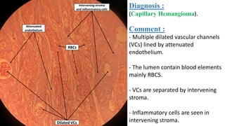
1. Capillary hemangioma, characterized by dilated vascular channels lined by endothelium containing red blood cells and separated by stroma.
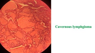
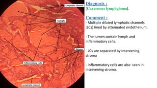
2. Cavernous lymphgioma, characterized by dilated lymphatic channels lined by endothelium containing lymph and inflammatory cells and separated by stroma.
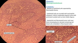
3. Hemangioendothelioma, characterized by nodules of epitheloid cells separated by stroma, containing intracytoplasmic vacuoles with red blood cells.
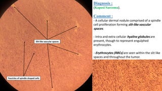
4. Kaposi Sarcoma, characterized by a dermal nodule of spindle cells forming slit-like vascular spaces containing erythrocytes.