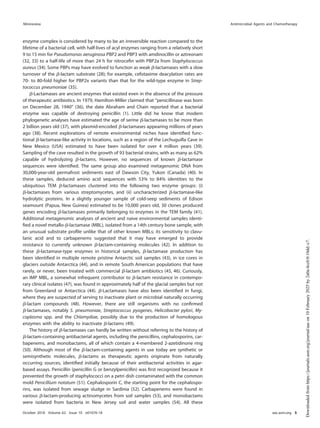
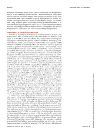
Past and Present Perspectives on ␤-Lactamases
Karen Busha
aDepartment of Biology, Indiana University Bloomington, Bloomington, Indiana, USA
ABSTRACT ␤-Lactamases, the major resistance determinant for ␤-lactam antibiotics in
Gram-negative bacteria, are ancient enzymes whose origins can be traced back millions
of years ago. These well-studied enzymes, currently numbering almost 2,800 unique pro-
teins, initially emerged from environmental sources, most likely to protect a producing
bacterium from attack by naturally occurring ␤-lactams. Their ancestors were presum-
ably penicillin-binding proteins that share sequence homology with ␤-lactamases pos-
sessing an active-site serine. Metallo-␤-lactamases also exist, with one or two catalytically
functional zinc ions. Although penicillinases in Gram-positive bacteria were reported
shortly after penicillin was introduced clinically, transmissible ␤-lactamases that could hy-
drolyze recently approved cephalosporins, monobactams, and carbapenems later be-
came important in Gram-negative pathogens. Nomenclature is based on one of two ma-
jor systems. Originally, functional classifications were used, based on substrate and
inhibitor profiles. A later scheme classifies ␤-lactamases according to amino acid se-
quences, resulting in class A, B, C, and D enzymes. A more recent nomenclature com-
bines the molecular and biochemical classifications into 17 functional groups that de-
scribe most ␤-lactamases. Some of the most problematic enzymes in the clinical
community include extended-spectrum ␤-lactamases (ESBLs) and the serine and metallo-
carbapenemases, all of which are at least partially addressed with new ␤-lactamase in-
hibitor combinations. New enzyme variants continue to be described, partly because of
the ease of obtaining sequence data from whole-genome sequencing studies. Often,
these new enzymes are devoid of any phenotypic descriptions, making it more difficult
for clinicians and antibiotic researchers to address new challenges that may be posed by
unusual ␤-lactamases.
KEYWORDS ESBL, MBL, ␤-lactam, ␤-lactamase, carbapenemase, cephalosporinase,
penicillinase
One of the most studied enzyme families is the group of enzymes known as
␤-lactamases, with more than 28,900 citations in Medline (https://www.ncbi.nlm.nih
.gov/pubmed/). These enzymes, whose most obvious role is to inactivate ␤-lactam
antibiotics, have driven research in academic laboratories since the early 1940s (1, 2).
Perhaps more importantly, they have been responsible for scores of pharmaceutical
research programs that have attempted to find ways to protect effective antibiotics
from destruction. ␤-Lactamases provide intellectual challenges for academic investiga-
tors with their deceptively simple mechanisms of action, as enzymes whose hydrolysis
activity approaches that of a “fully efficient enzyme” with diffusion-limited reaction
rates (3). Attempts to provide economically viable anti-infective agents that can cir-
cumvent the action of these enzymes have yielded substantial economic rewards to
successful pharmaceutical companies. As a result, the safe and effective ␤-lactam
antibiotics have become one of the most widely prescribed classes of antibacterial
agents (4). Many review articles have been published about various aspects of these
intriguing enzymes (5–11), but they have tended to focus on narrowly targeted sets of
␤-lactamases. In this minireview, a short history of the ␤-lactamases is presented,
emphasizing key points that have driven both academic and pharmaceutical science
Accepted manuscript posted online 30 July
2018
Citation Bush K. 2018. Past and present
perspectives on β-lactamases. Antimicrob
Agents Chemother 62:e01076-18. https://doi
.org/10.1128/AAC.01076-18.
Copyright © 2018 American Society for
Microbiology. All Rights Reserved.
Addresscorrespondencetokarbush@indiana.edu.
MINIREVIEW
crossm
October 2018 Volume 62 Issue 10 e01076-18 aac.asm.org 1
Antimicrobial Agents and Chemotherapy
Downloaded
from
https://journals.asm.org/journal/aac
on
10
February
2025
by
2a0a:4cc0:0:10dd::c7.
over the past 75 years. High points in their history will be discussed, based on the
“Table of Firsts” shown as Table 1. This history is not meant to be a comprehensive
review of all the current literature on ␤-lactamases but is meant to tell a story about
where these enzymes came from, how they have driven antibiotic discovery programs,
and what challenges they pose for today.
TOPLINE VIEW OF ␤-LACTAMASES
␤-Lactamases are versatile enzymes with a limited range of molecular structures
found in a diversity of bacterial sources. Their commonality is the ability to hydrolyze
chemical compounds containing a ␤-lactam ring. As shown in Fig. 1, they have been
classified biochemically into two broad divisions according to the mechanism by which
they perform hydrolysis, either through the formation of an acyl enzyme with an
active-site serine (Fig. 2) (12) or via a hydrolytic reaction facilitated by one or two
essential zinc ions in the active sites of metallo-␤-lactamases (MBLs) (13). After se-
quence analyses became available for key ␤-lactamases, four molecular classes, A, B, C,
and D, were eventually assigned based on molecular size and homology between
active-site amino acid motifs (14–16). However, biochemical differences between peni-
cillinases and cephalosporinases had been recognized well before sequences were
available (17), and known ␤-lactamases were distinguished based on functional capa-
bilities related to substrate and inhibitor profiles (Table 2). Early classification schemes
relied upon relative hydrolysis rates of penicillins and early cephalosporins, together
with the enzymatic response to protein-modifying agents (18, 19). As additional
substrates and inhibitors were introduced into clinical practice and gene sequencing
became inexpensive and routine, both molecular and functional characteristics were
combined into a more comprehensive classification scheme (20, 21). Today, at least 17
functional groups associated with the four molecular classes have been distinguished
(20), with the major groups shown in Fig. 1. Enzymes are still differentiated with respect
to the relative hydrolysis of the ␤-lactam substrates, penicillins, cephalosporins, car-
bapenems, and monobactams. Further differentiation is possible based on reactions
with the class A ␤-lactamase inhibitor clavulanic acid (22), the broad-spectrum serine
␤-lactamase inhibitor avibactam (23), and the metal ion chelator EDTA to identify MBLs
(24).
␤-LACTAMASE ORIGINS
The antibacterial effect of a ␤-lactam results in inhibition of the growth of replicating
bacteria by acylating an active-site serine in essential penicillin-binding proteins (PBPs)
(25, 26). During the terminal stages of cell wall biosynthesis, these enzymes are thus
prevented from effecting the cross-linking of peptide chains to form peptidoglycan,
TABLE 1 Table of Firsts: the dates, organisms, and locations of the first of a series of ␤-lactamase-producing isolates with long-term
clinical significance
Original ␤-lactamase name
(currently recognized name)
Yr of first
verified isolation Organism Location
First description
in literature Reference(s)
Penicillinase (chromosomal AmpC) 1940 Bacillus coli (Escherichia coli) England 1940 1
Penicillinase 1942 Staphylococcus aureus England 1942 65
OXA 1962 Salmonella enterica serovar
Typhimurium, Escherichia colia
England 1965 87, 215
1967
TEM-1 1963 Escherichia coli Greece 1965 85
SHV-1 1972 Klebsiella pneumoniae Unknown 1972 216
Transferable ESBL (SHV-2) Pre-1983 K. pneumoniae Germany 1983 217
Serine (class A, group 2f)
carbapenemase (SME-1)
1982 Serratia marcescens England (London)
USA (Minnesota)
1990 148, 150
1985 1986
Plasmid-encoded AmpC (MIR-1) 1988 K. pneumoniae USA (Massachusetts) 1990 141
Plasmid-encoded MBL (IMP-1) 1988 Pseudomonas aeruginosa Japan 1991 151
Inhibitor-resistant TEM (TEM-30) 1991 E. coli France (Paris) 1994 118
KPC-type (KPC-2) 1996 K. pneumoniae USA (North Carolina) 2000 158
NDM-1 2006 K. pneumoniae India (New Delhi) 2009 175, 176
aAnderson and Datta described a Salmonella Typhimurium isolate from 1962 that later was confirmed to produce the OXA-2 enzyme (215). Egawa et al. described an
E. coli isolate in 1967 that produced the OXA-1 enzyme (87).
Minireview Antimicrobial Agents and Chemotherapy
October 2018 Volume 62 Issue 10 e01076-18 aac.asm.org 2
Downloaded
from
https://journals.asm.org/journal/aac
on
10
February
2025
by
2a0a:4cc0:0:10dd::c7.
resulting in cell death. Tipper and Strominger proposed similarities between the
structures of penicillin and D-Ala-D-Ala, the terminal amino acids of the nascent
acetylmuramyl-pentapeptide fragment, to explain the antimicrobial activity of penicillin
(27). Because the majority of ␤-lactamases contain an active-site serine that also can be
acylated by ␤-lactam molecules, it is not unexpected to find mechanistic and structural
similarities between the two sets of enzymes (28, 29). Fisher and Mobashery have
cogently outlined a history of studies demonstrating the biochemical commonality
between the two acyl enzymes (30). Molecular modeling of various serine ␤-lactamases
and PBP structures has demonstrated three-dimensional similarities with conserved
folding patterns and preservation of topology at the active site, in spite of low amino
acid identities (28, 31).
The general reaction mechanism for both enzymes is shown in Fig. 2. It is commonly
assumed that PBPs were the precursors of the ␤-lactamases, with the k3 rate for
deacylation increased dramatically for ␤-lactamases, compared to PBPs that exhibit a
fast acylation step (k2) compared to a slow deacylation step. Formation of the PBP-acyl
FIG 1 Molecular and functional relationships among ␤-lactamases (adapted from references 20 and 201 with permission). AV, avibactam;
CA, clavulanic acid; Cb, carbapenem; Cp, cephalosporin; E, expanded-spectrum cephalosporin; M, monobactam; P, penicillin.
FIG 2 General reaction mechanism for binding of a ␤-lactam substrate (S) to a PBP (E) or a serine
␤-lactamase (E). Reversible formation of a Michaelis complex (E · S) which proceeds to a stable acyl
enzyme (E—S) caused by reaction with the active-site serine. Hydrolysis occurs to form the microbio-
logically inactive ring-opened ␤-lactam (P) and either enzymatically active PBP (slow hydrolysis of acyl
enzyme) or ␤-lactamase (Bla, high hydrolysis rate).
Minireview Antimicrobial Agents and Chemotherapy
October 2018 Volume 62 Issue 10 e01076-18 aac.asm.org 3
Downloaded
from
https://journals.asm.org/journal/aac
on
10
February
2025
by
2a0a:4cc0:0:10dd::c7.
TABLE
2
Characterization
of
␤
-lactamases
based
on
information
existing
at
the
time
of
publication
Yr
Basis
for
characterization
a
Conclusion(s)
Reference
or
source
1968
Substrate
profile,
reaction
with
antiserum
Three
different
groups
of
enzymes,
typical
cephalosporinases,
cephalosporinase/penicillinase,
and
penicillinases
(including
those
produced
by
R
factors)
Sawai
et
al.
(18)
1970
Substrate
profile,
inhibition
by
pCMB
and
cloxacillin,
reaction
with
antiserum,
electrical
charge
Eight
distinctive
types
of
␤
-lactamases
Jack
and
Richmond
(92)
1973
Substrate
profiles,
inhibition
by
cloxacillin
and
pCMB,
electrophoretic
mobility,
molecular
wt
Five
classes
of
␤
-lactamases
(I,
II,
III,
IV,
and
V)
Richmond
and
Sykes
(19)
1976
Substrate
profiles,
inhibition
by
cloxacillin
and
pCMB,
isoelectric
focusing,
immunological
relatedness,
molecular
wt;
chromosomal
or
plasmid
location
considered
Five
classes
of
␤
-lactamases
(distinct
from
Richmond
and
Sykes)
Sykes
and
Matthew
(218)
1980
Amino
acid
sequences
from
purified
proteins
Molecular
classes
A
and
B
defined
based
on
four
class
A
enzymes
and
one
class
B
enzyme
Ambler
(14)
1981
Amino
acid
sequence
translated
from
ampC
nucleotide
sequence
Molecular
class
C
defined
Jaurin
and
Grundström
(15)
1988
Amino
acid
sequence
translated
from
bla
PSE-2
nucleotide
sequence
Molecular
class
D
defined
Huovinen
et
al.
(16)
1988
Substrate
profile,
inhibition
by
clavulanic
acid,
aztreonam,
and
EDTA,
molecular
class
designations
for
eight
enzymes
based
on
active
site
or
full
amino
acid
sequence
data
Functional
classification
scheme
with
three
groupings
proposed
for
28
enzymes
Bush
(187)
1989
Substrate
profile,
inhibition
by
clavulanic
acid,
aztreonam,
and
EDTA,
isoelectric
point,
molecular
size,
molecular
class
designations
based
on
nucleotide
sequences
Functional
classification
assigned
to
groups
1,
2
(six
subgroups),
3,
and
4
for
84
enzymes,
aligned
with
molecular
classes
for
21
enzymes
Bush
(219-221)
1995
Substrate
profile,
inhibition
by
clavulanic
acid,
sulbactam,
tazobactam,
aztreonam,
cloxacillin,
pCMB,
and
EDTA,
molecular
size,
isoelectric
point,
molecular
class
designations
based
on
nucleotide
sequences
Functional
classification
for
groups
1,
2
(eight
subgroups),
3,
and
4,
for
217
enzymes,
aligned
with
molecular
classes
for
118
enzymes
Bush
et
al.
(21)
1996–2015
Amino
acid
sequences,
initially
for
naturally
occurring
acquired
TEM,
SHV,
and
OXA
ESBLs
and
IRTs;
later
expanded
to
all
␤
-
lactamase
families
Curated
numbering
assignments
for
sequential
␤
-lactamases
in
all
major
families
Lahey
database
(http://www.lahey.org/Studies/)
2010
Substrate
profile,
inhibition
by
clavulanic
acid,
tazobactam,
or
EDTA;
molecular
classification
Functional
classification
for
groups
1
(2
subgroups),
2
(12
subgroups),
3
(2
subgroups),
and
4
Bush
and
Jacoby
(20)
2012
Genomic
(DNA)
data
Genotypic
data
for
Klebsiella
spp.¸
including
curation
of
nomenclature
for
OKP,
LEN,
and
OXY
␤
-lactamases
Institut
Pasteur
(http://bigsdb.pasteur.fr/
klebsiella/klebsiella.html)
2013
Nucleotide
or
amino
acid
sequences
for
multiple
resistance
determinants
for
all
antibiotic
classes
Curated
collection
of
characterized
molecular
sequences
and
phenotypes
to
predict
functionality
associated
with
antibiotic
resistance
genes
Comprehensive
Antibiotic
Resistance
Database
(CARD;
https://card.mcmaster.ca)
(222,
223)
2015
DNA
sequences
in
NCBI
database
that
encode
proteins
conferring
or
contributing
to
resistance
to
␤
-lactam
and
other
antibiotics
Annotated
sequence
records
for
representative
DNA
sequences
in
the
NCBI
database,
including
both
naturally
occurring
and
laboratory-derived
␤
-lactamases
NCBI
(https://www.ncbi.nlm.nih.gov/bioproject/
PRJNA313047)
2016
Commonalities
of
amino
acid
sequences
of
285
class
A
␤
-lactamases
Separation
of
class
A
␤
-lactamases
into
two
distinct
subclasses,
A1
and
A2
Philippon
et
al.
(73)
2017
Amino
acid
sequences,
biochemical
data
(kinetics
of
hydrolysis),
X-ray
crystallographic
structures
Well-annotated
compilation
of
structure-function
relationships
of
␤
-lactamases
from
all
classes,
including
laboratory
mutants
BLDB
(http://bldb.eu)
(197)
a
pCMB,
p-chloromercuribenzoate.
Minireview Antimicrobial Agents and Chemotherapy
October 2018 Volume 62 Issue 10 e01076-18 aac.asm.org 4
Downloaded
from
https://journals.asm.org/journal/aac
on
10
February
2025
by
2a0a:4cc0:0:10dd::c7.
enzyme complex is considered by many to be an irreversible reaction compared to the
lifetime of a bacterial cell, with half-lives of acyl enzymes ranging from a relatively short
9 to 15 min for Pseudomonas aeruginosa PBP2 and PBP3 with amdinocillin or aztreonam
(32, 33) to a half-life of more than 24 h for nitrocefin with PBP2a from Staphylococcus
aureus (34). Some PBPs may have evolved to function as weak ␤-lactamases with a slow
turnover of the ␤-lactam substrate (28); for example, cefotaxime deacylation rates are
70- to 80-fold higher for PBP2x variants than that for the wild-type enzyme in Strep-
tococcus pneumoniae (35).
␤-Lactamases are ancient enzymes that existed even in the absence of the pressure
of therapeutic antibiotics. In 1979, Hamilton-Miller claimed that “penicillinase was born
on December 28, 1940” (36), the date Abraham and Chain reported that a bacterial
enzyme was capable of destroying penicillin (1). Little did he know that modern
phylogenetic analyses have estimated the age of serine ␤-lactamases to be more than
2 billion years old (37), with plasmid-encoded ␤-lactamases appearing millions of years
ago (38). Recent explorations of remote environmental niches have identified func-
tional ␤-lactamase-like activity in locations, such as a region of the Lechuguilla Cave in
New Mexico (USA) estimated to have been isolated for over 4 million years (39).
Sampling of the cave resulted in the growth of 93 bacterial strains, with as many as 62%
capable of hydrolyzing ␤-lactams. However, no sequences of known ␤-lactamase
sequences were identified. The same group also examined metagenomic DNA from
30,000-year-old permafrost sediments east of Dawson City, Yukon (Canada) (40). In
these samples, deduced amino acid sequences with 53% to 84% identities to the
ubiquitous TEM ␤-lactamases clustered into the following two enzyme groups: (i)
␤-lactamases from various streptomycetes, and (ii) uncharacterized ␤-lactamase-like
hydrolytic proteins. In a slightly younger sample of cold-seep sediments of Edison
seamount (Papua, New Guinea) estimated to be 10,000 years old, 30 clones produced
genes encoding ␤-lactamases primarily belonging to enzymes in the TEM family (41).
Additional metagenomic analyses of ancient and naive environmental samples identi-
fied a novel metallo-␤-lactamase (MBL), isolated from a 14th century bone sample, with
an unusual substrate profile unlike that of other known MBLs; its sensitivity to clavu-
lanic acid and to carbapenems suggested that it may have emerged to provide
resistance to currently unknown ␤-lactam-containing molecules (42). In addition to
these ␤-lactamase-type enzymes in historical samples, ␤-lactamase production has
been identified in multiple remote pristine Antarctic soil samples (43), in ice cores in
glaciers outside Antarctica (44), and in remote South American populations that have
rarely, or never, been treated with commercial ␤-lactam antibiotics (45, 46). Curiously,
an IMP MBL, a somewhat infrequent contributor to ␤-lactam resistance in contempo-
rary clinical isolates (47), was found in approximately half of the glacial samples but not
from Greenland or Antarctica (44). ␤-Lactamases have also been identified in fungi,
where they are suspected of serving to inactivate plant or microbial naturally occurring
␤-lactam compounds (48). However, there are still organisms with no confirmed
␤-lactamases, notably S. pneumoniae, Streptococcus pyogenes, Helicobacter pylori, My-
coplasma spp. and the Chlamydiae, possibly due to the production of homologous
enzymes with the ability to inactivate ␤-lactams (49).
The history of ␤-lactamases can hardly be written without referring to the history of
␤-lactam-containing antibacterial agents, including the penicillins, cephalosporins, car-
bapenems, and monobactams, all of which contain a 4-membered 2-azetidinone ring
(50). Although most of the ␤-lactam-containing agents in use today are synthetic or
semisynthetic molecules, ␤-lactams as therapeutic agents originate from naturally
occurring sources, identified initially because of their antibacterial activities in agar-
based assays. Penicillin (penicillin G or benzylpenicillin) was first recognized because it
prevented the growth of staphylococci on a petri dish contaminated with the common
mold Penicillium notatum (51). Cephalosporin C, the starting point for the cephalospo-
rins, was isolated from sewage sludge in Sardinia (52). Carbapenems were found in
various ␤-lactam-producing actinomycetes from soil samples (53), and monobactams
were isolated from bacteria in New Jersey soil and water samples (54). All these
Minireview Antimicrobial Agents and Chemotherapy
October 2018 Volume 62 Issue 10 e01076-18 aac.asm.org 5
Downloaded
from
https://journals.asm.org/journal/aac
on
10
February
2025
by
2a0a:4cc0:0:10dd::c7.
iterations of unmodified ␤-lactams existed in natural environments and placed selective
pressure on the neighboring bacteria to evade a milieu of deleterious agents. When
␤-lactam-producing organisms compete with nonproducing bacteria in the same
environmental niche, survival strategies are quickly developed. Because bacteria, pos-
sibly the first living organisms, have existed for 2.5 to 4 billion years (55, 56), there has
been a long incubation period for naturally occurring ␤-lactam biosynthesis to be
perfected and for neighboring bacteria to learn how to survive in the presence of these
molecules. Interestingly, mammalian enzymes, including serum proteins (57) and renal
dehydropeptidase (dipeptidase) (58), are also capable of hydrolyzing ␤-lactams.
␤-LACTAMASES IN GRAM-POSITIVE BACTERIA
Resistance to ␤-lactams can be mediated by multiple mechanisms. Because it is in
the best interest of the bacteria to maintain active PBPs, the first line of defense would
appear to be for PBPs to alter their affinities for ␤-lactams while maintaining their
physiological function (28). This approach has been utilized most effectively by Gram-
positive bacteria, where a primary resistance mechanism has been acquisition of PBPs
with decreased affinity for common ␤-lactams (59). However, it took over 20 years of
penicillin usage before clinical isolates of penicillin-resistant S. pneumoniae (60, 61) and
penicillin/methicillin-resistant S. aureus (MRSA) were reported as a result of production
of low-affinity PBP variants (62–64). Instead, penicillinases, or ␤-lactamases that hydro-
lyze penicillin, emerged as the initial resistance mechanism in S. aureus to challenge the
therapeutic use of this agent (65). The drug was used more extensively and promis-
cuously to treat nosocomial streptococcal infections previously associated with high
mortality (66), but, as collateral damage, staphylococci initially fully susceptible to
penicillin rapidly developed resistance as a result of penicillinase production. In a single
English hospital, the percentage of penicillinase-producing staphylococci increased
from 14% in 1946 to 59% in 1948 (67) and up to 80% in 1953 (66). More recently, a 2013
study of U.S. clinical isolates showed that 86.5% of all S. aureus isolates (75.6% of
methicillin-susceptible isolates) contained the penicillinase gene blaZ (68), indicating
that a subpopulation of methicillin-susceptible staphylococci has survived in the ab-
sence of either a low-affinity PBP or the production of penicillinase. A transferable
penicillinase was also reported in a few clinical isolates of Enterococcus faecalis in the
1980s (69), with identical or closely homologous amino acid sequences as staphylo-
coccal penicillinases (70). A genomic analysis in 2005 demonstrated at least two
divergent origins (71). However, penicillinase-producing enterococci have not been
observed for the past decade and appear to have disappeared from these pathogens
(72). Another analysis of recent genomic data noted similarities among a set of 88
class A ␤-lactamases from Gram-positive bacteria and two enzymes from Gram-
negative bacteria, the ROB-1 penicillinase from Haemophilus influenzae, and the
ACl-1 enzyme from Acidaminococcus fermentans (73), possibly due to “trans-Gram
transfer” of ␤-lactamase genes between Gram-positive and Gram-negative bacteria.
In addition to the observed penicillinase production in Gram-positive clinical iso-
lates, enzymes with ␤-lactam-hydrolyzing properties from numerous environmental
bacilli were used by theoretical enzymologists as prototypical biochemical examples of
well-behaved enzymes (74, 75). Early studies with ␤-lactamases from Bacillus cereus (76),
Bacillus anthracis (77), and Bacillus licheniformis (78) provided tools on which future
biochemical studies of ␤-lactamases were based. Assay development, enzyme purifi-
cation, induction properties, and identification of a metal-dependent ␤-lactamase from
these bacilli were activities critical to the establishment of ␤-lactamases as important
enzymological entities (75, 79, 80). It is notable that the classical structural classification
of ␤-lactamases initially proposed by Ambler in 1980 was based on five known amino
acid sequences, four of which were from enzymes originating in Gram-positive bacteria.
These included three class A penicillinases, PC1 from S. aureus, penicillinase from B.
licheniformis 749C, and the B. cereus 569/H ␤-lactamase I. The only class B metallo-␤-
lactamases (MBL) that had been sequenced and studied in any detail at the time was
the zinc-containing B. cereus 569/H ␤-lactamase II (14). The first report of ␤-lactamases
Minireview Antimicrobial Agents and Chemotherapy
October 2018 Volume 62 Issue 10 e01076-18 aac.asm.org 6
Downloaded
from
https://journals.asm.org/journal/aac
on
10
February
2025
by
2a0a:4cc0:0:10dd::c7.
other than class A and B enzymes in Gram-positive bacilli described a set of oxacillin-
hydrolyzing (class D) penicillinases from a set of environmental strains in 2016 (81).
␤-LACTAMASES IN GRAM-NEGATIVE BACTERIA
1940 to 1985 and the emergence of modern ␤-lactamases. In Gram-negative
bacteria, ␤-lactamases have played a critical clinical role and have served as the primary
resistance mechanism for the ␤-lactam antibiotics. The first enzyme with ␤-lactamase
activity reported in the literature in 1940 was from Bacillus coli (1), now assumed to be
the class C, AmpC chromosomal cephalosporinase from Escherichia coli (Table 1). As
␤-lactam resistance began to be more frequently recognized in Gram-negative patho-
gens, it was shown that many enteric bacteria and P. aeruginosa produced species-
specific inducible chromosomal ␤-lactamases (82, 83). However, it is the mobile
␤-lactamases in Gram-negative bacteria that have created a more insidious threat to
the ␤-lactams. Although plasmid-encoded penicillinases were first reported in staphy-
lococci (84), the genes encoding these enzymes were not readily transferred to other
species, other than for the few enterococcal strains that appeared in the 1980s carrying
a staphylococcal penicillinase gene (69). In contrast, transferable genetic elements
encoding a wide variety of ␤-lactamases became the most prevalent mechanism
leading to the emergence of ␤-lactam resistance among Gram-negative bacteria, with
few species barriers existing for their transmission. When ␤-lactamases on “R-factors”
were first described in 1965 (85), only a limited number of these enzymes were
identified, related either to the transferable penicillinase on RTEM (86), now known to be
TEM-1, or to a transferable enzyme that hydrolyzed cloxacillin (87), an enzyme in the
OXA family of ␤-lactamases. These mobile bla genes were soon disseminated among
most enteric bacteria (88). We now understand that ␤-lactamase-encoding genes can
be acquired horizontally by different means but mainly by plasmid acquisition. Gene
mobilization mechanisms may include genetic elements, such as transposons, gene
cassettes, integrons, and insertion sequences (89–91).
Sawai et al. in 1968 (18), Jack and Richmond in 1970 (92), and Richmond and Sykes
in 1973 (19) attempted to group the known ␤-lactamases from Gram-negative rods in
a meaningful way based on biochemical properties and functionality. Over time, these
approaches have seen considerable refinement. As shown in Table 2, a limited number
of different ␤-lactamases served as the starting point for a logical classification scheme
based on biochemical characteristics.
The consensus was that the ␤-lactamases known by the mid-1970s constituted the
range of ␤-lactam-hydrolyzing enzymes in relevant Gram-negative bacteria. The intro-
duction of isoelectric focusing (IEF) (82, 93) aided by the ease of ␤-lactamase detection
using nitrocefin as a colorimetric activity indicator (94) allowed investigators to ana-
lyze bacterial extracts for the presence of ␤-lactamase activity with minimal effort.
Until the mid-1980s, it was possible to make a reasonable estimate of the number
of ␤-lactamases produced per strain and the putative identity of an enzyme, based
on isoelectric points from IEF gels. Biochemical properties were determined using
purified enzymes; published molecular sizes, many of which were incorrect, were
based on data obtained from gel exclusion chromatography, and amino acid
composition was deduced from peptide analyses of purified proteins in a process
that took months to obtain a single enzyme sequence. This was the environment in
which the first definitions of molecular classes A, B, and C emerged (14, 15). Eventually,
class D was added, based on nucleotide sequencing of the blaPSE-2 gene (16), and it
represents the most diverse of the molecular classes.
During the late 1970s and early 1980s, a number of surveillance studies were
conducted to assess ␤-lactamase production in Gram-negative bacteria. In a set of five
studies published between 1979 and 1985, the distribution of ␤-lactamases from
almost 1,800 ampicillin-resistant enteric bacteria were evaluated, based primarily on IEF
profiles (95–99). The TEM-1 and TEM-2 penicillinases were the most common enzymes,
with an average of 63% (range, 42 to 85%) of the isolates producing one of these
enzymes. In these isolates, an enzyme designated SHV-1 was produced by an average
Minireview Antimicrobial Agents and Chemotherapy
October 2018 Volume 62 Issue 10 e01076-18 aac.asm.org 7
Downloaded
from
https://journals.asm.org/journal/aac
on
10
February
2025
by
2a0a:4cc0:0:10dd::c7.
of 9.9% of the isolates (range, 1.7 to 24.0%), primarily as a chromosomal enzyme in
Klebsiella pneumoniae, with OXA enzymes produced on average at 7.8% (range, 0.4 to
15.4%). As was common in the early years of ␤-lactam resistance due to transferable
␤-lactamases, few isolates produced more than one plasmid-encoded enzyme, with
multiple transferable ␤-lactamases generally reported in no more than 3% of the
isolates. An exception was a set of Klebsiella isolates from a 1983 Spanish study in which
30% of the strains produced at least two plasmidic enzymes, primarily combinations of
SHV-1 with TEM-1 or TEM-2 (97). This compilation set the stage for the future evolution
of ␤-lactamases.
The facile transmission of the R-factor encoded “RTEM” (TEM-1) into a multiplicity of
Gram-negative pathogens is associated with triggering one of the most productive
periods of antibiotic development, especially for the ␤-lactams. In 1974, reports of
ampicillin resistance in meningitis patients infected with Haemophilus influenzae led to
the discovery that these strains produced an acquired TEM-type ␤-lactamase (100). At
that time, ampicillin was the preferred treatment of sepsis or pediatric meningitis
caused by H. influenzae type b, and the new TEM-producing strains were no longer
responsive to therapeutic levels of ampicillin (101). This observation was followed quite
soon by the alarming finding that the TEM ␤-lactamase was freely being transferred
into Neisseria gonorrhoeae by conjugal mating when a 24.5-kDa gonococcal plasmid
was present (102). Two geographically distinct plasmids of different sizes emerged
almost simultaneously, each of which encoded the blaTEM gene in gonococci (103).
Epidemiologically, this was attributed in part to the promiscuity of naval populations
and prostitutes who were exchanging organisms with R-factor-associated TEM enzymes
during trips between the Philippines and Europe or between western Africa and
England (104). Prior to this, a single dose of penicillin had been demonstrated to cure
⬎95% of the cases of gonorrhea (105), so the loss of an effective, safe, and inexpensive
drug was devastating (106). To add insult to injury, the penicillin-resistant organisms
originating from the Asia-Pacific region also were tetracycline resistant due to the tet(M)
gene that appeared to travel on a different plasmid from blaTEM. This loss of useful
therapeutic approaches to a widespread venereal disease resulted in panic, not only
from military organizations operating in the western Pacific, but also from operators of
clinics designed to treat outpatients with a single injection of either penicillin or
tetracycline. As a result of concerns from the medical community (107), pharmaceutical
companies quickly took notice and increased research efforts to identify either inhib-
itors of the TEM ␤-lactamase or molecules that were stable to ␤-lactamases from
Gram-negative pathogens (4).
Because of the relatively small number of known ␤-lactamases in the mid-1970s,
TEM-stable molecules, or TEM inhibitors, were evaluated for antimicrobial activities
against a few standard ␤-lactamases and ␤-lactamase-producing strains that were
common in multiple pharmaceutical companies. The enzymes studied were those that
were easily purified in large amounts and that could be assayed with relatively rapid
spectrophotometric methods (79). Although TEM-1 was the primary target for many
research groups, other enzymes in the testing panels often included a class A penicil-
linase from S. aureus, a class C cephalosporinase from Enterobacter cloacae (frequently
the P99-hyperproducing strain), and the class A K1 enzyme from Klebsiella pneumoniae
(now Klebsiella oxytoca), the earliest extended-spectrum ␤-lactamase (ESBL) that served
as the most stringent enzyme to test the stability of oxyimino-cephalosporins and
monobactams (22, 108). As a result of this research, 24 new ␤-lactam-containing agents,
including 15 cephalosporins, were approved by the U.S. Food and Drug Administration
(FDA) in the 1980s (109). In addition to the TEM-stable oxyimino-substituted cephalo-
sporins (e.g., cefotaxime, cefuroxime, ceftriaxone, and ceftazidime), monobactam (az-
treonam) and carbapenem (imipenem), two ␤-lactamase inhibitors targeting the TEM
␤-lactamase and staphylococcal penicillinases, were also approved between 1981 and
1986 in three different combinations, as follows: clavulanic acid with amoxicillin (orally
administered) or ticarcillin (parenterally administered), and sulbactam with ampicillin as
an intravenous drug (4).
Minireview Antimicrobial Agents and Chemotherapy
October 2018 Volume 62 Issue 10 e01076-18 aac.asm.org 8
Downloaded
from
https://journals.asm.org/journal/aac
on
10
February
2025
by
2a0a:4cc0:0:10dd::c7.
1985 to 2000, new ␤-lactams as drivers of resistance. When microbiologists were
asked to predict how resistance would develop to these new agents, the most common
response involved the selection of derepressed AmpC mutants from the Enterobacte-
riaceae and P. aeruginosa, bacteria known to have inducible cephalosporinases whose
hyperproduction could be selected clinically by the oxyimino-cephalosporins, or
expanded-spectrum cephalosporins (110). E. cloacae in particular, as well as Citrobacter
spp. and P. aeruginosa (111), were reported to be prone to this kind of selection, with
clinical reports of 25% of patients treated with ceftazidime exhibiting resistance to the
drug as a result of a derepressed AmpC enzyme in E. cloacae (112). However, outbreaks
of cefotaxime- or ceftazidime-resistant Enterobacteriaceae reported in Clermont-
Ferrand, France, in the mid-1908s heralded a different broad-based resistance to these
new agents (113). These resistant isolates were the result of transferable ␤-lactamases,
now known as ESBLs, which were capable of hydrolyzing the new ␤-lactams substituted
with an oxyimino side chain, e.g., aztreonam and the third-generation or expanded-
spectrum cephalosporins. Initially, ESBLs were derived from the common SHV-1, TEM-1,
or TEM-2 ␤-lactamases and differed from the parental enzymes by no more than two
or three amino acids in the coding region (http://www.lahey.org/Studies/temtable.asp)
(5). In both Europe and the United States where these enzymes were identified almost
simultaneously in the late 1980s, the prominent ESBLs were almost all SHV or TEM
variants (114–117). Early ESBL-producing isolates were resistant to penicillins and most
cephalosporins but susceptible to ␤-lactamase inhibitor combinations, a differentiating
feature used to define ESBL producers phenotypically. As a result of the selective use
of agents, such as amoxicillin-clavulanic acid and piperacillin-tazobactam, “inhibitor-
resistant” TEM enzymes (IRTs) (118), followed by inhibitor-resistant SHV enzymes (119)
emerged. Curiously, organisms that produced many of these enzymes were resistant to
penicillin combinations with clavulanic acid or tazobactam but were susceptible to
cephalothin (120). Although these enzymes have not played a major role in resistance
to the inhibitor combinations in most parts of the world, a recent report from Spain
indicates that IRTs are still prevalent in Spanish E. coli isolates, from both community
and hospital sources (121).
Over the past decade, the common TEM, IRT, and SHV variants have diminished in
numbers, only to be replaced by the CTX-M family of ESBLs, a dominating contributor
to the multidrug-resistant profile in many Gram-negative bacteria (122, 123). In contrast
to the TEM and SHV variants that arose from prevalent European and North American
plasmid-encoded penicillinases, the CTX-M enzymes are closely related to chromo-
somal ␤-lactamases from the genus Kluyvera (124–126), a genus rarely associated with
clinical disease (127). SHV ESBL variants are still identified, but few new TEM-related
enzymes are now seen. Only recently have tazobactam-resistant CTX-M ␤-lactamases
been identified, unusual enzymes that are still inhibited by clavulanic acid (128, 129).
The family of GES enzymes was initially believed to represent another set of ESBLs, but
some GES variants with single point mutations have acquired the ability to hydrolyze
carbapenems (130, 131). The numbers of enzymes in major ␤-lactamase families are
reflected in Table 3, where the high rate of increase in TEM and SHV novel naturally
occurring variants from the 1990s/early 2000s has diminished over time compared to
other families. In current isolates, multiple ESBLs may be produced in the same
organism, together with enzymes from any of the other molecular classes (132–134).
Class C, AmpC-related, cephalosporinases also played a role in the resistance to
cephalosporins, cephamycins, and, to a lesser extent, to carbapenems in both enteric
bacteria and nonfermentative pathogens (135–137). Until the late 1980s, they were
most worrisome when derepressed AmpC production resulted in high levels of
chromosomal/species-specific enzymes. Even enzymes with poor hydrolysis rates for
these ␤-lactams could inactivate enough of the drug to cause clinical resistance,
particularly in strains with decreased permeability or increased efflux (138–140). How-
ever, when plasmid-encoded AmpC-type enzymes on high-copy-number plasmids
began to emerge by 1990 (141), a new threat was introduced, as these enzymes were
Minireview Antimicrobial Agents and Chemotherapy
October 2018 Volume 62 Issue 10 e01076-18 aac.asm.org 9
Downloaded
from
https://journals.asm.org/journal/aac
on
10
February
2025
by
2a0a:4cc0:0:10dd::c7.
freely transferable among species, resulting in increased resistance to multiple ␤-lactam
classes, including carbapenems (142).
As ESBL-producing isolates became a more prominent segment of the Gram-
negative population in hospitalized patients, carbapenems were used more frequently
in those health care centers with large outbreaks of cephalosporin-resistant infections
(143). This often occurred when isolates produced multiple ␤-lactamases and a
␤-lactamase inhibitor combination was not potent enough to overcome all the en-
zymes in the resistant isolate (144). Selection of carbapenem-resistant pathogens was
the fully predictable consequence. Resistance to carbapenems or alternative ␤-lactam-
containing agents emerged due to a multiplicity of the following factors: altered PBPs
in Acinetobacter spp. (145), overproduction of plasmid-encoded class C cephalospori-
nases together with porin deficiencies in the Enterobacteriaceae (144), and the produc-
tion of carbapenemases, both chromosomal and plasmid encoded (132).
Carbapenemases remain the major resistance mechanism for carbapenems in the
Gram-negative bacteria. Initially they were regarded as nontraditional clinically irrele-
vant zinc-containing ␤-lactamases occurring only in the occasional Stenotrophomonas
(Pseudomonas) maltophilia clinical isolate (146) and in isolates of Bacillus spp. (other
than B. anthracis) that are infrequent causes of nongastrointestinal human infections
(147). However, class A carbapenemases, such as the species-specific chromosomal SME
enzymes in Serratia marcescens, began to be identified intermittently in Europe (148,
149) and the United States (150) in the 1980s. Plasmid-encoded MBLs emerged in Japan
in 1990 with the IMP family of enzymes (151) and in Italy in 1997 with the VIM
␤-lactamases (152), causing some to predict a global epidemic of multidrug-resistant
MBL-producing Gram-negative bacteria. Although the IMP and VIM MBL families began
to expand after 2000 (Table 3), outbreaks associated with these enzymes have tended
to be small, limited in time, and localized to specific geographical regions (153–157).
2001 to 2018, major epidemic ␤-lactamases. Identification of the plasmid-
encoded KPC serine carbapenemases in the early 2000s (158, 159) soon led to major
epidemics caused by carbapenemase-producing Enterobacteriaceae in many areas of
the world (160–162). These enzymes can appear in almost any Gram-negative patho-
gen (9, 163, 164), although they are predominantly identified in K. pneumoniae. The
most prominent of the KPCs are KPC-2 and KPC-3, frequently found in K. pneumoniae
clonal complex 258 (CC258) (165, 166). Within CC258, the common sequence type 258
(ST258) found in the United States and Europe, are two major clades, clade I associated
with KPC-2 dissemination and clade II associated with KPC-3 (167). KPC-producing
TABLE 3 Increasing numbers of ␤-lactamases in well-characterized familiesa
Enzyme type Class Functional group
No. in class by yr
1961 1995 2000 2005 2010 2015 2018
CMY C 1 0 1 6 22 64 136 139
PDCb C 1 0 (1) (1) (1) 10 30 226
ADCc C 1 0 0 (1) 7 7 7 81
All TEMs A 2b, 2be, 2br 0 36 86 153 178 219 224
All SHVs A 2b, 2be, 2br 0 6 26 89 134 182 193
CTX-M A 2be 0 2 9 51 103 172 182
KPC A 2f 0 0 0 3 11 22 24
All OXAs D 2d 0 18 28 88 202 498 520
IMP B 3 0 1 3 23 29 48 53
VIM B 3 0 0 2 12 27 41 46
NDM B 3 0 0 0 0 1 12 14
Estimated total of all unique ␤-lactamases (⬍13)d 217 309 584 1,003 1,855 2,771
aSome data are from reference 224, as well as from http://www.lahey.org/Studies/ and https://www.ncbi.nlm.nih.gov/bioproject/PRJNA313047.
bPDC, Pseudomonas-derived cephalosporinase family, first named in 2009 (136). AmpC pseudomonal cephalosporinases were described as early as 1965 (137). This
family was not included in the Lahey database http://www.lahey.org/Studies/.
cADC, Acinetobacter-derived cephalosporinase, family first named in 2005 (190). The name ADC-1 was assigned to an enzyme first described in 2000 (135). This family
was not included in the Lahey database http://www.lahey.org/Studies/.
dEstimated, based on data from references 18 and 92 that most likely included some of the same enzymes.
Minireview Antimicrobial Agents and Chemotherapy
October 2018 Volume 62 Issue 10 e01076-18 aac.asm.org 10
Downloaded
from
https://journals.asm.org/journal/aac
on
10
February
2025
by
2a0a:4cc0:0:10dd::c7.
organisms have been associated with high rates of mortality, as high as 51% in patients
with infections caused by colistin-resistant K. pneumoniae strains (168). Other serine
carbapenemases have not become quite as prolific or deleterious as the KPCs. Only the
Serratia-specific chromosomal SME enzymes have occasionally caused small outbreaks
(169). A recent unusual multidrug-resistant S. marcescens isolate was selected during
therapy due to the selection of hyperproduction of both the AmpC and SME chromo-
somal ␤-lactamases (170).
Until recently, plasmid-encoded MBL dissemination had been a minor threat in most
geographical regions, even in countries that have recorded sporadic outbreaks with
enzymes, such as VIM-1 in Greece (171), IMP-8 in Taiwan (172), and IMP-1 in Japan (173,
174). However, MBLs became more menacing after the NDM-1 zinc-containing carbap-
enemase was identified in 2009 from an isolate originating from New Delhi, India (175).
Retrospective studies have traced the origins of this MBL to at least 2006 (176). In
contrast to the other MBLs, NDM-1 quickly spread worldwide, being the predominant
carbapenemase in the Indian subcontinent, but with major outbreaks also reported in
the Balkans and the Middle East (9, 166). Of great concern is the widespread occurrence
of the blaNDM gene that has been identified in environmental water samples in India
(177, 178).
A third transferable carbapenemase associated with outbreaks is the OXA-48 en-
zyme, originally identified as a class D oxacillinase from Turkey in 2001 (179). This
enzyme, found in multidrug-resistant Enterobacteriaceae, slowly hydrolyzes carbapen-
ems and expanded-spectrum cephalosporins and is poorly inhibited by most
␤-lactamase inhibitors, with the exception of avibactam (180). It is most prevalent in the
Mediterranean region and southern Europe. Outbreaks have been reported in France
(181) and Spain (182), where 74% and 32% of the carbapenemases in E. coli and
Citrobacter spp., respectively, were recently identified as OXA-48 (182, 183). Class D
oxacillinases are also frequently found in Acinetobacter spp. and are the primary cause
for carbapenem resistance in those organisms. As Acinetobacter-related infections
increased during the early 2000s, the contributions of the chromosomally encoded
OXA-51 in Acinetobacter baumannii (184) and plasmid-encoded OXA-23, OXA-24/33/40,
and OXA-58 enzymes were more fully appreciated (185). Although multidrug-resistant
Acinetobacter spp. have been associated with a number of outbreaks, the contribution
of ␤-lactamases to these outbreaks has been contributory, but not necessarily the
driving factor, due to the intrinsic resistance of these pathogens to most antibiotics
(185, 186).
␤-LACTAMASE CLASSIFICATIONS
With the variety of unique ␤-lactamases that have been identified in natural isolates
now exceeding 2,770 (Table 3 and Fig. 3), it is important to have reliable and easily
understandable nomenclature to refer to these enzymes. Beginning with Sawai and
colleagues, who classified ␤-lactamases as penicillinases or cephalosporinases accord-
ing to substrate profiles (18), other classification schemes arose (Table 2) based
traditionally on the functional characteristics of these enzymes (19). As nucleotide and
amino acid sequences became available, molecular relatedness was added as a defining
characteristic (14–16). One of the most cited classification schemes using functional
group designations was proposed by Bush, initially in 1988 (187), and then in collab-
oration with Jacoby and Medeiros (21), as discussed above. Their updated scheme
described in 2010 (20) has been further expanded, as shown in Fig. 1, with the addition
of avibactam as a differentiating inhibitor to separate serine carbapenemases from
MBLs, potentially a useful diagnostic characteristic in whole-cell phenotypic assays
(188). Fortuitously, assignments to functional groups generally aligned with molecular
classes, although exceptions were noted, particularly with the intrinsically diverse OXA
enzymes (189).
Numbering of variants within ␤-lactamase families has been a challenge, beginning
with the emergence of numerous ESBLs resulting from point mutations in both
nucleotide and amino acid sequences (5). As a result, in 1996, Jacoby offered to serve
Minireview Antimicrobial Agents and Chemotherapy
October 2018 Volume 62 Issue 10 e01076-18 aac.asm.org 11
Downloaded
from
https://journals.asm.org/journal/aac
on
10
February
2025
by
2a0a:4cc0:0:10dd::c7.
as the curator for assignments of new allele numbers for naturally occurring plasmid-
encoded ESBLs, with the establishment of the website at http://www.lahey.org/
Studies/. The site was later expanded to include assignments for natural alleles in all
major ␤-lactamase families containing more than three known variants. Functional
assignments were made for the TEM, SHV, and OXA enzymes if substrate and inhibitor
profiles were available. In 2015, the task of assigning new numbers for novel ␤-lactamases
was transferred to NCBI (https://www.ncbi.nlm.nih.gov/bioproject/PRJNA313047), with a
request for phenotypic information and genotype data. Tracking of the LEN, OXY, and
OKP enzyme families in Klebsiella spp. is being curated by investigators at the Institut Pasteur
(http://bigsdb.web.pasteur.fr/klebsiella/klebsiella.html). Bonomo and colleagues have estab-
lished numbering systems for the Acinetobacter-derived cephalosporinases (ADCs)
(190), followed by similar numbering of Pseudomonas-derived cephalosporinases
(PDCs) (136, 191), families whose numbers have increased greatly in the past 5 years as
a result of whole-genome sequencing projects (185) (Table 3 and Fig. 3).
The agreement within the ␤-lactamase community to provide a unique designation
for each unique (natural) allele has met with criticism from others in the antibiotic
resistance field who advocate that there should be at least a 2% change in nucleotide
or predicted amino acid sequence before a new enzyme or gene name is assigned
(192). However, a single amino acid substitution in a TEM or SHV ␤-lactamase can alter
its biochemical properties, such that the substrate spectrum is dramatically altered to
provide for an ESBL phenotype (5), or resistance to ␤-lactamase inhibitors can occur
(118). These enzymes clearly have different functional characteristics and as such
warrant a unique designation (193). A recent attempt to lump ESBLs and carbapen-
emases into a single ESBL category (194) was challenged by many in the ␤-lactamase
community (195). Although the original intention was to simplify ␤-lactamase nomen-
clature for the practicing physician, many thought that this would be confusing,
especially in situations in which carbapenems, which are routinely used to treat
traditional ESBL-producing pathogens (196), would not be effective against infections
caused by carbapenemase-producing organisms if carbapenemases were also named
ESBLs.
More recent classification schemes are based on the association between 3-dimensional
structures and functional information, particularly for the class A/group 2 ␤-lactamases (197–
200). As crystallographic analyses become available for additional ␤-lactamases, determi-
nation of structure-function relationships will become more common. However, for this
approach to be successful, high-quality phenotypic data must be provided for novel
␤-lactamases. The numbers of ␤-lactamases continue to increase almost exponentially
(see Fig. 3 and reference 201), partly because of easy access to inexpensive and rapid
FIG 3 Increase in numbers of unique, naturally occurring ␤-lactamases (some data from reference 224 as well as from http://www
.lahey.org/Studies/ and https://www.ncbi.nlm.nih.gov/bioproject/PRJNA313047). (A) ␤-Lactamases enumerated according to molec-
ular classes A, B, C, and D, with the total number of enzymes (all) equal to 2,771. (B) ␤-Lactamases enumerated according to major
functional groups with their trivial names, AmpC, group 1; ESBLs, group 2be; and carbapenemases, groups 2f and 3.
Minireview Antimicrobial Agents and Chemotherapy
October 2018 Volume 62 Issue 10 e01076-18 aac.asm.org 12
Downloaded
from
https://journals.asm.org/journal/aac
on
10
February
2025
by
2a0a:4cc0:0:10dd::c7.
gene sequencing. Laboratories now have the ability to identify dozens of “new”
␤-lactamase sequences (185), with no functional information provided. Unfortunately,
whole-genome sequencing provides more data than we perhaps need to know. Some
of the genes that are identified as ␤-lactamase-encoding genes are incomplete, some
are misannotated, and some are not expressed, so that assignments for new alleles are
not aligned with functionality (202). In addition, “␤-lactamase” may be included in the
annotation for a new protein sequence that has no ␤-lactamase hydrolytic capability.
An example is the NCBI annotation for sequences in the metallo-␤-lactamase super-
family, a family of more than 6,000 enzymes that includes oxidoreductases, as well as
enzymes which hydrolyze thiol-ester, phosphodiester, and sulfuric ester bonds (203)
but ⬍300 verified MBLs (Fig. 3). Naive investigators may incorrectly assume that this
commonly identified set of sequences signals the presence of an MBL, potentially
triggering aggressive medical treatment in the hospital laboratory. Even for legitimate
enumerations of new ␤-lactamases based only on sequence data, a lack of functional
data minimizes the usefulness of the information. Attempts by structural biologists to
align structures with function will suffer due to the lack of phenotypic information to
correlate with novel sequences. Although minimally, it is recommended that any novel
␤-lactamase gene be transferred to a non-␤-lactamase-producing strain and its resis-
tance phenotype confirmed, it is preferable that a novel enzyme be purified and its
biochemical properties determined and then correlated with microbiological properties
in the producing organism (20, 193). Ideally, both genomic and biochemical informa-
tion should be available before classifying enzymes into families (20).
DISCUSSION
Selective pressure from both naturally occurring ␤-lactams and clinically overused
␤-lactam-containing drugs has created an environment in which new ␤-lactamases
readily emerge, together with maintenance of some of the older, more fit, enzymes.
Recent laboratory studies have demonstrated that conjugal plasmids with antibiotic
resistance genes are stable in multispecies communities over long periods of time, even
in the absence of antibiotic pressure (204). This partially explains the continued
presence of antibiotic resistance genes in apparently naive environments (45, 46) and
the perpetuation of ␤-lactamase genes encoding common TEM, SHV, and OXA en-
zymes. It is notable that TEM-1 and, to a lesser extent, OXA-1, two of the first known
plasmid-encoded ␤-lactamases, have not disappeared and are frequently identified in
contemporary, multidrug-resistant, clinical, and environmental isolates (132, 205, 206),
perhaps because of their exquisite ability to hydrolyze inexpensive penicillins that are
still used therapeutically in community settings. However, there is great concern about
environmental contamination in public water supplies by ␤-lactamase-producing
antibiotic-resistant pathogens. A recent study in Hyderabad, India, in both urban and
rural areas, found that 100% of all water samples collected from bulk drug manufac-
turing facilities and multiple water sources, including those contaminated by sewage
treatment plants, were positive for ESBL genes, and 95% of them contained carbap-
enemase genes, often with multiple genes per sample (e.g., blaOXA-48, blaKPC, and
blaNDM) (178). Several studies have identified environmental sources of carbapenem-
resistant organisms carrying primarily blaKPC and blaVIM genes in hospital drains and
sinks, thereby providing sources for these pathogens to enter water supplies outside
health care centers (153, 207).
One of the most worrisome threats described by the Centers for Disease Control and
Prevention (CDC) is carbapenemase production in recent Gram-negative clinical iso-
lates, especially those enzymes with genes carried on mobile genetic elements (208).
Fortunately, serine carbapenemases are readily inhibited by new ␤-lactamase inhibi-
tors, including the diazabicyclooctanone (DBO) inhibitors, such as avibactam (209), and
the boronic acid derivative vaborbactam (210), both of which have been recently
approved for marketing in combination with ceftazidime or meropenem, respectively.
These inhibitor combinations allow relatively effective clinical treatment of infections
caused by organisms producing serine carbapenemases (211), thus lowering the risk of
Minireview Antimicrobial Agents and Chemotherapy
October 2018 Volume 62 Issue 10 e01076-18 aac.asm.org 13
Downloaded
from
https://journals.asm.org/journal/aac
on
10
February
2025
by
2a0a:4cc0:0:10dd::c7.
clinical failures. A combination of avibactam with the MBL-stable monobactam aztreo-
nam is currently in phase 3 clinical trials to treat Gram-negative bacterial infections,
including those with MBL-producing pathogens (https://clinicaltrials.gov/ct2/show/
NCT03329092?term⫽avibactam&draw⫽2&rank⫽19). Although resistance has been re-
ported in patients treated with avibactam-ceftazidime, the numbers are currently small
(212). As can be expected, however, resistance will increase as the new agents are used.
At this time, a ␤-lactam-containing agent inhibiting a broad spectrum of pathogens,
including nonfermentative and anaerobic pathogens, is still to be identified. This is due
in part to the increase in multidrug-resistant organisms producing MBLs and/or OXA-48
carbapenemases in combination with ␤-lactamases of different classes and the lack of
an effective inhibitor of all the enzymes (213). Thus, it is possible that MBLs rather than
serine ␤-lactamases may become the predominant carbapenemases in multidrug-
resistant bacteria. It is also possible that ␤-lactamase-stable ␤-lactams, such as cefidero-
col (S-649266), or non-␤-lactam-containing agents, such as plazomicin, omadacycline,
or eravacycline, will serve as reliable and safe therapeutic alternatives against the most
resistant carbapenem-resistant pathogens (214).
In conclusion, ␤-lactamases, some of our oldest enzymes, have emerged as perhaps
the most studied, and most troublesome, of the antibiotic resistance determinants. The
fact that many ␤-lactamase-encoding genes travel together on mobile elements with
transmissible resistance factors for other antibiotic classes means that resistance can
arise due to a multiplicity of selection pressures. One can anticipate that additional
unique ␤-lactamases with unusual properties will be identified due to the widespread
existence of these genes in both environmental and clinical sources and to continued
pressure from the use of ␤-lactam antibiotics. It will be up to the scientific community
to monitor their presence with detailed structural and functional studies to provide a
scientific basis for the design of new antimicrobial agents that may evade these
emerging enzymes, at least for the immediate future.
ACKNOWLEDGMENTS
This article is an updated version of the 2014 ICAAC Lecture “␤-Lactamases: Ubiq-
uitous and Formidable.” It is dedicated to the many investigators who set the stage for
those of us who have continued to study ␤-lactam resistance, especially as it drives the
discovery and development of new antibacterial agents.
I particularly thank my many collaborators and ␤-lactamase friends with whom I have
worked in this area, including those named below (in alphabetical order): Robert Bonomo,
Patricia Bradford, Jed Fisher, Jean-Marie Frère, Janet Hindler, Romney Humphries, George
Jacoby, Steve Jenkins, David Livermore, Antone Medeiros, Shahriar Mobashery, Malcolm
Page, Timothy Palzkill, Anne Marie Queenan, the late John Quinn, Gian Maria Rossolini,
Christine Sanders, Richard Sykes, the late Frank Tally, Fred Tenover, Carl Urban, and Youjun
Yang.
There was no financial support associated with the writing of this work.
REFERENCES
1. Abraham EP, Chain E. 1940. An enzyme from bacteria able to destroy
penicillin. Nature 146:837. https://doi.org/10.1038/146837a0.
2. Kirby WMN. 1944. Extraction of a highly potent penicillin inactivator
from penicillin resistant staphylococci. Science 99:452–453. https://doi
.org/10.1126/science.99.2579.452.
3. Fisher JF, Knowles JR. 1978. Bacterial resistance to ␤-lactams: the
␤-lactamases. Ann Rep Med Chem 13:239–248. https://doi.org/10
.1016/S0065-7743(08)60628-4.
4. Bush K, Bradford PA. 2016. ␤-Lactams and ␤-lactamase inhibitors: an
overview, p 23–44. In Silver LL, Bush K (ed), Antibiotics and antibi-
otic resistance. Cold Spring Harbor Laboratory Press, Cold Spring
Harbor, NY.
5. Bradford PA. 2001. Extended-spectrum ␤-lactamases in the 21st
century: characterization, epidemiology, and detection of this impor-
tant resistance threat. Clin Microbiol Rev 14:933–951. https://doi.org/
10.1128/CMR.14.4.933-951.2001.
6. Bonnet R. 2004. Growing group of extended-spectrum beta-
lactamases: the CTX-M enzymes. Antimicrob Agents Chemother 48:
1–14. https://doi.org/10.1128/AAC.48.1.1-14.2004.
7. Jacoby GA. 2009. AmpC beta-lactamases. Clin Microbiol Rev 22:
161–182. https://doi.org/10.1128/CMR.00036-08.
8. Poirel L, Potron A, Nordmann P. 2012. OXA-48-like carbapenemases:
the phantom menace. J Antimicrob Chemother 67:1597–1606. https://
doi.org/10.1093/jac/dks121.
9. Logan LK, Weinstein RA. 2017. The epidemiology of carbapenem-
resistant Enterobacteriaceae: the impact and evolution of a global
menace. J Infect Dis 215:S28–S36. https://doi.org/10.1093/infdis/
jiw282.
10. Palzkill T. 2018. Structural and mechanistic basis for extended-spectrum
drug-resistance mutations in altering the specificity of TEM, CTX-M, and
KPC beta-lactamases. Front Mol Biosci 5:16. https://doi.org/10.3389/
fmolb.2018.00016.
Minireview Antimicrobial Agents and Chemotherapy
October 2018 Volume 62 Issue 10 e01076-18 aac.asm.org 14
Downloaded
from
https://journals.asm.org/journal/aac
on
10
February
2025
by
2a0a:4cc0:0:10dd::c7.
11. Bonomo RA, Burd EM, Conly J, Limbago BM, Poirel L, Segre JA,
Westblade LF. 2018. Carbapenemase-producing organisms: a global
scourge. Clin Infect Dis 66:1290–1297. https://doi.org/10.1093/cid/
cix893.
12. Knott-Hunziker V, Waley SG, Orlek BS, Sammes PG. 1979. Penicillinase
active sites: labelling of serine-44 in beta-lactamase I by 6beta-
bromopenicillanic acid. FEBS Lett 99:59–61. https://doi.org/10.1016/
0014-5793(79)80248-3.
13. Zhang HM, Hao Q. 2011. Crystal structure of NDM-1 reveals a common
␤-lactam hydrolysis mechanism. FASEB J 25:2574–2582. https://doi
.org/10.1096/fj.11-184036.
14. Ambler RP. 1980. The structure of ␤-lactamases. Philos Trans R Soc
Lond B Biol Sci 289:321–331. https://doi.org/10.1098/rstb.1980.0049.
15. Jaurin B, Grundstrom T. 1981. ampC cephalosporinase of Escherichia
coli K-12 has a different evolutionary origin from that of ␤-lactamases
of the penicillinase type. Proc Natl Acad Sci U S A 78:4897–4901.
16. Huovinen P, Huovinen S, Jacoby GA. 1988. Sequence of PSE-2 beta-
lactamase. Antimicrob Agents Chemother 32:134–136. https://doi.org/
10.1128/AAC.32.1.134.
17. Kuwabara S, Abraham EP. 1967. Some properties of two extracellular
␤-lactamases from Bacillus cereus 569/H. Biochem J 103:27c–30c.
https://doi.org/10.1042/bj1030027C.
18. Sawai T, Mitsuhashi S, Yamagishi S. 1968. Drug resistance of enteric
bacteria. XIV. Comparison of ␤-lactamases in Gram-negative rod bac-
teria resistant to ␣-aminobenzylpenicillin. Jpn J Microbiol 12:423–434.
19. Richmond MH, Sykes RB. 1973. The ␤-lactamases of Gram-negative
bacteria and their possible physiological role, p 31–88. In Rose AH,
Tempest DW (ed), Advances in microbial physiology, vol 9. Academic
Press, New York, NY.
20. Bush K, Jacoby GA. 2010. Updated functional classification of
␤-lactamases. Antimicrob Agents Chemother 54:969–976. https://doi
.org/10.1128/AAC.01009-09.
21. Bush K, Jacoby GA, Medeiros AA. 1995. A functional classification
scheme for ␤-lactamases and its correlation with molecular structure.
Antimicrob Agents Chemother 39:1211–1233. https://doi.org/10.1128/
AAC.39.6.1211.
22. Reading C, Cole M. 1977. Clavulanic acid: a beta-lactamase inhibitor
from Streptomyces clavuligerus. Antimicrob Agents Chemother 11:
852–857. https://doi.org/10.1128/AAC.11.5.852.
23. Stachyra T, Pechereau MC, Bruneau JM, Claudon M, Frere JM, Miossec
C, Coleman K, Black MT. 2010. Mechanistic studies of the inactivation of
TEM-1 and P99 by NXL104, a novel non-beta-lactam beta-lactamase
inhibitor. Antimicrob Agents Chemother 54:5132–5138. https://doi.org/
10.1128/AAC.00568-10.
24. Saino Y, Kobayashi F, Inoue M, Mitsuhashi S. 1982. Purification and
properties of inducible penicillin beta-lactamase isolated from Pseu-
domonas maltophilia. Antimicrob Agents Chemother 22:564–570.
https://doi.org/10.1128/AAC.22.4.564.
25. Frère J-M, Dues C, Ghuysen J-M, Vandekerkhove J. 1976. Occurrence
of a serine in the penicillin-binding site of the exocellular DD-
carboxypeptidase-transpeptidase from Streptomyces R6 I. FEBS Lett
70:257–260. https://doi.org/10.1016/0014-5793(76)80770-3.
26. Spratt BG. 1983. Penicillin-binding proteins and the future of ß-lactam
antibiotics. J Gen Microbiol 129:1247–1260.
27. Tipper DJ, Strominger JL. 1965. Mechanism of action of penicillins: a
proposal based on their structural similarity to acyl-D-alanyl-D-alanine.
Proc Natl Acad Sci U S A 54:1133–1141.
28. Massova I, Mobashery S. 1998. Kinship and diversification of bacterial
penicillin-binding proteins and ␤-lactamases. Antimicrob Agents Che-
mother 42:1–17. https://doi.org/10.1093/jac/42.1.1.
29. Meroueh SO, Minasov G, Lee W, Shoichet BK, Mobashery S. 2003.
Structural aspects for evolution of beta-lactamases from penicillin-
binding proteins. J Am Chem Soc 125:9612–9618. https://doi.org/10
.1021/ja034861u.
30. Fisher JF, Mobashery S. 2009. Three decades of the class A beta-
lactamase acyl-enzyme. Curr Protein Pept Sci 10:401–407. https://doi
.org/10.2174/138920309789351967.
31. Smith JD, Kumarasiri M, Zhang W, Hesek D, Lee M, Toth M, Vakulenko
S, Fisher JF, Mobashery S, Chen Y. 2013. Structural analysis of the role
of Pseudomonas aeruginosa penicillin-binding protein 5 in beta-lactam
resistance. Antimicrob Agents Chemother 57:3137–3146. https://doi
.org/10.1128/AAC.00505-13.
32. Shapiro AB, Gu RF, Gao N, Livchak S, Thresher J. 2013. Continuous
fluorescence anisotropy-based assay of BOCILLIN FL penicillin reaction
with penicillin binding protein 3. Analyt Biochem 439:37–43. https://
doi.org/10.1016/j.ab.2013.04.009.
33. Shapiro AB, Gao N, Gu RF, Thresher J. 2014. Fluorescence anisotropy-
based measurement of Pseudomonas aeruginosa penicillin-binding
protein 2 transpeptidase inhibitor acylation rate constants. Analyt
Biochem 463:15–22. https://doi.org/10.1016/j.ab.2014.06.004.
34. Lemaire S, Fuda C, Van Bambeke F, Tulkens PM, Mobashery S. 2008.
Restoration of susceptibility of methicillin-resistant Staphylococcus au-
reus to beta-lactam antibiotics by acidic pH: role of penicillin-binding
protein PBP 2a. J Biol Chem 283:12769–12776. https://doi.org/10.1074/
jbc.M800079200.
35. Lu WP, Kincaid E, Sun Y, Bauer MD. 2001. Kinetics of beta-lactam
interactions with penicillin-susceptible and -resistant penicillin-binding
protein 2x proteins from Streptococcus pneumoniae. Involvement of
acylation and deacylation in beta-lactam resistance. J Biol Chem 276:
31494–31501.
36. Hamilton-Miller JMT. 1979. An historical introduction to beta-
lactamases, p 1–16. In Hamilton-Miller JMT, Smith JT (ed), Beta-
lactamases. Academic Press, London, United Kingdom.
37. Hall BG, Barlow M. 2004. Evolution of the serine beta-lactamases: past,
present and future. Drug Resist Updat 7:111–123. https://doi.org/10
.1016/j.drup.2004.02.003.
38. Barlow M, Hall BG. 2002. Phylogenetic analysis shows that the OXA
beta-lactamase genes have been on plasmids for millions of years. J
Mol Evol 55:314–321. https://doi.org/10.1007/s00239-002-2328-y.
39. Bhullar K, Waglechner N, Pawlowski A, Koteva K, Banks ED, Johnston
MD, Barton HA, Wright GD. 2012. Antibiotic resistance is prevalent in an
isolated cave microbiome. PLoS One 7:e34953. https://doi.org/10.1371/
journal.pone.0034953.
40. D’Costa VM, King CE, Kalan L, Morar M, Sung WW, Schwarz C, Froese D,
Zazula G, Calmels F, Debruyne R, Golding GB, Poinar HN, Wright GD.
2011. Antibiotic resistance is ancient. Nature 477:457–461. https://doi
.org/10.1038/nature10388.
41. Song JS, Jeon JH, Lee JH, Jeong SH, Jeong BC, Kim SJ, Lee JH, Lee SH.
2005. Molecular characterization of TEM-type beta-lactamases identi-
fied in cold-seep sediments of Edison Seamount (south of Lihir Island,
Papua New Guinea). J Microbiol 43:172–178.
42. Rascovan N, Telke A, Raoult D, Rolain JM, Desnues C. 2016. Exploring
divergent antibiotic resistance genes in ancient metagenomes and
discovery of a novel beta-lactamase family. Environ Microbiol Rep
8:886–895. https://doi.org/10.1111/1758-2229.12453.
43. Van Goethem MW, Pierneef R, Bezuidt OKI, Van De Peer Y, Cowan DA,
Makhalanyane TP. 2018. A reservoir of ‘historical’ antibiotic resistance
genes in remote pristine Antarctic soils. Microbiome 6:40. https://doi
.org/10.1186/s40168-018-0424-5.
44. Segawa T, Takeuchi N, Rivera A, Yamada A, Yoshimura Y, Barcaza G,
Shinbori K, Motoyama H, Kohshima S, Ushida K. 2013. Distribution of
antibiotic resistance genes in glacier environments. Environ Microbiol
Rep 5:127–134. https://doi.org/10.1111/1758-2229.12011.
45. Bartoloni A, Bartalesi F, Mantella A, Dell’Amico E, Roselli M, Strohmeyer
M, Barahona HG, Barron VP, Paradisi F, Rossolini GM. 2004. High prev-
alence of acquired antimicrobial resistance unrelated to heavy antimi-
crobial consumption. J Infect Dis 189:1291–1294. https://doi.org/10
.1086/382191.
46. Bartoloni A, Pallecchi L, Rodriguez H, Fernandez C, Mantella A, Bartalesi
F, Strohmeyer M, Kristiansson C, Gotuzzo E, Paradisi F, Rossolini GM.
2009. Antibiotic resistance in a very remote Amazonas community. Int
J Antimicrob Agents 33:125–129. https://doi.org/10.1016/j.ijantimicag
.2008.07.029.
47. Karlowsky JA, Lob SH, Kazmierczak KM, Badal RE, Young K, Motyl MR,
Sahm DF. 2017. In vitro activity of imipenem against carbapenemase-
positive Enterobacteriaceae isolates collected by the SMART global
surveillance program from 2008 to 2014. J Clin Microbiol 55:
1638–1649. https://doi.org/10.1128/JCM.02316-16.
48. Gao M, Glenn AE, Blacutt AA, Gold SE. 2017. Fungal lactamases: their
occurrence and function. Front Microbiol 8:1775. https://doi.org/10
.3389/fmicb.2017.01775.
49. Mittl PR, Luthy L, Hunziker P, Grutter MG. 2000. The cysteine-rich
protein A from Helicobacter pylori is a beta-lactamase. J Biol Chem
275:17693–17699. https://doi.org/10.1074/jbc.M001869200.
50. Abeylath SC, Turos E. 2008. Drug delivery approaches to overcome
bacterial resistance to beta-lactam antibiotics. Expert Opin Drug Deliv
5:931–949. https://doi.org/10.1517/17425247.5.9.931.
51. Fleming A. 1929. On the antibacterial action of cultures of a penicillium,
Minireview Antimicrobial Agents and Chemotherapy
October 2018 Volume 62 Issue 10 e01076-18 aac.asm.org 15
Downloaded
from
https://journals.asm.org/journal/aac
on
10
February
2025
by
2a0a:4cc0:0:10dd::c7.
with special reference to their use in the isolation of B. influenzae. Br J
Exp Pathol 10:226–236.
52. Newton GGF, Abraham EP. 1956. Isolation of cephalosporin C, a
penicillin-like antibiotic containing D-alpha aminoadipic acid. Biochem
J 62:651–658. https://doi.org/10.1042/bj0620651.
53. Kahan JS, Kahan FM, Goegelman R, Currie SA, Jackson M, Stapley EO,
Miller TW, Miller AK, Hendlin D, Mochales S, Hernandez S, Woodruff HB,
Birnbaum J. 1979. Thienamycin, a new beta-lactam antibiotic. I. Discov-
ery, taxonomy, isolation and physical properties. J Antibiot (Tokyo)
32:1–12.
54. Sykes RB, Wells JS, Parker WL, Koster WH, Cimarusti CM. 1986.
Aztreonam: discovery and development of the monobactams. N J Med
Spec No:8–15.
55. Moxon ER. 2011. Darwin, microbes and evolution by natural selection.
Adv Exp Med Biol 697:77–86. https://doi.org/10.1007/978-1-4419-7185
-2_6.
56. Knoll AH, Bergmann KD, Strauss JV. 2016. Life: the first two billion years.
Philos Trans R Soc Lond B Biol Sci 371:05. https://doi.org/10.1098/rstb
.2015.0493.
57. O’Callaghan CH. 1978. Irreversible effects of serum proteins on beta-
lactam antibiotics. Antimicrob Agents Chemother 13:628–633. https://
doi.org/10.1128/AAC.13.4.628.
58. Kropp H, Sundelof JG, Hadju R, Kahan FM. 1982. Metabolism of thiena-
mycin and related carbapenem antibiotics by the renal dipeptidase,
dehydropeptidase-I. Antimicrob Agents Chemother 22:62–70. https://
doi.org/10.1128/AAC.22.1.62.
59. Fisher JF, Mobashery S. 2016. ␤-Lactam resistance mechanisms: Gram-
positive bacteria and Mycobacterium tuberculosis, p 45–63. In Silver LL,
Bush K (ed), Antibiotics and antibiotic resistance. Cold Spring Harbor
Laboratory Press, Cold Spring Harbor, NY.
60. Hansman D, Bullen M. 1967. A resistant pneumococcus. Lancet ii:
264–265. https://doi.org/10.1016/S0140-6736(67)92346-X.
61. Dowson CG, Hutchison A, Brannigan JA, George RC, Hansman D,
Linares J, Tomasz A, Smith JM, Spratt BG. 1989. Horizontal transfer of
penicillin-binding protein genes in penicillin-resistant clinical isolates
of Streptococcus pneumoniae. Proc Natl Acad Sci U S A 86:8842–8846.
62. Hemmer RJ, Vaudaux P, Waldvogel FA. 1979. Methicillin potentiates the
effect of gentamicin on methicillin-resistant Staphylococcus aureus.
Antimicrob Agents Chemother 15:34–41. https://doi.org/10.1128/AAC
.15.1.34.
63. Saroglou G, Cromer M, Bisno AL. 1980. Methicillin-resistant Staphylo-
coccus aureus: interstate spread of nosocomial infections with emer-
gence of gentamicin-methicillin resistant strains. Infect Control
1:81–89. https://doi.org/10.1017/S0195941700052590.
64. Hartman BJ, Tomasz A. 1984. Low-affinity penicillin-binding protein
associated with ␤-lactam resistance in Staphylococcus aureus. J Bacte-
riol 158:513–516.
65. Rammelkamp CH, Maxon T. 1942. Resistance of Staphylococcus aureus
to the action of penicillin. Proc Soc Exp Biol Med 51:386–389. https://
doi.org/10.3181/00379727-51-13986.
66. Medeiros AA. 1997. Evolution and dissemination of ␤-lactamases ac-
celerated by generations of beta-lactam antibiotics. Clin Infect Dis
24:S19–S45. https://doi.org/10.1093/clinids/24.Supplement_1.S19.
67. Barber M, Whitehead JE. 1949. Bacteriophage types in penicillin-
resistant staphylococcal infection. Br Med J 2:565–569. https://doi.org/
10.1136/bmj.2.4627.565.
68. Richter SS, Doern GV, Heilmann KP, Miner S, Tendolkar S, Riahi F,
Diekema DJ. 2016. Detection and prevalence of penicillin-susceptible
Staphylococcus aureus in the United States in 2013. J Clin Microbiol
54:812–814. https://doi.org/10.1128/JCM.03109-15.
69. Murray BE, Mederski-Samaroj B. 1983. Transferable beta-lactamase. A
new mechanism for in vitro penicillin resistance in Streptococcus faeca-
lis. J Clin Invest 72:1168–1171. https://doi.org/10.1172/JCI111042.
70. Zscheck KK, Murray BE. 1991. Nucleotide sequence of the beta-
lactamase gene from Enterococcus faecalis HH22 and its similarity to
staphylococcal beta-lactamase genes. Antimicrob Agents Chemother
35:1736–1740. https://doi.org/10.1128/AAC.35.9.1736.
71. Nallapareddy SR, Wenxiang H, Weinstock GM, Murray BE. 2005. Molec-
ular characterization of a widespread, pathogenic, and antibiotic
resistance-receptive Enterococcus faecalis lineage and dissemination of
its putative pathogenicity island. J Bacteriol 187:5709–5718. https://doi
.org/10.1128/JB.187.16.5709-5718.2005.
72. Belhaj M, Boutiba-Ben Boubaker I, Slim A. 2016. Penicillin-binding
protein 5 sequence alteration and levels of plp5 mRNA expression in
clinical isolates of Enterococcus faecium with different levels of ampi-
cillin resistance. Microb Drug Resist 22:202–210. https://doi.org/10
.1089/mdr.2015.0211.
73. Philippon A, Slama P, Deny P, Labia R. 2016. A structure-based classi-
fication of class A beta-lactamases, a broadly diverse family of enzymes.
Clin Microbiol Rev 29:29–57. https://doi.org/10.1128/CMR.00019-15.
74. Pollock MR. 1965. Purification and properties of penicillinases from two
strains of Bacillus licheniformis: a chemical physicochemical and phys-
iological comparison. Biochem J 94:666–675. https://doi.org/10.1042/
bj0940666.
75. Sabath LD, Abraham EP. 1966. Zinc as a cofactor for cephalosporinase
from Bacillus cereus 569. Biochem J 98:11c–13c. https://doi.org/10
.1042/bj0980011C.
76. Benedict RG, Schmidt WH, Coghill RD. 1945. Penicillin VII. Penicillinase.
Arch Biochem 8:377–384.
77. Barnes JM. 1947. Penicillin and B. anthracis. J Pathol Bacteriol 59:
113–125.
78. Duthie ES. 1947. The production of stable potent preparations of
penicillinase. J Gen Microbiol 1:370–377. https://doi.org/10.1099/
00221287-1-3-370.
79. Jansson JA. 1965. A direct spectrophotometric assay for penicillin
␤-lactamase (penicillinase). Biochim Biophys Acta 99:171–172. https://
doi.org/10.1016/S0926-6593(65)80018-2.
80. Citri N, Pollock MR. 1966. The biochemistry and function of beta-
lactamase (penicillinase). Adv Enzymol Relat Areas Mol Biol 28:237–323.
81. Toth M, Antunes NT, Stewart NK, Frase H, Bhattacharya M, Smith CA,
Vakulenko SB. 2016. Class D beta-lactamases do exist in Gram-
positive bacteria. Nat Chem Biol 12:9–14. https://doi.org/10.1038/
nchembio.1950.
82. Matthew M, Harris AM. 1976. Identification of ␤-lactamases by analyt-
ical isoelectric focusing: correlation with bacterial taxonomy. J Gen
Microbiol 94:55–67. https://doi.org/10.1099/00221287-94-1-55.
83. Labia R, Guionie M, Masson JM, Philippon A, Barthelemy M. 1977.
Beta-lactamases produced by a Pseudomonas aeruginosa strain highly
resistant to carbenicillin. Antimicrob Agents Chemother 11:785–790.
https://doi.org/10.1128/AAC.11.5.785.
84. Mitsuhashi S, Hashimoto H, Kono M, Morimura M. 1965. Drug resistance
of staphylococci. II. Joint elimination and joint transduction of the
determinants of penicillinase production and resistance to macrolide
antibiotics. J Bacteriol 89:988–992.
85. Datta N, Kontomichalou P. 1965. Penicillinase synthesis controlled by
infectious R factors in Enterobacteriaceae. Nature 208:239–241. https://
doi.org/10.1038/208239a0.
86. Datta N, Richmond MH. 1966. The purification and properties of a
penicillinase whose synthesis is mediated by an R-factor in Escherichia
coli. Biochem J 98:204–209. https://doi.org/10.1042/bj0980204.
87. Egawa R, Sawai T, Mitsuhashi S. 1967. Drug resistance of enteric
bacteria. XII. Unique substrate specificity of penicillinase produced by
R-factor. Jpn J Microbiol 11:179–186.
88. Matthew M, Hedges RW. 1976. Analytical isoelectric focusing of R
factor-determined ␤-lactamases: correlation with plasmid compatibil-
ity. J Bacteriol 125:713–718.
89. Pfeifer Y, Cullik A, Witte W. 2010. Resistance to cephalosporins and
carbapenems in Gram-negative bacterial pathogens. Int J Med Micro-
biol 300:371–379. https://doi.org/10.1016/j.ijmm.2010.04.005.
90. Mathers A. 2016. Mobilization of carbapenemase-mediated resistance
in Enterobacteriaceae. Microbiol Spectr EI10-0010-2015. https://doi.org/
10.1128/microbiolspec.EI10-0010-2015.
91. Brolund A, Sandegren L. 2016. Characterization of ESBL disseminating
plasmids. Infect Dis 48:18–25. https://doi.org/10.3109/23744235.2015
.1062536.
92. Jack GW, Richmond MH. 1970. Comparative amino acid contents of
purified ␤-lactamases from enteric bacteria. FEBS Lett 12:30–32.
https://doi.org/10.1016/0014-5793(70)80587-7.
93. Matthew M, Harris AM, Marshall MJ, Ross GW. 1975. The use of
analytical isoelectric focusing for detection and identification of
beta-lactamases. J Gen Microbiol 88:169–178. https://doi.org/10
.1099/00221287-88-1-169.
94. O’Callaghan C, Morris A, Kirby SM, Shingler AH. 1972. Novel method for
detection of ␤-lactamases by using a chromogenic cephalosporin sub-
strate. Antimicrob Agents Chemother 1:283–288. https://doi.org/10
.1128/AAC.1.4.283.
95. Matthew M. 1979. Plasmid mediated ␤-lactamases of Gram-negative
Minireview Antimicrobial Agents and Chemotherapy
October 2018 Volume 62 Issue 10 e01076-18 aac.asm.org 16
Downloaded
from
https://journals.asm.org/journal/aac
on
10
February
2025
by
2a0a:4cc0:0:10dd::c7.
bacteria: properties and distribution. J Antimicrob Chemother
5:349–358. https://doi.org/10.1093/jac/5.4.349.
96. Simpson IN, Harper PB, O’Callaghan CH. 1980. Principal ␤-lactamases
responsible for resistance to ␤-lactam antibiotics in urinary tract infec-
tions. Antimicrob Agents Chemother 17:929–936. https://doi.org/10
.1128/AAC.17.6.929.
97. Roy C, Foz A, Segura C, Tirado M, Fuster C, Reig R. 1983. Plasmid-
determined ␤-lactamases identified in a group of 204 ampicillin-
resistant Enterobacteriaceae. J Antimicrob Chemother 12:507–510.
https://doi.org/10.1093/jac/12.5.507.
98. Medeiros AA. 1984. ␤-Lactamases. Br Med Bull 40:18–27. https://doi
.org/10.1093/oxfordjournals.bmb.a071942.
99. Roy C, Segura C, Tirado M, Reig R, Hermida M, Teruel D, Foz A. 1985.
Frequency of plasmid-determined beta-lactamases in 680 consecu-
tively isolated strains of Enterobacteriaceae. Eur J Clin Microbiol
4:146–147. https://doi.org/10.1007/BF02013586.
100. Medeiros AA, O’Brien TF. 1975. Ampicillin-resistant Haemophilus influ-
enzae type B possessing a TEM-type beta-lactamase but little perme-
ability barrier to ampicillin. Lancet i:716–719. https://doi.org/10.1016/
S0140-6736(75)91630-X.
101. Khan W, Ross S, Rodriguez W, Controni G, Saz AK. 1974. Haemophilus
influenzae type B resistant to ampicillin: a report of two cases. JAMA
229:298–301. https://doi.org/10.1001/jama.1974.03230410022016.
102. Eisenstein BI, Sox T, Biswas G, Blackman E, Sparling PF. 1977. Conjugal
transfer of the gonococcal penicillinase plasmid. Science 195:
998–1000. https://doi.org/10.1126/science.402693.
103. Roberts M, Elwell LP, Falkow S. 1977. Molecular characterization of two
beta-lactamase-specifying plasmids isolated from Neisseria gonor-
rhoeae. J Bacteriol 131:557–563.
104. Arya OP, Rees E, Turner GC, Percival A, Bartzokas CA, Annels EH, Carey PB,
Ghosh AK, Jephcott AE, Johnston NA. 1984. Epidemiology of penicillinase-
producing Neisseria gonorrhoeae in Liverpool from 1977 to 1982. J Infect
8:70–83. https://doi.org/10.1016/S0163-4453(84)93462-5.
105. McCormack WM. 1977. Treatment of gonorrhea–is penicillin passé? N Engl
J Med 296:934–936. https://doi.org/10.1056/NEJM197704212961610.
106. McCormack WM. 1982. Penicillinase-producing Neisseria gonorrhoeae–a
retrospective. N Engl J Med 307:438–439. https://doi.org/10.1056/
NEJM198208123070712.
107. Sparling PF, Holmes KK, Wiesner PJ, Puziss M. 1977. Summary of the
conference on the problem of penicillin-resistant gonococci. J Infect
Dis 135:865–867. https://doi.org/10.1093/infdis/135.5.865.
108. Bush K, Freudenberger JS, Sykes RB. 1982. Interaction of azthreonam
and related monobactams with ␤-lactamases from gram-negative bac-
teria. Antimicrob Agents Chemother 22:414–420. https://doi.org/10
.1128/AAC.22.3.414.
109. Powers JH. 2004. Antimicrobial drug development–the past, the pres-
ent, and the future. Clin Microbiol Infect 10(Suppl 4):S23–S31.
110. Sanders WE, Jr, Sanders CC. 1988. Inducible ␤-lactamases: clinical and
epidemiologic implications for use of newer cephalosporins. Rev Infect
Dis 10:830–838. https://doi.org/10.1093/clinids/10.4.830.
111. Livermore DM. 1987. Clinical significance of beta-lactamase induction
and stable derepression in Gram-negative rods. Eur J Clin Microbiol
6:439–445. https://doi.org/10.1007/BF02013107.
112. Quinn JP, DiVincenzo CA, Foster J. 1987. Emergence of resistance to
ceftazidime during therapy for Enterobacter cloacae infections. J Infect
Dis 155:942–947. https://doi.org/10.1093/infdis/155.5.942.
113. Sirot J, Chanal C, Petit A, Sirot D, Labia R, Gerbaud G. 1988. Klebsiella
pneumoniae and other Enterobacteriaceae producing novel plasmid-
mediated ␤-lactamases markedly active against third-generation
cephalosporins: epidemiological studies. Rev Infect Dis 10:850–859.
https://doi.org/10.1093/clinids/10.4.850.
114. Kliebe C, Nies BA, Meyer JF, Tolxdorff-Neutzling RM, Wiedemann B.
1985. Evolution of plasmid-coded resistance to broad spectrum ceph-
alosporins. Antimicrob Agents Chemother 28:302–307. https://doi.org/
10.1128/AAC.28.2.302.
115. Sirot D, Sirot J, Labia R, Morand A, Courvalin P, Darfeuille-Michaud A,
Perroux R, Cluzel R. 1987. Transferable resistance to third-generation
cephalosporins in clinical isolates of Klebsiella pneumoniae: identifica-
tion of CTX-1, a novel ␤-lactamase. J Antimicrob Chemother 20:
323–334. https://doi.org/10.1093/jac/20.3.323.
116. Jacoby GA, Medeiros AA, O’Brien TF, Pinto ME, Jiang H. 1988. Broad-
spectrum, transmissible ␤-lactamases. N Engl J Med 319:723–723.
https://doi.org/10.1056/NEJM198809153191114.
117. Meyer KS, Urban C, Eagan JA, Berger BJ, Rahal JJ. 1993. Nosocomial
outbreak of Klebsiella infection resistant to late-generation cephalospo-
rins. Ann Intern Med 119:353–358. https://doi.org/10.7326/0003-4819
-119-5-199309010-00001.
118. Zhou XY, Bordon F, Sirot D, Kitzis M-D, Gutmann L. 1994. Emergence of
clinical isolates of Escherichia coli producing TEM-1 derivatives or an
OXA-1␤-lactamase conferring resistance to ␤-lactamase inhibitors. An-
timicrob Agents Chemother 38:1085–1089. https://doi.org/10.1128/
AAC.38.5.1085.
119. Prinarakis EE, Miriagou V, Tzelepi E, Gazouli M, Tzouvelekis L. 1997.
Emergence of an inhibitor-resistant ␤-lactamase (SHV-10) derived from
an SHV-5 variant. Antimicrob Agents Chemother 41:838–840.
120. Bret L, Chaibi EB, Chanal-Claris C, Sirot D, Labia R, Sirot J. 1997.
Inhibitor-resistant TEM (IRT) ␤-lactamases with different substitutions
at position 244. Antimicrob Agents Chemother 41:2547–2549.
121. Ríos E, Lopez MC, Rodriguez-Avial I, Pena I, Picazo JJ. 2015. Character-
ization of inhibitor-resistant TEM ␤-lactamases and mechanisms of
fluoroquinolone resistance in Escherichia coli isolates. Microb Drug
Resist 21:512–515. https://doi.org/10.1089/mdr.2015.0039.
122. Peirano G, van der Bij AK, Gregson DB, Pitout JD. 2012. Molecular
epidemiology over an 11-year period (2000 to 2010) of extended-
spectrum beta-lactamase-producing Escherichia coli causing bactere-
mia in a centralized Canadian region. J Clin Microbiol 50:294–299.
https://doi.org/10.1128/JCM.06025-11.
123. Barrios H, Garza-Ramos U, Mejia-Miranda I, Reyna-Flores F, Sanchez-
Perez A, Mosqueda-Garcia D, Silva-Sanchez J, Bacterial Resistance Con-
sortium. 2017. ESBL-producing Escherichia coli and Klebsiella
pneumoniae: the most prevalent clinical isolates obtained between
2005 and 2012 in Mexico. J Glob Antimicrob Resist 10:243–246. https://
doi.org/10.1016/j.jgar.2017.06.008.
124. Decousser JW, Poirel L, Nordmann P. 2001. Characterization of a chro-
mosomally encoded extended-spectrum class A beta-lactamase from
Kluyvera cryocrescens. Antimicrob Agents Chemother 45:3595–3598.
125. Humeniuk C, Arlet G, Gautier V, Grimont P, Labia R, Philippon A. 2002.
Beta-lactamases of Kluyvera ascorbata, probable progenitors of some
plasmid-encoded CTX-M types. Antimicrob Agents Chemother 46:
3045–3049. https://doi.org/10.1128/AAC.46.9.3045-3049.2002.
126. Poirel L, Kampfer P, Nordmann P. 2002. Chromosome-encoded Ambler
class A beta-lactamase of Kluyvera georgiana, a probable progenitor of
a subgroup of CTX-M extended-spectrum beta-lactamases. Antimicrob
Agents Chemother 46:4038–4040. https://doi.org/10.1128/AAC.46.12
.4038-4040.2002.
127. Narchi H. 2005. Kluyvera urinary tract infection: case report and review
of the literature. Pediatr Infect Dis J 24:570–572. https://doi.org/10
.1097/01.inf.0000164702.21798.22.
128. Shen Z, Ding B, Bi Y, Wu S, Xu S, Xu X, Guo Q, Wang M. 2017.
CTX-M-190, a novel beta-lactamase resistant to tazobactam and sul-
bactam, identified in an Escherichia coli clinical isolate. Antimicrob
Agents Chemother 61:01. https://doi.org/10.1128/AAC.01848-16.
129. Cai J, Cheng Q, Shen Y, Gu D, Fang Y, Chan EW, Chen S. 2017. Genetic
and functional characterization of blaCTX-M-199, a novel tazobactam and
sulbactam resistance-encoding gene located in a conjugative mcr-1-
bearing IncI2 plasmid. Antimicrob Agents Chemother 61:e00562-17.
https://doi.org/10.1128/AAC.00562-17.
130. Bontron S, Poirel L, Nordmann P. 2015. In vitro prediction of the
evolution of GES-1 beta-lactamase hydrolytic activity. Antimicrob
Agents Chemother 59:1664 –1670. https://doi.org/10.1128/AAC
.04450-14.
131. Poirel L, Le Thomas I, Naas T, Karim A, Nordmann P. 2000. Biochemical
sequence analyses of GES-1, a novel class A extended-spectrum beta-
lactamase, and the class 1 integron In52 from Klebsiella pneumoniae.
Antimicrob Agents Chemother 44:622–632. https://doi.org/10.1128/
AAC.44.3.622-632.2000.
132. Bush K. 2013. Carbapenemases: partners in crime. J Glob Antimicrob
Resist 1:7–16. https://doi.org/10.1016/j.jgar.2013.01.005.
133. Wang JT, Chang SC, Chang FY, Fung CP, Chuang YC, Chen YS, Shiau YR,
Tan MC, Wang HY, Lai JF, Huang IW, Yang Lauderdale TL. 2015.
Antimicrobial non-susceptibility of Escherichia coli from outpatients
and patients visiting emergency rooms in Taiwan. PLoS One 10:
e0144103. https://doi.org/10.1371/journal.pone.0144103.
134. Shaw E, Rombauts A, Tubau F, Padulles A, Camara J, Lozano T, Cobo-
Sacristan S, Sabe N, Grau I, Rigo-Bonnin R, Dominguez MA, Carratala J.
2018. Clinical outcomes after combination treatment with ceftazidime/
avibactam and aztreonam for NDM-1/OXA-48/CTX-M-15-producing
Minireview Antimicrobial Agents and Chemotherapy
October 2018 Volume 62 Issue 10 e01076-18 aac.asm.org 17
Downloaded
from
https://journals.asm.org/journal/aac
on
10
February
2025
by
2a0a:4cc0:0:10dd::c7.
Klebsiella pneumoniae infection. J Antimicrob Chemother 73:
1104–1106. https://doi.org/10.1093/jac/dkx496.
135. Bou G, Martinez-Beltran J. 2000. Cloning, nucleotide sequencing, and
analysis of the gene encoding an AmpC ␤-lactamase in Acinetobacter
baumannii. Antimicrob Agents Chemother 44:428–432. https://doi
.org/10.1128/AAC.44.2.428-432.2000.
136. Rodríguez-Martínez JM, Poirel L, Nordmann P. 2009. Extended-
spectrum cephalosporinases in Pseudomonas aeruginosa. Antimi-
crob Agents Chemother 53:1766–1771. https://doi.org/10.1128/AAC
.01410-08.
137. Sabath LD, Jago M, Abraham EP. 1965. Cephalosporinase and penicil-
linase activity of beta-lactamase from Pseudomonas pyocyanea.
Biochem J 96:739–752. https://doi.org/10.1042/bj0960739.
138. Vu H, Nikaido H. 1985. Role of ␤-lactam hydrolysis in the mechanism of
resistance of a ␤-lactamase-constitutive Enterobacter cloacae strain to
expanded-spectrum ␤-lactams. Antimicrob Agents Chemother 27:
393–398. https://doi.org/10.1128/AAC.27.3.393.
139. Bush K, Tanaka SK, Bonner DP, Sykes RB. 1985. Resistance caused by
decreased penetration of ␤-lactam antibiotics into Enterobacter cloa-
cae. Antimicrob Agents Chemother 27:555–560. https://doi.org/10
.1128/AAC.27.4.555.
140. Lee EH, Nicolas MH, Kitzis MD, Pialoux G, Collatz E, Gutmann L. 1991.
Association of two resistance mechanisms in a high-level imipenem-
resistant clinical isolate of Enterobacter cloacae. Antimicrob Agents
Chemother 35:1093–1098. https://doi.org/10.1128/AAC.35.6.1093.
141. Papanicolaou G, Medeiros AA, Jacoby GA. 1990. Novel plasmid-
mediated ␤-lactamase (MIR-1) conferring resistance to oxyimino- and
␣-methoxy ␤-lactams in clinical isolates of Klebsiella pneumoniae. An-
timicrob Agents Chemother 34:2200–2209. https://doi.org/10.1128/
AAC.34.11.2200.
142. Cao VT, Arlet G, Ericsson BM, Tammelin A, Courvalin P, Lambert T.
2000. Emergence of imipenem resistance in Klebsiella pneumoniae
owing to combination of plasmid-mediated CMY-4 and permeability
alteration. J Antimicrob Chemother 46:895–900. https://doi.org/10
.1093/jac/46.6.895.
143. Rahal JJ, Urban C, Horn D, Freeman K, Segal-Maurer S, Maurer J,
Mariano N, Marks S, Burns JM, Dominick D, Lim M. 1998. Class restric-
tion of cephalosporin use to control total cephalosporin resistance in
nosocomial Klebsiella. JAMA 280:1233–1237. https://doi.org/10.1001/
jama.280.14.1233.
144. Bradford PA, Urban C, Mariano N, Projan SJ, Rahal JJ, Bush K. 1997.
Imipenem resistance in Klebsiella pneumoniae is associated with the
combination of ACT-1, a plasmid-mediated AmpC beta-lactamase, and
the loss of an outer membrane protein. Antimicrob Agents Chemother
41:563–569.
145. Urban C, Go E, Mariano N, Rahal JJ. 1995. Interaction of sulbactam,
clavulanic acid and tazobactam with penicillin-binding proteins of
imipenem-resistant and -susceptible Acinetobacter baumannii. FEMS
Microbiol Lett 125:193–198. https://doi.org/10.1111/j.1574-6968.1995
.tb07357.x.
146. Cullmann W, Dick W. 1990. Heterogeneity of beta-lactamase produc-
tion in Pseudomonas maltophilia, a nosocomial pathogen. Chemother-
apy 36:117–126. https://doi.org/10.1159/000238757.
147. Bottone EJ. 2010. Bacillus cereus, a volatile human pathogen. Clin
Microbiol Rev 23:382–398. https://doi.org/10.1128/CMR.00073-09.
148. Yang Y, Wu P, Livermore DM. 1990. Biochemical characterization of
a ␤-lactamase that hydrolyzes penems and carbapenems for two
Serratia marcescens isolates. Antimicrob Agents Chemother 34:
755–758. https://doi.org/10.1128/AAC.34.5.755.
149. Nordmann P, Mariotte S, Naas T, Labia R, Nicolas M-H. 1993. Biochem-
ical properties of a carbapenem-hydrolyzing ␤-lactamase from Entero-
bacter cloacae and cloning of the gene into Escherichia coli. Antimicrob
Agents Chemother 37:939–946. https://doi.org/10.1128/AAC.37.5.939.
150. Medeiros AA, Hare RS. 1986. Beta-lactamase mediated resistance to
penems and carbapenems amongst Enterobacteriaceae, abstr 116. 26th
Intersci Conf Antimicrob Agents Chemother, 28 September to 1 Octo-
ber 1986, New Orleans, LA.
151. Watanabe M, Iyobe S, Inoue M, Mitsuhashi S. 1991. Transferable imi-
penem resistance in Pseudomonas aeruginosa. Antimicrob Agents Che-
mother 35:147–151. https://doi.org/10.1128/AAC.35.1.147.
152. Lauretti L, Riccio ML, Mazzariol A, Cornaglia G, Amicosante G, Fon-
tana R, Rossolini GM. 1999. Cloning and characterization of blaVIM, a
new integron-borne metallo-beta-lactamase gene from a Pseudomo-
nas aeruginosa clinical isolate. Antimicrob Agents Chemother 43:
1584–1590.
153. Kizny Gordon AE, Mathers AJ, Cheong EYL, Gottlieb T, Kotay S, Walker
AS, Peto TEA, Crook DW, Stoesser N. 2017. The hospital water environ-
ment as a reservoir for carbapenem-resistant organisms causing
hospital-acquired infections-a systematic review of the literature. Clin
Infect Dis 64:1435–1444. https://doi.org/10.1093/cid/cix132.
154. Breathnach AS, Cubbon MD, Karunaharan RN, Pope CF, Planche TD.
2012. Multidrug-resistant Pseudomonas aeruginosa outbreaks in two
hospitals: association with contaminated hospital waste-water systems.
J Hosp Infect 82:19–24. https://doi.org/10.1016/j.jhin.2012.06.007.
155. Turton JF, Wright L, Underwood A, Witney AA, Chan YT, Al-Shahib A,
Arnold C, Doumith M, Patel B, Planche TD, Green J, Holliman R, Wood-
ford N. 2015. High-resolution analysis by whole-genome sequencing of
an international lineage (sequence type 111) of Pseudomonas aerugi-
nosa associated with metallo-carbapenemases in the United Kingdom.
J Clin Microbiol 53:2622–2631. https://doi.org/10.1128/JCM.00505-15.
156. Chouchani C, Marrakchi R, Ferchichi L, El Salabi A, Walsh TR. 2011. VIM
and IMP metallo-beta-lactamases and other extended-spectrum beta-
lactamases in Escherichia coli and Klebsiella pneumoniae from environ-
mental samples in a Tunisian hospital. APMIS 119:725–732. https://doi
.org/10.1111/j.1600-0463.2011.02793.x.
157. Herbert S, Halvorsen DS, Leong T, Franklin C, Harrington G, Spelman D.
2007. Large outbreak of infection and colonization with gram-negative
pathogens carrying the metallo-␤-lactamase gene blaIMP-4 at a 320-bed
tertiary hospital in Australia. Infect Control Hosp Epidemiol 28:98–101.
https://doi.org/10.1086/508841.
158. Yigit H, Queenan AM, Anderson GJ, Domenech-Sanchez A, Biddle JW,
Steward CD, Alberti S, Bush K, Tenover FC. 2001. Novel carbapenem-
hydrolyzing beta-lactamase, KPC-1, from a carbapenem-resistant strain
of Klebsiella pneumoniae. Antimicrob Agents Chemother 45:1151–1161.
https://doi.org/10.1128/AAC.45.4.1151-1161.2001.
159. Yigit H, Queenan AM, Rasheed JK, Biddle JW, Domenech-Sanchez A,
Alberti S, Bush K, Tenover FC. 2003. Carbapenem-resistant strain of
Klebsiella oxytoca harboring carbapenem-hydrolyzing beta-lactamase
KPC-2. Antimicrob Agents Chemother 47:3881–3889. https://doi.org/10
.1128/AAC.47.12.3881-3889.2003.
160. Bradford PA, Bratu S, Urban C, Visalli M, Mariano N, Landman D,
Rahal JJ, Brooks S, Cebular S, Quale J. 2004. Emergence of
carbapenem-resistant Klebsiella species possessing the class A
carbapenem-hydrolyzing KPC-2 and inhibitor-resistant TEM-30 beta-
lactamases in New York City. Clin Infect Dis 39:55–60. https://doi
.org/10.1086/421495.
161. Woodford N, Tierno PM, Jr, Young K, Tysall L, Palepou MF, Ward E,
Painter RE, Suber DF, Shungu D, Silver LL, Inglima K, Kornblum J,
Livermore DM. 2004. Outbreak of Klebsiella pneumoniae producing a
new carbapenem-hydrolyzing class A beta-lactamase, KPC-3, in a New
York medical center. Antimicrob Agents Chemother 48:4793–4799.
https://doi.org/10.1128/AAC.48.12.4793-4799.2004.
162. Leavitt A, Navon-Venezia S, Chmelnitsky I, Schwaber MJ, Carmeli Y.
2007. Emergence of KPC-2 and KPC-3 in carbapenem-resistant Kleb-
siella pneumoniae strains in an Israeli hospital. Antimicrob Agents
Chemother 51:3026–3029. https://doi.org/10.1128/AAC.00299-07.
163. Kazmierczak KM, Biedenbach DJ, Hackel M, Rabine S, de Jonge BL,
Bouchillon SK, Sahm DF, Bradford PA. 2016. Global dissemination of
blaKPC into bacterial species beyond Klebsiella pneumoniae and in vitro
susceptibility to ceftazidime-avibactam and aztreonam-avibactam. An-
timicrob Agents Chemother 60:4490–4500. https://doi.org/10.1128/
AAC.00107-16.
164. van Duin D, Doi Y. 2017. The global epidemiology of carbapenemase-
producing Enterobacteriaceae. Virulence 8:460–469. https://doi.org/10
.1080/21505594.2016.1222343.
165. Kitchel B, Rasheed JK, Patel JB, Srinivasan A, Navon-Venezia S, Carmeli
Y, Brolund A, Giske CG. 2009. Molecular epidemiology of KPC-
producing Klebsiella pneumoniae isolates in the United States: clonal
expansion of multilocus sequence type 258. Antimicrob Agents Che-
mother 53:3365–3370. https://doi.org/10.1128/AAC.00126-09.
166. Pitout JD, Nordmann P, Poirel L. 2015. Carbapenemase-producing Kleb-
siella pneumoniae, a key pathogen set for global nosocomial domi-
nance. Antimicrob Agents Chemother 59:5873–5884. https://doi.org/
10.1128/AAC.01019-15.
167. Deleo FR, Chen L, Porcella SF, Martens CA, Kobayashi SD, Porter AR,
Chavda KD, Jacobs MR, Mathema B, Olsen RJ, Bonomo RA, Musser JM,
Kreiswirth BN. 2014. Molecular dissection of the evolution of
Minireview Antimicrobial Agents and Chemotherapy
October 2018 Volume 62 Issue 10 e01076-18 aac.asm.org 18
Downloaded
from
https://journals.asm.org/journal/aac
on
10
February
2025
by
2a0a:4cc0:0:10dd::c7.
carbapenem-resistant multilocus sequence type 258 Klebsiella pneu-
moniae. Proc Natl Acad Sci U S A 111:4988–4993. https://doi.org/10
.1073/pnas.1321364111.
168. Giacobbe DR, Del Bono V, Trecarichi EM, De Rosa FG, Giannella M,
Bassetti M, Bartoloni A, Losito AR, Corcione S, Bartoletti M, Mantengoli
E, Saffioti C, Pagani N, Tedeschi S, Spanu T, Rossolini GM, Marchese A,
Ambretti S, Cauda R, Viale P, Viscoli C, Tumbarello M, ISGRI-SITA (Italian
Study Group on Resistant Infections of the Società Italiana Terapia
Antinfettiva). 2015. Risk factors for bloodstream infections due to
colistin-resistant KPC-producing Klebsiella pneumoniae: results from a
multicenter case-control-control study. Clin Microbiol Infect 21:
1106.e1–8. https://doi.org/10.1016/j.cmi.2015.08.001.
169. Queenan AM, Torres-Viera C, Gold HS, Carmeli Y, Eliopoulos GM,
Moellering RC, Quinn JP, Hindler J, Medeiros AA, Bush K. 2000.
SME-type carbapenem-hydrolyzing class A beta-lactamases from
geographically diverse Serratia marcescens strains. Antimicrob
Agents Chemother 44:3035–3039. https://doi.org/10.1128/AAC.44
.11.3035-3039.2000.
170. Hemarajata P, Amick T, Yang S, Gregson E, Holzmeyer C, Bush K,
Humphries RM. 2018. Selection of hyperproduction of AmpC and
SME-1 in a carbapenem-resistant Serratia marcescens isolate during
antibiotic therapy. J Antimicrob Chemother 73:1256–1262. https://doi
.org/10.1093/jac/dky028.
171. Zagorianou A, Sianou E, Iosifidis E, Dimou V, Protonotariou E, Miyakis S,
Roilides E, Sofianou D. 2012. Microbiological and molecular character-
istics of carbapenemase-producing Klebsiella pneumoniae endemic in a
tertiary Greek hospital during 2004–2010. Euro Surveill 17:pii⫽20088.
https://doi.org/10.2807/ese.17.07.20088-en.
172. Liao IC, Chen HM, Wu JJ, Tsai PF, Wang LR, Yan JJ. 2011. Metallo-beta-
lactamase-producing Enterobacteriaceae isolates at a Taiwanese
hospital: lack of distinctive phenotypes for screening. APMIS 119:
543–550. https://doi.org/10.1111/j.1600-0463.2011.02772.x.
173. Fukigai S, Alba J, Kimura S, Iida T, Nishikura N, Ishii Y, Yamaguchi K.
2007. Nosocomial outbreak of genetically related IMP-1 beta-
lactamase-producing Klebsiella pneumoniae in a general hospital in
Japan. Int J Antimicrob Agents 29:306–310. https://doi.org/10.1016/j
.ijantimicag.2006.10.011.
174. Mano Y, Saga T, Ishii Y, Yoshizumi A, Bonomo RA, Yamaguchi K, Tateda
K. 2015. Molecular analysis of the integrons of metallo-beta-lactamase-
producing Pseudomonas aeruginosa isolates collected by nationwide
surveillance programs across Japan. BMC Microbiol 15:41. https://doi
.org/10.1186/s12866-015-0378-8.
175. Yong D, Toleman MA, Giske CG, Cho HS, Sundman K, Lee K, Walsh TR.
2009. Characterization of a new metallo-beta-lactamase gene, blaNDM-1,
and a novel erythromycin esterase gene carried on a unique genetic
structure in Klebsiella pneumoniae sequence type 14 from India. Anti-
microb Agents Chemother 53:5046–5054. https://doi.org/10.1128/AAC
.00774-09.
176. Castanheira M, Deshpande LM, Mathai D, Bell JM, Jones RN, Mendes RE.
2011. Early dissemination of NDM-1- and OXA-181-producing Entero-
bacteriaceae in Indian hospitals: report from the SENTRY Antimicrobial
Surveillance Program (2006—2007). Antimicrob Agents Chemother
55:1274–1278. https://doi.org/10.1128/AAC.01497-10.
177. Walsh TR, Weeks J, Livermore DM, Toleman MA. 2011. Dissemination of
NDM-1 positive bacteria in the New Delhi environment and its implica-
tions for human health: an environmental point prevalence study. Lancet
Infect Dis 11:355–362. https://doi.org/10.1016/S1473-3099(11)70059-7.
178. Lübbert C, Baars C, Dayakar A, Lippmann N, Rodloff AC, Kinzig M, Sorgel
F. 2017. Environmental pollution with antimicrobial agents from bulk
drug manufacturing industries in Hyderabad, South India, is associated
with dissemination of extended-spectrum beta-lactamase and
carbapenemase-producing pathogens. Infection 45:479–491. https://
doi.org/10.1007/s15010-017-1007-2.
179. Poirel L, Heritier C, Tolun V, Nordmann P. 2004. Emergence of
oxacillinase-mediated resistance to imipenem in Klebsiella pneumoniae.
Antimicrob Agents Chemother 48:15–22. https://doi.org/10.1128/AAC
.48.1.15-22.2004.
180. Sader HS, Mendes RE, Pfaller MA, Shortridge D, Flamm RK, Castanheira
M. 2018. Antimicrobial activities of aztreonam-avibactam and compar-
ator agents against contemporary (2016) clinical Enterobacteriaceae
isolates. Antimicrob Agents Chemother 62:e01856-17. https://doi.org/
10.1128/AAC.00311-18.
181. Gauthier L, Dortet L, Cotellon G, Creton E, Cuzon G, Ponties V, Bonnin
RA, Naas T. 2018. Diversity of carbapenemase-producing Escherichia
coli isolates in France in 2012–2013. Antimicrob Agents Chemother
62:e00266-18. https://doi.org/10.1128/AAC.00266-18.
182. Ortega A, Saez D, Bautista V, Fernandez-Romero S, Lara N, Aracil B,
Perez-Vazquez M, Campos J, Oteo J, Spanish Collaborating Group for the
Antibiotic Resistance Surveillance Programme. 2016. Carbapenemase-
producing Escherichia coli is becoming more prevalent in Spain mainly
because of the polyclonal dissemination of OXA-48. J Antimicrob Che-
mother 71:2131–2138. https://doi.org/10.1093/jac/dkw148.
183. Arana DM, Ortega A, Gonzalez-Barbera E, Lara N, Bautista V, Gomez-
Ruiz D, Saez D, Fernandez-Romero S, Aracil B, Perez-Vazquez M, Cam-
pos J, Oteo J, Spanish Antibiotic Resistance Surveillance Programme
Collaborating Group. 2017. Carbapenem-resistant Citrobacter spp. iso-
lated in Spain from 2013 to 2015 produced a variety of carbapen-
emases including VIM-1, OXA-48, KPC-2, NDM-1 and VIM-2. J Antimi-
crob Chemother 72:3283–3287. https://doi.org/10.1093/jac/dkx325.
184. Turton JF, Woodford N, Glover J, Yarde S, Kaufmann ME, Pitt TL. 2006.
Identification of Acinetobacter baumannii by detection of the blaOXA-51-
like carbapenemase gene intrinsic to this species. J Clin Microbiol
44:2974–2976. https://doi.org/10.1128/JCM.01021-06.
185. Périchon B, Goussard S, Walewski V, Krizova L, Cerqueira G, Murphy C,
Feldgarden M, Wortman J, Clermont D, Nemec A, Courvalin P. 2014.
Identification of 50 class D ␤-lactamases and 65 Acinetobacter-derived
cephalosporinases in Acinetobacter spp. Antimicrob Agents Chemother
58:936–949. https://doi.org/10.1128/AAC.01261-13.
186. Bonomo RA, Szabo D. 2006. Mechanisms of multidrug resistance in
Acinetobacter species and Pseudomonas aeruginosa. Clin Infect Dis
43(Suppl 2):S49–S56. https://doi.org/10.1086/504477.
187. Bush K. 1988. Recent developments in ␤-lactamase research and their
implications for the future. Rev Infect Dis 10:681–690. https://doi.org/
10.1093/clinids/10.4.681.
188. Thomson G, Turner D, Brasso W, Kircher S, Guillet T, Thomson K. 2017.
High-stringency evaluation of the automated BD Phoenix CPO detect
and Rapidec Carba NP tests for detection and classification of carbap-
enemases. J Clin Microbiol 55:3437–3443. https://doi.org/10.1128/JCM
.01215-17.
189. Antunes NT, Lamoureaux TL, Toth M, Stewart NK, Frase H, Vakulenko
SB. 2014. Class D ␤-lactamases: are they all carbapenemases? Antimi-
crob Agents Chemother 58:2119–2125. https://doi.org/10.1128/AAC
.02522-13.
190. Hujer KM, Hamza NS, Hujer AM, Perez F, Helfand MS, Bethel CR,
Thomson JM, Anderson VE, Barlow M, Rice LB, Tenover FC, Bonomo RA.
2005. Identification of a new allelic variant of the Acinetobacter bau-
mannii cephalosporinase, ADC-7 beta-lactamase: defining a unique
family of class C enzymes. Antimicrob Agents Chemother 49:
2941–2948. https://doi.org/10.1128/AAC.49.7.2941-2948.2005.
191. Winkler ML, Papp-Wallace KM, Hujer AM, Domitrovic TN, Hujer KM,
Hurless KN, Tuohy M, Halld G, Bonomo RA. 2015. Unexpected chal-
lenges in treating multidrug-resistant Gram-negative bacteria: resis-
tance to ceftazidime-avibactam in archived isolates of Pseudomonas
aeruginosa. Antimicrob Agents Chemother 59:1020–1029. https://doi
.org/10.1128/AAC.04238-14.
192. Hall RM, Schwarz S. 2016. Resistance gene naming and numbering: is it
a new gene or not? J Antimicrob Chemother 71:569–571. https://doi
.org/10.1093/jac/dkv351.
193. Jacoby GA, Bonomo RA, Bradford PA, Bush K, Doi Y, Feldgarden M, Haft
D, Klimke W, Nordmann P, Palzkill T, Poirel L, Prasad A, Rossolini GM,
Walsh T. 2016. Comment on: resistance gene naming and numbering:
is it a new gene or not? J Antimicrob Chemother 71:2677–2678.
https://doi.org/10.1093/jac/dkw204.
194. Giske CG, Sundsfjord AS, Kahlmeter G, Woodford N, Nordmann P,
Paterson DL, Canton R, Walsh TR. 2009. Redefining extended-spectrum
␤-lactamases: balancing science and clinical need. J Antimicrob Che-
mother 63:1–4. https://doi.org/10.1093/jac/dkn444.
195. Bush K, Jacoby GA, Amicosante G, Bonomo RA, Bradford P, Cornaglia G,
Garau J, Giamarellou H, Jarlier V, Martinez-Martinez L, Miriagou V,
Palzkill T, Pascual A, Rodriguez-Bano J, Rossolini GM, Sougakoff W,
Vatopoulos A. 2009. Comment on: redefining extended-spectrum beta-
lactamases: balancing science and clinical need. J Antimicrob Che-
mother 64:212–213. https://doi.org/10.1093/jac/dkp104.
196. Bhavnani SM, Ambrose PG, Craig WA, Dudley MN, Jones RN, SENTRY
Antimicrobial Surveillance Program. 2006. Outcomes evaluation of pa-
tients with ESBL- and non-ESBL-producing Escherichia coli and Klebsiella
species as defined by CLSI reference methods: report from the SENTRY
Minireview Antimicrobial Agents and Chemotherapy
October 2018 Volume 62 Issue 10 e01076-18 aac.asm.org 19
Downloaded
from
https://journals.asm.org/journal/aac
on
10
February
2025
by
2a0a:4cc0:0:10dd::c7.
Antimicrobial Surveillance Program. Diagn Microbiol Infect Dis 54:
231–236. https://doi.org/10.1016/j.diagmicrobio.2005.09.011.
197. Naas T, Oueslati S, Bonnin RA, Dabos ML, Zavala A, Dortet L, Retailleau
P, Iorga BI. 2017. Beta-lactamase database (BLDB)–structure and func-
tion. J Enzyme Inhib Med Chem 32:917–919. https://doi.org/10.1080/
14756366.2017.1344235.
198. Lee D, Das S, Dawson NL, Dobrijevic D, Ward J, Orengo C. 2016. Novel
computational protocols for functionally classifying and characterising
serine ␤-lactamases. PLoS Comput Biol 12:e1004926. https://doi.org/10
.1371/journal.pcbi.1004926.
199. Zeil C, Widmann M, Fademrecht S, Vogel C, Pleiss J. 2016. Network
analysis of sequence-function relationships and exploration of se-
quence space of TEM ␤-lactamases. Antimicrob Agents Chemother
60:2709–2717. https://doi.org/10.1128/AAC.02930-15.
200. Öztürk H, Ozkirimli E, Özgür A. 2015. Classification of beta-lactamases
and penicillin binding proteins using ligand-centric network models.
PLoS One 10:e0117874. https://doi.org/10.1371/journal.pone.0117874.
201. Bush K. 2013. Proliferation and significance of clinically relevant
␤-lactamases. Ann N Y Acad Sci 1277:84–90. https://doi.org/10.1111/
nyas.12023.
202. Moran RA, Anantham S, Holt KE, Hall RM. 2017. Prediction of antibiotic
resistance from antibiotic resistance genes detected in antibiotic-
resistant commensal Escherichia coli using PCR or WGS. J Antimicrob
Chemother 72:700–704.
203. Bebrone C. 2007. Metallo-␤-lactamases (classification, activity, genetic
organization, structure, zinc coordination) and their superfamily.
Biochem Pharmacol 74:1686–1701. https://doi.org/10.1016/j.bcp.2007
.05.021.
204. Lopatkin AJ, Meredith HR, Srimani JK, Pfeiffer C, Durrett R, You L. 2017.
Persistence and reversal of plasmid-mediated antibiotic resistance. Nat
Commun 8:1689. https://doi.org/10.1038/s41467-017-01532-1.
205. Poirel L, Kieffer N, Nordmann P. 2016. In vitro evaluation of dual
carbapenem combinations against carbapenemase-producing Entero-
bacteriaceae. J Antimicrob Chemother 71:156–161. https://doi.org/10
.1093/jac/dkv294.
206. de Oliveira DV, Nunes LS, Barth AL, Van Der Sand ST. 2017. Genetic
background of ␤-lactamases in Enterobacteriaceae isolates from envi-
ronmental samples. Microb Ecol 74:599–607. https://doi.org/10.1007/
s00248-017-0970-6.
207. Vergara-López S, Dominguez MC, Conejo MC, Pascual A, Rodriguez-
Bano J. 2013. Wastewater drainage system as an occult reservoir in a
protracted clonal outbreak due to metallo-beta-lactamase-producing
Klebsiella oxytoca. Clin Microbiol Infect 19:E490–E498. https://doi.org/
10.1111/1469-0691.12288.
208. Centers for Disease Control. 2013. Antibiotic resistance threats in the
United States, 2013. Atlanta, GA. https://www.cdc.gov/drugresistance/
threat-report-2013/pdf/ar-threats-2013-508.pdf.
209. Stachyra T, Levasseur P, Pechereau MC, Girard AM, Claudon M, Miossec
C, Black MT. 2009. In vitro activity of the ␤-lactamase inhibitor NXL104
against KPC-2 carbapenemase and Enterobacteriaceae expressing KPC
carbapenemases. J Antimicrob Chemother 64:326–329. https://doi.org/
10.1093/jac/dkp197.
210. Lomovskaya O, Sun D, Rubio-Aparicio D, Nelson K, Tsivkovski R, Griffith
DC, Dudley MN. 2017. Vaborbactam: spectrum of ␤-lactamase inhibi-
tion and impact of resistance mechanisms on activity in Enterobacteri-
aceae. Antimicrob Agents Chemother 61:e01443-17. https://doi.org/10
.1128/AAC.01443-17.
211. Shields RK, Nguyen MH, Chen L, Press EG, Potoski BA, Marini RV, Doi Y,
Kreiswirth BN, Clancy CJ. 2017. Ceftazidime-avibactam is superior to
other treatment regimens against carbapenem-resistant Klebsiella
pneumoniae bacteremia. Antimicrob Agents Chemother 61:e00883-17.
https://doi.org/10.1128/AAC.00883-17.
212. Shields RK, Chen L, Cheng S, Chavda KD, Press EG, Snyder A, Pandey R,
Doi Y, Kreiswirth BN, Nguyen MH, Clancy CJ. 2017. Emergence of
ceftazidime-avibactam resistance due to plasmid-borne blaKPC-3 muta-
tions during treatment of carbapenem-resistant Klebsiella pneumoniae
infections. Antimicrob Agents Chemother 61:e02097-16. https://doi
.org/10.1128/AAC.02097-16.
213. Castanheira M, Huband MD, Mendes RE, Flamm RK. 2017.
Meropenem-vaborbactam tested against contemporary Gram-
negative isolates collected worldwide during 2014, including
carbapenem-resistant, KPC-producing, multidrug-resistant, and ex-
tensively drug-resistant Enterobacteriaceae. Antimicrob Agents Che-
mother 61:e00567-17. https://doi.org/10.1128/AAC.00567-17.
214. Bush K, Page MG. 2017. What we may expect from novel antibacterial
agents in the pipeline with respect to resistance and pharmacody-
namic principles. J Pharmacokinet Pharmacodyn 44:113–132. https://
doi.org/10.1007/s10928-017-9506-4.
215. Anderson ES, Datta N. 1965. Resistance to penicillins and its transfer in
Enterobacteriaceae. Lancet i:407–409.
216. Pitton JS. 1972. Mechanisms of bacterial resistance to antibiotics, p
15–93. In Adrian RH (ed), Reviews of physiology, vol 65. Springer, Berlin,
Germany.
217. Knothe H, Shah P, Krcmery V, Antal M, Mitsuhashi S. 1983. Transferable
resistance to cefotaxime, cefoxitin, cefamandole and cefuroxime in
clinical isolates of Klebsiella pneumoniae and Serratia marcescens. Infec-
tion 11:315–317. https://doi.org/10.1007/BF01641355.
218. Sykes RB, Matthew M. 1976. The ␤-lactamases of Gram-negative bac-
teria and their role in resistance to ␤-lactam antibiotics. J Antimicrob
Chemother 2:115–157. https://doi.org/10.1093/jac/2.2.115.
219. Bush K. 1989. Characterization of ␤-lactamases. Antimicrob Agents
Chemother 33:259–263. https://doi.org/10.1128/AAC.33.3.259.
220. Bush K. 1989. Classification of ␤-lactamases: groups 1, 2a, 2b, and 2b=.
Antimicrob Agents Chemother 33:264–270. https://doi.org/10.1128/
AAC.33.3.264.
221. Bush K. 1989. Classification of ␤-lactamases: groups 2c, 2d, 2e, 3, and 4.
Antimicrob Agents Chemother 33:271–276. https://doi.org/10.1128/
AAC.33.3.271.
222. Jia B, Raphenya AR, Alcock B, Waglechner N, Guo P, Tsang KK, Lago BA,
Dave BM, Pereira S, Sharma AN, Doshi S, Courtot M, Lo R, Williams LE,
Frye JG, Elsayegh T, Sardar D, Westman EL, Pawlowski AC, Johnson TA,
Brinkman FS, Wright GD, McArthur AG. 2017. CARD 2017: expansion
and model-centric curation of the comprehensive antibiotic resistance
database. Nucleic Acid Res 45:D566–D573. https://doi.org/10.1093/nar/
gkw1004.
223. McArthur AG, Waglechner N, Nizam F, Yan A, Azad MA, Baylay AJ,
Bhullar K, Canova MJ, De Pascale G, Ejim L, Kalan L, King AM, Koteva K,
Morar M, Mulvey MR, O’Brien JS, Pawlowski AC, Piddock LJ, Spanogi-
annopoulos P, Sutherland AD, Tang I, Taylor PL, Thaker M, Wang W, Yan
M, Yu T, Wright GD. 2013. The comprehensive antibiotic resistance
database. Antimicrob Agents Chemother 57:3348–3357. https://doi
.org/10.1128/AAC.00419-13.
224. Bush K, Fisher JF. 2011. Epidemiological expansion, structural studies,
and clinical challenges of new beta-lactamases from Gram-negative
bacteria. Annu Rev Microbiol 65:455–478. https://doi.org/10.1146/
annurev-micro-090110-102911.
Minireview Antimicrobial Agents and Chemotherapy
October 2018 Volume 62 Issue 10 e01076-18 aac.asm.org 20
Downloaded
from
https://journals.asm.org/journal/aac
on
10
February
2025
by
2a0a:4cc0:0:10dd::c7.

past-and-present-perspectives-on-β-lactamases

  • 1.
    Past and PresentPerspectives on ␤-Lactamases Karen Busha aDepartment of Biology, Indiana University Bloomington, Bloomington, Indiana, USA ABSTRACT ␤-Lactamases, the major resistance determinant for ␤-lactam antibiotics in Gram-negative bacteria, are ancient enzymes whose origins can be traced back millions of years ago. These well-studied enzymes, currently numbering almost 2,800 unique pro- teins, initially emerged from environmental sources, most likely to protect a producing bacterium from attack by naturally occurring ␤-lactams. Their ancestors were presum- ably penicillin-binding proteins that share sequence homology with ␤-lactamases pos- sessing an active-site serine. Metallo-␤-lactamases also exist, with one or two catalytically functional zinc ions. Although penicillinases in Gram-positive bacteria were reported shortly after penicillin was introduced clinically, transmissible ␤-lactamases that could hy- drolyze recently approved cephalosporins, monobactams, and carbapenems later be- came important in Gram-negative pathogens. Nomenclature is based on one of two ma- jor systems. Originally, functional classifications were used, based on substrate and inhibitor profiles. A later scheme classifies ␤-lactamases according to amino acid se- quences, resulting in class A, B, C, and D enzymes. A more recent nomenclature com- bines the molecular and biochemical classifications into 17 functional groups that de- scribe most ␤-lactamases. Some of the most problematic enzymes in the clinical community include extended-spectrum ␤-lactamases (ESBLs) and the serine and metallo- carbapenemases, all of which are at least partially addressed with new ␤-lactamase in- hibitor combinations. New enzyme variants continue to be described, partly because of the ease of obtaining sequence data from whole-genome sequencing studies. Often, these new enzymes are devoid of any phenotypic descriptions, making it more difficult for clinicians and antibiotic researchers to address new challenges that may be posed by unusual ␤-lactamases. KEYWORDS ESBL, MBL, ␤-lactam, ␤-lactamase, carbapenemase, cephalosporinase, penicillinase One of the most studied enzyme families is the group of enzymes known as ␤-lactamases, with more than 28,900 citations in Medline (https://www.ncbi.nlm.nih .gov/pubmed/). These enzymes, whose most obvious role is to inactivate ␤-lactam antibiotics, have driven research in academic laboratories since the early 1940s (1, 2). Perhaps more importantly, they have been responsible for scores of pharmaceutical research programs that have attempted to find ways to protect effective antibiotics from destruction. ␤-Lactamases provide intellectual challenges for academic investiga- tors with their deceptively simple mechanisms of action, as enzymes whose hydrolysis activity approaches that of a “fully efficient enzyme” with diffusion-limited reaction rates (3). Attempts to provide economically viable anti-infective agents that can cir- cumvent the action of these enzymes have yielded substantial economic rewards to successful pharmaceutical companies. As a result, the safe and effective ␤-lactam antibiotics have become one of the most widely prescribed classes of antibacterial agents (4). Many review articles have been published about various aspects of these intriguing enzymes (5–11), but they have tended to focus on narrowly targeted sets of ␤-lactamases. In this minireview, a short history of the ␤-lactamases is presented, emphasizing key points that have driven both academic and pharmaceutical science Accepted manuscript posted online 30 July 2018 Citation Bush K. 2018. Past and present perspectives on β-lactamases. Antimicrob Agents Chemother 62:e01076-18. https://doi .org/10.1128/AAC.01076-18. Copyright © 2018 American Society for Microbiology. All Rights Reserved. Addresscorrespondencetokarbush@indiana.edu. MINIREVIEW crossm October 2018 Volume 62 Issue 10 e01076-18 aac.asm.org 1 Antimicrobial Agents and Chemotherapy Downloaded from https://journals.asm.org/journal/aac on 10 February 2025 by 2a0a:4cc0:0:10dd::c7.
  • 2.
    over the past75 years. High points in their history will be discussed, based on the “Table of Firsts” shown as Table 1. This history is not meant to be a comprehensive review of all the current literature on ␤-lactamases but is meant to tell a story about where these enzymes came from, how they have driven antibiotic discovery programs, and what challenges they pose for today. TOPLINE VIEW OF ␤-LACTAMASES ␤-Lactamases are versatile enzymes with a limited range of molecular structures found in a diversity of bacterial sources. Their commonality is the ability to hydrolyze chemical compounds containing a ␤-lactam ring. As shown in Fig. 1, they have been classified biochemically into two broad divisions according to the mechanism by which they perform hydrolysis, either through the formation of an acyl enzyme with an active-site serine (Fig. 2) (12) or via a hydrolytic reaction facilitated by one or two essential zinc ions in the active sites of metallo-␤-lactamases (MBLs) (13). After se- quence analyses became available for key ␤-lactamases, four molecular classes, A, B, C, and D, were eventually assigned based on molecular size and homology between active-site amino acid motifs (14–16). However, biochemical differences between peni- cillinases and cephalosporinases had been recognized well before sequences were available (17), and known ␤-lactamases were distinguished based on functional capa- bilities related to substrate and inhibitor profiles (Table 2). Early classification schemes relied upon relative hydrolysis rates of penicillins and early cephalosporins, together with the enzymatic response to protein-modifying agents (18, 19). As additional substrates and inhibitors were introduced into clinical practice and gene sequencing became inexpensive and routine, both molecular and functional characteristics were combined into a more comprehensive classification scheme (20, 21). Today, at least 17 functional groups associated with the four molecular classes have been distinguished (20), with the major groups shown in Fig. 1. Enzymes are still differentiated with respect to the relative hydrolysis of the ␤-lactam substrates, penicillins, cephalosporins, car- bapenems, and monobactams. Further differentiation is possible based on reactions with the class A ␤-lactamase inhibitor clavulanic acid (22), the broad-spectrum serine ␤-lactamase inhibitor avibactam (23), and the metal ion chelator EDTA to identify MBLs (24). ␤-LACTAMASE ORIGINS The antibacterial effect of a ␤-lactam results in inhibition of the growth of replicating bacteria by acylating an active-site serine in essential penicillin-binding proteins (PBPs) (25, 26). During the terminal stages of cell wall biosynthesis, these enzymes are thus prevented from effecting the cross-linking of peptide chains to form peptidoglycan, TABLE 1 Table of Firsts: the dates, organisms, and locations of the first of a series of ␤-lactamase-producing isolates with long-term clinical significance Original ␤-lactamase name (currently recognized name) Yr of first verified isolation Organism Location First description in literature Reference(s) Penicillinase (chromosomal AmpC) 1940 Bacillus coli (Escherichia coli) England 1940 1 Penicillinase 1942 Staphylococcus aureus England 1942 65 OXA 1962 Salmonella enterica serovar Typhimurium, Escherichia colia England 1965 87, 215 1967 TEM-1 1963 Escherichia coli Greece 1965 85 SHV-1 1972 Klebsiella pneumoniae Unknown 1972 216 Transferable ESBL (SHV-2) Pre-1983 K. pneumoniae Germany 1983 217 Serine (class A, group 2f) carbapenemase (SME-1) 1982 Serratia marcescens England (London) USA (Minnesota) 1990 148, 150 1985 1986 Plasmid-encoded AmpC (MIR-1) 1988 K. pneumoniae USA (Massachusetts) 1990 141 Plasmid-encoded MBL (IMP-1) 1988 Pseudomonas aeruginosa Japan 1991 151 Inhibitor-resistant TEM (TEM-30) 1991 E. coli France (Paris) 1994 118 KPC-type (KPC-2) 1996 K. pneumoniae USA (North Carolina) 2000 158 NDM-1 2006 K. pneumoniae India (New Delhi) 2009 175, 176 aAnderson and Datta described a Salmonella Typhimurium isolate from 1962 that later was confirmed to produce the OXA-2 enzyme (215). Egawa et al. described an E. coli isolate in 1967 that produced the OXA-1 enzyme (87). Minireview Antimicrobial Agents and Chemotherapy October 2018 Volume 62 Issue 10 e01076-18 aac.asm.org 2 Downloaded from https://journals.asm.org/journal/aac on 10 February 2025 by 2a0a:4cc0:0:10dd::c7.
  • 3.
    resulting in celldeath. Tipper and Strominger proposed similarities between the structures of penicillin and D-Ala-D-Ala, the terminal amino acids of the nascent acetylmuramyl-pentapeptide fragment, to explain the antimicrobial activity of penicillin (27). Because the majority of ␤-lactamases contain an active-site serine that also can be acylated by ␤-lactam molecules, it is not unexpected to find mechanistic and structural similarities between the two sets of enzymes (28, 29). Fisher and Mobashery have cogently outlined a history of studies demonstrating the biochemical commonality between the two acyl enzymes (30). Molecular modeling of various serine ␤-lactamases and PBP structures has demonstrated three-dimensional similarities with conserved folding patterns and preservation of topology at the active site, in spite of low amino acid identities (28, 31). The general reaction mechanism for both enzymes is shown in Fig. 2. It is commonly assumed that PBPs were the precursors of the ␤-lactamases, with the k3 rate for deacylation increased dramatically for ␤-lactamases, compared to PBPs that exhibit a fast acylation step (k2) compared to a slow deacylation step. Formation of the PBP-acyl FIG 1 Molecular and functional relationships among ␤-lactamases (adapted from references 20 and 201 with permission). AV, avibactam; CA, clavulanic acid; Cb, carbapenem; Cp, cephalosporin; E, expanded-spectrum cephalosporin; M, monobactam; P, penicillin. FIG 2 General reaction mechanism for binding of a ␤-lactam substrate (S) to a PBP (E) or a serine ␤-lactamase (E). Reversible formation of a Michaelis complex (E · S) which proceeds to a stable acyl enzyme (E—S) caused by reaction with the active-site serine. Hydrolysis occurs to form the microbio- logically inactive ring-opened ␤-lactam (P) and either enzymatically active PBP (slow hydrolysis of acyl enzyme) or ␤-lactamase (Bla, high hydrolysis rate). Minireview Antimicrobial Agents and Chemotherapy October 2018 Volume 62 Issue 10 e01076-18 aac.asm.org 3 Downloaded from https://journals.asm.org/journal/aac on 10 February 2025 by 2a0a:4cc0:0:10dd::c7.
  • 4.
    TABLE 2 Characterization of ␤ -lactamases based on information existing at the time of publication Yr Basis for characterization a Conclusion(s) Reference or source 1968 Substrate profile, reaction with antiserum Three different groups of enzymes, typical cephalosporinases, cephalosporinase/penicillinase, and penicillinases (including those produced by R factors) Sawai et al. (18) 1970 Substrate profile, inhibition by pCMB and cloxacillin, reaction with antiserum, electrical charge Eight distinctive types of ␤ -lactamases Jack and Richmond (92) 1973 Substrate profiles, inhibition by cloxacillin and pCMB, electrophoretic mobility, molecular wt Five classes of ␤ -lactamases (I, II, III, IV, and V) Richmond and Sykes (19) 1976 Substrate profiles, inhibition by cloxacillin and pCMB, isoelectric focusing, immunological relatedness, molecular wt; chromosomal or plasmid location considered Five classes of ␤ -lactamases (distinct from Richmond and Sykes) Sykes and Matthew (218) 1980 Amino acid sequences from purified proteins Molecular classes A and B defined based on four class A enzymes and one class B enzyme Ambler (14) 1981 Amino acid sequence translated from ampC nucleotide sequence Molecular class C defined Jaurin and Grundström (15) 1988 Amino acid sequence translated from bla PSE-2 nucleotide sequence Molecular class D defined Huovinen et al. (16) 1988 Substrate profile, inhibition by clavulanic acid, aztreonam, and EDTA, molecular class designations for eight enzymes based on active site or full amino acid sequence data Functional classification scheme with three groupings proposed for 28 enzymes Bush (187) 1989 Substrate profile, inhibition by clavulanic acid, aztreonam, and EDTA, isoelectric point, molecular size, molecular class designations based on nucleotide sequences Functional classification assigned to groups 1, 2 (six subgroups), 3, and 4 for 84 enzymes, aligned with molecular classes for 21 enzymes Bush (219-221) 1995 Substrate profile, inhibition by clavulanic acid, sulbactam, tazobactam, aztreonam, cloxacillin, pCMB, and EDTA, molecular size, isoelectric point, molecular class designations based on nucleotide sequences Functional classification for groups 1, 2 (eight subgroups), 3, and 4, for 217 enzymes, aligned with molecular classes for 118 enzymes Bush et al. (21) 1996–2015 Amino acid sequences, initially for naturally occurring acquired TEM, SHV, and OXA ESBLs and IRTs; later expanded to all ␤ - lactamase families Curated numbering assignments for sequential ␤ -lactamases in all major families Lahey database (http://www.lahey.org/Studies/) 2010 Substrate profile, inhibition by clavulanic acid, tazobactam, or EDTA; molecular classification Functional classification for groups 1 (2 subgroups), 2 (12 subgroups), 3 (2 subgroups), and 4 Bush and Jacoby (20) 2012 Genomic (DNA) data Genotypic data for Klebsiella spp.¸ including curation of nomenclature for OKP, LEN, and OXY ␤ -lactamases Institut Pasteur (http://bigsdb.pasteur.fr/ klebsiella/klebsiella.html) 2013 Nucleotide or amino acid sequences for multiple resistance determinants for all antibiotic classes Curated collection of characterized molecular sequences and phenotypes to predict functionality associated with antibiotic resistance genes Comprehensive Antibiotic Resistance Database (CARD; https://card.mcmaster.ca) (222, 223) 2015 DNA sequences in NCBI database that encode proteins conferring or contributing to resistance to ␤ -lactam and other antibiotics Annotated sequence records for representative DNA sequences in the NCBI database, including both naturally occurring and laboratory-derived ␤ -lactamases NCBI (https://www.ncbi.nlm.nih.gov/bioproject/ PRJNA313047) 2016 Commonalities of amino acid sequences of 285 class A ␤ -lactamases Separation of class A ␤ -lactamases into two distinct subclasses, A1 and A2 Philippon et al. (73) 2017 Amino acid sequences, biochemical data (kinetics of hydrolysis), X-ray crystallographic structures Well-annotated compilation of structure-function relationships of ␤ -lactamases from all classes, including laboratory mutants BLDB (http://bldb.eu) (197) a pCMB, p-chloromercuribenzoate. Minireview Antimicrobial Agentsand Chemotherapy October 2018 Volume 62 Issue 10 e01076-18 aac.asm.org 4 Downloaded from https://journals.asm.org/journal/aac on 10 February 2025 by 2a0a:4cc0:0:10dd::c7.
  • 5.
    enzyme complex isconsidered by many to be an irreversible reaction compared to the lifetime of a bacterial cell, with half-lives of acyl enzymes ranging from a relatively short 9 to 15 min for Pseudomonas aeruginosa PBP2 and PBP3 with amdinocillin or aztreonam (32, 33) to a half-life of more than 24 h for nitrocefin with PBP2a from Staphylococcus aureus (34). Some PBPs may have evolved to function as weak ␤-lactamases with a slow turnover of the ␤-lactam substrate (28); for example, cefotaxime deacylation rates are 70- to 80-fold higher for PBP2x variants than that for the wild-type enzyme in Strep- tococcus pneumoniae (35). ␤-Lactamases are ancient enzymes that existed even in the absence of the pressure of therapeutic antibiotics. In 1979, Hamilton-Miller claimed that “penicillinase was born on December 28, 1940” (36), the date Abraham and Chain reported that a bacterial enzyme was capable of destroying penicillin (1). Little did he know that modern phylogenetic analyses have estimated the age of serine ␤-lactamases to be more than 2 billion years old (37), with plasmid-encoded ␤-lactamases appearing millions of years ago (38). Recent explorations of remote environmental niches have identified func- tional ␤-lactamase-like activity in locations, such as a region of the Lechuguilla Cave in New Mexico (USA) estimated to have been isolated for over 4 million years (39). Sampling of the cave resulted in the growth of 93 bacterial strains, with as many as 62% capable of hydrolyzing ␤-lactams. However, no sequences of known ␤-lactamase sequences were identified. The same group also examined metagenomic DNA from 30,000-year-old permafrost sediments east of Dawson City, Yukon (Canada) (40). In these samples, deduced amino acid sequences with 53% to 84% identities to the ubiquitous TEM ␤-lactamases clustered into the following two enzyme groups: (i) ␤-lactamases from various streptomycetes, and (ii) uncharacterized ␤-lactamase-like hydrolytic proteins. In a slightly younger sample of cold-seep sediments of Edison seamount (Papua, New Guinea) estimated to be 10,000 years old, 30 clones produced genes encoding ␤-lactamases primarily belonging to enzymes in the TEM family (41). Additional metagenomic analyses of ancient and naive environmental samples identi- fied a novel metallo-␤-lactamase (MBL), isolated from a 14th century bone sample, with an unusual substrate profile unlike that of other known MBLs; its sensitivity to clavu- lanic acid and to carbapenems suggested that it may have emerged to provide resistance to currently unknown ␤-lactam-containing molecules (42). In addition to these ␤-lactamase-type enzymes in historical samples, ␤-lactamase production has been identified in multiple remote pristine Antarctic soil samples (43), in ice cores in glaciers outside Antarctica (44), and in remote South American populations that have rarely, or never, been treated with commercial ␤-lactam antibiotics (45, 46). Curiously, an IMP MBL, a somewhat infrequent contributor to ␤-lactam resistance in contempo- rary clinical isolates (47), was found in approximately half of the glacial samples but not from Greenland or Antarctica (44). ␤-Lactamases have also been identified in fungi, where they are suspected of serving to inactivate plant or microbial naturally occurring ␤-lactam compounds (48). However, there are still organisms with no confirmed ␤-lactamases, notably S. pneumoniae, Streptococcus pyogenes, Helicobacter pylori, My- coplasma spp. and the Chlamydiae, possibly due to the production of homologous enzymes with the ability to inactivate ␤-lactams (49). The history of ␤-lactamases can hardly be written without referring to the history of ␤-lactam-containing antibacterial agents, including the penicillins, cephalosporins, car- bapenems, and monobactams, all of which contain a 4-membered 2-azetidinone ring (50). Although most of the ␤-lactam-containing agents in use today are synthetic or semisynthetic molecules, ␤-lactams as therapeutic agents originate from naturally occurring sources, identified initially because of their antibacterial activities in agar- based assays. Penicillin (penicillin G or benzylpenicillin) was first recognized because it prevented the growth of staphylococci on a petri dish contaminated with the common mold Penicillium notatum (51). Cephalosporin C, the starting point for the cephalospo- rins, was isolated from sewage sludge in Sardinia (52). Carbapenems were found in various ␤-lactam-producing actinomycetes from soil samples (53), and monobactams were isolated from bacteria in New Jersey soil and water samples (54). All these Minireview Antimicrobial Agents and Chemotherapy October 2018 Volume 62 Issue 10 e01076-18 aac.asm.org 5 Downloaded from https://journals.asm.org/journal/aac on 10 February 2025 by 2a0a:4cc0:0:10dd::c7.
  • 6.
    iterations of unmodified␤-lactams existed in natural environments and placed selective pressure on the neighboring bacteria to evade a milieu of deleterious agents. When ␤-lactam-producing organisms compete with nonproducing bacteria in the same environmental niche, survival strategies are quickly developed. Because bacteria, pos- sibly the first living organisms, have existed for 2.5 to 4 billion years (55, 56), there has been a long incubation period for naturally occurring ␤-lactam biosynthesis to be perfected and for neighboring bacteria to learn how to survive in the presence of these molecules. Interestingly, mammalian enzymes, including serum proteins (57) and renal dehydropeptidase (dipeptidase) (58), are also capable of hydrolyzing ␤-lactams. ␤-LACTAMASES IN GRAM-POSITIVE BACTERIA Resistance to ␤-lactams can be mediated by multiple mechanisms. Because it is in the best interest of the bacteria to maintain active PBPs, the first line of defense would appear to be for PBPs to alter their affinities for ␤-lactams while maintaining their physiological function (28). This approach has been utilized most effectively by Gram- positive bacteria, where a primary resistance mechanism has been acquisition of PBPs with decreased affinity for common ␤-lactams (59). However, it took over 20 years of penicillin usage before clinical isolates of penicillin-resistant S. pneumoniae (60, 61) and penicillin/methicillin-resistant S. aureus (MRSA) were reported as a result of production of low-affinity PBP variants (62–64). Instead, penicillinases, or ␤-lactamases that hydro- lyze penicillin, emerged as the initial resistance mechanism in S. aureus to challenge the therapeutic use of this agent (65). The drug was used more extensively and promis- cuously to treat nosocomial streptococcal infections previously associated with high mortality (66), but, as collateral damage, staphylococci initially fully susceptible to penicillin rapidly developed resistance as a result of penicillinase production. In a single English hospital, the percentage of penicillinase-producing staphylococci increased from 14% in 1946 to 59% in 1948 (67) and up to 80% in 1953 (66). More recently, a 2013 study of U.S. clinical isolates showed that 86.5% of all S. aureus isolates (75.6% of methicillin-susceptible isolates) contained the penicillinase gene blaZ (68), indicating that a subpopulation of methicillin-susceptible staphylococci has survived in the ab- sence of either a low-affinity PBP or the production of penicillinase. A transferable penicillinase was also reported in a few clinical isolates of Enterococcus faecalis in the 1980s (69), with identical or closely homologous amino acid sequences as staphylo- coccal penicillinases (70). A genomic analysis in 2005 demonstrated at least two divergent origins (71). However, penicillinase-producing enterococci have not been observed for the past decade and appear to have disappeared from these pathogens (72). Another analysis of recent genomic data noted similarities among a set of 88 class A ␤-lactamases from Gram-positive bacteria and two enzymes from Gram- negative bacteria, the ROB-1 penicillinase from Haemophilus influenzae, and the ACl-1 enzyme from Acidaminococcus fermentans (73), possibly due to “trans-Gram transfer” of ␤-lactamase genes between Gram-positive and Gram-negative bacteria. In addition to the observed penicillinase production in Gram-positive clinical iso- lates, enzymes with ␤-lactam-hydrolyzing properties from numerous environmental bacilli were used by theoretical enzymologists as prototypical biochemical examples of well-behaved enzymes (74, 75). Early studies with ␤-lactamases from Bacillus cereus (76), Bacillus anthracis (77), and Bacillus licheniformis (78) provided tools on which future biochemical studies of ␤-lactamases were based. Assay development, enzyme purifi- cation, induction properties, and identification of a metal-dependent ␤-lactamase from these bacilli were activities critical to the establishment of ␤-lactamases as important enzymological entities (75, 79, 80). It is notable that the classical structural classification of ␤-lactamases initially proposed by Ambler in 1980 was based on five known amino acid sequences, four of which were from enzymes originating in Gram-positive bacteria. These included three class A penicillinases, PC1 from S. aureus, penicillinase from B. licheniformis 749C, and the B. cereus 569/H ␤-lactamase I. The only class B metallo-␤- lactamases (MBL) that had been sequenced and studied in any detail at the time was the zinc-containing B. cereus 569/H ␤-lactamase II (14). The first report of ␤-lactamases Minireview Antimicrobial Agents and Chemotherapy October 2018 Volume 62 Issue 10 e01076-18 aac.asm.org 6 Downloaded from https://journals.asm.org/journal/aac on 10 February 2025 by 2a0a:4cc0:0:10dd::c7.
  • 7.
    other than classA and B enzymes in Gram-positive bacilli described a set of oxacillin- hydrolyzing (class D) penicillinases from a set of environmental strains in 2016 (81). ␤-LACTAMASES IN GRAM-NEGATIVE BACTERIA 1940 to 1985 and the emergence of modern ␤-lactamases. In Gram-negative bacteria, ␤-lactamases have played a critical clinical role and have served as the primary resistance mechanism for the ␤-lactam antibiotics. The first enzyme with ␤-lactamase activity reported in the literature in 1940 was from Bacillus coli (1), now assumed to be the class C, AmpC chromosomal cephalosporinase from Escherichia coli (Table 1). As ␤-lactam resistance began to be more frequently recognized in Gram-negative patho- gens, it was shown that many enteric bacteria and P. aeruginosa produced species- specific inducible chromosomal ␤-lactamases (82, 83). However, it is the mobile ␤-lactamases in Gram-negative bacteria that have created a more insidious threat to the ␤-lactams. Although plasmid-encoded penicillinases were first reported in staphy- lococci (84), the genes encoding these enzymes were not readily transferred to other species, other than for the few enterococcal strains that appeared in the 1980s carrying a staphylococcal penicillinase gene (69). In contrast, transferable genetic elements encoding a wide variety of ␤-lactamases became the most prevalent mechanism leading to the emergence of ␤-lactam resistance among Gram-negative bacteria, with few species barriers existing for their transmission. When ␤-lactamases on “R-factors” were first described in 1965 (85), only a limited number of these enzymes were identified, related either to the transferable penicillinase on RTEM (86), now known to be TEM-1, or to a transferable enzyme that hydrolyzed cloxacillin (87), an enzyme in the OXA family of ␤-lactamases. These mobile bla genes were soon disseminated among most enteric bacteria (88). We now understand that ␤-lactamase-encoding genes can be acquired horizontally by different means but mainly by plasmid acquisition. Gene mobilization mechanisms may include genetic elements, such as transposons, gene cassettes, integrons, and insertion sequences (89–91). Sawai et al. in 1968 (18), Jack and Richmond in 1970 (92), and Richmond and Sykes in 1973 (19) attempted to group the known ␤-lactamases from Gram-negative rods in a meaningful way based on biochemical properties and functionality. Over time, these approaches have seen considerable refinement. As shown in Table 2, a limited number of different ␤-lactamases served as the starting point for a logical classification scheme based on biochemical characteristics. The consensus was that the ␤-lactamases known by the mid-1970s constituted the range of ␤-lactam-hydrolyzing enzymes in relevant Gram-negative bacteria. The intro- duction of isoelectric focusing (IEF) (82, 93) aided by the ease of ␤-lactamase detection using nitrocefin as a colorimetric activity indicator (94) allowed investigators to ana- lyze bacterial extracts for the presence of ␤-lactamase activity with minimal effort. Until the mid-1980s, it was possible to make a reasonable estimate of the number of ␤-lactamases produced per strain and the putative identity of an enzyme, based on isoelectric points from IEF gels. Biochemical properties were determined using purified enzymes; published molecular sizes, many of which were incorrect, were based on data obtained from gel exclusion chromatography, and amino acid composition was deduced from peptide analyses of purified proteins in a process that took months to obtain a single enzyme sequence. This was the environment in which the first definitions of molecular classes A, B, and C emerged (14, 15). Eventually, class D was added, based on nucleotide sequencing of the blaPSE-2 gene (16), and it represents the most diverse of the molecular classes. During the late 1970s and early 1980s, a number of surveillance studies were conducted to assess ␤-lactamase production in Gram-negative bacteria. In a set of five studies published between 1979 and 1985, the distribution of ␤-lactamases from almost 1,800 ampicillin-resistant enteric bacteria were evaluated, based primarily on IEF profiles (95–99). The TEM-1 and TEM-2 penicillinases were the most common enzymes, with an average of 63% (range, 42 to 85%) of the isolates producing one of these enzymes. In these isolates, an enzyme designated SHV-1 was produced by an average Minireview Antimicrobial Agents and Chemotherapy October 2018 Volume 62 Issue 10 e01076-18 aac.asm.org 7 Downloaded from https://journals.asm.org/journal/aac on 10 February 2025 by 2a0a:4cc0:0:10dd::c7.
  • 8.
    of 9.9% ofthe isolates (range, 1.7 to 24.0%), primarily as a chromosomal enzyme in Klebsiella pneumoniae, with OXA enzymes produced on average at 7.8% (range, 0.4 to 15.4%). As was common in the early years of ␤-lactam resistance due to transferable ␤-lactamases, few isolates produced more than one plasmid-encoded enzyme, with multiple transferable ␤-lactamases generally reported in no more than 3% of the isolates. An exception was a set of Klebsiella isolates from a 1983 Spanish study in which 30% of the strains produced at least two plasmidic enzymes, primarily combinations of SHV-1 with TEM-1 or TEM-2 (97). This compilation set the stage for the future evolution of ␤-lactamases. The facile transmission of the R-factor encoded “RTEM” (TEM-1) into a multiplicity of Gram-negative pathogens is associated with triggering one of the most productive periods of antibiotic development, especially for the ␤-lactams. In 1974, reports of ampicillin resistance in meningitis patients infected with Haemophilus influenzae led to the discovery that these strains produced an acquired TEM-type ␤-lactamase (100). At that time, ampicillin was the preferred treatment of sepsis or pediatric meningitis caused by H. influenzae type b, and the new TEM-producing strains were no longer responsive to therapeutic levels of ampicillin (101). This observation was followed quite soon by the alarming finding that the TEM ␤-lactamase was freely being transferred into Neisseria gonorrhoeae by conjugal mating when a 24.5-kDa gonococcal plasmid was present (102). Two geographically distinct plasmids of different sizes emerged almost simultaneously, each of which encoded the blaTEM gene in gonococci (103). Epidemiologically, this was attributed in part to the promiscuity of naval populations and prostitutes who were exchanging organisms with R-factor-associated TEM enzymes during trips between the Philippines and Europe or between western Africa and England (104). Prior to this, a single dose of penicillin had been demonstrated to cure ⬎95% of the cases of gonorrhea (105), so the loss of an effective, safe, and inexpensive drug was devastating (106). To add insult to injury, the penicillin-resistant organisms originating from the Asia-Pacific region also were tetracycline resistant due to the tet(M) gene that appeared to travel on a different plasmid from blaTEM. This loss of useful therapeutic approaches to a widespread venereal disease resulted in panic, not only from military organizations operating in the western Pacific, but also from operators of clinics designed to treat outpatients with a single injection of either penicillin or tetracycline. As a result of concerns from the medical community (107), pharmaceutical companies quickly took notice and increased research efforts to identify either inhib- itors of the TEM ␤-lactamase or molecules that were stable to ␤-lactamases from Gram-negative pathogens (4). Because of the relatively small number of known ␤-lactamases in the mid-1970s, TEM-stable molecules, or TEM inhibitors, were evaluated for antimicrobial activities against a few standard ␤-lactamases and ␤-lactamase-producing strains that were common in multiple pharmaceutical companies. The enzymes studied were those that were easily purified in large amounts and that could be assayed with relatively rapid spectrophotometric methods (79). Although TEM-1 was the primary target for many research groups, other enzymes in the testing panels often included a class A penicil- linase from S. aureus, a class C cephalosporinase from Enterobacter cloacae (frequently the P99-hyperproducing strain), and the class A K1 enzyme from Klebsiella pneumoniae (now Klebsiella oxytoca), the earliest extended-spectrum ␤-lactamase (ESBL) that served as the most stringent enzyme to test the stability of oxyimino-cephalosporins and monobactams (22, 108). As a result of this research, 24 new ␤-lactam-containing agents, including 15 cephalosporins, were approved by the U.S. Food and Drug Administration (FDA) in the 1980s (109). In addition to the TEM-stable oxyimino-substituted cephalo- sporins (e.g., cefotaxime, cefuroxime, ceftriaxone, and ceftazidime), monobactam (az- treonam) and carbapenem (imipenem), two ␤-lactamase inhibitors targeting the TEM ␤-lactamase and staphylococcal penicillinases, were also approved between 1981 and 1986 in three different combinations, as follows: clavulanic acid with amoxicillin (orally administered) or ticarcillin (parenterally administered), and sulbactam with ampicillin as an intravenous drug (4). Minireview Antimicrobial Agents and Chemotherapy October 2018 Volume 62 Issue 10 e01076-18 aac.asm.org 8 Downloaded from https://journals.asm.org/journal/aac on 10 February 2025 by 2a0a:4cc0:0:10dd::c7.
  • 9.
    1985 to 2000,new ␤-lactams as drivers of resistance. When microbiologists were asked to predict how resistance would develop to these new agents, the most common response involved the selection of derepressed AmpC mutants from the Enterobacte- riaceae and P. aeruginosa, bacteria known to have inducible cephalosporinases whose hyperproduction could be selected clinically by the oxyimino-cephalosporins, or expanded-spectrum cephalosporins (110). E. cloacae in particular, as well as Citrobacter spp. and P. aeruginosa (111), were reported to be prone to this kind of selection, with clinical reports of 25% of patients treated with ceftazidime exhibiting resistance to the drug as a result of a derepressed AmpC enzyme in E. cloacae (112). However, outbreaks of cefotaxime- or ceftazidime-resistant Enterobacteriaceae reported in Clermont- Ferrand, France, in the mid-1908s heralded a different broad-based resistance to these new agents (113). These resistant isolates were the result of transferable ␤-lactamases, now known as ESBLs, which were capable of hydrolyzing the new ␤-lactams substituted with an oxyimino side chain, e.g., aztreonam and the third-generation or expanded- spectrum cephalosporins. Initially, ESBLs were derived from the common SHV-1, TEM-1, or TEM-2 ␤-lactamases and differed from the parental enzymes by no more than two or three amino acids in the coding region (http://www.lahey.org/Studies/temtable.asp) (5). In both Europe and the United States where these enzymes were identified almost simultaneously in the late 1980s, the prominent ESBLs were almost all SHV or TEM variants (114–117). Early ESBL-producing isolates were resistant to penicillins and most cephalosporins but susceptible to ␤-lactamase inhibitor combinations, a differentiating feature used to define ESBL producers phenotypically. As a result of the selective use of agents, such as amoxicillin-clavulanic acid and piperacillin-tazobactam, “inhibitor- resistant” TEM enzymes (IRTs) (118), followed by inhibitor-resistant SHV enzymes (119) emerged. Curiously, organisms that produced many of these enzymes were resistant to penicillin combinations with clavulanic acid or tazobactam but were susceptible to cephalothin (120). Although these enzymes have not played a major role in resistance to the inhibitor combinations in most parts of the world, a recent report from Spain indicates that IRTs are still prevalent in Spanish E. coli isolates, from both community and hospital sources (121). Over the past decade, the common TEM, IRT, and SHV variants have diminished in numbers, only to be replaced by the CTX-M family of ESBLs, a dominating contributor to the multidrug-resistant profile in many Gram-negative bacteria (122, 123). In contrast to the TEM and SHV variants that arose from prevalent European and North American plasmid-encoded penicillinases, the CTX-M enzymes are closely related to chromo- somal ␤-lactamases from the genus Kluyvera (124–126), a genus rarely associated with clinical disease (127). SHV ESBL variants are still identified, but few new TEM-related enzymes are now seen. Only recently have tazobactam-resistant CTX-M ␤-lactamases been identified, unusual enzymes that are still inhibited by clavulanic acid (128, 129). The family of GES enzymes was initially believed to represent another set of ESBLs, but some GES variants with single point mutations have acquired the ability to hydrolyze carbapenems (130, 131). The numbers of enzymes in major ␤-lactamase families are reflected in Table 3, where the high rate of increase in TEM and SHV novel naturally occurring variants from the 1990s/early 2000s has diminished over time compared to other families. In current isolates, multiple ESBLs may be produced in the same organism, together with enzymes from any of the other molecular classes (132–134). Class C, AmpC-related, cephalosporinases also played a role in the resistance to cephalosporins, cephamycins, and, to a lesser extent, to carbapenems in both enteric bacteria and nonfermentative pathogens (135–137). Until the late 1980s, they were most worrisome when derepressed AmpC production resulted in high levels of chromosomal/species-specific enzymes. Even enzymes with poor hydrolysis rates for these ␤-lactams could inactivate enough of the drug to cause clinical resistance, particularly in strains with decreased permeability or increased efflux (138–140). How- ever, when plasmid-encoded AmpC-type enzymes on high-copy-number plasmids began to emerge by 1990 (141), a new threat was introduced, as these enzymes were Minireview Antimicrobial Agents and Chemotherapy October 2018 Volume 62 Issue 10 e01076-18 aac.asm.org 9 Downloaded from https://journals.asm.org/journal/aac on 10 February 2025 by 2a0a:4cc0:0:10dd::c7.
  • 10.
    freely transferable amongspecies, resulting in increased resistance to multiple ␤-lactam classes, including carbapenems (142). As ESBL-producing isolates became a more prominent segment of the Gram- negative population in hospitalized patients, carbapenems were used more frequently in those health care centers with large outbreaks of cephalosporin-resistant infections (143). This often occurred when isolates produced multiple ␤-lactamases and a ␤-lactamase inhibitor combination was not potent enough to overcome all the en- zymes in the resistant isolate (144). Selection of carbapenem-resistant pathogens was the fully predictable consequence. Resistance to carbapenems or alternative ␤-lactam- containing agents emerged due to a multiplicity of the following factors: altered PBPs in Acinetobacter spp. (145), overproduction of plasmid-encoded class C cephalospori- nases together with porin deficiencies in the Enterobacteriaceae (144), and the produc- tion of carbapenemases, both chromosomal and plasmid encoded (132). Carbapenemases remain the major resistance mechanism for carbapenems in the Gram-negative bacteria. Initially they were regarded as nontraditional clinically irrele- vant zinc-containing ␤-lactamases occurring only in the occasional Stenotrophomonas (Pseudomonas) maltophilia clinical isolate (146) and in isolates of Bacillus spp. (other than B. anthracis) that are infrequent causes of nongastrointestinal human infections (147). However, class A carbapenemases, such as the species-specific chromosomal SME enzymes in Serratia marcescens, began to be identified intermittently in Europe (148, 149) and the United States (150) in the 1980s. Plasmid-encoded MBLs emerged in Japan in 1990 with the IMP family of enzymes (151) and in Italy in 1997 with the VIM ␤-lactamases (152), causing some to predict a global epidemic of multidrug-resistant MBL-producing Gram-negative bacteria. Although the IMP and VIM MBL families began to expand after 2000 (Table 3), outbreaks associated with these enzymes have tended to be small, limited in time, and localized to specific geographical regions (153–157). 2001 to 2018, major epidemic ␤-lactamases. Identification of the plasmid- encoded KPC serine carbapenemases in the early 2000s (158, 159) soon led to major epidemics caused by carbapenemase-producing Enterobacteriaceae in many areas of the world (160–162). These enzymes can appear in almost any Gram-negative patho- gen (9, 163, 164), although they are predominantly identified in K. pneumoniae. The most prominent of the KPCs are KPC-2 and KPC-3, frequently found in K. pneumoniae clonal complex 258 (CC258) (165, 166). Within CC258, the common sequence type 258 (ST258) found in the United States and Europe, are two major clades, clade I associated with KPC-2 dissemination and clade II associated with KPC-3 (167). KPC-producing TABLE 3 Increasing numbers of ␤-lactamases in well-characterized familiesa Enzyme type Class Functional group No. in class by yr 1961 1995 2000 2005 2010 2015 2018 CMY C 1 0 1 6 22 64 136 139 PDCb C 1 0 (1) (1) (1) 10 30 226 ADCc C 1 0 0 (1) 7 7 7 81 All TEMs A 2b, 2be, 2br 0 36 86 153 178 219 224 All SHVs A 2b, 2be, 2br 0 6 26 89 134 182 193 CTX-M A 2be 0 2 9 51 103 172 182 KPC A 2f 0 0 0 3 11 22 24 All OXAs D 2d 0 18 28 88 202 498 520 IMP B 3 0 1 3 23 29 48 53 VIM B 3 0 0 2 12 27 41 46 NDM B 3 0 0 0 0 1 12 14 Estimated total of all unique ␤-lactamases (⬍13)d 217 309 584 1,003 1,855 2,771 aSome data are from reference 224, as well as from http://www.lahey.org/Studies/ and https://www.ncbi.nlm.nih.gov/bioproject/PRJNA313047. bPDC, Pseudomonas-derived cephalosporinase family, first named in 2009 (136). AmpC pseudomonal cephalosporinases were described as early as 1965 (137). This family was not included in the Lahey database http://www.lahey.org/Studies/. cADC, Acinetobacter-derived cephalosporinase, family first named in 2005 (190). The name ADC-1 was assigned to an enzyme first described in 2000 (135). This family was not included in the Lahey database http://www.lahey.org/Studies/. dEstimated, based on data from references 18 and 92 that most likely included some of the same enzymes. Minireview Antimicrobial Agents and Chemotherapy October 2018 Volume 62 Issue 10 e01076-18 aac.asm.org 10 Downloaded from https://journals.asm.org/journal/aac on 10 February 2025 by 2a0a:4cc0:0:10dd::c7.
  • 11.
    organisms have beenassociated with high rates of mortality, as high as 51% in patients with infections caused by colistin-resistant K. pneumoniae strains (168). Other serine carbapenemases have not become quite as prolific or deleterious as the KPCs. Only the Serratia-specific chromosomal SME enzymes have occasionally caused small outbreaks (169). A recent unusual multidrug-resistant S. marcescens isolate was selected during therapy due to the selection of hyperproduction of both the AmpC and SME chromo- somal ␤-lactamases (170). Until recently, plasmid-encoded MBL dissemination had been a minor threat in most geographical regions, even in countries that have recorded sporadic outbreaks with enzymes, such as VIM-1 in Greece (171), IMP-8 in Taiwan (172), and IMP-1 in Japan (173, 174). However, MBLs became more menacing after the NDM-1 zinc-containing carbap- enemase was identified in 2009 from an isolate originating from New Delhi, India (175). Retrospective studies have traced the origins of this MBL to at least 2006 (176). In contrast to the other MBLs, NDM-1 quickly spread worldwide, being the predominant carbapenemase in the Indian subcontinent, but with major outbreaks also reported in the Balkans and the Middle East (9, 166). Of great concern is the widespread occurrence of the blaNDM gene that has been identified in environmental water samples in India (177, 178). A third transferable carbapenemase associated with outbreaks is the OXA-48 en- zyme, originally identified as a class D oxacillinase from Turkey in 2001 (179). This enzyme, found in multidrug-resistant Enterobacteriaceae, slowly hydrolyzes carbapen- ems and expanded-spectrum cephalosporins and is poorly inhibited by most ␤-lactamase inhibitors, with the exception of avibactam (180). It is most prevalent in the Mediterranean region and southern Europe. Outbreaks have been reported in France (181) and Spain (182), where 74% and 32% of the carbapenemases in E. coli and Citrobacter spp., respectively, were recently identified as OXA-48 (182, 183). Class D oxacillinases are also frequently found in Acinetobacter spp. and are the primary cause for carbapenem resistance in those organisms. As Acinetobacter-related infections increased during the early 2000s, the contributions of the chromosomally encoded OXA-51 in Acinetobacter baumannii (184) and plasmid-encoded OXA-23, OXA-24/33/40, and OXA-58 enzymes were more fully appreciated (185). Although multidrug-resistant Acinetobacter spp. have been associated with a number of outbreaks, the contribution of ␤-lactamases to these outbreaks has been contributory, but not necessarily the driving factor, due to the intrinsic resistance of these pathogens to most antibiotics (185, 186). ␤-LACTAMASE CLASSIFICATIONS With the variety of unique ␤-lactamases that have been identified in natural isolates now exceeding 2,770 (Table 3 and Fig. 3), it is important to have reliable and easily understandable nomenclature to refer to these enzymes. Beginning with Sawai and colleagues, who classified ␤-lactamases as penicillinases or cephalosporinases accord- ing to substrate profiles (18), other classification schemes arose (Table 2) based traditionally on the functional characteristics of these enzymes (19). As nucleotide and amino acid sequences became available, molecular relatedness was added as a defining characteristic (14–16). One of the most cited classification schemes using functional group designations was proposed by Bush, initially in 1988 (187), and then in collab- oration with Jacoby and Medeiros (21), as discussed above. Their updated scheme described in 2010 (20) has been further expanded, as shown in Fig. 1, with the addition of avibactam as a differentiating inhibitor to separate serine carbapenemases from MBLs, potentially a useful diagnostic characteristic in whole-cell phenotypic assays (188). Fortuitously, assignments to functional groups generally aligned with molecular classes, although exceptions were noted, particularly with the intrinsically diverse OXA enzymes (189). Numbering of variants within ␤-lactamase families has been a challenge, beginning with the emergence of numerous ESBLs resulting from point mutations in both nucleotide and amino acid sequences (5). As a result, in 1996, Jacoby offered to serve Minireview Antimicrobial Agents and Chemotherapy October 2018 Volume 62 Issue 10 e01076-18 aac.asm.org 11 Downloaded from https://journals.asm.org/journal/aac on 10 February 2025 by 2a0a:4cc0:0:10dd::c7.
  • 12.
    as the curatorfor assignments of new allele numbers for naturally occurring plasmid- encoded ESBLs, with the establishment of the website at http://www.lahey.org/ Studies/. The site was later expanded to include assignments for natural alleles in all major ␤-lactamase families containing more than three known variants. Functional assignments were made for the TEM, SHV, and OXA enzymes if substrate and inhibitor profiles were available. In 2015, the task of assigning new numbers for novel ␤-lactamases was transferred to NCBI (https://www.ncbi.nlm.nih.gov/bioproject/PRJNA313047), with a request for phenotypic information and genotype data. Tracking of the LEN, OXY, and OKP enzyme families in Klebsiella spp. is being curated by investigators at the Institut Pasteur (http://bigsdb.web.pasteur.fr/klebsiella/klebsiella.html). Bonomo and colleagues have estab- lished numbering systems for the Acinetobacter-derived cephalosporinases (ADCs) (190), followed by similar numbering of Pseudomonas-derived cephalosporinases (PDCs) (136, 191), families whose numbers have increased greatly in the past 5 years as a result of whole-genome sequencing projects (185) (Table 3 and Fig. 3). The agreement within the ␤-lactamase community to provide a unique designation for each unique (natural) allele has met with criticism from others in the antibiotic resistance field who advocate that there should be at least a 2% change in nucleotide or predicted amino acid sequence before a new enzyme or gene name is assigned (192). However, a single amino acid substitution in a TEM or SHV ␤-lactamase can alter its biochemical properties, such that the substrate spectrum is dramatically altered to provide for an ESBL phenotype (5), or resistance to ␤-lactamase inhibitors can occur (118). These enzymes clearly have different functional characteristics and as such warrant a unique designation (193). A recent attempt to lump ESBLs and carbapen- emases into a single ESBL category (194) was challenged by many in the ␤-lactamase community (195). Although the original intention was to simplify ␤-lactamase nomen- clature for the practicing physician, many thought that this would be confusing, especially in situations in which carbapenems, which are routinely used to treat traditional ESBL-producing pathogens (196), would not be effective against infections caused by carbapenemase-producing organisms if carbapenemases were also named ESBLs. More recent classification schemes are based on the association between 3-dimensional structures and functional information, particularly for the class A/group 2 ␤-lactamases (197– 200). As crystallographic analyses become available for additional ␤-lactamases, determi- nation of structure-function relationships will become more common. However, for this approach to be successful, high-quality phenotypic data must be provided for novel ␤-lactamases. The numbers of ␤-lactamases continue to increase almost exponentially (see Fig. 3 and reference 201), partly because of easy access to inexpensive and rapid FIG 3 Increase in numbers of unique, naturally occurring ␤-lactamases (some data from reference 224 as well as from http://www .lahey.org/Studies/ and https://www.ncbi.nlm.nih.gov/bioproject/PRJNA313047). (A) ␤-Lactamases enumerated according to molec- ular classes A, B, C, and D, with the total number of enzymes (all) equal to 2,771. (B) ␤-Lactamases enumerated according to major functional groups with their trivial names, AmpC, group 1; ESBLs, group 2be; and carbapenemases, groups 2f and 3. Minireview Antimicrobial Agents and Chemotherapy October 2018 Volume 62 Issue 10 e01076-18 aac.asm.org 12 Downloaded from https://journals.asm.org/journal/aac on 10 February 2025 by 2a0a:4cc0:0:10dd::c7.
  • 13.
    gene sequencing. Laboratoriesnow have the ability to identify dozens of “new” ␤-lactamase sequences (185), with no functional information provided. Unfortunately, whole-genome sequencing provides more data than we perhaps need to know. Some of the genes that are identified as ␤-lactamase-encoding genes are incomplete, some are misannotated, and some are not expressed, so that assignments for new alleles are not aligned with functionality (202). In addition, “␤-lactamase” may be included in the annotation for a new protein sequence that has no ␤-lactamase hydrolytic capability. An example is the NCBI annotation for sequences in the metallo-␤-lactamase super- family, a family of more than 6,000 enzymes that includes oxidoreductases, as well as enzymes which hydrolyze thiol-ester, phosphodiester, and sulfuric ester bonds (203) but ⬍300 verified MBLs (Fig. 3). Naive investigators may incorrectly assume that this commonly identified set of sequences signals the presence of an MBL, potentially triggering aggressive medical treatment in the hospital laboratory. Even for legitimate enumerations of new ␤-lactamases based only on sequence data, a lack of functional data minimizes the usefulness of the information. Attempts by structural biologists to align structures with function will suffer due to the lack of phenotypic information to correlate with novel sequences. Although minimally, it is recommended that any novel ␤-lactamase gene be transferred to a non-␤-lactamase-producing strain and its resis- tance phenotype confirmed, it is preferable that a novel enzyme be purified and its biochemical properties determined and then correlated with microbiological properties in the producing organism (20, 193). Ideally, both genomic and biochemical informa- tion should be available before classifying enzymes into families (20). DISCUSSION Selective pressure from both naturally occurring ␤-lactams and clinically overused ␤-lactam-containing drugs has created an environment in which new ␤-lactamases readily emerge, together with maintenance of some of the older, more fit, enzymes. Recent laboratory studies have demonstrated that conjugal plasmids with antibiotic resistance genes are stable in multispecies communities over long periods of time, even in the absence of antibiotic pressure (204). This partially explains the continued presence of antibiotic resistance genes in apparently naive environments (45, 46) and the perpetuation of ␤-lactamase genes encoding common TEM, SHV, and OXA en- zymes. It is notable that TEM-1 and, to a lesser extent, OXA-1, two of the first known plasmid-encoded ␤-lactamases, have not disappeared and are frequently identified in contemporary, multidrug-resistant, clinical, and environmental isolates (132, 205, 206), perhaps because of their exquisite ability to hydrolyze inexpensive penicillins that are still used therapeutically in community settings. However, there is great concern about environmental contamination in public water supplies by ␤-lactamase-producing antibiotic-resistant pathogens. A recent study in Hyderabad, India, in both urban and rural areas, found that 100% of all water samples collected from bulk drug manufac- turing facilities and multiple water sources, including those contaminated by sewage treatment plants, were positive for ESBL genes, and 95% of them contained carbap- enemase genes, often with multiple genes per sample (e.g., blaOXA-48, blaKPC, and blaNDM) (178). Several studies have identified environmental sources of carbapenem- resistant organisms carrying primarily blaKPC and blaVIM genes in hospital drains and sinks, thereby providing sources for these pathogens to enter water supplies outside health care centers (153, 207). One of the most worrisome threats described by the Centers for Disease Control and Prevention (CDC) is carbapenemase production in recent Gram-negative clinical iso- lates, especially those enzymes with genes carried on mobile genetic elements (208). Fortunately, serine carbapenemases are readily inhibited by new ␤-lactamase inhibi- tors, including the diazabicyclooctanone (DBO) inhibitors, such as avibactam (209), and the boronic acid derivative vaborbactam (210), both of which have been recently approved for marketing in combination with ceftazidime or meropenem, respectively. These inhibitor combinations allow relatively effective clinical treatment of infections caused by organisms producing serine carbapenemases (211), thus lowering the risk of Minireview Antimicrobial Agents and Chemotherapy October 2018 Volume 62 Issue 10 e01076-18 aac.asm.org 13 Downloaded from https://journals.asm.org/journal/aac on 10 February 2025 by 2a0a:4cc0:0:10dd::c7.
  • 14.
    clinical failures. Acombination of avibactam with the MBL-stable monobactam aztreo- nam is currently in phase 3 clinical trials to treat Gram-negative bacterial infections, including those with MBL-producing pathogens (https://clinicaltrials.gov/ct2/show/ NCT03329092?term⫽avibactam&draw⫽2&rank⫽19). Although resistance has been re- ported in patients treated with avibactam-ceftazidime, the numbers are currently small (212). As can be expected, however, resistance will increase as the new agents are used. At this time, a ␤-lactam-containing agent inhibiting a broad spectrum of pathogens, including nonfermentative and anaerobic pathogens, is still to be identified. This is due in part to the increase in multidrug-resistant organisms producing MBLs and/or OXA-48 carbapenemases in combination with ␤-lactamases of different classes and the lack of an effective inhibitor of all the enzymes (213). Thus, it is possible that MBLs rather than serine ␤-lactamases may become the predominant carbapenemases in multidrug- resistant bacteria. It is also possible that ␤-lactamase-stable ␤-lactams, such as cefidero- col (S-649266), or non-␤-lactam-containing agents, such as plazomicin, omadacycline, or eravacycline, will serve as reliable and safe therapeutic alternatives against the most resistant carbapenem-resistant pathogens (214). In conclusion, ␤-lactamases, some of our oldest enzymes, have emerged as perhaps the most studied, and most troublesome, of the antibiotic resistance determinants. The fact that many ␤-lactamase-encoding genes travel together on mobile elements with transmissible resistance factors for other antibiotic classes means that resistance can arise due to a multiplicity of selection pressures. One can anticipate that additional unique ␤-lactamases with unusual properties will be identified due to the widespread existence of these genes in both environmental and clinical sources and to continued pressure from the use of ␤-lactam antibiotics. It will be up to the scientific community to monitor their presence with detailed structural and functional studies to provide a scientific basis for the design of new antimicrobial agents that may evade these emerging enzymes, at least for the immediate future. ACKNOWLEDGMENTS This article is an updated version of the 2014 ICAAC Lecture “␤-Lactamases: Ubiq- uitous and Formidable.” It is dedicated to the many investigators who set the stage for those of us who have continued to study ␤-lactam resistance, especially as it drives the discovery and development of new antibacterial agents. I particularly thank my many collaborators and ␤-lactamase friends with whom I have worked in this area, including those named below (in alphabetical order): Robert Bonomo, Patricia Bradford, Jed Fisher, Jean-Marie Frère, Janet Hindler, Romney Humphries, George Jacoby, Steve Jenkins, David Livermore, Antone Medeiros, Shahriar Mobashery, Malcolm Page, Timothy Palzkill, Anne Marie Queenan, the late John Quinn, Gian Maria Rossolini, Christine Sanders, Richard Sykes, the late Frank Tally, Fred Tenover, Carl Urban, and Youjun Yang. There was no financial support associated with the writing of this work. REFERENCES 1. Abraham EP, Chain E. 1940. An enzyme from bacteria able to destroy penicillin. Nature 146:837. https://doi.org/10.1038/146837a0. 2. Kirby WMN. 1944. Extraction of a highly potent penicillin inactivator from penicillin resistant staphylococci. Science 99:452–453. https://doi .org/10.1126/science.99.2579.452. 3. Fisher JF, Knowles JR. 1978. Bacterial resistance to ␤-lactams: the ␤-lactamases. Ann Rep Med Chem 13:239–248. https://doi.org/10 .1016/S0065-7743(08)60628-4. 4. Bush K, Bradford PA. 2016. ␤-Lactams and ␤-lactamase inhibitors: an overview, p 23–44. In Silver LL, Bush K (ed), Antibiotics and antibi- otic resistance. Cold Spring Harbor Laboratory Press, Cold Spring Harbor, NY. 5. Bradford PA. 2001. Extended-spectrum ␤-lactamases in the 21st century: characterization, epidemiology, and detection of this impor- tant resistance threat. Clin Microbiol Rev 14:933–951. https://doi.org/ 10.1128/CMR.14.4.933-951.2001. 6. Bonnet R. 2004. Growing group of extended-spectrum beta- lactamases: the CTX-M enzymes. Antimicrob Agents Chemother 48: 1–14. https://doi.org/10.1128/AAC.48.1.1-14.2004. 7. Jacoby GA. 2009. AmpC beta-lactamases. Clin Microbiol Rev 22: 161–182. https://doi.org/10.1128/CMR.00036-08. 8. Poirel L, Potron A, Nordmann P. 2012. OXA-48-like carbapenemases: the phantom menace. J Antimicrob Chemother 67:1597–1606. https:// doi.org/10.1093/jac/dks121. 9. Logan LK, Weinstein RA. 2017. The epidemiology of carbapenem- resistant Enterobacteriaceae: the impact and evolution of a global menace. J Infect Dis 215:S28–S36. https://doi.org/10.1093/infdis/ jiw282. 10. Palzkill T. 2018. Structural and mechanistic basis for extended-spectrum drug-resistance mutations in altering the specificity of TEM, CTX-M, and KPC beta-lactamases. Front Mol Biosci 5:16. https://doi.org/10.3389/ fmolb.2018.00016. Minireview Antimicrobial Agents and Chemotherapy October 2018 Volume 62 Issue 10 e01076-18 aac.asm.org 14 Downloaded from https://journals.asm.org/journal/aac on 10 February 2025 by 2a0a:4cc0:0:10dd::c7.
  • 15.
    11. Bonomo RA,Burd EM, Conly J, Limbago BM, Poirel L, Segre JA, Westblade LF. 2018. Carbapenemase-producing organisms: a global scourge. Clin Infect Dis 66:1290–1297. https://doi.org/10.1093/cid/ cix893. 12. Knott-Hunziker V, Waley SG, Orlek BS, Sammes PG. 1979. Penicillinase active sites: labelling of serine-44 in beta-lactamase I by 6beta- bromopenicillanic acid. FEBS Lett 99:59–61. https://doi.org/10.1016/ 0014-5793(79)80248-3. 13. Zhang HM, Hao Q. 2011. Crystal structure of NDM-1 reveals a common ␤-lactam hydrolysis mechanism. FASEB J 25:2574–2582. https://doi .org/10.1096/fj.11-184036. 14. Ambler RP. 1980. The structure of ␤-lactamases. Philos Trans R Soc Lond B Biol Sci 289:321–331. https://doi.org/10.1098/rstb.1980.0049. 15. Jaurin B, Grundstrom T. 1981. ampC cephalosporinase of Escherichia coli K-12 has a different evolutionary origin from that of ␤-lactamases of the penicillinase type. Proc Natl Acad Sci U S A 78:4897–4901. 16. Huovinen P, Huovinen S, Jacoby GA. 1988. Sequence of PSE-2 beta- lactamase. Antimicrob Agents Chemother 32:134–136. https://doi.org/ 10.1128/AAC.32.1.134. 17. Kuwabara S, Abraham EP. 1967. Some properties of two extracellular ␤-lactamases from Bacillus cereus 569/H. Biochem J 103:27c–30c. https://doi.org/10.1042/bj1030027C. 18. Sawai T, Mitsuhashi S, Yamagishi S. 1968. Drug resistance of enteric bacteria. XIV. Comparison of ␤-lactamases in Gram-negative rod bac- teria resistant to ␣-aminobenzylpenicillin. Jpn J Microbiol 12:423–434. 19. Richmond MH, Sykes RB. 1973. The ␤-lactamases of Gram-negative bacteria and their possible physiological role, p 31–88. In Rose AH, Tempest DW (ed), Advances in microbial physiology, vol 9. Academic Press, New York, NY. 20. Bush K, Jacoby GA. 2010. Updated functional classification of ␤-lactamases. Antimicrob Agents Chemother 54:969–976. https://doi .org/10.1128/AAC.01009-09. 21. Bush K, Jacoby GA, Medeiros AA. 1995. A functional classification scheme for ␤-lactamases and its correlation with molecular structure. Antimicrob Agents Chemother 39:1211–1233. https://doi.org/10.1128/ AAC.39.6.1211. 22. Reading C, Cole M. 1977. Clavulanic acid: a beta-lactamase inhibitor from Streptomyces clavuligerus. Antimicrob Agents Chemother 11: 852–857. https://doi.org/10.1128/AAC.11.5.852. 23. Stachyra T, Pechereau MC, Bruneau JM, Claudon M, Frere JM, Miossec C, Coleman K, Black MT. 2010. Mechanistic studies of the inactivation of TEM-1 and P99 by NXL104, a novel non-beta-lactam beta-lactamase inhibitor. Antimicrob Agents Chemother 54:5132–5138. https://doi.org/ 10.1128/AAC.00568-10. 24. Saino Y, Kobayashi F, Inoue M, Mitsuhashi S. 1982. Purification and properties of inducible penicillin beta-lactamase isolated from Pseu- domonas maltophilia. Antimicrob Agents Chemother 22:564–570. https://doi.org/10.1128/AAC.22.4.564. 25. Frère J-M, Dues C, Ghuysen J-M, Vandekerkhove J. 1976. Occurrence of a serine in the penicillin-binding site of the exocellular DD- carboxypeptidase-transpeptidase from Streptomyces R6 I. FEBS Lett 70:257–260. https://doi.org/10.1016/0014-5793(76)80770-3. 26. Spratt BG. 1983. Penicillin-binding proteins and the future of ß-lactam antibiotics. J Gen Microbiol 129:1247–1260. 27. Tipper DJ, Strominger JL. 1965. Mechanism of action of penicillins: a proposal based on their structural similarity to acyl-D-alanyl-D-alanine. Proc Natl Acad Sci U S A 54:1133–1141. 28. Massova I, Mobashery S. 1998. Kinship and diversification of bacterial penicillin-binding proteins and ␤-lactamases. Antimicrob Agents Che- mother 42:1–17. https://doi.org/10.1093/jac/42.1.1. 29. Meroueh SO, Minasov G, Lee W, Shoichet BK, Mobashery S. 2003. Structural aspects for evolution of beta-lactamases from penicillin- binding proteins. J Am Chem Soc 125:9612–9618. https://doi.org/10 .1021/ja034861u. 30. Fisher JF, Mobashery S. 2009. Three decades of the class A beta- lactamase acyl-enzyme. Curr Protein Pept Sci 10:401–407. https://doi .org/10.2174/138920309789351967. 31. Smith JD, Kumarasiri M, Zhang W, Hesek D, Lee M, Toth M, Vakulenko S, Fisher JF, Mobashery S, Chen Y. 2013. Structural analysis of the role of Pseudomonas aeruginosa penicillin-binding protein 5 in beta-lactam resistance. Antimicrob Agents Chemother 57:3137–3146. https://doi .org/10.1128/AAC.00505-13. 32. Shapiro AB, Gu RF, Gao N, Livchak S, Thresher J. 2013. Continuous fluorescence anisotropy-based assay of BOCILLIN FL penicillin reaction with penicillin binding protein 3. Analyt Biochem 439:37–43. https:// doi.org/10.1016/j.ab.2013.04.009. 33. Shapiro AB, Gao N, Gu RF, Thresher J. 2014. Fluorescence anisotropy- based measurement of Pseudomonas aeruginosa penicillin-binding protein 2 transpeptidase inhibitor acylation rate constants. Analyt Biochem 463:15–22. https://doi.org/10.1016/j.ab.2014.06.004. 34. Lemaire S, Fuda C, Van Bambeke F, Tulkens PM, Mobashery S. 2008. Restoration of susceptibility of methicillin-resistant Staphylococcus au- reus to beta-lactam antibiotics by acidic pH: role of penicillin-binding protein PBP 2a. J Biol Chem 283:12769–12776. https://doi.org/10.1074/ jbc.M800079200. 35. Lu WP, Kincaid E, Sun Y, Bauer MD. 2001. Kinetics of beta-lactam interactions with penicillin-susceptible and -resistant penicillin-binding protein 2x proteins from Streptococcus pneumoniae. Involvement of acylation and deacylation in beta-lactam resistance. J Biol Chem 276: 31494–31501. 36. Hamilton-Miller JMT. 1979. An historical introduction to beta- lactamases, p 1–16. In Hamilton-Miller JMT, Smith JT (ed), Beta- lactamases. Academic Press, London, United Kingdom. 37. Hall BG, Barlow M. 2004. Evolution of the serine beta-lactamases: past, present and future. Drug Resist Updat 7:111–123. https://doi.org/10 .1016/j.drup.2004.02.003. 38. Barlow M, Hall BG. 2002. Phylogenetic analysis shows that the OXA beta-lactamase genes have been on plasmids for millions of years. J Mol Evol 55:314–321. https://doi.org/10.1007/s00239-002-2328-y. 39. Bhullar K, Waglechner N, Pawlowski A, Koteva K, Banks ED, Johnston MD, Barton HA, Wright GD. 2012. Antibiotic resistance is prevalent in an isolated cave microbiome. PLoS One 7:e34953. https://doi.org/10.1371/ journal.pone.0034953. 40. D’Costa VM, King CE, Kalan L, Morar M, Sung WW, Schwarz C, Froese D, Zazula G, Calmels F, Debruyne R, Golding GB, Poinar HN, Wright GD. 2011. Antibiotic resistance is ancient. Nature 477:457–461. https://doi .org/10.1038/nature10388. 41. Song JS, Jeon JH, Lee JH, Jeong SH, Jeong BC, Kim SJ, Lee JH, Lee SH. 2005. Molecular characterization of TEM-type beta-lactamases identi- fied in cold-seep sediments of Edison Seamount (south of Lihir Island, Papua New Guinea). J Microbiol 43:172–178. 42. Rascovan N, Telke A, Raoult D, Rolain JM, Desnues C. 2016. Exploring divergent antibiotic resistance genes in ancient metagenomes and discovery of a novel beta-lactamase family. Environ Microbiol Rep 8:886–895. https://doi.org/10.1111/1758-2229.12453. 43. Van Goethem MW, Pierneef R, Bezuidt OKI, Van De Peer Y, Cowan DA, Makhalanyane TP. 2018. A reservoir of ‘historical’ antibiotic resistance genes in remote pristine Antarctic soils. Microbiome 6:40. https://doi .org/10.1186/s40168-018-0424-5. 44. Segawa T, Takeuchi N, Rivera A, Yamada A, Yoshimura Y, Barcaza G, Shinbori K, Motoyama H, Kohshima S, Ushida K. 2013. Distribution of antibiotic resistance genes in glacier environments. Environ Microbiol Rep 5:127–134. https://doi.org/10.1111/1758-2229.12011. 45. Bartoloni A, Bartalesi F, Mantella A, Dell’Amico E, Roselli M, Strohmeyer M, Barahona HG, Barron VP, Paradisi F, Rossolini GM. 2004. High prev- alence of acquired antimicrobial resistance unrelated to heavy antimi- crobial consumption. J Infect Dis 189:1291–1294. https://doi.org/10 .1086/382191. 46. Bartoloni A, Pallecchi L, Rodriguez H, Fernandez C, Mantella A, Bartalesi F, Strohmeyer M, Kristiansson C, Gotuzzo E, Paradisi F, Rossolini GM. 2009. Antibiotic resistance in a very remote Amazonas community. Int J Antimicrob Agents 33:125–129. https://doi.org/10.1016/j.ijantimicag .2008.07.029. 47. Karlowsky JA, Lob SH, Kazmierczak KM, Badal RE, Young K, Motyl MR, Sahm DF. 2017. In vitro activity of imipenem against carbapenemase- positive Enterobacteriaceae isolates collected by the SMART global surveillance program from 2008 to 2014. J Clin Microbiol 55: 1638–1649. https://doi.org/10.1128/JCM.02316-16. 48. Gao M, Glenn AE, Blacutt AA, Gold SE. 2017. Fungal lactamases: their occurrence and function. Front Microbiol 8:1775. https://doi.org/10 .3389/fmicb.2017.01775. 49. Mittl PR, Luthy L, Hunziker P, Grutter MG. 2000. The cysteine-rich protein A from Helicobacter pylori is a beta-lactamase. J Biol Chem 275:17693–17699. https://doi.org/10.1074/jbc.M001869200. 50. Abeylath SC, Turos E. 2008. Drug delivery approaches to overcome bacterial resistance to beta-lactam antibiotics. Expert Opin Drug Deliv 5:931–949. https://doi.org/10.1517/17425247.5.9.931. 51. Fleming A. 1929. On the antibacterial action of cultures of a penicillium, Minireview Antimicrobial Agents and Chemotherapy October 2018 Volume 62 Issue 10 e01076-18 aac.asm.org 15 Downloaded from https://journals.asm.org/journal/aac on 10 February 2025 by 2a0a:4cc0:0:10dd::c7.
  • 16.
    with special referenceto their use in the isolation of B. influenzae. Br J Exp Pathol 10:226–236. 52. Newton GGF, Abraham EP. 1956. Isolation of cephalosporin C, a penicillin-like antibiotic containing D-alpha aminoadipic acid. Biochem J 62:651–658. https://doi.org/10.1042/bj0620651. 53. Kahan JS, Kahan FM, Goegelman R, Currie SA, Jackson M, Stapley EO, Miller TW, Miller AK, Hendlin D, Mochales S, Hernandez S, Woodruff HB, Birnbaum J. 1979. Thienamycin, a new beta-lactam antibiotic. I. Discov- ery, taxonomy, isolation and physical properties. J Antibiot (Tokyo) 32:1–12. 54. Sykes RB, Wells JS, Parker WL, Koster WH, Cimarusti CM. 1986. Aztreonam: discovery and development of the monobactams. N J Med Spec No:8–15. 55. Moxon ER. 2011. Darwin, microbes and evolution by natural selection. Adv Exp Med Biol 697:77–86. https://doi.org/10.1007/978-1-4419-7185 -2_6. 56. Knoll AH, Bergmann KD, Strauss JV. 2016. Life: the first two billion years. Philos Trans R Soc Lond B Biol Sci 371:05. https://doi.org/10.1098/rstb .2015.0493. 57. O’Callaghan CH. 1978. Irreversible effects of serum proteins on beta- lactam antibiotics. Antimicrob Agents Chemother 13:628–633. https:// doi.org/10.1128/AAC.13.4.628. 58. Kropp H, Sundelof JG, Hadju R, Kahan FM. 1982. Metabolism of thiena- mycin and related carbapenem antibiotics by the renal dipeptidase, dehydropeptidase-I. Antimicrob Agents Chemother 22:62–70. https:// doi.org/10.1128/AAC.22.1.62. 59. Fisher JF, Mobashery S. 2016. ␤-Lactam resistance mechanisms: Gram- positive bacteria and Mycobacterium tuberculosis, p 45–63. In Silver LL, Bush K (ed), Antibiotics and antibiotic resistance. Cold Spring Harbor Laboratory Press, Cold Spring Harbor, NY. 60. Hansman D, Bullen M. 1967. A resistant pneumococcus. Lancet ii: 264–265. https://doi.org/10.1016/S0140-6736(67)92346-X. 61. Dowson CG, Hutchison A, Brannigan JA, George RC, Hansman D, Linares J, Tomasz A, Smith JM, Spratt BG. 1989. Horizontal transfer of penicillin-binding protein genes in penicillin-resistant clinical isolates of Streptococcus pneumoniae. Proc Natl Acad Sci U S A 86:8842–8846. 62. Hemmer RJ, Vaudaux P, Waldvogel FA. 1979. Methicillin potentiates the effect of gentamicin on methicillin-resistant Staphylococcus aureus. Antimicrob Agents Chemother 15:34–41. https://doi.org/10.1128/AAC .15.1.34. 63. Saroglou G, Cromer M, Bisno AL. 1980. Methicillin-resistant Staphylo- coccus aureus: interstate spread of nosocomial infections with emer- gence of gentamicin-methicillin resistant strains. Infect Control 1:81–89. https://doi.org/10.1017/S0195941700052590. 64. Hartman BJ, Tomasz A. 1984. Low-affinity penicillin-binding protein associated with ␤-lactam resistance in Staphylococcus aureus. J Bacte- riol 158:513–516. 65. Rammelkamp CH, Maxon T. 1942. Resistance of Staphylococcus aureus to the action of penicillin. Proc Soc Exp Biol Med 51:386–389. https:// doi.org/10.3181/00379727-51-13986. 66. Medeiros AA. 1997. Evolution and dissemination of ␤-lactamases ac- celerated by generations of beta-lactam antibiotics. Clin Infect Dis 24:S19–S45. https://doi.org/10.1093/clinids/24.Supplement_1.S19. 67. Barber M, Whitehead JE. 1949. Bacteriophage types in penicillin- resistant staphylococcal infection. Br Med J 2:565–569. https://doi.org/ 10.1136/bmj.2.4627.565. 68. Richter SS, Doern GV, Heilmann KP, Miner S, Tendolkar S, Riahi F, Diekema DJ. 2016. Detection and prevalence of penicillin-susceptible Staphylococcus aureus in the United States in 2013. J Clin Microbiol 54:812–814. https://doi.org/10.1128/JCM.03109-15. 69. Murray BE, Mederski-Samaroj B. 1983. Transferable beta-lactamase. A new mechanism for in vitro penicillin resistance in Streptococcus faeca- lis. J Clin Invest 72:1168–1171. https://doi.org/10.1172/JCI111042. 70. Zscheck KK, Murray BE. 1991. Nucleotide sequence of the beta- lactamase gene from Enterococcus faecalis HH22 and its similarity to staphylococcal beta-lactamase genes. Antimicrob Agents Chemother 35:1736–1740. https://doi.org/10.1128/AAC.35.9.1736. 71. Nallapareddy SR, Wenxiang H, Weinstock GM, Murray BE. 2005. Molec- ular characterization of a widespread, pathogenic, and antibiotic resistance-receptive Enterococcus faecalis lineage and dissemination of its putative pathogenicity island. J Bacteriol 187:5709–5718. https://doi .org/10.1128/JB.187.16.5709-5718.2005. 72. Belhaj M, Boutiba-Ben Boubaker I, Slim A. 2016. Penicillin-binding protein 5 sequence alteration and levels of plp5 mRNA expression in clinical isolates of Enterococcus faecium with different levels of ampi- cillin resistance. Microb Drug Resist 22:202–210. https://doi.org/10 .1089/mdr.2015.0211. 73. Philippon A, Slama P, Deny P, Labia R. 2016. A structure-based classi- fication of class A beta-lactamases, a broadly diverse family of enzymes. Clin Microbiol Rev 29:29–57. https://doi.org/10.1128/CMR.00019-15. 74. Pollock MR. 1965. Purification and properties of penicillinases from two strains of Bacillus licheniformis: a chemical physicochemical and phys- iological comparison. Biochem J 94:666–675. https://doi.org/10.1042/ bj0940666. 75. Sabath LD, Abraham EP. 1966. Zinc as a cofactor for cephalosporinase from Bacillus cereus 569. Biochem J 98:11c–13c. https://doi.org/10 .1042/bj0980011C. 76. Benedict RG, Schmidt WH, Coghill RD. 1945. Penicillin VII. Penicillinase. Arch Biochem 8:377–384. 77. Barnes JM. 1947. Penicillin and B. anthracis. J Pathol Bacteriol 59: 113–125. 78. Duthie ES. 1947. The production of stable potent preparations of penicillinase. J Gen Microbiol 1:370–377. https://doi.org/10.1099/ 00221287-1-3-370. 79. Jansson JA. 1965. A direct spectrophotometric assay for penicillin ␤-lactamase (penicillinase). Biochim Biophys Acta 99:171–172. https:// doi.org/10.1016/S0926-6593(65)80018-2. 80. Citri N, Pollock MR. 1966. The biochemistry and function of beta- lactamase (penicillinase). Adv Enzymol Relat Areas Mol Biol 28:237–323. 81. Toth M, Antunes NT, Stewart NK, Frase H, Bhattacharya M, Smith CA, Vakulenko SB. 2016. Class D beta-lactamases do exist in Gram- positive bacteria. Nat Chem Biol 12:9–14. https://doi.org/10.1038/ nchembio.1950. 82. Matthew M, Harris AM. 1976. Identification of ␤-lactamases by analyt- ical isoelectric focusing: correlation with bacterial taxonomy. J Gen Microbiol 94:55–67. https://doi.org/10.1099/00221287-94-1-55. 83. Labia R, Guionie M, Masson JM, Philippon A, Barthelemy M. 1977. Beta-lactamases produced by a Pseudomonas aeruginosa strain highly resistant to carbenicillin. Antimicrob Agents Chemother 11:785–790. https://doi.org/10.1128/AAC.11.5.785. 84. Mitsuhashi S, Hashimoto H, Kono M, Morimura M. 1965. Drug resistance of staphylococci. II. Joint elimination and joint transduction of the determinants of penicillinase production and resistance to macrolide antibiotics. J Bacteriol 89:988–992. 85. Datta N, Kontomichalou P. 1965. Penicillinase synthesis controlled by infectious R factors in Enterobacteriaceae. Nature 208:239–241. https:// doi.org/10.1038/208239a0. 86. Datta N, Richmond MH. 1966. The purification and properties of a penicillinase whose synthesis is mediated by an R-factor in Escherichia coli. Biochem J 98:204–209. https://doi.org/10.1042/bj0980204. 87. Egawa R, Sawai T, Mitsuhashi S. 1967. Drug resistance of enteric bacteria. XII. Unique substrate specificity of penicillinase produced by R-factor. Jpn J Microbiol 11:179–186. 88. Matthew M, Hedges RW. 1976. Analytical isoelectric focusing of R factor-determined ␤-lactamases: correlation with plasmid compatibil- ity. J Bacteriol 125:713–718. 89. Pfeifer Y, Cullik A, Witte W. 2010. Resistance to cephalosporins and carbapenems in Gram-negative bacterial pathogens. Int J Med Micro- biol 300:371–379. https://doi.org/10.1016/j.ijmm.2010.04.005. 90. Mathers A. 2016. Mobilization of carbapenemase-mediated resistance in Enterobacteriaceae. Microbiol Spectr EI10-0010-2015. https://doi.org/ 10.1128/microbiolspec.EI10-0010-2015. 91. Brolund A, Sandegren L. 2016. Characterization of ESBL disseminating plasmids. Infect Dis 48:18–25. https://doi.org/10.3109/23744235.2015 .1062536. 92. Jack GW, Richmond MH. 1970. Comparative amino acid contents of purified ␤-lactamases from enteric bacteria. FEBS Lett 12:30–32. https://doi.org/10.1016/0014-5793(70)80587-7. 93. Matthew M, Harris AM, Marshall MJ, Ross GW. 1975. The use of analytical isoelectric focusing for detection and identification of beta-lactamases. J Gen Microbiol 88:169–178. https://doi.org/10 .1099/00221287-88-1-169. 94. O’Callaghan C, Morris A, Kirby SM, Shingler AH. 1972. Novel method for detection of ␤-lactamases by using a chromogenic cephalosporin sub- strate. Antimicrob Agents Chemother 1:283–288. https://doi.org/10 .1128/AAC.1.4.283. 95. Matthew M. 1979. Plasmid mediated ␤-lactamases of Gram-negative Minireview Antimicrobial Agents and Chemotherapy October 2018 Volume 62 Issue 10 e01076-18 aac.asm.org 16 Downloaded from https://journals.asm.org/journal/aac on 10 February 2025 by 2a0a:4cc0:0:10dd::c7.
  • 17.
    bacteria: properties anddistribution. J Antimicrob Chemother 5:349–358. https://doi.org/10.1093/jac/5.4.349. 96. Simpson IN, Harper PB, O’Callaghan CH. 1980. Principal ␤-lactamases responsible for resistance to ␤-lactam antibiotics in urinary tract infec- tions. Antimicrob Agents Chemother 17:929–936. https://doi.org/10 .1128/AAC.17.6.929. 97. Roy C, Foz A, Segura C, Tirado M, Fuster C, Reig R. 1983. Plasmid- determined ␤-lactamases identified in a group of 204 ampicillin- resistant Enterobacteriaceae. J Antimicrob Chemother 12:507–510. https://doi.org/10.1093/jac/12.5.507. 98. Medeiros AA. 1984. ␤-Lactamases. Br Med Bull 40:18–27. https://doi .org/10.1093/oxfordjournals.bmb.a071942. 99. Roy C, Segura C, Tirado M, Reig R, Hermida M, Teruel D, Foz A. 1985. Frequency of plasmid-determined beta-lactamases in 680 consecu- tively isolated strains of Enterobacteriaceae. Eur J Clin Microbiol 4:146–147. https://doi.org/10.1007/BF02013586. 100. Medeiros AA, O’Brien TF. 1975. Ampicillin-resistant Haemophilus influ- enzae type B possessing a TEM-type beta-lactamase but little perme- ability barrier to ampicillin. Lancet i:716–719. https://doi.org/10.1016/ S0140-6736(75)91630-X. 101. Khan W, Ross S, Rodriguez W, Controni G, Saz AK. 1974. Haemophilus influenzae type B resistant to ampicillin: a report of two cases. JAMA 229:298–301. https://doi.org/10.1001/jama.1974.03230410022016. 102. Eisenstein BI, Sox T, Biswas G, Blackman E, Sparling PF. 1977. Conjugal transfer of the gonococcal penicillinase plasmid. Science 195: 998–1000. https://doi.org/10.1126/science.402693. 103. Roberts M, Elwell LP, Falkow S. 1977. Molecular characterization of two beta-lactamase-specifying plasmids isolated from Neisseria gonor- rhoeae. J Bacteriol 131:557–563. 104. Arya OP, Rees E, Turner GC, Percival A, Bartzokas CA, Annels EH, Carey PB, Ghosh AK, Jephcott AE, Johnston NA. 1984. Epidemiology of penicillinase- producing Neisseria gonorrhoeae in Liverpool from 1977 to 1982. J Infect 8:70–83. https://doi.org/10.1016/S0163-4453(84)93462-5. 105. McCormack WM. 1977. Treatment of gonorrhea–is penicillin passé? N Engl J Med 296:934–936. https://doi.org/10.1056/NEJM197704212961610. 106. McCormack WM. 1982. Penicillinase-producing Neisseria gonorrhoeae–a retrospective. N Engl J Med 307:438–439. https://doi.org/10.1056/ NEJM198208123070712. 107. Sparling PF, Holmes KK, Wiesner PJ, Puziss M. 1977. Summary of the conference on the problem of penicillin-resistant gonococci. J Infect Dis 135:865–867. https://doi.org/10.1093/infdis/135.5.865. 108. Bush K, Freudenberger JS, Sykes RB. 1982. Interaction of azthreonam and related monobactams with ␤-lactamases from gram-negative bac- teria. Antimicrob Agents Chemother 22:414–420. https://doi.org/10 .1128/AAC.22.3.414. 109. Powers JH. 2004. Antimicrobial drug development–the past, the pres- ent, and the future. Clin Microbiol Infect 10(Suppl 4):S23–S31. 110. Sanders WE, Jr, Sanders CC. 1988. Inducible ␤-lactamases: clinical and epidemiologic implications for use of newer cephalosporins. Rev Infect Dis 10:830–838. https://doi.org/10.1093/clinids/10.4.830. 111. Livermore DM. 1987. Clinical significance of beta-lactamase induction and stable derepression in Gram-negative rods. Eur J Clin Microbiol 6:439–445. https://doi.org/10.1007/BF02013107. 112. Quinn JP, DiVincenzo CA, Foster J. 1987. Emergence of resistance to ceftazidime during therapy for Enterobacter cloacae infections. J Infect Dis 155:942–947. https://doi.org/10.1093/infdis/155.5.942. 113. Sirot J, Chanal C, Petit A, Sirot D, Labia R, Gerbaud G. 1988. Klebsiella pneumoniae and other Enterobacteriaceae producing novel plasmid- mediated ␤-lactamases markedly active against third-generation cephalosporins: epidemiological studies. Rev Infect Dis 10:850–859. https://doi.org/10.1093/clinids/10.4.850. 114. Kliebe C, Nies BA, Meyer JF, Tolxdorff-Neutzling RM, Wiedemann B. 1985. Evolution of plasmid-coded resistance to broad spectrum ceph- alosporins. Antimicrob Agents Chemother 28:302–307. https://doi.org/ 10.1128/AAC.28.2.302. 115. Sirot D, Sirot J, Labia R, Morand A, Courvalin P, Darfeuille-Michaud A, Perroux R, Cluzel R. 1987. Transferable resistance to third-generation cephalosporins in clinical isolates of Klebsiella pneumoniae: identifica- tion of CTX-1, a novel ␤-lactamase. J Antimicrob Chemother 20: 323–334. https://doi.org/10.1093/jac/20.3.323. 116. Jacoby GA, Medeiros AA, O’Brien TF, Pinto ME, Jiang H. 1988. Broad- spectrum, transmissible ␤-lactamases. N Engl J Med 319:723–723. https://doi.org/10.1056/NEJM198809153191114. 117. Meyer KS, Urban C, Eagan JA, Berger BJ, Rahal JJ. 1993. Nosocomial outbreak of Klebsiella infection resistant to late-generation cephalospo- rins. Ann Intern Med 119:353–358. https://doi.org/10.7326/0003-4819 -119-5-199309010-00001. 118. Zhou XY, Bordon F, Sirot D, Kitzis M-D, Gutmann L. 1994. Emergence of clinical isolates of Escherichia coli producing TEM-1 derivatives or an OXA-1␤-lactamase conferring resistance to ␤-lactamase inhibitors. An- timicrob Agents Chemother 38:1085–1089. https://doi.org/10.1128/ AAC.38.5.1085. 119. Prinarakis EE, Miriagou V, Tzelepi E, Gazouli M, Tzouvelekis L. 1997. Emergence of an inhibitor-resistant ␤-lactamase (SHV-10) derived from an SHV-5 variant. Antimicrob Agents Chemother 41:838–840. 120. Bret L, Chaibi EB, Chanal-Claris C, Sirot D, Labia R, Sirot J. 1997. Inhibitor-resistant TEM (IRT) ␤-lactamases with different substitutions at position 244. Antimicrob Agents Chemother 41:2547–2549. 121. Ríos E, Lopez MC, Rodriguez-Avial I, Pena I, Picazo JJ. 2015. Character- ization of inhibitor-resistant TEM ␤-lactamases and mechanisms of fluoroquinolone resistance in Escherichia coli isolates. Microb Drug Resist 21:512–515. https://doi.org/10.1089/mdr.2015.0039. 122. Peirano G, van der Bij AK, Gregson DB, Pitout JD. 2012. Molecular epidemiology over an 11-year period (2000 to 2010) of extended- spectrum beta-lactamase-producing Escherichia coli causing bactere- mia in a centralized Canadian region. J Clin Microbiol 50:294–299. https://doi.org/10.1128/JCM.06025-11. 123. Barrios H, Garza-Ramos U, Mejia-Miranda I, Reyna-Flores F, Sanchez- Perez A, Mosqueda-Garcia D, Silva-Sanchez J, Bacterial Resistance Con- sortium. 2017. ESBL-producing Escherichia coli and Klebsiella pneumoniae: the most prevalent clinical isolates obtained between 2005 and 2012 in Mexico. J Glob Antimicrob Resist 10:243–246. https:// doi.org/10.1016/j.jgar.2017.06.008. 124. Decousser JW, Poirel L, Nordmann P. 2001. Characterization of a chro- mosomally encoded extended-spectrum class A beta-lactamase from Kluyvera cryocrescens. Antimicrob Agents Chemother 45:3595–3598. 125. Humeniuk C, Arlet G, Gautier V, Grimont P, Labia R, Philippon A. 2002. Beta-lactamases of Kluyvera ascorbata, probable progenitors of some plasmid-encoded CTX-M types. Antimicrob Agents Chemother 46: 3045–3049. https://doi.org/10.1128/AAC.46.9.3045-3049.2002. 126. Poirel L, Kampfer P, Nordmann P. 2002. Chromosome-encoded Ambler class A beta-lactamase of Kluyvera georgiana, a probable progenitor of a subgroup of CTX-M extended-spectrum beta-lactamases. Antimicrob Agents Chemother 46:4038–4040. https://doi.org/10.1128/AAC.46.12 .4038-4040.2002. 127. Narchi H. 2005. Kluyvera urinary tract infection: case report and review of the literature. Pediatr Infect Dis J 24:570–572. https://doi.org/10 .1097/01.inf.0000164702.21798.22. 128. Shen Z, Ding B, Bi Y, Wu S, Xu S, Xu X, Guo Q, Wang M. 2017. CTX-M-190, a novel beta-lactamase resistant to tazobactam and sul- bactam, identified in an Escherichia coli clinical isolate. Antimicrob Agents Chemother 61:01. https://doi.org/10.1128/AAC.01848-16. 129. Cai J, Cheng Q, Shen Y, Gu D, Fang Y, Chan EW, Chen S. 2017. Genetic and functional characterization of blaCTX-M-199, a novel tazobactam and sulbactam resistance-encoding gene located in a conjugative mcr-1- bearing IncI2 plasmid. Antimicrob Agents Chemother 61:e00562-17. https://doi.org/10.1128/AAC.00562-17. 130. Bontron S, Poirel L, Nordmann P. 2015. In vitro prediction of the evolution of GES-1 beta-lactamase hydrolytic activity. Antimicrob Agents Chemother 59:1664 –1670. https://doi.org/10.1128/AAC .04450-14. 131. Poirel L, Le Thomas I, Naas T, Karim A, Nordmann P. 2000. Biochemical sequence analyses of GES-1, a novel class A extended-spectrum beta- lactamase, and the class 1 integron In52 from Klebsiella pneumoniae. Antimicrob Agents Chemother 44:622–632. https://doi.org/10.1128/ AAC.44.3.622-632.2000. 132. Bush K. 2013. Carbapenemases: partners in crime. J Glob Antimicrob Resist 1:7–16. https://doi.org/10.1016/j.jgar.2013.01.005. 133. Wang JT, Chang SC, Chang FY, Fung CP, Chuang YC, Chen YS, Shiau YR, Tan MC, Wang HY, Lai JF, Huang IW, Yang Lauderdale TL. 2015. Antimicrobial non-susceptibility of Escherichia coli from outpatients and patients visiting emergency rooms in Taiwan. PLoS One 10: e0144103. https://doi.org/10.1371/journal.pone.0144103. 134. Shaw E, Rombauts A, Tubau F, Padulles A, Camara J, Lozano T, Cobo- Sacristan S, Sabe N, Grau I, Rigo-Bonnin R, Dominguez MA, Carratala J. 2018. Clinical outcomes after combination treatment with ceftazidime/ avibactam and aztreonam for NDM-1/OXA-48/CTX-M-15-producing Minireview Antimicrobial Agents and Chemotherapy October 2018 Volume 62 Issue 10 e01076-18 aac.asm.org 17 Downloaded from https://journals.asm.org/journal/aac on 10 February 2025 by 2a0a:4cc0:0:10dd::c7.
  • 18.
    Klebsiella pneumoniae infection.J Antimicrob Chemother 73: 1104–1106. https://doi.org/10.1093/jac/dkx496. 135. Bou G, Martinez-Beltran J. 2000. Cloning, nucleotide sequencing, and analysis of the gene encoding an AmpC ␤-lactamase in Acinetobacter baumannii. Antimicrob Agents Chemother 44:428–432. https://doi .org/10.1128/AAC.44.2.428-432.2000. 136. Rodríguez-Martínez JM, Poirel L, Nordmann P. 2009. Extended- spectrum cephalosporinases in Pseudomonas aeruginosa. Antimi- crob Agents Chemother 53:1766–1771. https://doi.org/10.1128/AAC .01410-08. 137. Sabath LD, Jago M, Abraham EP. 1965. Cephalosporinase and penicil- linase activity of beta-lactamase from Pseudomonas pyocyanea. Biochem J 96:739–752. https://doi.org/10.1042/bj0960739. 138. Vu H, Nikaido H. 1985. Role of ␤-lactam hydrolysis in the mechanism of resistance of a ␤-lactamase-constitutive Enterobacter cloacae strain to expanded-spectrum ␤-lactams. Antimicrob Agents Chemother 27: 393–398. https://doi.org/10.1128/AAC.27.3.393. 139. Bush K, Tanaka SK, Bonner DP, Sykes RB. 1985. Resistance caused by decreased penetration of ␤-lactam antibiotics into Enterobacter cloa- cae. Antimicrob Agents Chemother 27:555–560. https://doi.org/10 .1128/AAC.27.4.555. 140. Lee EH, Nicolas MH, Kitzis MD, Pialoux G, Collatz E, Gutmann L. 1991. Association of two resistance mechanisms in a high-level imipenem- resistant clinical isolate of Enterobacter cloacae. Antimicrob Agents Chemother 35:1093–1098. https://doi.org/10.1128/AAC.35.6.1093. 141. Papanicolaou G, Medeiros AA, Jacoby GA. 1990. Novel plasmid- mediated ␤-lactamase (MIR-1) conferring resistance to oxyimino- and ␣-methoxy ␤-lactams in clinical isolates of Klebsiella pneumoniae. An- timicrob Agents Chemother 34:2200–2209. https://doi.org/10.1128/ AAC.34.11.2200. 142. Cao VT, Arlet G, Ericsson BM, Tammelin A, Courvalin P, Lambert T. 2000. Emergence of imipenem resistance in Klebsiella pneumoniae owing to combination of plasmid-mediated CMY-4 and permeability alteration. J Antimicrob Chemother 46:895–900. https://doi.org/10 .1093/jac/46.6.895. 143. Rahal JJ, Urban C, Horn D, Freeman K, Segal-Maurer S, Maurer J, Mariano N, Marks S, Burns JM, Dominick D, Lim M. 1998. Class restric- tion of cephalosporin use to control total cephalosporin resistance in nosocomial Klebsiella. JAMA 280:1233–1237. https://doi.org/10.1001/ jama.280.14.1233. 144. Bradford PA, Urban C, Mariano N, Projan SJ, Rahal JJ, Bush K. 1997. Imipenem resistance in Klebsiella pneumoniae is associated with the combination of ACT-1, a plasmid-mediated AmpC beta-lactamase, and the loss of an outer membrane protein. Antimicrob Agents Chemother 41:563–569. 145. Urban C, Go E, Mariano N, Rahal JJ. 1995. Interaction of sulbactam, clavulanic acid and tazobactam with penicillin-binding proteins of imipenem-resistant and -susceptible Acinetobacter baumannii. FEMS Microbiol Lett 125:193–198. https://doi.org/10.1111/j.1574-6968.1995 .tb07357.x. 146. Cullmann W, Dick W. 1990. Heterogeneity of beta-lactamase produc- tion in Pseudomonas maltophilia, a nosocomial pathogen. Chemother- apy 36:117–126. https://doi.org/10.1159/000238757. 147. Bottone EJ. 2010. Bacillus cereus, a volatile human pathogen. Clin Microbiol Rev 23:382–398. https://doi.org/10.1128/CMR.00073-09. 148. Yang Y, Wu P, Livermore DM. 1990. Biochemical characterization of a ␤-lactamase that hydrolyzes penems and carbapenems for two Serratia marcescens isolates. Antimicrob Agents Chemother 34: 755–758. https://doi.org/10.1128/AAC.34.5.755. 149. Nordmann P, Mariotte S, Naas T, Labia R, Nicolas M-H. 1993. Biochem- ical properties of a carbapenem-hydrolyzing ␤-lactamase from Entero- bacter cloacae and cloning of the gene into Escherichia coli. Antimicrob Agents Chemother 37:939–946. https://doi.org/10.1128/AAC.37.5.939. 150. Medeiros AA, Hare RS. 1986. Beta-lactamase mediated resistance to penems and carbapenems amongst Enterobacteriaceae, abstr 116. 26th Intersci Conf Antimicrob Agents Chemother, 28 September to 1 Octo- ber 1986, New Orleans, LA. 151. Watanabe M, Iyobe S, Inoue M, Mitsuhashi S. 1991. Transferable imi- penem resistance in Pseudomonas aeruginosa. Antimicrob Agents Che- mother 35:147–151. https://doi.org/10.1128/AAC.35.1.147. 152. Lauretti L, Riccio ML, Mazzariol A, Cornaglia G, Amicosante G, Fon- tana R, Rossolini GM. 1999. Cloning and characterization of blaVIM, a new integron-borne metallo-beta-lactamase gene from a Pseudomo- nas aeruginosa clinical isolate. Antimicrob Agents Chemother 43: 1584–1590. 153. Kizny Gordon AE, Mathers AJ, Cheong EYL, Gottlieb T, Kotay S, Walker AS, Peto TEA, Crook DW, Stoesser N. 2017. The hospital water environ- ment as a reservoir for carbapenem-resistant organisms causing hospital-acquired infections-a systematic review of the literature. Clin Infect Dis 64:1435–1444. https://doi.org/10.1093/cid/cix132. 154. Breathnach AS, Cubbon MD, Karunaharan RN, Pope CF, Planche TD. 2012. Multidrug-resistant Pseudomonas aeruginosa outbreaks in two hospitals: association with contaminated hospital waste-water systems. J Hosp Infect 82:19–24. https://doi.org/10.1016/j.jhin.2012.06.007. 155. Turton JF, Wright L, Underwood A, Witney AA, Chan YT, Al-Shahib A, Arnold C, Doumith M, Patel B, Planche TD, Green J, Holliman R, Wood- ford N. 2015. High-resolution analysis by whole-genome sequencing of an international lineage (sequence type 111) of Pseudomonas aerugi- nosa associated with metallo-carbapenemases in the United Kingdom. J Clin Microbiol 53:2622–2631. https://doi.org/10.1128/JCM.00505-15. 156. Chouchani C, Marrakchi R, Ferchichi L, El Salabi A, Walsh TR. 2011. VIM and IMP metallo-beta-lactamases and other extended-spectrum beta- lactamases in Escherichia coli and Klebsiella pneumoniae from environ- mental samples in a Tunisian hospital. APMIS 119:725–732. https://doi .org/10.1111/j.1600-0463.2011.02793.x. 157. Herbert S, Halvorsen DS, Leong T, Franklin C, Harrington G, Spelman D. 2007. Large outbreak of infection and colonization with gram-negative pathogens carrying the metallo-␤-lactamase gene blaIMP-4 at a 320-bed tertiary hospital in Australia. Infect Control Hosp Epidemiol 28:98–101. https://doi.org/10.1086/508841. 158. Yigit H, Queenan AM, Anderson GJ, Domenech-Sanchez A, Biddle JW, Steward CD, Alberti S, Bush K, Tenover FC. 2001. Novel carbapenem- hydrolyzing beta-lactamase, KPC-1, from a carbapenem-resistant strain of Klebsiella pneumoniae. Antimicrob Agents Chemother 45:1151–1161. https://doi.org/10.1128/AAC.45.4.1151-1161.2001. 159. Yigit H, Queenan AM, Rasheed JK, Biddle JW, Domenech-Sanchez A, Alberti S, Bush K, Tenover FC. 2003. Carbapenem-resistant strain of Klebsiella oxytoca harboring carbapenem-hydrolyzing beta-lactamase KPC-2. Antimicrob Agents Chemother 47:3881–3889. https://doi.org/10 .1128/AAC.47.12.3881-3889.2003. 160. Bradford PA, Bratu S, Urban C, Visalli M, Mariano N, Landman D, Rahal JJ, Brooks S, Cebular S, Quale J. 2004. Emergence of carbapenem-resistant Klebsiella species possessing the class A carbapenem-hydrolyzing KPC-2 and inhibitor-resistant TEM-30 beta- lactamases in New York City. Clin Infect Dis 39:55–60. https://doi .org/10.1086/421495. 161. Woodford N, Tierno PM, Jr, Young K, Tysall L, Palepou MF, Ward E, Painter RE, Suber DF, Shungu D, Silver LL, Inglima K, Kornblum J, Livermore DM. 2004. Outbreak of Klebsiella pneumoniae producing a new carbapenem-hydrolyzing class A beta-lactamase, KPC-3, in a New York medical center. Antimicrob Agents Chemother 48:4793–4799. https://doi.org/10.1128/AAC.48.12.4793-4799.2004. 162. Leavitt A, Navon-Venezia S, Chmelnitsky I, Schwaber MJ, Carmeli Y. 2007. Emergence of KPC-2 and KPC-3 in carbapenem-resistant Kleb- siella pneumoniae strains in an Israeli hospital. Antimicrob Agents Chemother 51:3026–3029. https://doi.org/10.1128/AAC.00299-07. 163. Kazmierczak KM, Biedenbach DJ, Hackel M, Rabine S, de Jonge BL, Bouchillon SK, Sahm DF, Bradford PA. 2016. Global dissemination of blaKPC into bacterial species beyond Klebsiella pneumoniae and in vitro susceptibility to ceftazidime-avibactam and aztreonam-avibactam. An- timicrob Agents Chemother 60:4490–4500. https://doi.org/10.1128/ AAC.00107-16. 164. van Duin D, Doi Y. 2017. The global epidemiology of carbapenemase- producing Enterobacteriaceae. Virulence 8:460–469. https://doi.org/10 .1080/21505594.2016.1222343. 165. Kitchel B, Rasheed JK, Patel JB, Srinivasan A, Navon-Venezia S, Carmeli Y, Brolund A, Giske CG. 2009. Molecular epidemiology of KPC- producing Klebsiella pneumoniae isolates in the United States: clonal expansion of multilocus sequence type 258. Antimicrob Agents Che- mother 53:3365–3370. https://doi.org/10.1128/AAC.00126-09. 166. Pitout JD, Nordmann P, Poirel L. 2015. Carbapenemase-producing Kleb- siella pneumoniae, a key pathogen set for global nosocomial domi- nance. Antimicrob Agents Chemother 59:5873–5884. https://doi.org/ 10.1128/AAC.01019-15. 167. Deleo FR, Chen L, Porcella SF, Martens CA, Kobayashi SD, Porter AR, Chavda KD, Jacobs MR, Mathema B, Olsen RJ, Bonomo RA, Musser JM, Kreiswirth BN. 2014. Molecular dissection of the evolution of Minireview Antimicrobial Agents and Chemotherapy October 2018 Volume 62 Issue 10 e01076-18 aac.asm.org 18 Downloaded from https://journals.asm.org/journal/aac on 10 February 2025 by 2a0a:4cc0:0:10dd::c7.
  • 19.
    carbapenem-resistant multilocus sequencetype 258 Klebsiella pneu- moniae. Proc Natl Acad Sci U S A 111:4988–4993. https://doi.org/10 .1073/pnas.1321364111. 168. Giacobbe DR, Del Bono V, Trecarichi EM, De Rosa FG, Giannella M, Bassetti M, Bartoloni A, Losito AR, Corcione S, Bartoletti M, Mantengoli E, Saffioti C, Pagani N, Tedeschi S, Spanu T, Rossolini GM, Marchese A, Ambretti S, Cauda R, Viale P, Viscoli C, Tumbarello M, ISGRI-SITA (Italian Study Group on Resistant Infections of the Società Italiana Terapia Antinfettiva). 2015. Risk factors for bloodstream infections due to colistin-resistant KPC-producing Klebsiella pneumoniae: results from a multicenter case-control-control study. Clin Microbiol Infect 21: 1106.e1–8. https://doi.org/10.1016/j.cmi.2015.08.001. 169. Queenan AM, Torres-Viera C, Gold HS, Carmeli Y, Eliopoulos GM, Moellering RC, Quinn JP, Hindler J, Medeiros AA, Bush K. 2000. SME-type carbapenem-hydrolyzing class A beta-lactamases from geographically diverse Serratia marcescens strains. Antimicrob Agents Chemother 44:3035–3039. https://doi.org/10.1128/AAC.44 .11.3035-3039.2000. 170. Hemarajata P, Amick T, Yang S, Gregson E, Holzmeyer C, Bush K, Humphries RM. 2018. Selection of hyperproduction of AmpC and SME-1 in a carbapenem-resistant Serratia marcescens isolate during antibiotic therapy. J Antimicrob Chemother 73:1256–1262. https://doi .org/10.1093/jac/dky028. 171. Zagorianou A, Sianou E, Iosifidis E, Dimou V, Protonotariou E, Miyakis S, Roilides E, Sofianou D. 2012. Microbiological and molecular character- istics of carbapenemase-producing Klebsiella pneumoniae endemic in a tertiary Greek hospital during 2004–2010. Euro Surveill 17:pii⫽20088. https://doi.org/10.2807/ese.17.07.20088-en. 172. Liao IC, Chen HM, Wu JJ, Tsai PF, Wang LR, Yan JJ. 2011. Metallo-beta- lactamase-producing Enterobacteriaceae isolates at a Taiwanese hospital: lack of distinctive phenotypes for screening. APMIS 119: 543–550. https://doi.org/10.1111/j.1600-0463.2011.02772.x. 173. Fukigai S, Alba J, Kimura S, Iida T, Nishikura N, Ishii Y, Yamaguchi K. 2007. Nosocomial outbreak of genetically related IMP-1 beta- lactamase-producing Klebsiella pneumoniae in a general hospital in Japan. Int J Antimicrob Agents 29:306–310. https://doi.org/10.1016/j .ijantimicag.2006.10.011. 174. Mano Y, Saga T, Ishii Y, Yoshizumi A, Bonomo RA, Yamaguchi K, Tateda K. 2015. Molecular analysis of the integrons of metallo-beta-lactamase- producing Pseudomonas aeruginosa isolates collected by nationwide surveillance programs across Japan. BMC Microbiol 15:41. https://doi .org/10.1186/s12866-015-0378-8. 175. Yong D, Toleman MA, Giske CG, Cho HS, Sundman K, Lee K, Walsh TR. 2009. Characterization of a new metallo-beta-lactamase gene, blaNDM-1, and a novel erythromycin esterase gene carried on a unique genetic structure in Klebsiella pneumoniae sequence type 14 from India. Anti- microb Agents Chemother 53:5046–5054. https://doi.org/10.1128/AAC .00774-09. 176. Castanheira M, Deshpande LM, Mathai D, Bell JM, Jones RN, Mendes RE. 2011. Early dissemination of NDM-1- and OXA-181-producing Entero- bacteriaceae in Indian hospitals: report from the SENTRY Antimicrobial Surveillance Program (2006—2007). Antimicrob Agents Chemother 55:1274–1278. https://doi.org/10.1128/AAC.01497-10. 177. Walsh TR, Weeks J, Livermore DM, Toleman MA. 2011. Dissemination of NDM-1 positive bacteria in the New Delhi environment and its implica- tions for human health: an environmental point prevalence study. Lancet Infect Dis 11:355–362. https://doi.org/10.1016/S1473-3099(11)70059-7. 178. Lübbert C, Baars C, Dayakar A, Lippmann N, Rodloff AC, Kinzig M, Sorgel F. 2017. Environmental pollution with antimicrobial agents from bulk drug manufacturing industries in Hyderabad, South India, is associated with dissemination of extended-spectrum beta-lactamase and carbapenemase-producing pathogens. Infection 45:479–491. https:// doi.org/10.1007/s15010-017-1007-2. 179. Poirel L, Heritier C, Tolun V, Nordmann P. 2004. Emergence of oxacillinase-mediated resistance to imipenem in Klebsiella pneumoniae. Antimicrob Agents Chemother 48:15–22. https://doi.org/10.1128/AAC .48.1.15-22.2004. 180. Sader HS, Mendes RE, Pfaller MA, Shortridge D, Flamm RK, Castanheira M. 2018. Antimicrobial activities of aztreonam-avibactam and compar- ator agents against contemporary (2016) clinical Enterobacteriaceae isolates. Antimicrob Agents Chemother 62:e01856-17. https://doi.org/ 10.1128/AAC.00311-18. 181. Gauthier L, Dortet L, Cotellon G, Creton E, Cuzon G, Ponties V, Bonnin RA, Naas T. 2018. Diversity of carbapenemase-producing Escherichia coli isolates in France in 2012–2013. Antimicrob Agents Chemother 62:e00266-18. https://doi.org/10.1128/AAC.00266-18. 182. Ortega A, Saez D, Bautista V, Fernandez-Romero S, Lara N, Aracil B, Perez-Vazquez M, Campos J, Oteo J, Spanish Collaborating Group for the Antibiotic Resistance Surveillance Programme. 2016. Carbapenemase- producing Escherichia coli is becoming more prevalent in Spain mainly because of the polyclonal dissemination of OXA-48. J Antimicrob Che- mother 71:2131–2138. https://doi.org/10.1093/jac/dkw148. 183. Arana DM, Ortega A, Gonzalez-Barbera E, Lara N, Bautista V, Gomez- Ruiz D, Saez D, Fernandez-Romero S, Aracil B, Perez-Vazquez M, Cam- pos J, Oteo J, Spanish Antibiotic Resistance Surveillance Programme Collaborating Group. 2017. Carbapenem-resistant Citrobacter spp. iso- lated in Spain from 2013 to 2015 produced a variety of carbapen- emases including VIM-1, OXA-48, KPC-2, NDM-1 and VIM-2. J Antimi- crob Chemother 72:3283–3287. https://doi.org/10.1093/jac/dkx325. 184. Turton JF, Woodford N, Glover J, Yarde S, Kaufmann ME, Pitt TL. 2006. Identification of Acinetobacter baumannii by detection of the blaOXA-51- like carbapenemase gene intrinsic to this species. J Clin Microbiol 44:2974–2976. https://doi.org/10.1128/JCM.01021-06. 185. Périchon B, Goussard S, Walewski V, Krizova L, Cerqueira G, Murphy C, Feldgarden M, Wortman J, Clermont D, Nemec A, Courvalin P. 2014. Identification of 50 class D ␤-lactamases and 65 Acinetobacter-derived cephalosporinases in Acinetobacter spp. Antimicrob Agents Chemother 58:936–949. https://doi.org/10.1128/AAC.01261-13. 186. Bonomo RA, Szabo D. 2006. Mechanisms of multidrug resistance in Acinetobacter species and Pseudomonas aeruginosa. Clin Infect Dis 43(Suppl 2):S49–S56. https://doi.org/10.1086/504477. 187. Bush K. 1988. Recent developments in ␤-lactamase research and their implications for the future. Rev Infect Dis 10:681–690. https://doi.org/ 10.1093/clinids/10.4.681. 188. Thomson G, Turner D, Brasso W, Kircher S, Guillet T, Thomson K. 2017. High-stringency evaluation of the automated BD Phoenix CPO detect and Rapidec Carba NP tests for detection and classification of carbap- enemases. J Clin Microbiol 55:3437–3443. https://doi.org/10.1128/JCM .01215-17. 189. Antunes NT, Lamoureaux TL, Toth M, Stewart NK, Frase H, Vakulenko SB. 2014. Class D ␤-lactamases: are they all carbapenemases? Antimi- crob Agents Chemother 58:2119–2125. https://doi.org/10.1128/AAC .02522-13. 190. Hujer KM, Hamza NS, Hujer AM, Perez F, Helfand MS, Bethel CR, Thomson JM, Anderson VE, Barlow M, Rice LB, Tenover FC, Bonomo RA. 2005. Identification of a new allelic variant of the Acinetobacter bau- mannii cephalosporinase, ADC-7 beta-lactamase: defining a unique family of class C enzymes. Antimicrob Agents Chemother 49: 2941–2948. https://doi.org/10.1128/AAC.49.7.2941-2948.2005. 191. Winkler ML, Papp-Wallace KM, Hujer AM, Domitrovic TN, Hujer KM, Hurless KN, Tuohy M, Halld G, Bonomo RA. 2015. Unexpected chal- lenges in treating multidrug-resistant Gram-negative bacteria: resis- tance to ceftazidime-avibactam in archived isolates of Pseudomonas aeruginosa. Antimicrob Agents Chemother 59:1020–1029. https://doi .org/10.1128/AAC.04238-14. 192. Hall RM, Schwarz S. 2016. Resistance gene naming and numbering: is it a new gene or not? J Antimicrob Chemother 71:569–571. https://doi .org/10.1093/jac/dkv351. 193. Jacoby GA, Bonomo RA, Bradford PA, Bush K, Doi Y, Feldgarden M, Haft D, Klimke W, Nordmann P, Palzkill T, Poirel L, Prasad A, Rossolini GM, Walsh T. 2016. Comment on: resistance gene naming and numbering: is it a new gene or not? J Antimicrob Chemother 71:2677–2678. https://doi.org/10.1093/jac/dkw204. 194. Giske CG, Sundsfjord AS, Kahlmeter G, Woodford N, Nordmann P, Paterson DL, Canton R, Walsh TR. 2009. Redefining extended-spectrum ␤-lactamases: balancing science and clinical need. J Antimicrob Che- mother 63:1–4. https://doi.org/10.1093/jac/dkn444. 195. Bush K, Jacoby GA, Amicosante G, Bonomo RA, Bradford P, Cornaglia G, Garau J, Giamarellou H, Jarlier V, Martinez-Martinez L, Miriagou V, Palzkill T, Pascual A, Rodriguez-Bano J, Rossolini GM, Sougakoff W, Vatopoulos A. 2009. Comment on: redefining extended-spectrum beta- lactamases: balancing science and clinical need. J Antimicrob Che- mother 64:212–213. https://doi.org/10.1093/jac/dkp104. 196. Bhavnani SM, Ambrose PG, Craig WA, Dudley MN, Jones RN, SENTRY Antimicrobial Surveillance Program. 2006. Outcomes evaluation of pa- tients with ESBL- and non-ESBL-producing Escherichia coli and Klebsiella species as defined by CLSI reference methods: report from the SENTRY Minireview Antimicrobial Agents and Chemotherapy October 2018 Volume 62 Issue 10 e01076-18 aac.asm.org 19 Downloaded from https://journals.asm.org/journal/aac on 10 February 2025 by 2a0a:4cc0:0:10dd::c7.
  • 20.
    Antimicrobial Surveillance Program.Diagn Microbiol Infect Dis 54: 231–236. https://doi.org/10.1016/j.diagmicrobio.2005.09.011. 197. Naas T, Oueslati S, Bonnin RA, Dabos ML, Zavala A, Dortet L, Retailleau P, Iorga BI. 2017. Beta-lactamase database (BLDB)–structure and func- tion. J Enzyme Inhib Med Chem 32:917–919. https://doi.org/10.1080/ 14756366.2017.1344235. 198. Lee D, Das S, Dawson NL, Dobrijevic D, Ward J, Orengo C. 2016. Novel computational protocols for functionally classifying and characterising serine ␤-lactamases. PLoS Comput Biol 12:e1004926. https://doi.org/10 .1371/journal.pcbi.1004926. 199. Zeil C, Widmann M, Fademrecht S, Vogel C, Pleiss J. 2016. Network analysis of sequence-function relationships and exploration of se- quence space of TEM ␤-lactamases. Antimicrob Agents Chemother 60:2709–2717. https://doi.org/10.1128/AAC.02930-15. 200. Öztürk H, Ozkirimli E, Özgür A. 2015. Classification of beta-lactamases and penicillin binding proteins using ligand-centric network models. PLoS One 10:e0117874. https://doi.org/10.1371/journal.pone.0117874. 201. Bush K. 2013. Proliferation and significance of clinically relevant ␤-lactamases. Ann N Y Acad Sci 1277:84–90. https://doi.org/10.1111/ nyas.12023. 202. Moran RA, Anantham S, Holt KE, Hall RM. 2017. Prediction of antibiotic resistance from antibiotic resistance genes detected in antibiotic- resistant commensal Escherichia coli using PCR or WGS. J Antimicrob Chemother 72:700–704. 203. Bebrone C. 2007. Metallo-␤-lactamases (classification, activity, genetic organization, structure, zinc coordination) and their superfamily. Biochem Pharmacol 74:1686–1701. https://doi.org/10.1016/j.bcp.2007 .05.021. 204. Lopatkin AJ, Meredith HR, Srimani JK, Pfeiffer C, Durrett R, You L. 2017. Persistence and reversal of plasmid-mediated antibiotic resistance. Nat Commun 8:1689. https://doi.org/10.1038/s41467-017-01532-1. 205. Poirel L, Kieffer N, Nordmann P. 2016. In vitro evaluation of dual carbapenem combinations against carbapenemase-producing Entero- bacteriaceae. J Antimicrob Chemother 71:156–161. https://doi.org/10 .1093/jac/dkv294. 206. de Oliveira DV, Nunes LS, Barth AL, Van Der Sand ST. 2017. Genetic background of ␤-lactamases in Enterobacteriaceae isolates from envi- ronmental samples. Microb Ecol 74:599–607. https://doi.org/10.1007/ s00248-017-0970-6. 207. Vergara-López S, Dominguez MC, Conejo MC, Pascual A, Rodriguez- Bano J. 2013. Wastewater drainage system as an occult reservoir in a protracted clonal outbreak due to metallo-beta-lactamase-producing Klebsiella oxytoca. Clin Microbiol Infect 19:E490–E498. https://doi.org/ 10.1111/1469-0691.12288. 208. Centers for Disease Control. 2013. Antibiotic resistance threats in the United States, 2013. Atlanta, GA. https://www.cdc.gov/drugresistance/ threat-report-2013/pdf/ar-threats-2013-508.pdf. 209. Stachyra T, Levasseur P, Pechereau MC, Girard AM, Claudon M, Miossec C, Black MT. 2009. In vitro activity of the ␤-lactamase inhibitor NXL104 against KPC-2 carbapenemase and Enterobacteriaceae expressing KPC carbapenemases. J Antimicrob Chemother 64:326–329. https://doi.org/ 10.1093/jac/dkp197. 210. Lomovskaya O, Sun D, Rubio-Aparicio D, Nelson K, Tsivkovski R, Griffith DC, Dudley MN. 2017. Vaborbactam: spectrum of ␤-lactamase inhibi- tion and impact of resistance mechanisms on activity in Enterobacteri- aceae. Antimicrob Agents Chemother 61:e01443-17. https://doi.org/10 .1128/AAC.01443-17. 211. Shields RK, Nguyen MH, Chen L, Press EG, Potoski BA, Marini RV, Doi Y, Kreiswirth BN, Clancy CJ. 2017. Ceftazidime-avibactam is superior to other treatment regimens against carbapenem-resistant Klebsiella pneumoniae bacteremia. Antimicrob Agents Chemother 61:e00883-17. https://doi.org/10.1128/AAC.00883-17. 212. Shields RK, Chen L, Cheng S, Chavda KD, Press EG, Snyder A, Pandey R, Doi Y, Kreiswirth BN, Nguyen MH, Clancy CJ. 2017. Emergence of ceftazidime-avibactam resistance due to plasmid-borne blaKPC-3 muta- tions during treatment of carbapenem-resistant Klebsiella pneumoniae infections. Antimicrob Agents Chemother 61:e02097-16. https://doi .org/10.1128/AAC.02097-16. 213. Castanheira M, Huband MD, Mendes RE, Flamm RK. 2017. Meropenem-vaborbactam tested against contemporary Gram- negative isolates collected worldwide during 2014, including carbapenem-resistant, KPC-producing, multidrug-resistant, and ex- tensively drug-resistant Enterobacteriaceae. Antimicrob Agents Che- mother 61:e00567-17. https://doi.org/10.1128/AAC.00567-17. 214. Bush K, Page MG. 2017. What we may expect from novel antibacterial agents in the pipeline with respect to resistance and pharmacody- namic principles. J Pharmacokinet Pharmacodyn 44:113–132. https:// doi.org/10.1007/s10928-017-9506-4. 215. Anderson ES, Datta N. 1965. Resistance to penicillins and its transfer in Enterobacteriaceae. Lancet i:407–409. 216. Pitton JS. 1972. Mechanisms of bacterial resistance to antibiotics, p 15–93. In Adrian RH (ed), Reviews of physiology, vol 65. Springer, Berlin, Germany. 217. Knothe H, Shah P, Krcmery V, Antal M, Mitsuhashi S. 1983. Transferable resistance to cefotaxime, cefoxitin, cefamandole and cefuroxime in clinical isolates of Klebsiella pneumoniae and Serratia marcescens. Infec- tion 11:315–317. https://doi.org/10.1007/BF01641355. 218. Sykes RB, Matthew M. 1976. The ␤-lactamases of Gram-negative bac- teria and their role in resistance to ␤-lactam antibiotics. J Antimicrob Chemother 2:115–157. https://doi.org/10.1093/jac/2.2.115. 219. Bush K. 1989. Characterization of ␤-lactamases. Antimicrob Agents Chemother 33:259–263. https://doi.org/10.1128/AAC.33.3.259. 220. Bush K. 1989. Classification of ␤-lactamases: groups 1, 2a, 2b, and 2b=. Antimicrob Agents Chemother 33:264–270. https://doi.org/10.1128/ AAC.33.3.264. 221. Bush K. 1989. Classification of ␤-lactamases: groups 2c, 2d, 2e, 3, and 4. Antimicrob Agents Chemother 33:271–276. https://doi.org/10.1128/ AAC.33.3.271. 222. Jia B, Raphenya AR, Alcock B, Waglechner N, Guo P, Tsang KK, Lago BA, Dave BM, Pereira S, Sharma AN, Doshi S, Courtot M, Lo R, Williams LE, Frye JG, Elsayegh T, Sardar D, Westman EL, Pawlowski AC, Johnson TA, Brinkman FS, Wright GD, McArthur AG. 2017. CARD 2017: expansion and model-centric curation of the comprehensive antibiotic resistance database. Nucleic Acid Res 45:D566–D573. https://doi.org/10.1093/nar/ gkw1004. 223. McArthur AG, Waglechner N, Nizam F, Yan A, Azad MA, Baylay AJ, Bhullar K, Canova MJ, De Pascale G, Ejim L, Kalan L, King AM, Koteva K, Morar M, Mulvey MR, O’Brien JS, Pawlowski AC, Piddock LJ, Spanogi- annopoulos P, Sutherland AD, Tang I, Taylor PL, Thaker M, Wang W, Yan M, Yu T, Wright GD. 2013. The comprehensive antibiotic resistance database. Antimicrob Agents Chemother 57:3348–3357. https://doi .org/10.1128/AAC.00419-13. 224. Bush K, Fisher JF. 2011. Epidemiological expansion, structural studies, and clinical challenges of new beta-lactamases from Gram-negative bacteria. Annu Rev Microbiol 65:455–478. https://doi.org/10.1146/ annurev-micro-090110-102911. Minireview Antimicrobial Agents and Chemotherapy October 2018 Volume 62 Issue 10 e01076-18 aac.asm.org 20 Downloaded from https://journals.asm.org/journal/aac on 10 February 2025 by 2a0a:4cc0:0:10dd::c7.