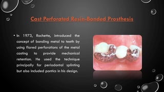
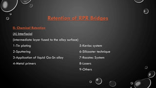
The partial coverage restoration is a conservative restoration that requires less destruction of tooth structure than does a full coverage crown.
Its use is based on the premise that an intact surface of tooth structure should not be covered by a crown if its inclusion is not essential to the retention, strength, or esthetic result of the definitive restoration.
Laminate veneers by student at faculty of oral and dental medcine Ahram canad...Menna-Allah Ashraf
this document will supply you with all you need to know about laminate veneers :
1) Advantages and disadvantages of laminate veneers.
2) Indications and contraindications of laminate veneers
3) Types of laminate veneer and their advantages and disadvantages.
4) Porcelain laminate veneers : features and preparations.
5) lumineers
In the last decades the development of the porcelain materials, the reliable bonding strength to enamel and dentin, and the bonding of resin cement to the porcelain through the silane, Porcelain laminates become trusted type of treatment in the daily practice.
It is an aesthetic treatment that concerns mainly the labial face of the anterior teeth, its thickness is about 0.3 mm in the cervical area to 0.4 -7 at the incisal third, in certain cases it can be done without any prep or just little touch of the enamel (lumineer), but in most prep is indicated to improve the adaptation in the cervical area also to remove the aprismatic enamel layer which which has low bonding strength with the resin cement, however prep should be in the enamel limits, 3 different type of prep are practiced, however, they are the same on the labial surface but the but the difference concerns the incisal edge.
In this lecture, indications and contraindications are exposed. All the materials in use and their indications as well as the clinical procedures are detailed.
Retainers in FIXED PARTIAL DENTURES(FPDS) AND RESIN BONDED FPDNAMITHA ANAND
DIFFERENT RETAINERS IN FPD ARE DISCUSSED WITH PICTURES AND REFERENCES AND SPECIAL CONSIDERATION FOR RESIN BONDED FPDS PARTIAL COVERAGE RESTORATIONS AND INTRACORONAL RESTORATIONS
Failures in Fixed Partial Denture
(Prosthodontics FPD- Dental science)
Various types of failures in the fabrication of fixed partial denture
Dr.Sachin Sunny Otta
St.Gregorios Dental College,Kothamangalam,Ernakulam
Description :
The Indian Dental Academy is the Leader in continuing dental education , training dentists in all aspects of dentistry and
offering a wide range of dental certified courses in different formats.for more details please visit
www.indiandentalacademy.com
3- Basic principles for designing the removable partial denture Amal Kaddah
Clinical course of Partial Denture
3- Basic principles for designing the removable partial denture
a- Problems and General Principles Applied for Kennedy Class I
This seminar is of postgraduate level, which will be helpful for students. The presenter has added the information from various sources and the references are quoted in the last few slides of the seminar to gather more information about the seminar.
2- b. Basic principles for designing Kennedy class II, III and IV Removable P...AmalKaddah1
1-a. Basic principles for designing the removable partial denture (class I partial denture design)
Introduction.
Objectives and Functions of RPD.
Factors that affect RPD design.
Basic principles for designing Kennedy class I partial denture.
2- b. Basic principles for designing Kennedy class II, III and IV Removable Partial Denture(RPD)
Indian Dental Academy: will be one of the most relevant and exciting training center with best faculty and flexible training programs for dental professionals who wish to advance in their dental practice,Offers certified courses in Dental implants,Orthodontics,Endodontics,Cosmetic Dentistry, Prosthetic Dentistry, Periodontics and General Dentistry.
It include proximal stripping, Diagnostic aids, advantages, disadvantages, periodontal consideration, procedure for proximal stripping. Expansion, extraction, Distalization in detail as method of gaining space, Extra-oral, Intra-oral method for gaining space. uprighting, derotation of posterior teeth. proclination of anterior teeth.
Posterior tooth preparationscertified fixed orthodontic courses by Indian den...Indian dental academy
The Indian Dental Academy is the Leader in continuing dental education , training dentists in all aspects of dentistry and offering a wide range of dental certified courses in different formats.
Indian dental academy provides dental crown & Bridge,rotary endodontics,fixed orthodontics,
Dental implants courses.for details pls visit www.indiandentalacademy.com ,or call
0091-9248678078
The pediatric dentistry in the restorative to the damaged tooth by the caries and the prevention for the further shedding and erupting of the permanent tooth.
Design of a fixed Partial Denture (with Abutment Tooth Preparation)Taseef Hasan Farook
A simplified take on the steps to designing a Fixed partial denture. This presentation also includes an overview of abutment preparation, associated finishes and methods of impression taking prior to the designing of the prosthesis itself
Rehabilitation of endodontically treated teeth : Post & CoreNaveed AnJum
These days we often come across mutilated or badly broken teeth in our practice. However various factors are involved for a better prognosis of such a teeth. This presentation mainly focuses on post and core treatment of such a teeth.
This presentation includes brief history, classification and definition of overdentures and explains in details about the various tooth supported overdentures. It explains about bar attachments, ball attachments, telecsopic dentures etc.
history, classification, types of veneers, indications and contraindications, working procedure, preparation, ipmpression taking for veneers, surface treatment and cementation, veneers vs crowns
This pdf is about the Schizophrenia.
For more details visit on YouTube; @SELF-EXPLANATORY;
https://www.youtube.com/channel/UCAiarMZDNhe1A3Rnpr_WkzA/videos
Thanks...!
Earliest Galaxies in the JADES Origins Field: Luminosity Function and Cosmic ...Sérgio Sacani
We characterize the earliest galaxy population in the JADES Origins Field (JOF), the deepest
imaging field observed with JWST. We make use of the ancillary Hubble optical images (5 filters
spanning 0.4−0.9µm) and novel JWST images with 14 filters spanning 0.8−5µm, including 7 mediumband filters, and reaching total exposure times of up to 46 hours per filter. We combine all our data
at > 2.3µm to construct an ultradeep image, reaching as deep as ≈ 31.4 AB mag in the stack and
30.3-31.0 AB mag (5σ, r = 0.1” circular aperture) in individual filters. We measure photometric
redshifts and use robust selection criteria to identify a sample of eight galaxy candidates at redshifts
z = 11.5 − 15. These objects show compact half-light radii of R1/2 ∼ 50 − 200pc, stellar masses of
M⋆ ∼ 107−108M⊙, and star-formation rates of SFR ∼ 0.1−1 M⊙ yr−1
. Our search finds no candidates
at 15 < z < 20, placing upper limits at these redshifts. We develop a forward modeling approach to
infer the properties of the evolving luminosity function without binning in redshift or luminosity that
marginalizes over the photometric redshift uncertainty of our candidate galaxies and incorporates the
impact of non-detections. We find a z = 12 luminosity function in good agreement with prior results,
and that the luminosity function normalization and UV luminosity density decline by a factor of ∼ 2.5
from z = 12 to z = 14. We discuss the possible implications of our results in the context of theoretical
models for evolution of the dark matter halo mass function.
Richard's aventures in two entangled wonderlandsRichard Gill
Since the loophole-free Bell experiments of 2020 and the Nobel prizes in physics of 2022, critics of Bell's work have retreated to the fortress of super-determinism. Now, super-determinism is a derogatory word - it just means "determinism". Palmer, Hance and Hossenfelder argue that quantum mechanics and determinism are not incompatible, using a sophisticated mathematical construction based on a subtle thinning of allowed states and measurements in quantum mechanics, such that what is left appears to make Bell's argument fail, without altering the empirical predictions of quantum mechanics. I think however that it is a smoke screen, and the slogan "lost in math" comes to my mind. I will discuss some other recent disproofs of Bell's theorem using the language of causality based on causal graphs. Causal thinking is also central to law and justice. I will mention surprising connections to my work on serial killer nurse cases, in particular the Dutch case of Lucia de Berk and the current UK case of Lucy Letby.
Seminar of U.V. Spectroscopy by SAMIR PANDASAMIR PANDA
Spectroscopy is a branch of science dealing the study of interaction of electromagnetic radiation with matter.
Ultraviolet-visible spectroscopy refers to absorption spectroscopy or reflect spectroscopy in the UV-VIS spectral region.
Ultraviolet-visible spectroscopy is an analytical method that can measure the amount of light received by the analyte.
A brief information about the SCOP protein database used in bioinformatics.
The Structural Classification of Proteins (SCOP) database is a comprehensive and authoritative resource for the structural and evolutionary relationships of proteins. It provides a detailed and curated classification of protein structures, grouping them into families, superfamilies, and folds based on their structural and sequence similarities.
Introduction:
RNA interference (RNAi) or Post-Transcriptional Gene Silencing (PTGS) is an important biological process for modulating eukaryotic gene expression.
It is highly conserved process of posttranscriptional gene silencing by which double stranded RNA (dsRNA) causes sequence-specific degradation of mRNA sequences.
dsRNA-induced gene silencing (RNAi) is reported in a wide range of eukaryotes ranging from worms, insects, mammals and plants.
This process mediates resistance to both endogenous parasitic and exogenous pathogenic nucleic acids, and regulates the expression of protein-coding genes.
What are small ncRNAs?
micro RNA (miRNA)
short interfering RNA (siRNA)
Properties of small non-coding RNA:
Involved in silencing mRNA transcripts.
Called “small” because they are usually only about 21-24 nucleotides long.
Synthesized by first cutting up longer precursor sequences (like the 61nt one that Lee discovered).
Silence an mRNA by base pairing with some sequence on the mRNA.
Discovery of siRNA?
The first small RNA:
In 1993 Rosalind Lee (Victor Ambros lab) was studying a non- coding gene in C. elegans, lin-4, that was involved in silencing of another gene, lin-14, at the appropriate time in the
development of the worm C. elegans.
Two small transcripts of lin-4 (22nt and 61nt) were found to be complementary to a sequence in the 3' UTR of lin-14.
Because lin-4 encoded no protein, she deduced that it must be these transcripts that are causing the silencing by RNA-RNA interactions.
Types of RNAi ( non coding RNA)
MiRNA
Length (23-25 nt)
Trans acting
Binds with target MRNA in mismatch
Translation inhibition
Si RNA
Length 21 nt.
Cis acting
Bind with target Mrna in perfect complementary sequence
Piwi-RNA
Length ; 25 to 36 nt.
Expressed in Germ Cells
Regulates trnasposomes activity
MECHANISM OF RNAI:
First the double-stranded RNA teams up with a protein complex named Dicer, which cuts the long RNA into short pieces.
Then another protein complex called RISC (RNA-induced silencing complex) discards one of the two RNA strands.
The RISC-docked, single-stranded RNA then pairs with the homologous mRNA and destroys it.
THE RISC COMPLEX:
RISC is large(>500kD) RNA multi- protein Binding complex which triggers MRNA degradation in response to MRNA
Unwinding of double stranded Si RNA by ATP independent Helicase
Active component of RISC is Ago proteins( ENDONUCLEASE) which cleave target MRNA.
DICER: endonuclease (RNase Family III)
Argonaute: Central Component of the RNA-Induced Silencing Complex (RISC)
One strand of the dsRNA produced by Dicer is retained in the RISC complex in association with Argonaute
ARGONAUTE PROTEIN :
1.PAZ(PIWI/Argonaute/ Zwille)- Recognition of target MRNA
2.PIWI (p-element induced wimpy Testis)- breaks Phosphodiester bond of mRNA.)RNAse H activity.
MiRNA:
The Double-stranded RNAs are naturally produced in eukaryotic cells during development, and they have a key role in regulating gene expression .
(May 29th, 2024) Advancements in Intravital Microscopy- Insights for Preclini...Scintica Instrumentation
Intravital microscopy (IVM) is a powerful tool utilized to study cellular behavior over time and space in vivo. Much of our understanding of cell biology has been accomplished using various in vitro and ex vivo methods; however, these studies do not necessarily reflect the natural dynamics of biological processes. Unlike traditional cell culture or fixed tissue imaging, IVM allows for the ultra-fast high-resolution imaging of cellular processes over time and space and were studied in its natural environment. Real-time visualization of biological processes in the context of an intact organism helps maintain physiological relevance and provide insights into the progression of disease, response to treatments or developmental processes.
In this webinar we give an overview of advanced applications of the IVM system in preclinical research. IVIM technology is a provider of all-in-one intravital microscopy systems and solutions optimized for in vivo imaging of live animal models at sub-micron resolution. The system’s unique features and user-friendly software enables researchers to probe fast dynamic biological processes such as immune cell tracking, cell-cell interaction as well as vascularization and tumor metastasis with exceptional detail. This webinar will also give an overview of IVM being utilized in drug development, offering a view into the intricate interaction between drugs/nanoparticles and tissues in vivo and allows for the evaluation of therapeutic intervention in a variety of tissues and organs. This interdisciplinary collaboration continues to drive the advancements of novel therapeutic strategies.
2. Minimally Invasive Dentistry
• Traditionally, if a patient requires restorative work in the posterior region,
conventional thought has been to treatment plan full-coverage restorations, such
as crowns. The trend in recent years has been “minimally invasive” dentistry, which
means preserving as much tooth structure as possible whenever feasible.
• This inherently signifies moving away from procedures, such as crown placement,
that require destruction of sound enamel and dentin if other, less invasive options
are available and will be equally effective.
3. Partial Coverage Restorations
• The partial coverage restoration is a conservative restoration that
requires less destruction of tooth structure than does a full coverage
crown.
• Its use is based on the premise that an intact surface of tooth structure
should not be covered by a crown if its inclusion is not essential to the
retention, strength, or esthetic result of the definitive restoration.
5. Partial Coverage Crown
An extra-coronal restoration that restores all but one coronal surface of a tooth or
dental implant, usually not covering the facial surface.
6. Classification
1- Retention By Grooves
• ¾ Crown
• R ¾ crown
• Mesial ½ crown
• 7/8 crown
2- Retention By Pins
• Pin-ledge
3- Modified Type
• Grooves + pins
7. Characteristics
• Not as retentive as full coverage crown.
• A preparation feature must be substituted to
compensate for the retention and resistance lost
when an axial surface is not covered.
• The most commonly used feature is a groove.
• Grooves must have definite lingual walls (lingual
hook or a lock effect).
• A v-shaped groove, without a definite lingual wall,
provides only 68% of the retention and 57% of
the resistance of a concave groove with a lingual
wall.
8. Indications
1. If the buccal surface is intact and supported by sound tooth structure.
2. Minimal caries and good oral hygiene.
3. Short span FPD.
4. Where restorations or alteration of the occlusal surface is needed.
5. Anterior partial veneer crown can be used as a retainer, to reestablish anterior
guidance and to splint teeth.
6. Teeth with crown length that is average or exceeds average.
7. Teeth with normal anatomic crown form.
8. Anterior teeth with adequate labio-lingual thickness.
9. Contraindications
1. Short clinical crown.
2. Long span FPD.
3. Rarely suitable for root canal treated
teeth, specially anterior teeth.
4. Posterior root canal treated teeth where
cusps are weakened.
5. Active caries and periodontal diseases.
6. Poor oral hygiene.
7. Deep cervical caries.
8. Teeth with extensive core restorations.
9. Bell shaped teeth.
10. Thin teeth.
11. Poorly aligned abutments.
10. Advantages
1. Tooth structure is spared.
2. Much of the margin is accessible to the dentist for finishing and to the patient
for cleaning.
3. Less restoration margin is in proximity to the gingival crevice, reducing the
possibility of periodontal irritation.
4. Easily seated completely during cementation.
5. If an electric pulp test ever needs to be conducted on the tooth, a portion of
enamel is not veneered and accessible.
6. Excess cement removed easily.
7. Better esthetics.
11. Disadvantages
1. Less retention and resistance
2. Limited adjustments can be made in the path of placement
3. Skillful preparation is critical
4. Limited indications
5. Metal display in completed restorations
12. Maxillary Posterior
Three-Quarter Crown
• Most commonly used partial
coverage crown.
• Facial surface is left uncovered.
• The occlusal finish line on a
maxillary tooth terminates near the
facio-occlusal angle.
• If designed skillfully, the three-
quarter crown can be very esthetic.
13. Three-Quarter Crown With
Proximal Boxes
• More retentive than standard preparation with
grooves.
• Boxes are more destructive.
• Used in cases of proximal caries or previous
restoration.
• Less destructive way to gain more retention is to
use four grooves.
14. Three-Quarter Crown
On A Mandibular
Molar
• Occlusal finish line on the facial
surface is gingival to the occlusal
contacts.
• Occlusal shoulder on the facial
aspects of the facial cusps tying the
grooves together and strengthening
the margins.
15. Seven-Eights Crown
• Used when distal cusp must be
covered.
• Most commonly used in
maxillary molars, but can also
be used in mandibular
premolars and molars.
• Its an excellent retainer for FPD.
16. Reverse Three-Quarter
Crown
• Used in mandibular molars to
preserve an intact lingual surface.
• Useful in FPD abutments with severs
lingua inclination.
• Grooves on the linguo-proximal line
angles are joined by occlusal offset
on the facial slopes of the lingual
cusps.
17. Proximal Half Crown
• It’s a ¾ crown that is rotated 90°, with
the distal, rather than the facial
surface, left intact.
• Can be used as a retainer on a tilted
mandibular molar FPD abutment.
• Needs excellent oral hygiene and low
incidence of proximal caries.
• Mesial surface is parallel to the path
of insertion of the mesial abutment.
18. Anterior Three-Quarter
Crown
• Its rarely used.
• Usually used in pin modified ¾
crown form.
• Can be used as a retainer in short
span FPD on a restoration- and
caries-free abutment.
• Path of insertion is parallel to the
incisal 2/3 of the labial surface. In
order to be longer and more
retentive.
19. Pin Modified Three-
Quarter Crown
• Used for short span FPD.
• It preserves the facial and one proximal
surface.
• Minimal subginival margins.
• Good to repair incisors and canines with
severe lingual abrasion.
• Not used on small, thin malpostioned
teeth or those with large pulp.
• Needs a skilled operator.
• Pins should be 4 mm deep for optimum
retention and resistance.
21. Definition
• It’s a layer of tooth colored material that’s applied to a tooth to restore localized
or generalized defects and/or intrinsic discoloration. (Sturdevant)
• A protective or ornamental facing or a superficial attractive display in multiple
layers. (Rosensteil)
• A conservative esthetic restoration of anterior teeth to mask discoloration, restore
malformed teeth & correct minor tooth alignment. (Mosbey dental dictionary)
23. Advantages Of Direct Composite System
1. One appointment
2. The dentist directly controls form and color.
3. Reduced cost.
4. Repairable
24. Indirect Porcelain Technique
1. Time saving for the dentist.
2. Less chair time.
3. Color stability.
4. More esthetic.
5. Better wear resistant.
6. Better tissue compatibility.
32. Preparation Design
1. Type 1 (Window Or Contact Lens):
• Doesn’t involve the incisal edge
2. Type 2 (Feather Edge):
• Most commonly used.
• Terminates at the incisal edge.
3. Type 3 (Incisal Bevel Or Butt Joint):
• Covers the incisal edge and terminates lingually.
• Used in lower anteriors and upper canine.
4. Type 4 (Incisal Overlap):
• Terminates I mm before the centric contact.
• Indicated for extensive changes in color and contour.
35. Inlays And Onlays
• Are forms of indirect restoration used when a
posterior tooth is too damaged to support a
basic filling, but not so severely that it needs a
crown.
• Essentially, restore structural integrity to a
tooth to serve both as a functional and an
aesthetic benefit.
36. Inlay
• Is an intra-coronal dental rest. That
made out of the oral cavity to restore
some of the occlusal surface of the
tooth but does not restore any of the
cusps.
37. Types Of Inlay
• Acc. To Color:
1. Metal
2. Tooth colored
• Acc. To Technique:
1. Direct
2. Indirect
38.
39. Inlay-Retained Fixed Partial Denture
• Luted by use of adhesive procedures offer
a clinical alternative for the restoration of
single missing posterior teeth.
• They offering good aesthetic and
functional results. The procedure is
minimally invasive and conservative.
40. Inlay-Retained Fixed Partial Denture
• Indications:-
1. Patients with good oral hygiene and low
susceptibility to caries
2. Minimum coronal tooth height of 5 mm,
parallel abutments and a maximum
mesiodistal edentulous gap of 12 mm
3. Teeth with previous restoration
41. Inlay-Retained Fixed Partial Denture
• Advantages :-
1. The main advantage of inlay bridge is minimal tooth reduction . preparing the
teeth for the traditional bridge prosthesis (FPD), 63-72% of healthy tooth structure
is removed (edelhoff et al., 2002) compared to the removal of only 3-30% of
healthy tooth structure for the inlay prosthesis preparation.
2. These restorations are attractive for young people with healthy teeth.
3. In addition, after the cementation of inlay bridge , there is less postoperative
sensitivity because retention is made by linking the prosthesis with natural
remaining enamel.
42. Inlay-Retained Fixed Partial Denture
• Disadvantages:-
1. Inlay bridges are associated with an increased risk of falling than the
traditional prosthesis (Priest, 1996).
2. The retention of these prostheses is 70-80% (from 4 till 6 years), less than
the normal bridge prosthesis.
3. The most common failure is discementation or development of secondary
caries (Invisible discementation of the retentive element may lead to
increased amounts of dental plaque and resultant periodontal
inflammation. )
43. Inlay-Retained Fixed Partial Denture
• Preparation:-
1. The depth of the cavity is 2-4 mm.;
2. Should be enough space - 9 mm 2 for connective element ;
3. The angle of vertical walls of the cavity 2-3 0;
4. Borders clearly visible;
5. Rounded angles, without sharp edges;
6. Minimal width of the step 0.4 mm;
7. The depth of mesial and distal (proximal) box 4 mm;
44. Inlay-Retained Fixed Partial Denture
• Preparation:-
8. The width of mesial and distal (proximal) box 4 mm;
9. The maximal length of intermediate part is 10 mm;
10. It is necessary to use traditional layered ceramics to avoid
deterioration of opposing teeth.
11. Preparation for ceramic inlay (interproximal surfaces):
12. The depth of mesial and distal (proximal) box 4 mm,
13. The depth of cavity 2 mm
14. Occlusal view, the width of mesial and distal (proximal) box 4 mm.
45. Inlay-retained FPDs allow for single-tooth substitution when implant placement is not possible or not
indicated. The bone-tissue loss during implantation and tooth-tissue loss during preparation of
abutment teeth needed for full coverage FPDs can be avoided.
47. What Is An Onlay?
• It is a posterior restoration that
is made to protect the tooth and
involves the proximal surfaces
(class II) of a posterior tooth
and covers one or more of the
cusps.
• It spans the gap between inlays
(intra-coronal restorations) and
full coverage crown.
48. Indications & Contraindications
Indications
1. Large restorations
2. Cuspal protection
3. Endodontically treated teeth
4. Teeth at risk for fracture
5. Diastema closure and occlusal plane
correction
6. Abutment for PD
Contraindications
1. High caries rate
2. Intact facial and lingual surfaces
3. Small restorations
4. Extensive occlusal facet
49. Advantages & Disadvantages
Advantages
1. High strength
2. Biocompatibility
3. Low wear
4. Control of contours and contacts
5. Less chance of voids and internal
stresses
Disadvantages
1. Micro leakage
2. More chair time
3. High cost
4. Technique sensitive
5. Difficult to repair
51. Types Acc. To Technique
1. Direct Tech.: Some of the steps are done on the patient mouth.
2. Indirect Tech.: Require impression taking and detailed laboratory steps.
3. Indirect-direct Tech.: Initial fabrication is done on a model.
52. Preparation
1. Preparation walls should be 6-10° occlusally divergent.
2. Cuspal reduction of 1.5-2 mm in functional cusp and 1-1.5 mm on non-functional
cusp.
3. All line angles and bevels are smoothly joined with no interruption.
4. Gingival, occlusal bevels and flares are prepared in a manner such that a
marginal cast gold metal of 40° is obtained.
5. A carbide bur or diamond bur is used to create a rougher preparation to aid in
bonding of the final restoration.
54. Other Preparation Designs
• Carvalho et al, 2021 found that the
non retentive design preparation
showed the best mechanical behavior
compared to all-ceramic onlay
preparation design with occlusal
isthmus reduction and traditional all-
ceramic onlay preparation without
occlusal isthmus reduction.
55. Types Of Ceramic Inlays And Onlays
• Although some laboratory-processed composite systems have been available, and at
least one machinable composite (paradigm MZ100, 3M ESPE, st. Paul, MN) is available
for CAD/CAM, most tooth-colored indirect posterior restorations are fabricated from
ceramic materials.
• Ceramic inlays and onlays have become popular not only because of patient demand for
esthetic, durable restorative materials but also because of improvements in materials,
fabrication techniques, adhesives, and resin-based cements.
56. Types Of Ceramic Inlays And Onlays
• Among the ceramic materials used are feldspathic porcelain, leucite-
reinforced pressed ceramics, lithium disilicate, and various types of
machinable (milled) ceramics designed for use with either chair side or
laboratory CAD/CAM systems.
57. What Is An Overlay?
• It is a posterior restoration that
is made to protect the tooth
and covers all cusps of the
occlusal surface.
58. Advantages
1. A biomimetic alternative to the traditional crown preparation in situations
that require full occlusal coverage.
2. Less time consuming.
3. Preservation of enamel body of the teeth.
4. Less invasive which avoid risks of conventional preparation.
5. Work in harmony with the natural biomechanics of the tooth.
6. Help extend the restorative life cycle of the tooth.
7. Maximize the fracture resistance of ceramic materials.
59. Indications
1. Severe worn occlusal enamel.
2. When there is need to strengthen
occlusal surface while axial walls
are intact.
Contraindications
1. When a full coverage crown is
indicated.
2. Extensively damaged molars
3. Over post supported restoration
Indications And Contraindications
61. What Is An Occlusal Veneer?
• Conservative alternative to conventional overlay or
complete crown restorations.
• The tooth preparation for occlusal veneers is
straightforward as the anatomic structure of the teeth is
maintained and the interocclusal distance is taken into
consideration.
• They have a minimal thickness of 0.4 mm to 0.6 mm at
the developmental grooves and 1.0 mm to 1.3 mm at
the cusp tips.
62. Advantages
• More conservative than conventional full coverage crown
• Less time consuming
• Preservation of enamel body of the teeth
• Less invasive which avoid risks of conventional preparation
63. Indication
• Occlusal veneers are indicated when
occlusal enamel is worn down or
severely eroded, reducing its thickness
and even exposing the underlying
dentin at the occlusal surface.
64. Contra-Indications
1. When a full coverage crown is indicated .
2. Large restorations requiring onlays
3. Extensively damaged molars
4. Over post supported restoration
5. Isolation can not be achieved
65. Preparation
• At least 1 mm of occlusal clearence should be done to provide enough thickness for
the occlusal veneer restoration.
• Sharp margin preparation: 120 degrees was the angle between the buccal and
lingual cusp slopes, mesiobuccal and distobuccal slopes, mesiolingual and distolingual
slopes , with margin surface angel less than 90 degree.
66. Preparation
• Hollow chamfer preparartion:
• 120 degrees is the angle between the
buccal and lingual cusp slopes,
mesiobuccal and distobuccal slopes,
mesiolingual and distolingual slopes ,
with margin surface angel equals or
more than 90 degree
67.
68. Material Of Choice
1. Lithium disilicate
2. Zirconia-reinforced lithium silicate (vita suprinity),
3. Polymer-infiltrated ceramic (vita enamic),
4. Polymethylmethacrylate PMMA or composite resin (paradigm MZ100)
69. Role Of Occlusal Veneer In Dentistry
• Occlusal veneers are considered a treatment option for many cases since it is
the most conservative method for restoration of occlusal surface wear ,
restoration of the occlusal surface will achieve the following:
1. Treatment of sensitive posterior teeth Large restorations requiring onlays.
2. Prevention of TMJ dysfunction Over post supported restoration.
3. Correct occlusal interference for treatment of traumatic occlusion.
70. Occlusal Veneer Vs. Full Coverage Crown
• Schlichting et al. Stated that ultra-thin composite resin (MZ100) and lithium
disilicate (e.Max CAD) occlusal veneers represent a conservative alternative to
traditional onlays and complete coverage crowns for the treatment of severe
erosive lesions in the posterior dentition. (Modeling of ultrathin occlusal veneers)
71. Occlusal Veneer Vs. Overlay
• Gürpınar wt al. evaluated the fracture resistance of occlusal veneer and overlay
CAD/CAM restorations made of polymer-infiltrated ceramic and lithium disilicate
ceramic. They found that for restorative treatment of posterior worn teeth, minimal
invasive occlusal veneer restorations made of LDC or PIC materials are successful
alternatives.
72. Vonlay
• A vonlay is a hybrid of an onlay with an extended
buccal veneer surface for use in posterior teeth
where there is mostly enamel to bond to.
• This restorative option requires a much less invasive
preparation than a full-coverage crown but
provides the same structural benefits.
• Crowneer: It’s a modification of vonlay that covers
more than the buccal surface. (It’s a crown/veneer
hybrid)
73. Advantages
1. Enhances the strength and esthetics of the remaining tooth.
2. Excellent for patients presenting with wear, decay, or
occlusal problems in posterior teeth.
3. Less invasive
4. More readily repairable
5. Less technique-sensitive to attain adequate bonding.
6. Leave more sound tooth structure remaining if further
treatment is required in the future.
75. Endocrown
• It’s a modification of onlay restoration that indicated in extensively damaged
molars, it gains its retention from pulp chamber and coronal walls.
• The restoration gains its retention from the coronal pulpal walls of pulp chamber
using adhesion with etched ceramic restoration providing cuspal coverage.
• It is supported by pulpal floor and wide cervical butt joint.
76. Endocrown
• Proposed in 1999 by Bindl and Mörmann as an alternative to the full post-and-
core supported crown, the “endocrown” is a one-piece ceramic construction, based
on concepts developed by Pissis.
• In 2008, Lander and Dietschi presented a clinical report on endocrowns, and, in
2009, Magne and Knezevic, who were concerned about the choice of
reconstruction materials, considered ceramics versus composites for endocrown
molar restorations.
• Various studies suggested extending the concept to maxillary premolars and
maxillary incisors, but these proposals remain controversial.
77. Indications & Contraindications
• The endocrown is indicated for all molars, particularly those with clinically short
crowns, calcified root canals or very slender roots.
• The endocrown is contraindicated if adhesion cannot be assured, if the pulpal
chamber is less than 3 mm deep or if the cervical margin is less than 2 mm wide
for most of its circumference.
78. Preparation
• Occlusal Preparation:
• At least 2 mm reduction in the axial direction.
• Cervical margin should be supra gingival.
• However in certain cases it may follow the gingival margin.
• Differences in level between the various parts of the cervical
margin must be linked by a slope of no more than 60° to
avoid a staircase effect.
• Enamel walls less than 2 mm thick should be removed.
79. Preparation
Axial Preparation
• Elimination undercuts in the access cavity with a 7° occlusal
divergence.
• The depth of the cavity should be at least 3 mm without touching
the pulpal floor.
Preparation Of The Cavity Floor
• The entrance to the pulpal canal is opened gutta percha is
removed to a depth not exceeding 2 mm to take advantage of
the saddle-like anatomy of the cavity floor.
80. Endocrown Vs. Post And Core
• Chang, Chia-Yu, et al. Tested fracture resistance and failure modes of CEREC endocrowns
and conventional post and core-supported CEREC crowns and concluded that the bonded
ceramic endocrowns showed a significantly higher fracture resistance than the classic
reinforced and designed group and, therefore, offer a feasible alternative for severely
damaged teeth.
• Sedrez-Porto et al. Suggests that endocrowns may perform similarly or better than the
conventional treatments using intraradicular posts, direct composite resin or inlay/onlay
restorations.
81. Ceramic Thickness & Fracture Resistance
• Today, we are moving toward minimally invasive dentistry with the philosophy that
less is more. Less tooth reduction means more adhesion and clinical longevity.
• Bakeman et al, evaluated the influence of ceramic thickness and ceramic materials
on fracture resistance of posterior partial coverage ceramic restorations.
• They found that lithium disilicate glass ceramic significantly improved fracture
resistance when compared to a leucite-reinforced glass ceramic, even at a
thickness below the manufacturer’s suggested minimum. The use of lithium disilicate
may have advantages in clinical situations of minimal occlusal clearance.
83. Resin Bonded Bridges
• The restoration is simple in concept
and consists of one or more pontics
supported by thin retainers bonded
lingually and proximally to the enamel
of the abutment teeth.
84. Cast Perforated Resin-Bonded Prosthesis
• In 1973, Rochette, introduced the
concept of bonding metal to teeth by
using flared perforations of the metal
casting to provide mechanical
retention. He used the technique
principally for periodontal splinting
but also included pontics in his design.
85. Cast Perforated Resin-Bonded Prosthesis
• Howe and Denehy recognized the
metal framework’s improved retention
(over bonded pontics) and began
using FDPs with cast perforated metal
retainers bonded to abutment teeth
and metal-ceramic pontics to replace
missing anterior teeth.
86. Cast Perforated Resin-Bonded Prosthesis
• Their design recommendation, extending the framework to cover a maximum area
of the lingual surface, suggested little or no tooth preparation.
• Use of these FDPs was limited to mandibular teeth or situations with minimal
occlusal contact.
• The restorations were bonded with a heavily filled composite resin as a luting
medium.
87. Cast Perforated Resin-Bonded Prosthesis
• Livaditis expanded this concept to replacement of posterior teeth, Perforated retainers
were used to increase resistance and retention.
• Cast retainers were extended inter-proximally into the edentulous areas and onto
occlusal surfaces.
• The design included a defined occluso-gingival path of placement by tooth modification,
which involved lowering the proximal and lingual height of contour of the enamel on the
abutment teeth.
• These restorations were placed in normal occlusion; many have survived and have been
seen on recall for up to 13 years.
88. Cast Perforated Resin-Bonded Prosthesis
• Despite this success, the perforation
technique presents the following
limitations:
1. Weakening of the metal retainer by the
perforations
2. Exposure to wear of the resin at the
perforations
3. Limited adhesion of the metal provided
by the perforations
89. Maryland Bridge
• Etched-Cast Resin-Bonded Fixed
Dental Prostheses (Micromechanical
Retention)
• A technique for the electrolytic etching
of cast base metal
• retainers was developed at the
University of Maryland by Thompson
and Livaditis.
90. Maryland Bridge
• Etched-cast retainers have definite advantages over cast-perforated restorations:
1. Retention is improved because the resin-to-etched metal bond can be substantially
stronger than the resin-to-etched enamel.
2. The retainers can be thinner yet still resist flexing.
3. The oral surface of the cast retainers is highly polished and resists plaque
accumulation.
4. the need for a composite resin with a low film thickness for luting the casting became
apparent.
91. Maryland Bridge
• Etched-cast retainers have definite advantages over cast-perforated restorations:
5. which allowed micromechanical bonding into the undercuts in the metal casting
created by etching and simultaneously provided adequate strength.
6. composite resin filled (60% by weight) with a film thickness of approximately
20μm.
7. Such cements do not adhere chemically to the metal.
92. Maryland Bridge
• Electrolytic etching of base metal alloys proved to
be critically dependent on the base metal alloy.
• Initial etching methods were developed for a
nickel-chromium (Ni-Cr) alloy and a nickel-
chromium-molybdenum-aluminum-beryllium (Ni-Cr-
Mo-Al-Be) alloy.
• These methods were followed by simplified
techniques, chemical etching, or gel etching.
93. Ceramic Retainers
• High-strength ceramics, particularly zirconia have
been used as retainers for resin-bonded FDPs.
• These restorations exhibit:
• Better esthetics than do metal retainers, which can
discolor, particularly with thin abutment teeth.
• Good medium-term clinical performance has been
demonstrated.
94. Preparation of the Anterior Abutment Teeth
• The amount of reduction is Minimal because the enamel must not be
penetrated.
• Non-noble alloys are usually used because they provide a strong framework
in thin metal sections, also provides a strong margin.
• There must be sufficient enamel area for successful bonding.
• Removal of proximal undercuts.
95. Preparation of the Anterior Abutment Teeth
• Retention can be improved by:
1. Two additional grooves are placed on the lingual surface of the abutment
tooth,these grooves run in the incisogingival direction, They should be 0.75mm
wide, 1mm deep.
2. An additional groove is placed on the interproximal surface next to the pontic
space, cingulum rest seat
3. Make an accurate impression and marginal fit is as crucial for a resin-bonded
restoration
97. Preparation of the Posterior Abutment Teeth
• Three major components:
1. The occlusal rest (for resistance to gingival
displacement)
2. The retentive surface (for resistance to
occlusal displacement)
3. The proximal wrap and proximal slots (for
resistance to torquing forces)
98. Preparation of the Posterior Abutment Teeth
• Retention improved by:
1. A spoon-shaped occlusal rest seat, is placed in the
proximal marginal ridge area of the abutments
adjacent to the edentulous space. the rest is an
important retention feature and simultaneously provides
resistance to both occlusal and lateral forces.
2. To resist occlusal displacement, the restoration is
designed to maximize the bonding area without
unnecessarily compromising periodontal health or
esthetics.
99. Preparation of the Posterior Abutment Teeth
• Interproximal area, a gingival chamfer
margin is not desirable; a knife-edge
margin is better for avoiding enamel
penetration.
• Occlusally, the framework should be
extended high on the cuspal slope
• A single path of placement should exist.
100. Retention of RPR Bridges
I-Mechanical Retention
(A) Macro-Mechanical Retention
1. Rochette (perforations)
2. Virginia bridge (lost salt crystal)
3. Cast mesh pattern.
4. Retention acrylic beads
I-mechanical Retention
(B) Micro-mechanical Retention
1. Maryland bridge (electrolytic etching )
2. Chemical etching
3. Porous metal coating
4. Sand-blasting
101. Retention of RPR Bridges
II- Chemical Retention
(A) Interfacial
(intermediate layer fused to the alloy surface)
1-Tin plating
2-Sputtering
3-Application of liquid Ga-Sn alloy
4-Metal primers
5-Kevloc system
6-Silicoater technique
7-Rocatec System
8-Lasers
9-Others
102. Retention of RPR Bridges
II- Chemical Retention
(B) Adhesive
(reactive groups of the resin cements bond to the oxide layer on the alloy
1-Heating the alloys
- Single heating - Double heating
2- Immersion in oxidizing solution
3-Immersion in conc. Nitric acid
103. Advantages
1. Minimal removal of tooth structure
2. Minimal potential for pulpal trauma
3. Anesthesia not usually required
4. Supra-gingival preparation
5. Easy impression making
6. Interim restoration not usually required
7. Reduced chair time
8. Reduced patient expense
9. Rebonding possible
104. Disadvantages
1. Reduced restoration longevity.
2. Enamel modifications: required Space correction: difficult.
3. Good alignment of abutment teeth: required.
4. Esthetics compromised on posterior teeth.
105. Indications
1. Replacement of missing anterior teeth in children and adolescents
2. Short edentulous span
3. Unrestored abutments
4. Single posterior tooth replacement
5. Significant clinical crown length
6. Excellent moisture control
106. Contraindications
1. Parafunctional habits
2. Long edentulous span
3. Restored or damaged abutments
4. Compromised enamel
5. Significant pontic width discrepancy
6. Deep vertical overlap
7. Nickel allergy
107. Failure Causes
(A) Improper Patient Selection:
1. Poor alignment of teeth.
2. Short clinical crown.
3. Inadequate enamel for bonding.
4. Metal sensitivity.
108. Failure Causes
(B) Incomplete Tooth Preparation:
1- Insufficient proximal and lingual surface reduction.
2- Incomplete or less than 180° extension of wraparounds.
3- Lack of accommodation to mandibular protrusive movements.
109. Failure Causes
(C) Bonding Of The Resin Bonded Retainers:
1- Contamination of the metal.
2- Improper selection or manipulation of the luting cement.
110. Less is More
• With the availability of newer high-strength materials like lithium disilicate and
processing technologies like CAD/CAM and heat pressing, dental professionals
are now able to offer patients highly esthetic, high-strength restorations that not
only blend seamlessly with the natural dentition, but that also can withstand even
posterior occlusal forces.
• As a result, these restorative materials can be used in innovative ways to provide
the minimally invasive dentistry that today’s patients demand.
111. Everything Is Possible
• In modern dentistry, materials and fabrication processes have advanced to a
point where there is little that can’t be accomplished restoratively, even in the
posterior region.
• Both chair side and through the laboratory, highly esthetic, high-strength
restorations are becoming more cost-effective and are able to be more quickly
fabricated.