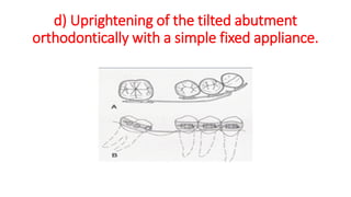
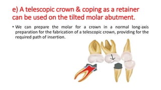
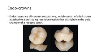
Good Morning
Atypical Tooth Preparation
By:
Osama Tarek Ali
MSc Fixed Prosthodontics
Minya University
Atypical Tooth Preparation
• Most teeth requiring cemented restorations may suffer from many
problems that necessitate deviation from the classic preparation form.
• Unmodified classic tooth preparations can be used in relatively few
clinical situations.
.
Different Situations Requiring Atypical Tooth
Preparations:
• A) Problems encountered in the abutment teeth:
1. Malaligned teeth
2. Short teeth
3. Mutilated teeth
4. Periodontally compromised teeth
• B) Atypical preparations for endodontically treated teeth.
• C) Other Recent Techniques.
I) Malaligned teeth:
• The mesio-lingually tilted second molar is commonly encountered
especially in cases of long-standing extraction of the first molar.
• A further complication may occur if the third molar is present and
tipped.
Several solutions have been suggested for this
problem:
a) Restoring or recontouring the mesial surface
of the third molar.
b) Proximal half-crown as a retainer on the
tilted molar abutment
c) A non-rigid connector on the distal aspect of
the premolar retainer.
d) Uprightening of the tilted abutment
orthodontically with a simple fixed appliance.
e) A telescopic crown & coping as a retainer
can be used on the tilted molar abutment.
• We can prepare the molar for a crown in a normal long-axis
preparation for the fabrication of a telescopic crown, providing for the
required path of insertion.
II) Short teeth:
• The main problem encountered in these teeth is the
limited amount of retention they afford to the fixed
prosthesis.
Sleeve Designed Preparation
• The Sleeve retainer is a partial-coverage cast crown with an intact occlusal
surface.
• The preparation of the abutment tooth consists of axial reduction of the
facial, lingual and proximal surfaces.
• The reduction of all surfaces is completed with a chamfer finish line,
established supragingival and ends in a knife edge occlusal finish line
approximately 0.5 mm from the occluding areas of the functional cusps.
THE JOURNAL OF PROSTHETIC DENTISTRY 2000
DIMASHKIEH AND AL-SHAMMERY
Advantages:
• preserving the length of the axial walls with minimum tooth reduction.
• Intact occlusal surfaces of abutments aid the establishment of occlusal
relationships and maintain pulpal integrity.
• Supragingival placement of the finish line.
• Eliminates the use of grooves, boxes, and retentive pins which may violate
pulpal integrity.
• There is also a substantial loss of tooth structure, and these forms of
retention result in an involved tooth preparation that severely complicates
the fabrication of the prosthesis.
• Pulpal testing is facilitated and root canal treatment is accessible.
Altering tooth preparation design:
• Retention and resistance factors in tooth preparation are related
primarily to surface area and height of the preparation, axial wall
convergence, and texture of the prepared surface
• And secondarily to intracoronary retentive devices. The walls of a short
clinical crown should be as parallel as possible.
• Increased tapering of the walls decreases resistance and retention
form, necessitating the use of secondary retention factors (pins,
grooves, or boxes).
III) Mutilated teeth:
Since every damaged tooth differs, it would be impossible to describe
the correct preparation for each circumstance. However, there are
certain guidelines that should be taken into consideration during the
modification of the preparation.
Guidelines for Preparation of Mutilated Teeth
I. Conversion of defects into retentive features
II. Grooves and box forms
III. Orientation of sloping surfaces
IV. Pin holes
I) Conversion of defects into retentive features:
• In cases of non-extensive defects, any carious lesion or previous
restoration that extends deeper than 1.5 mm into a vital tooth should
be filled to that level.
The walls of the remaining defect should be
shaped to
• Remove undercuts
• Provide vertical walls nearly parallel with the path of insertion
In the case of two opposite defects. Ideally, no less than 180 degrees of
sound tooth structure should remain between them. Otherwise, the
remaining tooth structure may be susceptible to fracture.
II) Grooves and box forms:
• Grooves placed in the vertical walls of sound tooth structure should
be at least 1 mm wide and deep and as long as possible to improve
retention and resistance.
• Grooves should be used accurately as the presence of multiple
grooves in full crown preparations may adversely affect the seating of
the crown.
• While boxes are indicated in cases of carious lesions or prior
restorations
Conclusion
• Decreasing from 22° to 8° in the cervical 1.5 mm of the reduced axial
surfaces significantly increased the resistance form.
• Two interproximal boxes that followed the existing convergence of the
axial walls (22°) significantly increased the resistance form.
• Two interproximal grooves that followed the existing convergence of
the axial walls (22°) did not significantly increase the resistance form.
• The most effective method of enhancing resistance form is to decrease
the taper of the cervical portion of the prepared axial walls.
III) Orientation of sloping surfaces:
• In case of sloping surfaces that are left after cusp fracture or
excavation of large carious lesions, It is better to form multiple small
steps in the form of:
1. Horizontal surfaces are perpendicular to the path of insertion.
2. Vertical surfaces should be aligned with the path of insertion to
assist retention and resistance to tipping than make a single vertical
plane that weakens the tooth and endangers pulp vitality.
IV) Pin holes :
• Indicated in cases of insufficient axial wall length for placement of
other retentive features. They generate additional length internally
and apically.
2 types :
1. Parallel with the path of insertion in case of cast restorations
2. Non-parallel to retain an amalgam or composite resin core, they can
be either cemented, threaded or pressed into tooth structure
IV) Periodontally Compromised Teeth:
• Restoration of a tooth around which there has been a change in the
gingival architecture frequently requires modification in tooth
preparation.
Tooth preparation and crown configuration
Finish line:
1-Location:
The optimum location for F.L. of a crown preparation is on enamel,
away from the gingival sulcus.
It may be necessary to extend the margin apically to cover the expense
of root surface to include caries and erosion.
2- Type:
• A shoulder F.L. is a poor choice when the margin must be placed on
the root surface for the following reasons:
1. The axial reduction will be extended into the tooth to a pulp-
threatening depth to achieve the same 1mm wide shoulder.
2. This gross destruction of axial tooth structure weakens the natural
structural durability of the tooth.
3. The shoulder has greater potential for concentrating stresses that
could lead to a fracture of the tooth.
• A chamfer F.L. in this apical position will result in the same depth of
axial reduction.
• A metal-ceramic crown fabricated in such circumstances should have
a wide metal gingival collar. Extension of ceramic veneer to the
gingival margin will create over-contouring or will require the use of
the shoulder.
Furcation flutes
• Sometimes, the crown margins on a molar must extend enough
apically so that the preparation of F.L. approaches the furcation,
where the common root trunk divides into two or three roots.
• There will be an intersection of F.L. with vertical flutes and concavities
in the common root trunk extending from the actual furcation in the
direction of CEJ. The axial S. occlusal to this gingival F.L. must also
have vertical concavities or flutes until it meets the facial groove in
the occlusal 1/3 of the facial surface.
• The concavities usually merge with features originating on the
occlusal surface.
• Any horizontal ridge in the facial or lingual surface of the tooth that
intersects with the concavity and blocks, it will result in a plaque
retaining area.
• There also will be concavities on the mesial and distal aspects of
upper molars arising from their furcations, they should be blended
into the surrounding axial surfaces of the crown. This will minimize
the difficulty of cleaning.
Mandibular Hemi-section
• Frequently one root is removed, while the other remains. Saving the
mesial segment would be desirable if the molar were the last tooth in
the arch.
• Distal root could be used as an abutment for short-span FPD replacing
mesial root. One root may also be used as an abutment for a longer-
span bridge replacing an entire molar.
• If an effort is made to save both roots of the molar following the
resection, the process is called "Bicuspidization premolarization".
• If both roots are maintained, it is important to be separated correctly
to allow normal gingival embrasure spaces.
This separation is accomplished either:
Naturally or Orthodontically
B) Atypical Preparations for Endodontically
Treated Teeth
Endo-crowns
• Endocrowns are all-ceramic restorations, which consist of a full crown
attached to a protruding retention section that sits tightly in the pulp
chamber of a reduced tooth.
Indications :
• I ) Complete loss of the clinical crown.
• ii) Limited inter-arch space
where traditional post-and-core rehabilitation prior to crown
cementation is not possible because of inadequate ceramic thickness.
• The alternative treatment method for this type of clinical situation is
to use an “endo-crown” which allows the desired 2.0 mm thickness of
ceramic on the occlusal surface
Tooth preparation:
• For macro retention and as a positioning aid, a central pulp chamber
inlay cavity is prepared.
• The shoulder finish line is prepared for seating of the crown
• dv
Do We Still Need Intraradicular Retainers? Current
Perspectives on the Treatment of Endodontically
Treated Teeth
• Recent Findings:
➢Recent evidence points to the benefits of endocrown restorations for the
posterior region, being a promising option for molars and premolars
➢however, intraradicular retainers are still suggested for the anterior region
due to the incidence of lateral loads.
• Machry RV, Dapieve KS, Pereira GK, Valandro LF. Do We Still Need Intraradicular Retainers?
Current Perspectives on the Treatment of Endodontically Treated Teeth. Current Oral Health
Reports. 2023 Feb 6:1-6.
Occlusal veneers
• They are extra-coronal restorations requiring a simpler preparation
driven by interocclusal clearance and anatomical considerations.
• The development of stronger materials in combination with
CAD/CAM techniques AND innovative adhesive technology such as
immediate dentin sealing, more conservative approaches are
explored as potential treatment options.
• Used in case of raising the bite
• Constructed from lithium disilicate or Hybrid ceramics
• Occlusal clearance of minimum 1 mm
Prep:
Vonlay
• A hybrid of an onlay with an extended buccal veneer
surface for use in bicuspid regions where there is mostly
enamel to bond to
• Hazzaa MA, El Mahallawi OS, Anwar E, Badran A. Clinical Outcomes of
Premolars Restored with Ceramic Vonlay Restorations versus Onlay Using
Modified USPHS Criteria (Randomized Clinical Trial). Journal of
Pharmaceutical Negative Results. 2023 Feb 7:1010-21.
Conclusion:
• Vonlays represents a very hopeful treatment alternative for
endodontically treated premolars, it allows maintaining of tooth
structure, it is compatible with minimally invasive dentistry, and it is
adequate for the concept of bio-integration. It is a conservative
approach to the mechanical and aesthetic restoration of nonvital
posterior teeth.
Sultan S, Al Garni H, Al Onazi M, Gangi KK, Al Otha S, Al Ruwaili F, Al Anazi S, Al Shammari SM,
Fandi A, Fayad M. Minimally Invasive Posterior Full Crown Competitors: Onlays, Occlusal Veneers,
Vonlays and Endocrowns: A Review and Proposed Classification. Journal of International Dental and
Medical Research. 2021;14(4):1617-22.
• Hazzaa MA, El Mahallawi OS, Anwar E, Badran A. Clinical Outcomes of
Premolars Restored with Ceramic Vonlay Restorations versus Onlay Using
Modified USPHS Criteria (Randomized Clinical Trial). Journal of
Pharmaceutical Negative Results. 2023 Feb 7:1010-21.
CONCLUSIONS
1. Both lithium disilicate (e-max press) onlay and vonlay restorations
are reliable treatment options with highly successful clinical
performance in terms of fracture, marginal integrity and marginal
discolouration for restoring defects in maxillary premolar dentition
over a period of one year.
2. Vonlay restorations can effectively repair weakened tooth
structure without considerable removal of healthy dental tissue
RECOMMENDATIONS
• Whenever the clinical situation permits (cervical caries or labial
defect), the preparation for vonlay restorations is recommended
Vertical Preparation
• The BOPT "Biologically Oriented Preparation Technique
• is a feather-edge vertical tooth preparation technique and involves the removal of the
anatomical cementoenamel junction, respectively the remodelling of pre-existing
marginal endings in previously prepared teeth.
• a concept with a vertical tooth preparation, gingitage, an immediate interim
restoration preserving the clot, and a specific laboratory technique aiming to adapt the
marginal periodontal tissue to a remodelled emergence profile of the crown
The BOPT "Biologically Oriented Preparation
Technique" concept in fixed prosthodontics. A
systematic review.
• Source: dentalTarget. apr2022, Vol. 17 Issue 1, p1-4. 4p.
• Author(s): Drafta, Sergiu; König, Arina Mara; Cristache, Corina Marilena;
Măru, Nicoleta
• BOPT introduces a new concept based on the fact that the profile of
the gingival margin can adapt to crowns margins.
• Conclusions:
Although the BOPT technique looks very promising, the reduced
number of published studies prevents it from being recommended for
daily practice and currently remains a niche technique.
Results
• Probing depth (greater than 3 mm) increased in only 2.3%, gingival
inflammation was present in 22.8%, a gingival recession occurred in
1.7% (decreased gingival stability), and mechanical and biological
failures occurred in 4.4% of the teeth.
Conclusions
• Following the concept of the biologically oriented preparation
technique did not increase the probing depth and showed a
moderate rate of gingival inflammation, lower recession rates, and
lower mechanical and biological failures at the 5-year follow-up.
• The biologically oriented preparation technique appears to be a
viable alternative technique for obtaining satisfactory and stable
clinical results for up to 5 years.
Comparison of horizontal and vertical methods
of tooth preparation for a prosthetic crown
• https://www.jpccr.eu/Comparison-of-horizontal-and-vertical-methods-of-tooth-
preparation-for-a-prosthetic,116672,0,2.html
• Łabno P, Drobnik K. Comparison of horizontal and vertical methods of tooth preparation
for a prosthetic crown. Journal of Pre-Clinical and Clinical Research. 2020;14(1).
• Objectives: This clinical case report describes the rehabilitation of
central incisors with veneers in a patient with gingival asymmetry. The
teeth were prepared without a finish line, applying BOPT concepts to
correct asymmetry, and obtained a harmoniously integrated
restoration with optimal periodontal health.
Results
The application of ceramic veneers after tooth preparation without a
finish line, following BOPT concepts made it possible to place
restorations that were harmoniously integrated both esthetically and
periodontally.
The chromatic continuity was adequate, the dental proportions,
gingival health, and symmetry of the two gingival zeniths were correct
and the integration of the restorations was good thanks to correct soft
tissue management.
At the one-year follow-up visit, the soft tissues had stabilized correctly,
and good gingival health was observed despite the correction of
gingival asymmetry.
Gingival veneer
• Also called removable artificial gingiva or gingival mask
• Consists of a prosthesis made of thermoactivated acrylic resin in a colour
similar to the gum tissue.
• It is placed on the labial surface of the teeth. The veneer's function is to
restore the mucogingival contour and esthetics in areas where periodontal
tissues are deficient.
• Gingival veneers were first introduced by Emslie
• A conservative, simple, and inexpensive treatment.
• It is indicated in cases of gingival unevenness in the contour of the concave
labial arc, poor esthetics characterized by interdental “black triangles,”
exposed root surfaces and/or crown margins, food packing in interproximal
spaces, lack of saliva control, impaired speech, and root–dentin sensitivity.
• The use of these veneers is contraindicated in situations in which patients
present poor oral hygiene, limited manual dexterity, high caries activity/risk,
incomplete periodontal therapy, and allergy to fabrication materials.
Gingival Veneers
Custom-made veneers are designed to make the teeth look shorter and
cover the signs of gum recession.
Final Conclusion
• Atypical word means any deviation from the normal approach to any
procedure that gave us an alternative idea about what else we can do
to have the same result as the typical approach.
Atypical Tooth Preparation.pdf

Atypical Tooth Preparation.pdf

  • 1.
  • 2.
    Atypical Tooth Preparation By: OsamaTarek Ali MSc Fixed Prosthodontics Minya University
  • 3.
    Atypical Tooth Preparation •Most teeth requiring cemented restorations may suffer from many problems that necessitate deviation from the classic preparation form. • Unmodified classic tooth preparations can be used in relatively few clinical situations. .
  • 4.
    Different Situations RequiringAtypical Tooth Preparations: • A) Problems encountered in the abutment teeth: 1. Malaligned teeth 2. Short teeth 3. Mutilated teeth 4. Periodontally compromised teeth • B) Atypical preparations for endodontically treated teeth. • C) Other Recent Techniques.
  • 5.
    I) Malaligned teeth: •The mesio-lingually tilted second molar is commonly encountered especially in cases of long-standing extraction of the first molar. • A further complication may occur if the third molar is present and tipped.
  • 6.
    Several solutions havebeen suggested for this problem:
  • 7.
    a) Restoring orrecontouring the mesial surface of the third molar.
  • 8.
    b) Proximal half-crownas a retainer on the tilted molar abutment
  • 9.
    c) A non-rigidconnector on the distal aspect of the premolar retainer.
  • 10.
    d) Uprightening ofthe tilted abutment orthodontically with a simple fixed appliance.
  • 11.
    e) A telescopiccrown & coping as a retainer can be used on the tilted molar abutment. • We can prepare the molar for a crown in a normal long-axis preparation for the fabrication of a telescopic crown, providing for the required path of insertion.
  • 12.
    II) Short teeth: •The main problem encountered in these teeth is the limited amount of retention they afford to the fixed prosthesis.
  • 13.
    Sleeve Designed Preparation •The Sleeve retainer is a partial-coverage cast crown with an intact occlusal surface. • The preparation of the abutment tooth consists of axial reduction of the facial, lingual and proximal surfaces. • The reduction of all surfaces is completed with a chamfer finish line, established supragingival and ends in a knife edge occlusal finish line approximately 0.5 mm from the occluding areas of the functional cusps.
  • 14.
    THE JOURNAL OFPROSTHETIC DENTISTRY 2000 DIMASHKIEH AND AL-SHAMMERY
  • 15.
    Advantages: • preserving thelength of the axial walls with minimum tooth reduction. • Intact occlusal surfaces of abutments aid the establishment of occlusal relationships and maintain pulpal integrity. • Supragingival placement of the finish line. • Eliminates the use of grooves, boxes, and retentive pins which may violate pulpal integrity. • There is also a substantial loss of tooth structure, and these forms of retention result in an involved tooth preparation that severely complicates the fabrication of the prosthesis. • Pulpal testing is facilitated and root canal treatment is accessible.
  • 16.
    Altering tooth preparationdesign: • Retention and resistance factors in tooth preparation are related primarily to surface area and height of the preparation, axial wall convergence, and texture of the prepared surface • And secondarily to intracoronary retentive devices. The walls of a short clinical crown should be as parallel as possible. • Increased tapering of the walls decreases resistance and retention form, necessitating the use of secondary retention factors (pins, grooves, or boxes).
  • 17.
    III) Mutilated teeth: Sinceevery damaged tooth differs, it would be impossible to describe the correct preparation for each circumstance. However, there are certain guidelines that should be taken into consideration during the modification of the preparation.
  • 18.
    Guidelines for Preparationof Mutilated Teeth I. Conversion of defects into retentive features II. Grooves and box forms III. Orientation of sloping surfaces IV. Pin holes
  • 19.
    I) Conversion ofdefects into retentive features: • In cases of non-extensive defects, any carious lesion or previous restoration that extends deeper than 1.5 mm into a vital tooth should be filled to that level.
  • 20.
    The walls ofthe remaining defect should be shaped to • Remove undercuts • Provide vertical walls nearly parallel with the path of insertion In the case of two opposite defects. Ideally, no less than 180 degrees of sound tooth structure should remain between them. Otherwise, the remaining tooth structure may be susceptible to fracture.
  • 23.
    II) Grooves andbox forms: • Grooves placed in the vertical walls of sound tooth structure should be at least 1 mm wide and deep and as long as possible to improve retention and resistance. • Grooves should be used accurately as the presence of multiple grooves in full crown preparations may adversely affect the seating of the crown. • While boxes are indicated in cases of carious lesions or prior restorations
  • 25.
    Conclusion • Decreasing from22° to 8° in the cervical 1.5 mm of the reduced axial surfaces significantly increased the resistance form. • Two interproximal boxes that followed the existing convergence of the axial walls (22°) significantly increased the resistance form. • Two interproximal grooves that followed the existing convergence of the axial walls (22°) did not significantly increase the resistance form. • The most effective method of enhancing resistance form is to decrease the taper of the cervical portion of the prepared axial walls.
  • 26.
    III) Orientation ofsloping surfaces: • In case of sloping surfaces that are left after cusp fracture or excavation of large carious lesions, It is better to form multiple small steps in the form of: 1. Horizontal surfaces are perpendicular to the path of insertion. 2. Vertical surfaces should be aligned with the path of insertion to assist retention and resistance to tipping than make a single vertical plane that weakens the tooth and endangers pulp vitality.
  • 28.
    IV) Pin holes: • Indicated in cases of insufficient axial wall length for placement of other retentive features. They generate additional length internally and apically.
  • 29.
    2 types : 1.Parallel with the path of insertion in case of cast restorations 2. Non-parallel to retain an amalgam or composite resin core, they can be either cemented, threaded or pressed into tooth structure
  • 30.
    IV) Periodontally CompromisedTeeth: • Restoration of a tooth around which there has been a change in the gingival architecture frequently requires modification in tooth preparation.
  • 31.
    Tooth preparation andcrown configuration Finish line: 1-Location: The optimum location for F.L. of a crown preparation is on enamel, away from the gingival sulcus. It may be necessary to extend the margin apically to cover the expense of root surface to include caries and erosion.
  • 33.
    2- Type: • Ashoulder F.L. is a poor choice when the margin must be placed on the root surface for the following reasons: 1. The axial reduction will be extended into the tooth to a pulp- threatening depth to achieve the same 1mm wide shoulder. 2. This gross destruction of axial tooth structure weakens the natural structural durability of the tooth. 3. The shoulder has greater potential for concentrating stresses that could lead to a fracture of the tooth.
  • 34.
    • A chamferF.L. in this apical position will result in the same depth of axial reduction. • A metal-ceramic crown fabricated in such circumstances should have a wide metal gingival collar. Extension of ceramic veneer to the gingival margin will create over-contouring or will require the use of the shoulder.
  • 36.
    Furcation flutes • Sometimes,the crown margins on a molar must extend enough apically so that the preparation of F.L. approaches the furcation, where the common root trunk divides into two or three roots. • There will be an intersection of F.L. with vertical flutes and concavities in the common root trunk extending from the actual furcation in the direction of CEJ. The axial S. occlusal to this gingival F.L. must also have vertical concavities or flutes until it meets the facial groove in the occlusal 1/3 of the facial surface.
  • 37.
    • The concavitiesusually merge with features originating on the occlusal surface. • Any horizontal ridge in the facial or lingual surface of the tooth that intersects with the concavity and blocks, it will result in a plaque retaining area. • There also will be concavities on the mesial and distal aspects of upper molars arising from their furcations, they should be blended into the surrounding axial surfaces of the crown. This will minimize the difficulty of cleaning.
  • 39.
    Mandibular Hemi-section • Frequentlyone root is removed, while the other remains. Saving the mesial segment would be desirable if the molar were the last tooth in the arch. • Distal root could be used as an abutment for short-span FPD replacing mesial root. One root may also be used as an abutment for a longer- span bridge replacing an entire molar.
  • 40.
    • If aneffort is made to save both roots of the molar following the resection, the process is called "Bicuspidization premolarization". • If both roots are maintained, it is important to be separated correctly to allow normal gingival embrasure spaces.
  • 41.
    This separation isaccomplished either: Naturally or Orthodontically
  • 42.
    B) Atypical Preparationsfor Endodontically Treated Teeth
  • 43.
    Endo-crowns • Endocrowns areall-ceramic restorations, which consist of a full crown attached to a protruding retention section that sits tightly in the pulp chamber of a reduced tooth.
  • 44.
    Indications : • I) Complete loss of the clinical crown. • ii) Limited inter-arch space where traditional post-and-core rehabilitation prior to crown cementation is not possible because of inadequate ceramic thickness. • The alternative treatment method for this type of clinical situation is to use an “endo-crown” which allows the desired 2.0 mm thickness of ceramic on the occlusal surface
  • 45.
    Tooth preparation: • Formacro retention and as a positioning aid, a central pulp chamber inlay cavity is prepared. • The shoulder finish line is prepared for seating of the crown
  • 49.
  • 52.
    Do We StillNeed Intraradicular Retainers? Current Perspectives on the Treatment of Endodontically Treated Teeth • Recent Findings: ➢Recent evidence points to the benefits of endocrown restorations for the posterior region, being a promising option for molars and premolars ➢however, intraradicular retainers are still suggested for the anterior region due to the incidence of lateral loads. • Machry RV, Dapieve KS, Pereira GK, Valandro LF. Do We Still Need Intraradicular Retainers? Current Perspectives on the Treatment of Endodontically Treated Teeth. Current Oral Health Reports. 2023 Feb 6:1-6.
  • 53.
    Occlusal veneers • Theyare extra-coronal restorations requiring a simpler preparation driven by interocclusal clearance and anatomical considerations. • The development of stronger materials in combination with CAD/CAM techniques AND innovative adhesive technology such as immediate dentin sealing, more conservative approaches are explored as potential treatment options.
  • 54.
    • Used incase of raising the bite • Constructed from lithium disilicate or Hybrid ceramics • Occlusal clearance of minimum 1 mm
  • 56.
  • 59.
    Vonlay • A hybridof an onlay with an extended buccal veneer surface for use in bicuspid regions where there is mostly enamel to bond to
  • 61.
    • Hazzaa MA,El Mahallawi OS, Anwar E, Badran A. Clinical Outcomes of Premolars Restored with Ceramic Vonlay Restorations versus Onlay Using Modified USPHS Criteria (Randomized Clinical Trial). Journal of Pharmaceutical Negative Results. 2023 Feb 7:1010-21.
  • 64.
    Conclusion: • Vonlays representsa very hopeful treatment alternative for endodontically treated premolars, it allows maintaining of tooth structure, it is compatible with minimally invasive dentistry, and it is adequate for the concept of bio-integration. It is a conservative approach to the mechanical and aesthetic restoration of nonvital posterior teeth.
  • 65.
    Sultan S, AlGarni H, Al Onazi M, Gangi KK, Al Otha S, Al Ruwaili F, Al Anazi S, Al Shammari SM, Fandi A, Fayad M. Minimally Invasive Posterior Full Crown Competitors: Onlays, Occlusal Veneers, Vonlays and Endocrowns: A Review and Proposed Classification. Journal of International Dental and Medical Research. 2021;14(4):1617-22.
  • 67.
    • Hazzaa MA,El Mahallawi OS, Anwar E, Badran A. Clinical Outcomes of Premolars Restored with Ceramic Vonlay Restorations versus Onlay Using Modified USPHS Criteria (Randomized Clinical Trial). Journal of Pharmaceutical Negative Results. 2023 Feb 7:1010-21.
  • 71.
    CONCLUSIONS 1. Both lithiumdisilicate (e-max press) onlay and vonlay restorations are reliable treatment options with highly successful clinical performance in terms of fracture, marginal integrity and marginal discolouration for restoring defects in maxillary premolar dentition over a period of one year. 2. Vonlay restorations can effectively repair weakened tooth structure without considerable removal of healthy dental tissue
  • 72.
    RECOMMENDATIONS • Whenever theclinical situation permits (cervical caries or labial defect), the preparation for vonlay restorations is recommended
  • 73.
    Vertical Preparation • TheBOPT "Biologically Oriented Preparation Technique • is a feather-edge vertical tooth preparation technique and involves the removal of the anatomical cementoenamel junction, respectively the remodelling of pre-existing marginal endings in previously prepared teeth. • a concept with a vertical tooth preparation, gingitage, an immediate interim restoration preserving the clot, and a specific laboratory technique aiming to adapt the marginal periodontal tissue to a remodelled emergence profile of the crown
  • 74.
    The BOPT "BiologicallyOriented Preparation Technique" concept in fixed prosthodontics. A systematic review. • Source: dentalTarget. apr2022, Vol. 17 Issue 1, p1-4. 4p. • Author(s): Drafta, Sergiu; König, Arina Mara; Cristache, Corina Marilena; Măru, Nicoleta
  • 75.
    • BOPT introducesa new concept based on the fact that the profile of the gingival margin can adapt to crowns margins. • Conclusions: Although the BOPT technique looks very promising, the reduced number of published studies prevents it from being recommended for daily practice and currently remains a niche technique.
  • 77.
    Results • Probing depth(greater than 3 mm) increased in only 2.3%, gingival inflammation was present in 22.8%, a gingival recession occurred in 1.7% (decreased gingival stability), and mechanical and biological failures occurred in 4.4% of the teeth.
  • 78.
    Conclusions • Following theconcept of the biologically oriented preparation technique did not increase the probing depth and showed a moderate rate of gingival inflammation, lower recession rates, and lower mechanical and biological failures at the 5-year follow-up. • The biologically oriented preparation technique appears to be a viable alternative technique for obtaining satisfactory and stable clinical results for up to 5 years.
  • 79.
    Comparison of horizontaland vertical methods of tooth preparation for a prosthetic crown • https://www.jpccr.eu/Comparison-of-horizontal-and-vertical-methods-of-tooth- preparation-for-a-prosthetic,116672,0,2.html • Łabno P, Drobnik K. Comparison of horizontal and vertical methods of tooth preparation for a prosthetic crown. Journal of Pre-Clinical and Clinical Research. 2020;14(1).
  • 84.
    • Objectives: Thisclinical case report describes the rehabilitation of central incisors with veneers in a patient with gingival asymmetry. The teeth were prepared without a finish line, applying BOPT concepts to correct asymmetry, and obtained a harmoniously integrated restoration with optimal periodontal health.
  • 94.
    Results The application ofceramic veneers after tooth preparation without a finish line, following BOPT concepts made it possible to place restorations that were harmoniously integrated both esthetically and periodontally. The chromatic continuity was adequate, the dental proportions, gingival health, and symmetry of the two gingival zeniths were correct and the integration of the restorations was good thanks to correct soft tissue management. At the one-year follow-up visit, the soft tissues had stabilized correctly, and good gingival health was observed despite the correction of gingival asymmetry.
  • 95.
    Gingival veneer • Alsocalled removable artificial gingiva or gingival mask • Consists of a prosthesis made of thermoactivated acrylic resin in a colour similar to the gum tissue. • It is placed on the labial surface of the teeth. The veneer's function is to restore the mucogingival contour and esthetics in areas where periodontal tissues are deficient. • Gingival veneers were first introduced by Emslie
  • 96.
    • A conservative,simple, and inexpensive treatment. • It is indicated in cases of gingival unevenness in the contour of the concave labial arc, poor esthetics characterized by interdental “black triangles,” exposed root surfaces and/or crown margins, food packing in interproximal spaces, lack of saliva control, impaired speech, and root–dentin sensitivity. • The use of these veneers is contraindicated in situations in which patients present poor oral hygiene, limited manual dexterity, high caries activity/risk, incomplete periodontal therapy, and allergy to fabrication materials.
  • 97.
    Gingival Veneers Custom-made veneersare designed to make the teeth look shorter and cover the signs of gum recession.
  • 99.
    Final Conclusion • Atypicalword means any deviation from the normal approach to any procedure that gave us an alternative idea about what else we can do to have the same result as the typical approach.