SlideShare a Scribd company logo
RADIO-ANATOMY

PAROTID GLAND
AND
SUBMANDIBULAR GLAND
OVERVIEW OF ANATOMY
1)- DEVELOPMENTAL AND GROSS ANATOMY
2)- ULTRASOUND FEATURES
3)- X RAY
4)- CT AND MRI ANATOMICAL LOCATION
PAROTID GLAND
Development
• Appear early - sixth week of prenatal age.
• The epithelial buds of these glands are located on the inner
part of the cheek, near the labial commissures of the
primitive mouth
• Grow posteriorly toward the otic placodes of the ears and
branch to form solid cords with rounded terminal ends near
the developing facial nerve.
• 10 weeks of prenatal development, these cords are
canalized and form ducts, with the largest becoming the
parotid duct for the parotid gland.
• Secretion by the parotid glands via the parotid duct begins
at approximately 18 weeks of gestation.
Location
•

Inferior and anterior to the external acoustic meatus and
posterior to the mandibular ramus and anterior to the
mastoid process of temporal bone.

Draining
• The vestibule of oral cavity through Stensen duct or
parotid duct that emerges from the anterior border of the
gland, superficial to the masseter muscle pierces the
buccinator muscle, then opening up into the oral cavity on
the inner surface of the cheek, usually opposite the
maxillary second molar.
• The parotid papilla is a small elevation of tissue that marks
the opening of the parotid duct.
Surfaces and borders
• The gland has four surfaces superficial or
lateral,superior, anteromedial and
posteromedial.
• The gland has three borders anterior, medial and
posterior.
• The Parotid gland has two ends: superior end in
the form of small surface and an inferior end
(apex).
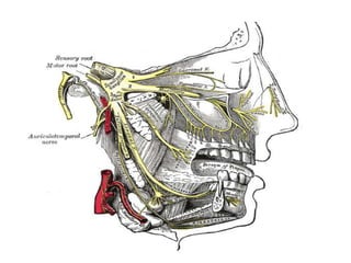
Structures that pass through the gland
These are from lateral to medial:

(1) Facial nerve
(2) Retromandibular vein
(3) External Carotid artery
(4) Superficial temporal artery
(5) branches of the great auricular nerve
Blood Supply
The gland is mainly irrigated by External Carotid
artery via the posterior auricular artery and the
transverse facial.
Venous Drainage
Venous return is to the Retromandibular vein.
Lymphatic drainage
The gland is mainly drained into the preauricular or
parotid lymph nodes which ultimately drain to the
deep cervical chain.
SUBMENDIBULAR GLAND
• Develop later than the parotid glands and appear late in the sixth
week of prenatal development.
• They develop bilaterally from epithelial buds in the sulcus
surrounding the sublingual folds on the floor of the primitive
mouth.
• Arise of solid cords branch from the buds and grow
posteriorly, lateral to the developing tongue. The cords of the
submandibular gland later branch and then become canalized to
form the ductal part.
• The submandibular gland acini develop from the cords’ rounded
terminal ends at 12 weeks, and secretory activity via the
submandibular duct begins at 16 weeks. Growth of the
submandibular gland continues after birth.
LOCTION
• Lying superior to the digastric muscles, divided into
superficial and deep lobes, which are separated by the
mylohyoid muscle.
• The superficial lobe comprises most of the gland, with the
mylohyoid muscle runs under it. The deep lobe is the
smaller part.
Sialography
Indications
•
•
•
•
•
•

Indications include:
In the evaluation of the functional integrity of the salivary glands
In case of obstructions
To evaluate the ductal pattern
In case of facial swellings, to rule out salivary gland pathology
In case of intra-glandular neoplasms.

Containdications
•
•
•
•

Persons who are allergic to iodine and/or contrast medium.
Cases where there is acute infection,
patients with thyroid function tests
When calculi are located in anterior part of the salivary gland duct
CT OF SLIVERY GLAND
It is a fatty glandular tissue that is encased in a dense
capsule. Because of this, the parotid gland on CT is
consistently more lucent (-25 to 10 Hounsfield units
[H]) than surrounding muscles (35-60 H) and likewise
is distinctly more radiodense than adjacent fat in the
subcutaneous tissues, infratemponal fossa, and
lateral pharyngeal space(-125 to -50 H)(figs.
1A, 1B, and 2B).
Parotid
Parotid
Parotid
Parotid
Parotid
Parotid
Parotid
Parotid
Parotid
Parotid
Parotid
Parotid
Parotid
Parotid

More Related Content

What's hot

Basic physics of ultrasound.JH
Basic physics of ultrasound.JHBasic physics of ultrasound.JH
Basic physics of ultrasound.JHhari baskar
 
Line focus-principle-and-anode-heel-effect
Line focus-principle-and-anode-heel-effectLine focus-principle-and-anode-heel-effect
Line focus-principle-and-anode-heel-effect
JoaquinLuisRinehart1
 
Skull anatomy and positioning
Skull anatomy and positioningSkull anatomy and positioning
Skull anatomy and positioning
Amit Rauniyar
 
Presentation1.pptx, radiological anatomy of the abdomen and pelvis.
Presentation1.pptx, radiological anatomy of the abdomen and pelvis.Presentation1.pptx, radiological anatomy of the abdomen and pelvis.
Presentation1.pptx, radiological anatomy of the abdomen and pelvis.Abdellah Nazeer
 
Skull radiography
Skull radiography Skull radiography
Skull radiography
Dr Sudhir Patel
 
Presentation1.pptx, radiological anatomy of the naso, oro and hypopharynx.
Presentation1.pptx, radiological anatomy of the naso, oro and hypopharynx.Presentation1.pptx, radiological anatomy of the naso, oro and hypopharynx.
Presentation1.pptx, radiological anatomy of the naso, oro and hypopharynx.Abdellah Nazeer
 
Line focus principle.
Line focus principle.Line focus principle.
Line focus principle.
mohammed althaf
 
Mammography physics and technique
Mammography  physics and techniqueMammography  physics and technique
Mammography physics and technique
Archana Koshy
 
Basics Of MRI
Basics Of MRIBasics Of MRI
Basics Of MRI
Keshav Kulkarni
 
Deep neck spaces and surgical anatomy
Deep neck spaces and surgical anatomyDeep neck spaces and surgical anatomy
Deep neck spaces and surgical anatomy
Himanshu Mishra
 
Basics of mri physics Dr. Muhammad Bin Zulfiqar
Basics of mri physics Dr. Muhammad Bin ZulfiqarBasics of mri physics Dr. Muhammad Bin Zulfiqar
Basics of mri physics Dr. Muhammad Bin Zulfiqar
Dr. Muhammad Bin Zulfiqar
 
Brain CT Anatomy and Basic Interpretation Part I
Brain CT Anatomy and Basic Interpretation Part IBrain CT Anatomy and Basic Interpretation Part I
Brain CT Anatomy and Basic Interpretation Part I
Sakher Alkhaderi
 
Ultrasound Physics
Ultrasound PhysicsUltrasound Physics
Ultrasound Physicsu.surgery
 
History of radiology
History of radiologyHistory of radiology
History of radiology
Abhinaya Luitel
 
Contrast Radiography
Contrast RadiographyContrast Radiography
Contrast Radiography
Vibhuti Kaul
 
Radiology of nose and paranasal sinuses
Radiology of nose and paranasal sinuses Radiology of nose and paranasal sinuses
Radiology of nose and paranasal sinuses
Andrea R Salins
 
Vagus nerve
Vagus nerveVagus nerve
Vagus nerve
Idris Siddiqui
 
Barium meal follow through
Barium meal follow throughBarium meal follow through
Barium meal follow through
Shiva Prakash
 
Sialography & dacrocystography
Sialography  & dacrocystographySialography  & dacrocystography
Sialography & dacrocystography
airwave12
 
Dental radiography
Dental radiographyDental radiography
Dental radiography
Mfoniso Akpaniwo
 

What's hot (20)

Basic physics of ultrasound.JH
Basic physics of ultrasound.JHBasic physics of ultrasound.JH
Basic physics of ultrasound.JH
 
Line focus-principle-and-anode-heel-effect
Line focus-principle-and-anode-heel-effectLine focus-principle-and-anode-heel-effect
Line focus-principle-and-anode-heel-effect
 
Skull anatomy and positioning
Skull anatomy and positioningSkull anatomy and positioning
Skull anatomy and positioning
 
Presentation1.pptx, radiological anatomy of the abdomen and pelvis.
Presentation1.pptx, radiological anatomy of the abdomen and pelvis.Presentation1.pptx, radiological anatomy of the abdomen and pelvis.
Presentation1.pptx, radiological anatomy of the abdomen and pelvis.
 
Skull radiography
Skull radiography Skull radiography
Skull radiography
 
Presentation1.pptx, radiological anatomy of the naso, oro and hypopharynx.
Presentation1.pptx, radiological anatomy of the naso, oro and hypopharynx.Presentation1.pptx, radiological anatomy of the naso, oro and hypopharynx.
Presentation1.pptx, radiological anatomy of the naso, oro and hypopharynx.
 
Line focus principle.
Line focus principle.Line focus principle.
Line focus principle.
 
Mammography physics and technique
Mammography  physics and techniqueMammography  physics and technique
Mammography physics and technique
 
Basics Of MRI
Basics Of MRIBasics Of MRI
Basics Of MRI
 
Deep neck spaces and surgical anatomy
Deep neck spaces and surgical anatomyDeep neck spaces and surgical anatomy
Deep neck spaces and surgical anatomy
 
Basics of mri physics Dr. Muhammad Bin Zulfiqar
Basics of mri physics Dr. Muhammad Bin ZulfiqarBasics of mri physics Dr. Muhammad Bin Zulfiqar
Basics of mri physics Dr. Muhammad Bin Zulfiqar
 
Brain CT Anatomy and Basic Interpretation Part I
Brain CT Anatomy and Basic Interpretation Part IBrain CT Anatomy and Basic Interpretation Part I
Brain CT Anatomy and Basic Interpretation Part I
 
Ultrasound Physics
Ultrasound PhysicsUltrasound Physics
Ultrasound Physics
 
History of radiology
History of radiologyHistory of radiology
History of radiology
 
Contrast Radiography
Contrast RadiographyContrast Radiography
Contrast Radiography
 
Radiology of nose and paranasal sinuses
Radiology of nose and paranasal sinuses Radiology of nose and paranasal sinuses
Radiology of nose and paranasal sinuses
 
Vagus nerve
Vagus nerveVagus nerve
Vagus nerve
 
Barium meal follow through
Barium meal follow throughBarium meal follow through
Barium meal follow through
 
Sialography & dacrocystography
Sialography  & dacrocystographySialography  & dacrocystography
Sialography & dacrocystography
 
Dental radiography
Dental radiographyDental radiography
Dental radiography
 

Viewers also liked

Parotid gland
Parotid glandParotid gland
Parotid gland
nadia omer
 
Parotid ppt
Parotid pptParotid ppt
Parotid ppt
Nikesh Gosrani
 
Salivary glands antomy
Salivary glands antomySalivary glands antomy
Salivary glands antomy
Dr Ahmed Fathy
 
Anatomy of Submandibular Gland
Anatomy of Submandibular GlandAnatomy of Submandibular Gland
Anatomy of Submandibular GlandFuad Ridha Mahabot
 
Submandibular Region (Anatomy of the Neck)
Submandibular Region (Anatomy of the Neck)Submandibular Region (Anatomy of the Neck)
Submandibular Region (Anatomy of the Neck)
Dr. Sherif Fahmy
 
Diagnostic imaging in head and neck pathology
Diagnostic imaging in head and neck pathologyDiagnostic imaging in head and neck pathology
Diagnostic imaging in head and neck pathologyHayat Youssef
 
Basics of mri
Basics of mriBasics of mri
Basics of mri
Ganesh Nair
 
BASICS OF MRI
BASICS OF MRIBASICS OF MRI
BASICS OF MRI
Krishna Kiran Karanth
 
Magnetic Resonance Imaging
Magnetic Resonance ImagingMagnetic Resonance Imaging
Magnetic Resonance Imaging
guest2d52f2
 
Parotid salivary gland
Parotid salivary glandParotid salivary gland
Parotid salivary gland
drasarma1947
 

Viewers also liked (12)

Parotid gland
Parotid glandParotid gland
Parotid gland
 
Parotid ppt
Parotid pptParotid ppt
Parotid ppt
 
Salivary glands antomy
Salivary glands antomySalivary glands antomy
Salivary glands antomy
 
Anatomy of Submandibular Gland
Anatomy of Submandibular GlandAnatomy of Submandibular Gland
Anatomy of Submandibular Gland
 
Submandibular Region (Anatomy of the Neck)
Submandibular Region (Anatomy of the Neck)Submandibular Region (Anatomy of the Neck)
Submandibular Region (Anatomy of the Neck)
 
Parotid gland
Parotid glandParotid gland
Parotid gland
 
MRI in neck imaging
MRI in neck imaging MRI in neck imaging
MRI in neck imaging
 
Diagnostic imaging in head and neck pathology
Diagnostic imaging in head and neck pathologyDiagnostic imaging in head and neck pathology
Diagnostic imaging in head and neck pathology
 
Basics of mri
Basics of mriBasics of mri
Basics of mri
 
BASICS OF MRI
BASICS OF MRIBASICS OF MRI
BASICS OF MRI
 
Magnetic Resonance Imaging
Magnetic Resonance ImagingMagnetic Resonance Imaging
Magnetic Resonance Imaging
 
Parotid salivary gland
Parotid salivary glandParotid salivary gland
Parotid salivary gland
 

Similar to Parotid

lacrimal system
lacrimal systemlacrimal system
lacrimal system
Samten Dorji
 
Congenital nasolacrimal duct obstruction
Congenital nasolacrimal duct obstructionCongenital nasolacrimal duct obstruction
Congenital nasolacrimal duct obstruction
Raju Kaiti
 
Saliva and salivary glands.pptx
Saliva and salivary glands.pptxSaliva and salivary glands.pptx
Saliva and salivary glands.pptx
malti19
 
hypospadias .pptx
hypospadias .pptxhypospadias .pptx
hypospadias .pptx
RenuSingla3
 
Anatomy and physiology of salivary gland
Anatomy and physiology of salivary glandAnatomy and physiology of salivary gland
Anatomy and physiology of salivary gland
Sandeep Shrestha
 
Anatomy and physiology of salivary glands
Anatomy and physiology of salivary glandsAnatomy and physiology of salivary glands
Anatomy and physiology of salivary glands
Prem Davis
 
Salivary glands /certified fixed orthodontic courses by Indian dental academy
Salivary glands /certified fixed orthodontic courses by Indian dental academy Salivary glands /certified fixed orthodontic courses by Indian dental academy
Salivary glands /certified fixed orthodontic courses by Indian dental academy
Indian dental academy
 
Growth and development of facial structures
Growth and development of facial structures Growth and development of facial structures
Growth and development of facial structures
Kunal Ajay Patankar
 
Anatomy of pns
Anatomy of pnsAnatomy of pns
Anatomy of pns
raju kafle
 
Surgical anantomy of thyroid gland
Surgical anantomy of thyroid glandSurgical anantomy of thyroid gland
Surgical anantomy of thyroid gland
Soumen Kanjilal
 
Digestive system
Digestive systemDigestive system
Digestive systemFarhan Ali
 
Salivary gland and its importance in dentistry
Salivary gland and its importance in dentistrySalivary gland and its importance in dentistry
Salivary gland and its importance in dentistry
lipiprakash01
 
Development of face, palate, tongue and mandible
Development of face,  palate, tongue and mandibleDevelopment of face,  palate, tongue and mandible
Development of face, palate, tongue and mandible
PankajGoyal79
 
Salivary gland ppt - Kanato Assumi
Salivary gland ppt  - Kanato AssumiSalivary gland ppt  - Kanato Assumi
Salivary gland ppt - Kanato Assumi
Kanato Assumi
 
Salivary glands seminar
Salivary glands seminarSalivary glands seminar
Salivary glands seminar
ANIL KUMAR
 
Anatomy of pns dr rk
Anatomy of pns dr rkAnatomy of pns dr rk
Anatomy of pns dr rk
raju kafle
 
Larynx
LarynxLarynx
Larynx
anup bhatta
 
Maxilla and Mandible - its applied aspect - Copy.pptx
Maxilla and Mandible - its applied aspect - Copy.pptxMaxilla and Mandible - its applied aspect - Copy.pptx
Maxilla and Mandible - its applied aspect - Copy.pptx
RiddhikaKochar1
 
Saliva and salivary glands by Dr.Nilesh Vaidya
Saliva and salivary glands by Dr.Nilesh VaidyaSaliva and salivary glands by Dr.Nilesh Vaidya
Saliva and salivary glands by Dr.Nilesh Vaidya
Nilesh Vaidya
 
Development and congenital anomalies of urogenital system
Development and congenital anomalies of urogenital systemDevelopment and congenital anomalies of urogenital system
Development and congenital anomalies of urogenital systemJayeta Choudhury
 

Similar to Parotid (20)

lacrimal system
lacrimal systemlacrimal system
lacrimal system
 
Congenital nasolacrimal duct obstruction
Congenital nasolacrimal duct obstructionCongenital nasolacrimal duct obstruction
Congenital nasolacrimal duct obstruction
 
Saliva and salivary glands.pptx
Saliva and salivary glands.pptxSaliva and salivary glands.pptx
Saliva and salivary glands.pptx
 
hypospadias .pptx
hypospadias .pptxhypospadias .pptx
hypospadias .pptx
 
Anatomy and physiology of salivary gland
Anatomy and physiology of salivary glandAnatomy and physiology of salivary gland
Anatomy and physiology of salivary gland
 
Anatomy and physiology of salivary glands
Anatomy and physiology of salivary glandsAnatomy and physiology of salivary glands
Anatomy and physiology of salivary glands
 
Salivary glands /certified fixed orthodontic courses by Indian dental academy
Salivary glands /certified fixed orthodontic courses by Indian dental academy Salivary glands /certified fixed orthodontic courses by Indian dental academy
Salivary glands /certified fixed orthodontic courses by Indian dental academy
 
Growth and development of facial structures
Growth and development of facial structures Growth and development of facial structures
Growth and development of facial structures
 
Anatomy of pns
Anatomy of pnsAnatomy of pns
Anatomy of pns
 
Surgical anantomy of thyroid gland
Surgical anantomy of thyroid glandSurgical anantomy of thyroid gland
Surgical anantomy of thyroid gland
 
Digestive system
Digestive systemDigestive system
Digestive system
 
Salivary gland and its importance in dentistry
Salivary gland and its importance in dentistrySalivary gland and its importance in dentistry
Salivary gland and its importance in dentistry
 
Development of face, palate, tongue and mandible
Development of face,  palate, tongue and mandibleDevelopment of face,  palate, tongue and mandible
Development of face, palate, tongue and mandible
 
Salivary gland ppt - Kanato Assumi
Salivary gland ppt  - Kanato AssumiSalivary gland ppt  - Kanato Assumi
Salivary gland ppt - Kanato Assumi
 
Salivary glands seminar
Salivary glands seminarSalivary glands seminar
Salivary glands seminar
 
Anatomy of pns dr rk
Anatomy of pns dr rkAnatomy of pns dr rk
Anatomy of pns dr rk
 
Larynx
LarynxLarynx
Larynx
 
Maxilla and Mandible - its applied aspect - Copy.pptx
Maxilla and Mandible - its applied aspect - Copy.pptxMaxilla and Mandible - its applied aspect - Copy.pptx
Maxilla and Mandible - its applied aspect - Copy.pptx
 
Saliva and salivary glands by Dr.Nilesh Vaidya
Saliva and salivary glands by Dr.Nilesh VaidyaSaliva and salivary glands by Dr.Nilesh Vaidya
Saliva and salivary glands by Dr.Nilesh Vaidya
 
Development and congenital anomalies of urogenital system
Development and congenital anomalies of urogenital systemDevelopment and congenital anomalies of urogenital system
Development and congenital anomalies of urogenital system
 

More from Narendra Singh Patel

Lacture varicose
Lacture  varicose Lacture  varicose
Lacture varicose
Narendra Singh Patel
 
HIV EXPOSURE PROPHYLAXICS
HIV EXPOSURE  PROPHYLAXICSHIV EXPOSURE  PROPHYLAXICS
HIV EXPOSURE PROPHYLAXICS
Narendra Singh Patel
 
OPns 1 11-13 final
OPns 1 11-13 finalOPns 1 11-13 final
OPns 1 11-13 final
Narendra Singh Patel
 
Fracture HEALING
Fracture HEALINGFracture HEALING
Fracture HEALING
Narendra Singh Patel
 

More from Narendra Singh Patel (8)

Lacture varicose
Lacture  varicose Lacture  varicose
Lacture varicose
 
HIV EXPOSURE PROPHYLAXICS
HIV EXPOSURE  PROPHYLAXICSHIV EXPOSURE  PROPHYLAXICS
HIV EXPOSURE PROPHYLAXICS
 
Degenerative spine
Degenerative  spineDegenerative  spine
Degenerative spine
 
OPns 1 11-13 final
OPns 1 11-13 finalOPns 1 11-13 final
OPns 1 11-13 final
 
SCROTAL ULTRASOUND
SCROTAL ULTRASOUNDSCROTAL ULTRASOUND
SCROTAL ULTRASOUND
 
W4 physics 2003
W4 physics 2003W4 physics 2003
W4 physics 2003
 
Fracture HEALING
Fracture HEALINGFracture HEALING
Fracture HEALING
 
Name fractures
Name fracturesName fractures
Name fractures
 

Recently uploaded

Cervical & Brachial Plexus By Dr. RIG.pptx
Cervical & Brachial Plexus By Dr. RIG.pptxCervical & Brachial Plexus By Dr. RIG.pptx
Cervical & Brachial Plexus By Dr. RIG.pptx
Dr. Rabia Inam Gandapore
 
ARTHROLOGY PPT NCISM SYLLABUS AYURVEDA STUDENTS
ARTHROLOGY PPT NCISM SYLLABUS AYURVEDA STUDENTSARTHROLOGY PPT NCISM SYLLABUS AYURVEDA STUDENTS
ARTHROLOGY PPT NCISM SYLLABUS AYURVEDA STUDENTS
Dr. Vinay Pareek
 
TEST BANK for Operations Management, 14th Edition by William J. Stevenson, Ve...
TEST BANK for Operations Management, 14th Edition by William J. Stevenson, Ve...TEST BANK for Operations Management, 14th Edition by William J. Stevenson, Ve...
TEST BANK for Operations Management, 14th Edition by William J. Stevenson, Ve...
kevinkariuki227
 
For Better Surat #ℂall #Girl Service ❤85270-49040❤ Surat #ℂall #Girls
For Better Surat #ℂall #Girl Service ❤85270-49040❤ Surat #ℂall #GirlsFor Better Surat #ℂall #Girl Service ❤85270-49040❤ Surat #ℂall #Girls
For Better Surat #ℂall #Girl Service ❤85270-49040❤ Surat #ℂall #Girls
Savita Shen $i11
 
263778731218 Abortion Clinic /Pills In Harare ,
263778731218 Abortion Clinic /Pills In Harare ,263778731218 Abortion Clinic /Pills In Harare ,
263778731218 Abortion Clinic /Pills In Harare ,
sisternakatoto
 
Thyroid Gland- Gross Anatomy by Dr. Rabia Inam Gandapore.pptx
Thyroid Gland- Gross Anatomy by Dr. Rabia Inam Gandapore.pptxThyroid Gland- Gross Anatomy by Dr. Rabia Inam Gandapore.pptx
Thyroid Gland- Gross Anatomy by Dr. Rabia Inam Gandapore.pptx
Dr. Rabia Inam Gandapore
 
Surat @ℂall @Girls ꧁❤8527049040❤꧂@ℂall @Girls Service Vip Top Model Safe
Surat @ℂall @Girls ꧁❤8527049040❤꧂@ℂall @Girls Service Vip Top Model SafeSurat @ℂall @Girls ꧁❤8527049040❤꧂@ℂall @Girls Service Vip Top Model Safe
Surat @ℂall @Girls ꧁❤8527049040❤꧂@ℂall @Girls Service Vip Top Model Safe
Savita Shen $i11
 
Ocular injury ppt Upendra pal optometrist upums saifai etawah
Ocular injury  ppt  Upendra pal  optometrist upums saifai etawahOcular injury  ppt  Upendra pal  optometrist upums saifai etawah
Ocular injury ppt Upendra pal optometrist upums saifai etawah
pal078100
 
Light House Retreats: Plant Medicine Retreat Europe
Light House Retreats: Plant Medicine Retreat EuropeLight House Retreats: Plant Medicine Retreat Europe
Light House Retreats: Plant Medicine Retreat Europe
Lighthouse Retreat
 
Knee anatomy and clinical tests 2024.pdf
Knee anatomy and clinical tests 2024.pdfKnee anatomy and clinical tests 2024.pdf
Knee anatomy and clinical tests 2024.pdf
vimalpl1234
 
MANAGEMENT OF ATRIOVENTRICULAR CONDUCTION BLOCK.pdf
MANAGEMENT OF ATRIOVENTRICULAR CONDUCTION BLOCK.pdfMANAGEMENT OF ATRIOVENTRICULAR CONDUCTION BLOCK.pdf
MANAGEMENT OF ATRIOVENTRICULAR CONDUCTION BLOCK.pdf
Jim Jacob Roy
 
Pharynx and Clinical Correlations BY Dr.Rabia Inam Gandapore.pptx
Pharynx and Clinical Correlations BY Dr.Rabia Inam Gandapore.pptxPharynx and Clinical Correlations BY Dr.Rabia Inam Gandapore.pptx
Pharynx and Clinical Correlations BY Dr.Rabia Inam Gandapore.pptx
Dr. Rabia Inam Gandapore
 
Lung Cancer: Artificial Intelligence, Synergetics, Complex System Analysis, S...
Lung Cancer: Artificial Intelligence, Synergetics, Complex System Analysis, S...Lung Cancer: Artificial Intelligence, Synergetics, Complex System Analysis, S...
Lung Cancer: Artificial Intelligence, Synergetics, Complex System Analysis, S...
Oleg Kshivets
 
ARTIFICIAL INTELLIGENCE IN HEALTHCARE.pdf
ARTIFICIAL INTELLIGENCE IN  HEALTHCARE.pdfARTIFICIAL INTELLIGENCE IN  HEALTHCARE.pdf
ARTIFICIAL INTELLIGENCE IN HEALTHCARE.pdf
Anujkumaranit
 
Superficial & Deep Fascia of the NECK.pptx
Superficial & Deep Fascia of the NECK.pptxSuperficial & Deep Fascia of the NECK.pptx
Superficial & Deep Fascia of the NECK.pptx
Dr. Rabia Inam Gandapore
 
Basavarajeeyam - Ayurvedic heritage book of Andhra pradesh
Basavarajeeyam - Ayurvedic heritage book of Andhra pradeshBasavarajeeyam - Ayurvedic heritage book of Andhra pradesh
Basavarajeeyam - Ayurvedic heritage book of Andhra pradesh
Dr. Madduru Muni Haritha
 
Triangles of Neck and Clinical Correlation by Dr. RIG.pptx
Triangles of Neck and Clinical Correlation by Dr. RIG.pptxTriangles of Neck and Clinical Correlation by Dr. RIG.pptx
Triangles of Neck and Clinical Correlation by Dr. RIG.pptx
Dr. Rabia Inam Gandapore
 
Physiology of Chemical Sensation of smell.pdf
Physiology of Chemical Sensation of smell.pdfPhysiology of Chemical Sensation of smell.pdf
Physiology of Chemical Sensation of smell.pdf
MedicoseAcademics
 
HOT NEW PRODUCT! BIG SALES FAST SHIPPING NOW FROM CHINA!! EU KU DB BK substit...
HOT NEW PRODUCT! BIG SALES FAST SHIPPING NOW FROM CHINA!! EU KU DB BK substit...HOT NEW PRODUCT! BIG SALES FAST SHIPPING NOW FROM CHINA!! EU KU DB BK substit...
HOT NEW PRODUCT! BIG SALES FAST SHIPPING NOW FROM CHINA!! EU KU DB BK substit...
GL Anaacs
 
heat stroke and heat exhaustion in children
heat stroke and heat exhaustion in childrenheat stroke and heat exhaustion in children
heat stroke and heat exhaustion in children
SumeraAhmad5
 

Recently uploaded (20)

Cervical & Brachial Plexus By Dr. RIG.pptx
Cervical & Brachial Plexus By Dr. RIG.pptxCervical & Brachial Plexus By Dr. RIG.pptx
Cervical & Brachial Plexus By Dr. RIG.pptx
 
ARTHROLOGY PPT NCISM SYLLABUS AYURVEDA STUDENTS
ARTHROLOGY PPT NCISM SYLLABUS AYURVEDA STUDENTSARTHROLOGY PPT NCISM SYLLABUS AYURVEDA STUDENTS
ARTHROLOGY PPT NCISM SYLLABUS AYURVEDA STUDENTS
 
TEST BANK for Operations Management, 14th Edition by William J. Stevenson, Ve...
TEST BANK for Operations Management, 14th Edition by William J. Stevenson, Ve...TEST BANK for Operations Management, 14th Edition by William J. Stevenson, Ve...
TEST BANK for Operations Management, 14th Edition by William J. Stevenson, Ve...
 
For Better Surat #ℂall #Girl Service ❤85270-49040❤ Surat #ℂall #Girls
For Better Surat #ℂall #Girl Service ❤85270-49040❤ Surat #ℂall #GirlsFor Better Surat #ℂall #Girl Service ❤85270-49040❤ Surat #ℂall #Girls
For Better Surat #ℂall #Girl Service ❤85270-49040❤ Surat #ℂall #Girls
 
263778731218 Abortion Clinic /Pills In Harare ,
263778731218 Abortion Clinic /Pills In Harare ,263778731218 Abortion Clinic /Pills In Harare ,
263778731218 Abortion Clinic /Pills In Harare ,
 
Thyroid Gland- Gross Anatomy by Dr. Rabia Inam Gandapore.pptx
Thyroid Gland- Gross Anatomy by Dr. Rabia Inam Gandapore.pptxThyroid Gland- Gross Anatomy by Dr. Rabia Inam Gandapore.pptx
Thyroid Gland- Gross Anatomy by Dr. Rabia Inam Gandapore.pptx
 
Surat @ℂall @Girls ꧁❤8527049040❤꧂@ℂall @Girls Service Vip Top Model Safe
Surat @ℂall @Girls ꧁❤8527049040❤꧂@ℂall @Girls Service Vip Top Model SafeSurat @ℂall @Girls ꧁❤8527049040❤꧂@ℂall @Girls Service Vip Top Model Safe
Surat @ℂall @Girls ꧁❤8527049040❤꧂@ℂall @Girls Service Vip Top Model Safe
 
Ocular injury ppt Upendra pal optometrist upums saifai etawah
Ocular injury  ppt  Upendra pal  optometrist upums saifai etawahOcular injury  ppt  Upendra pal  optometrist upums saifai etawah
Ocular injury ppt Upendra pal optometrist upums saifai etawah
 
Light House Retreats: Plant Medicine Retreat Europe
Light House Retreats: Plant Medicine Retreat EuropeLight House Retreats: Plant Medicine Retreat Europe
Light House Retreats: Plant Medicine Retreat Europe
 
Knee anatomy and clinical tests 2024.pdf
Knee anatomy and clinical tests 2024.pdfKnee anatomy and clinical tests 2024.pdf
Knee anatomy and clinical tests 2024.pdf
 
MANAGEMENT OF ATRIOVENTRICULAR CONDUCTION BLOCK.pdf
MANAGEMENT OF ATRIOVENTRICULAR CONDUCTION BLOCK.pdfMANAGEMENT OF ATRIOVENTRICULAR CONDUCTION BLOCK.pdf
MANAGEMENT OF ATRIOVENTRICULAR CONDUCTION BLOCK.pdf
 
Pharynx and Clinical Correlations BY Dr.Rabia Inam Gandapore.pptx
Pharynx and Clinical Correlations BY Dr.Rabia Inam Gandapore.pptxPharynx and Clinical Correlations BY Dr.Rabia Inam Gandapore.pptx
Pharynx and Clinical Correlations BY Dr.Rabia Inam Gandapore.pptx
 
Lung Cancer: Artificial Intelligence, Synergetics, Complex System Analysis, S...
Lung Cancer: Artificial Intelligence, Synergetics, Complex System Analysis, S...Lung Cancer: Artificial Intelligence, Synergetics, Complex System Analysis, S...
Lung Cancer: Artificial Intelligence, Synergetics, Complex System Analysis, S...
 
ARTIFICIAL INTELLIGENCE IN HEALTHCARE.pdf
ARTIFICIAL INTELLIGENCE IN  HEALTHCARE.pdfARTIFICIAL INTELLIGENCE IN  HEALTHCARE.pdf
ARTIFICIAL INTELLIGENCE IN HEALTHCARE.pdf
 
Superficial & Deep Fascia of the NECK.pptx
Superficial & Deep Fascia of the NECK.pptxSuperficial & Deep Fascia of the NECK.pptx
Superficial & Deep Fascia of the NECK.pptx
 
Basavarajeeyam - Ayurvedic heritage book of Andhra pradesh
Basavarajeeyam - Ayurvedic heritage book of Andhra pradeshBasavarajeeyam - Ayurvedic heritage book of Andhra pradesh
Basavarajeeyam - Ayurvedic heritage book of Andhra pradesh
 
Triangles of Neck and Clinical Correlation by Dr. RIG.pptx
Triangles of Neck and Clinical Correlation by Dr. RIG.pptxTriangles of Neck and Clinical Correlation by Dr. RIG.pptx
Triangles of Neck and Clinical Correlation by Dr. RIG.pptx
 
Physiology of Chemical Sensation of smell.pdf
Physiology of Chemical Sensation of smell.pdfPhysiology of Chemical Sensation of smell.pdf
Physiology of Chemical Sensation of smell.pdf
 
HOT NEW PRODUCT! BIG SALES FAST SHIPPING NOW FROM CHINA!! EU KU DB BK substit...
HOT NEW PRODUCT! BIG SALES FAST SHIPPING NOW FROM CHINA!! EU KU DB BK substit...HOT NEW PRODUCT! BIG SALES FAST SHIPPING NOW FROM CHINA!! EU KU DB BK substit...
HOT NEW PRODUCT! BIG SALES FAST SHIPPING NOW FROM CHINA!! EU KU DB BK substit...
 
heat stroke and heat exhaustion in children
heat stroke and heat exhaustion in childrenheat stroke and heat exhaustion in children
heat stroke and heat exhaustion in children
 

Parotid

  • 2. OVERVIEW OF ANATOMY 1)- DEVELOPMENTAL AND GROSS ANATOMY 2)- ULTRASOUND FEATURES 3)- X RAY 4)- CT AND MRI ANATOMICAL LOCATION
  • 3. PAROTID GLAND Development • Appear early - sixth week of prenatal age. • The epithelial buds of these glands are located on the inner part of the cheek, near the labial commissures of the primitive mouth • Grow posteriorly toward the otic placodes of the ears and branch to form solid cords with rounded terminal ends near the developing facial nerve. • 10 weeks of prenatal development, these cords are canalized and form ducts, with the largest becoming the parotid duct for the parotid gland. • Secretion by the parotid glands via the parotid duct begins at approximately 18 weeks of gestation.
  • 4. Location • Inferior and anterior to the external acoustic meatus and posterior to the mandibular ramus and anterior to the mastoid process of temporal bone. Draining • The vestibule of oral cavity through Stensen duct or parotid duct that emerges from the anterior border of the gland, superficial to the masseter muscle pierces the buccinator muscle, then opening up into the oral cavity on the inner surface of the cheek, usually opposite the maxillary second molar. • The parotid papilla is a small elevation of tissue that marks the opening of the parotid duct.
  • 5. Surfaces and borders • The gland has four surfaces superficial or lateral,superior, anteromedial and posteromedial. • The gland has three borders anterior, medial and posterior. • The Parotid gland has two ends: superior end in the form of small surface and an inferior end (apex).
  • 6. Structures that pass through the gland These are from lateral to medial: (1) Facial nerve (2) Retromandibular vein (3) External Carotid artery (4) Superficial temporal artery (5) branches of the great auricular nerve
  • 7. Blood Supply The gland is mainly irrigated by External Carotid artery via the posterior auricular artery and the transverse facial. Venous Drainage Venous return is to the Retromandibular vein. Lymphatic drainage The gland is mainly drained into the preauricular or parotid lymph nodes which ultimately drain to the deep cervical chain.
  • 8. SUBMENDIBULAR GLAND • Develop later than the parotid glands and appear late in the sixth week of prenatal development. • They develop bilaterally from epithelial buds in the sulcus surrounding the sublingual folds on the floor of the primitive mouth. • Arise of solid cords branch from the buds and grow posteriorly, lateral to the developing tongue. The cords of the submandibular gland later branch and then become canalized to form the ductal part. • The submandibular gland acini develop from the cords’ rounded terminal ends at 12 weeks, and secretory activity via the submandibular duct begins at 16 weeks. Growth of the submandibular gland continues after birth.
  • 9. LOCTION • Lying superior to the digastric muscles, divided into superficial and deep lobes, which are separated by the mylohyoid muscle. • The superficial lobe comprises most of the gland, with the mylohyoid muscle runs under it. The deep lobe is the smaller part.
  • 10.
  • 11.
  • 12.
  • 13.
  • 14.
  • 15.
  • 16.
  • 17.
  • 18.
  • 19.
  • 20.
  • 21.
  • 22.
  • 23. Sialography Indications • • • • • • Indications include: In the evaluation of the functional integrity of the salivary glands In case of obstructions To evaluate the ductal pattern In case of facial swellings, to rule out salivary gland pathology In case of intra-glandular neoplasms. Containdications • • • • Persons who are allergic to iodine and/or contrast medium. Cases where there is acute infection, patients with thyroid function tests When calculi are located in anterior part of the salivary gland duct
  • 24.
  • 25.
  • 26.
  • 27.
  • 28. CT OF SLIVERY GLAND It is a fatty glandular tissue that is encased in a dense capsule. Because of this, the parotid gland on CT is consistently more lucent (-25 to 10 Hounsfield units [H]) than surrounding muscles (35-60 H) and likewise is distinctly more radiodense than adjacent fat in the subcutaneous tissues, infratemponal fossa, and lateral pharyngeal space(-125 to -50 H)(figs. 1A, 1B, and 2B).

Editor's Notes

  1. Note the submandibular ganglion, nerve to mylohyoid and how the lingual nerve swerves around the duct
  2. The normal parotid gland appears homogeneous andof increased echogenicity relative to adjacent muscle onultrasound. This increased echogenicity is related to thefatty glandular tissue composition of the gland.
  3. Sonography of the parotid glands in this patient reveal: a) bilateral microabscess formation with b) swollen glands c) hypoechoic lesions. These ultrasound images suggest inflammation s/o parotitis.ParotitisInflammation of one or both parotid glands is known as parotitis. The most common cause of parotitis is mumps. Widespread vaccination against mumps has markedly reduced the incidence of mumps parotitis. The pain of mumps is due to the swelling of the gland wlithin its fibrous capsule (see histology).[2]
  4. 1) bilateral hypoechoic, well defined masses (larger on the right side) which show no significant acoustic enhancement. 2)The lesion in the right parotid measures 2.5 x 1.6 x 1.2 cms. and that on the left side measures 0.7 cms.3) Fine septae are seen within the masses. 4) Color doppler imaging shows multiple vessels within the mass with typical low velocity flow. These findings suggest either pleomorphic adenoma of the parotids or Warthin'stimour. Absence of posterior acoustic enhancement in these ultrasound images suggests that this is a Warthin's tumour of the parotid gland. Histopathological study confirmed this to be Warthin's tumour.
  5. Sonography of the parotid gland s was done in this 2 yr. old child, to evaluate a swelling in the right parotid region. Ultrasound and Power Doppler / Color Doppler images reveal: a) Marked swelling of the right parotid gland b) multiple anechoic and hypoechoic cystic spaces within the right parotid gland c) marked augmentation of vascularity in the right parotid gland. These ultrasound images suggest right parotid abscess. The child had earlier episodes of pain and swelling in this region. The left parotid gland appears normal
  6. ultrasound image shows a large calculus (stone) in the left submandibular duct (Wharton duct), close to its opening under the tongue (the sublingual caruncle). This orifice is close to the sublingual salivary gland. The Wharton duct calculus (CALC) is seen as a markedly hyperechoic linear structure within the dilated duct of the left submandibular salivary gland. Note also the dilatation of the intraglandular part (within the submandibular gland) of the Wharton duct. Chronic obstruction can cause infections and subsequent atrophy of the submandibular salivary gland, if left unresolved. Ultrasound image of Wharton duct calculus is courtesy of Dr. Ravi Kadasne, MD, UAE.
  7. This elderly patient showed a gradually enlarging mass of the right parotid area. Sonography of the right parotid gland showed a 14 x 22 mm. hypoechoic, well defined mass within the parotid gland. It showed a homogenous and well encapsulated appearance. These ultrasound images favor a diagnosis of a benign tumor of the parotid salivary gland. Biopsy of the mass showed it to be Parotid gland Schwannoma (the tumor having arisen from the facial nerve within the parotid). Ultrasound and other imaging methods may not be able to accurately differentiate Schwannoma from other parotid tumors. Ultrasound images of parotid schwannoma is courtesy of Mr. Shlomo Gobi, Israel.
  8. Conventional sialogram showing that some branch ducts arising superiorly from the hilum of the submandibular gland seemed todistribute in the cavity area
  9. A, Plane throughsuperior part of parotid gland. Note relation of deep part of gland as it wrapsaround back of mandible passing lust anterior to tip of mastoid process andlying adjacent to styloid process which separates it from neurovascularbundle.
  10. B, Plane through middle of parotid gland showing its relative radiolucencycompared to adjacent muscles. Retromandibular vein and externalcarotid artery branches are distinctly seen posterior to mandible. Deep lobelies immediately lateral to fat-filled lateral pharyngeal space and posterolateralto pterygoid muscles.
  11. C, Plane through hyoid bone and middle of superficialpart of submandibular gland. Gland lies just anterior to sternocleidomastoidmuscle and is of about same radiodensity as adjacent muscle soft tissues,although shoulder artifacts make sternocleidomastoid muscle appear moredense on this illustration.
  12. A, Plane through posteriormaxillary sinus, through middle of masseter muscle and therefore anteriorto parotid gland. Slightly irregular radiolucency of sublingual gland (andadjacent submandibular duct) just above mylohyoid muscle and lateral tointrinsic tongue muscles. (This scan is not normal; normal appearance ofmaxillary sinus is solid black.)
  13. B, Plane through posterior part of mandibularramus includes anterior part of relatively lucent parotid gland and posteriorinferior part of masseter muscle. Uncinate part of submandibular gland isseen as it passes around back of mylohyoid muscle.
  14. C. Plane throughposterior part of parotid gland where it is intimately related to externalauditory canal. Note its close approximation to skull base and C1.
  15. A pleomorphic adenoma in the superficial lobe of the left parotid gland of a 70-year-old woman. (a) An axial T1W image (500/14, TR/TE) showing a mass lesion that is hypointense (arrow) to gland parenchyma and isointense compared with muscle. (b) An axial T2W image (3800/90, TR/TE) showing a mass lesion that is isointense (arrow) with gland parenchyma and hyperintense compared with muscle. (c) An apparentdiffusion coefficient (ADC) map showing a solid lesion (arrow) with high ADC (1.5461023 mm2 s21).
  16. An adenocarcinoma located in the deep lobe of the left parotid gland of a 62-year-old man. (a) A non-contrast axial T2W image (3800/90, TR/TE) showing a heterogeneous hypointense mass (arrows) relative to the gland parenchyma. Note the ill-defined contour of the lesion. (b)A non-contrast axial T1W image (500/14, TR/TE) showing mass (arrows), hypointense to gland parenchyma. (c) An apparent diffusion coefficient (ADC) map image showing the lesion (arrows) with an intermediate ADC of 1.19 6 1023 mm2 s21.