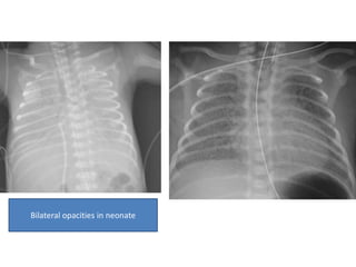
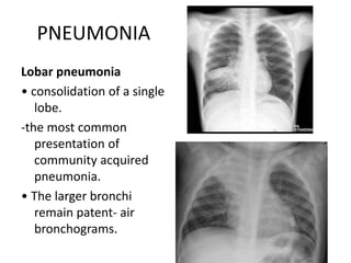
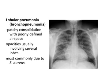
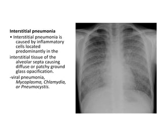
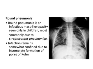
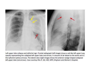
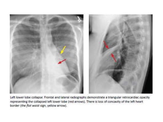
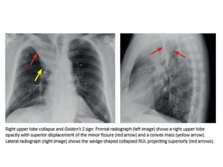
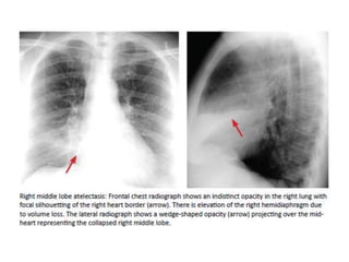
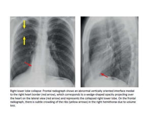
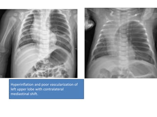
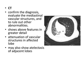
PAEDIATRIC RADIOLGOY
-DR.ANJAAN AUGUSTINE
HYALINE MEMBRANE DISEASE/RDS
Radiographic features
• Plain radiograph
• low lung volumes
• diffuse, bilateral and symmetrical granular opacities
• bell-shaped thorax
• air bronchograms
• surfactant therapy-asymmetric improvement as more
surfactant may reach certain parts of the lungs than
others.
• Ultrasound
• retrodiaphragmatic hyperechogenicity
Bilateral opacities in neonate
• Differential diagnosis
• TTN: lung volumes are normal to slightly
hyperinflated in transient tachypnea of the
newborn and decreased in respiratory distress
syndrome
• Pulmonary edema/hemmorage
• Pulmonary venous congetsion
PNEUMONIA
Lobar pneumonia
• consolidation of a single
lobe.
-the most common
presentation of
community acquired
pneumonia.
• The larger bronchi
remain patent- air
bronchograms.
Lobular pneumonia
(bronchopneumonia)
-patchy consolidation
with poorly defined
airspace
opacities usually
involving several
lobes
most commonly due to
S. aureus.
Interstitial pneumonia
• Interstitial pneumonia is
caused by inflammatory
cells located
predominantly in the
interstitial tissue of the
alveolar septa causing
diffuse or patchy ground
glass opacification.
-viral pneumonia,
Mycoplasma, Chlamydia,
or Pneumocystis.
Round pneumonia
• Round pneumonia is an
infectious mass-like opacity
seen only in children, most
commonly due to
streptococcus pneumoniae.
• Infection remains
somewhat confined due to
incomplete formation of
pores of Kohn
COLLAPSE
• Direct signs of atelectasis are from lobar volume loss
and include:
-Displacement of the fissures
-Vascular crowding.
• Indirect signs of atelectasis are due to the effect of
volume loss on adjacent structures
-Elevation of the diaphragm.
-Rib crowding on the side with volume loss.
-Mediastinal shift to the side with volume loss.
-Overinflation of adjacent or c/l lobes
-hilar displacement
CLE
• Congenital Lobar Emphysema
• (CLE) is the overinflation of one or more lung
lobes. The left upper lobe is most frequently
affected.
• X-RAY:
• Immediate postpartum period
• affected lobe -opaque and homogeneous because of fetal lung
fluid diffuse reticular pattern that represents distended lymphatic
channels filled with fetal lung fluid.
• Later findings
• appears as an area of hyperlucency in the lung with oligemia
(i.e. paucity of vessels)
• mass effect with mediastinal shift and hemidiaphragmatic
depression
• Lateral film with the patient lying on the affected side will show
little or no change in lung volume
• lateral film may show posterior displacement of the heart
Hyperinflation and poor vascularization of
left upper lobe with contralateral
mediastinal shift.
• CT
• confirm the diagnosis,
evaluate the mediastinal
vascular structures, and
to rule out other
abnormalities.
• shows above features in
greater detail
• attenuation of vascular
structures in affected
lobe
• may also show atelectasis
of adjacent lobes
• Differential diagnosis
• Bronchial atresia
• CPAM
• Pulmonary artery hypoplasia
• filamin A mutation
THANKYOU

PAEDIATRIC RADIOLGOY.pptx

  • 1.
  • 2.
    HYALINE MEMBRANE DISEASE/RDS Radiographicfeatures • Plain radiograph • low lung volumes • diffuse, bilateral and symmetrical granular opacities • bell-shaped thorax • air bronchograms • surfactant therapy-asymmetric improvement as more surfactant may reach certain parts of the lungs than others. • Ultrasound • retrodiaphragmatic hyperechogenicity
  • 3.
  • 4.
    • Differential diagnosis •TTN: lung volumes are normal to slightly hyperinflated in transient tachypnea of the newborn and decreased in respiratory distress syndrome • Pulmonary edema/hemmorage • Pulmonary venous congetsion
  • 5.
    PNEUMONIA Lobar pneumonia • consolidationof a single lobe. -the most common presentation of community acquired pneumonia. • The larger bronchi remain patent- air bronchograms.
  • 6.
    Lobular pneumonia (bronchopneumonia) -patchy consolidation withpoorly defined airspace opacities usually involving several lobes most commonly due to S. aureus.
  • 7.
    Interstitial pneumonia • Interstitialpneumonia is caused by inflammatory cells located predominantly in the interstitial tissue of the alveolar septa causing diffuse or patchy ground glass opacification. -viral pneumonia, Mycoplasma, Chlamydia, or Pneumocystis.
  • 8.
    Round pneumonia • Roundpneumonia is an infectious mass-like opacity seen only in children, most commonly due to streptococcus pneumoniae. • Infection remains somewhat confined due to incomplete formation of pores of Kohn
  • 9.
    COLLAPSE • Direct signsof atelectasis are from lobar volume loss and include: -Displacement of the fissures -Vascular crowding. • Indirect signs of atelectasis are due to the effect of volume loss on adjacent structures -Elevation of the diaphragm. -Rib crowding on the side with volume loss. -Mediastinal shift to the side with volume loss. -Overinflation of adjacent or c/l lobes -hilar displacement
  • 15.
    CLE • Congenital LobarEmphysema • (CLE) is the overinflation of one or more lung lobes. The left upper lobe is most frequently affected.
  • 16.
    • X-RAY: • Immediatepostpartum period • affected lobe -opaque and homogeneous because of fetal lung fluid diffuse reticular pattern that represents distended lymphatic channels filled with fetal lung fluid. • Later findings • appears as an area of hyperlucency in the lung with oligemia (i.e. paucity of vessels) • mass effect with mediastinal shift and hemidiaphragmatic depression • Lateral film with the patient lying on the affected side will show little or no change in lung volume • lateral film may show posterior displacement of the heart
  • 17.
    Hyperinflation and poorvascularization of left upper lobe with contralateral mediastinal shift.
  • 18.
    • CT • confirmthe diagnosis, evaluate the mediastinal vascular structures, and to rule out other abnormalities. • shows above features in greater detail • attenuation of vascular structures in affected lobe • may also show atelectasis of adjacent lobes
  • 19.
    • Differential diagnosis •Bronchial atresia • CPAM • Pulmonary artery hypoplasia • filamin A mutation
  • 20.