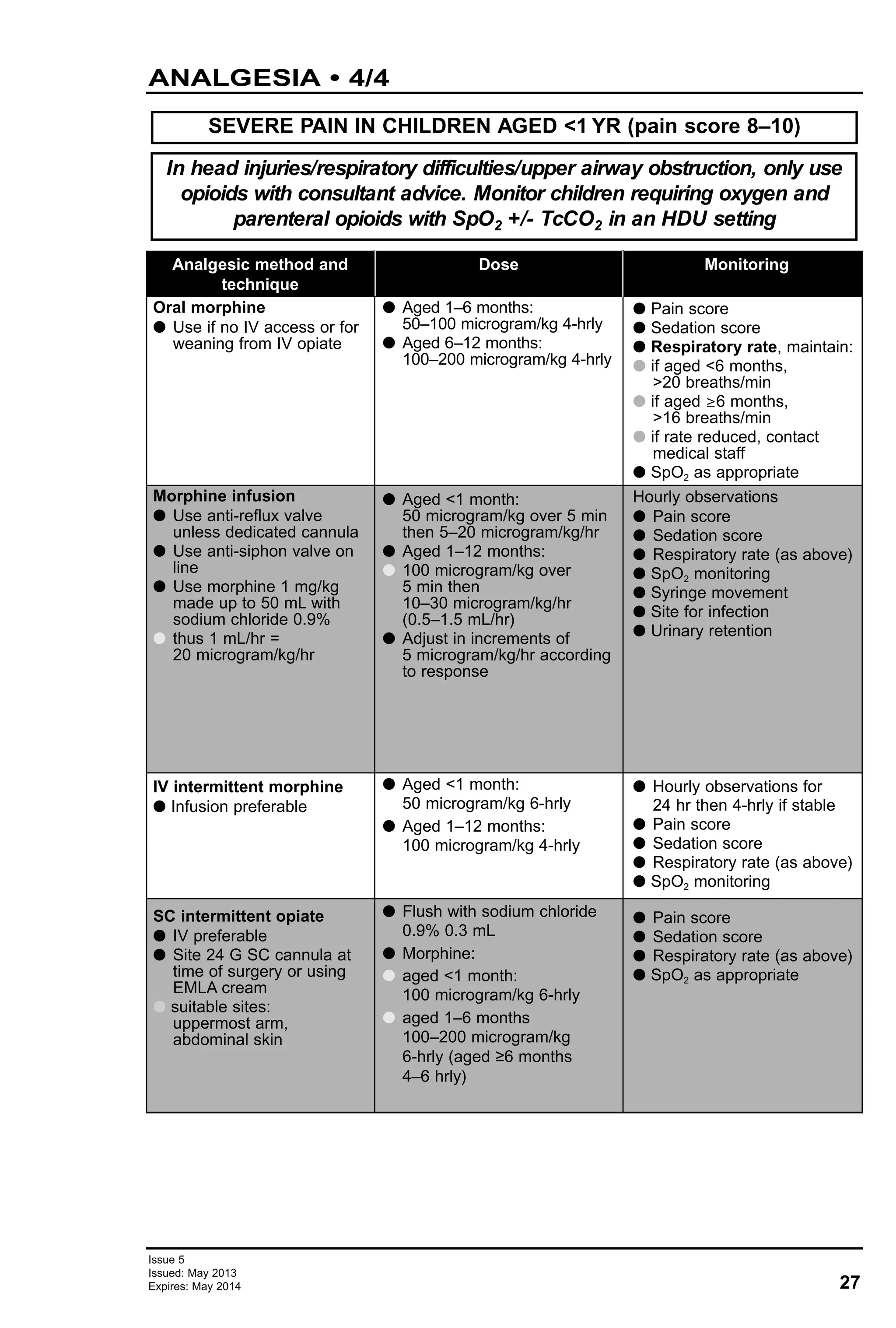
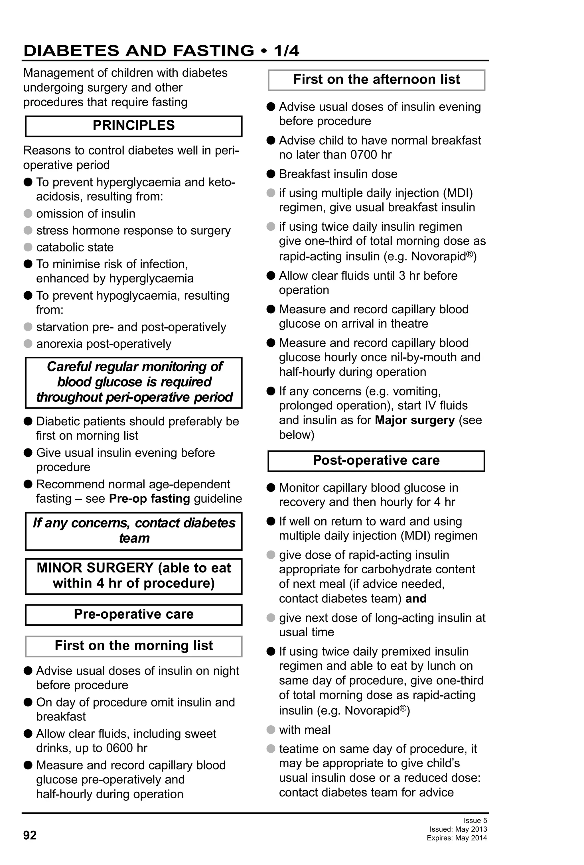
These guidelines provide advice on cardiopulmonary resuscitation for children, including:
- Initial steps of establishing airway, breathing and circulation
- Obtaining vascular access via intravenous or intraosseous routes
- Recommended doses of adrenaline for different age groups
- Details on ventilation rates, cardiac compression rates, and equipment such as endotracheal tubes
- Guidance on establishing and maintaining an airway, and obtaining intravenous or intraosseous access
- Use of an ECG monitor to identify shockable vs. non-shockable rhythms and guide treatment according to algorithm
![9
Issue 5
Issued: May 2013
Expires: May 2014
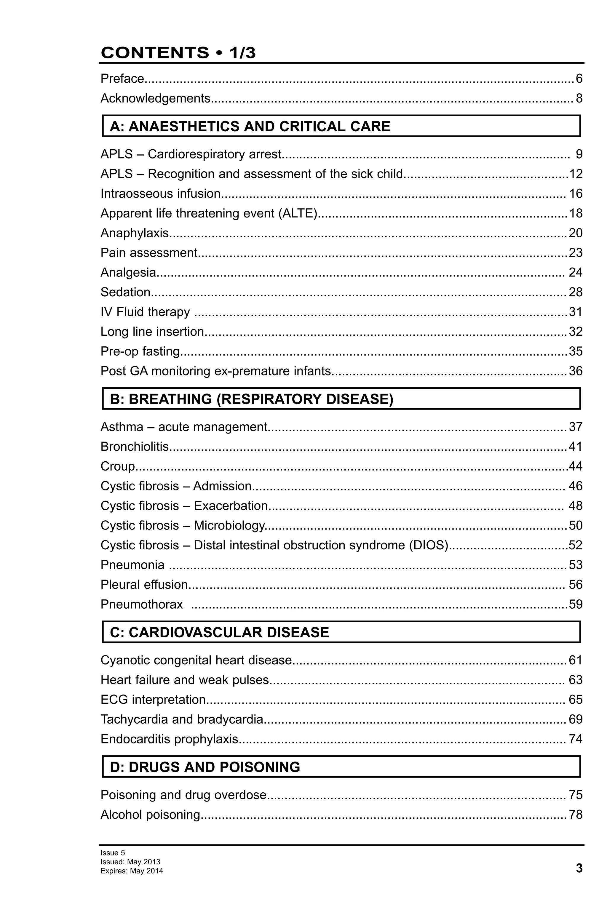
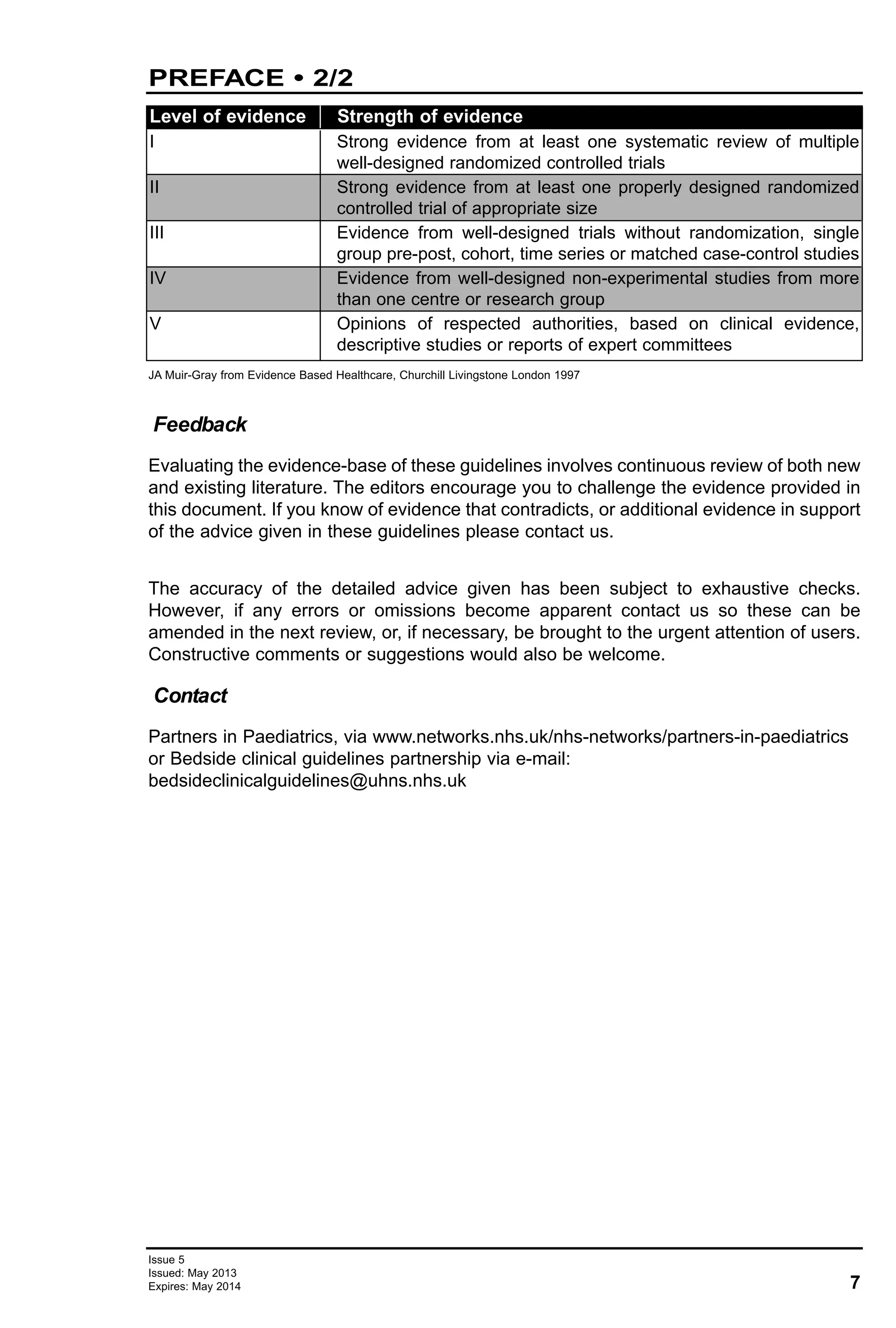
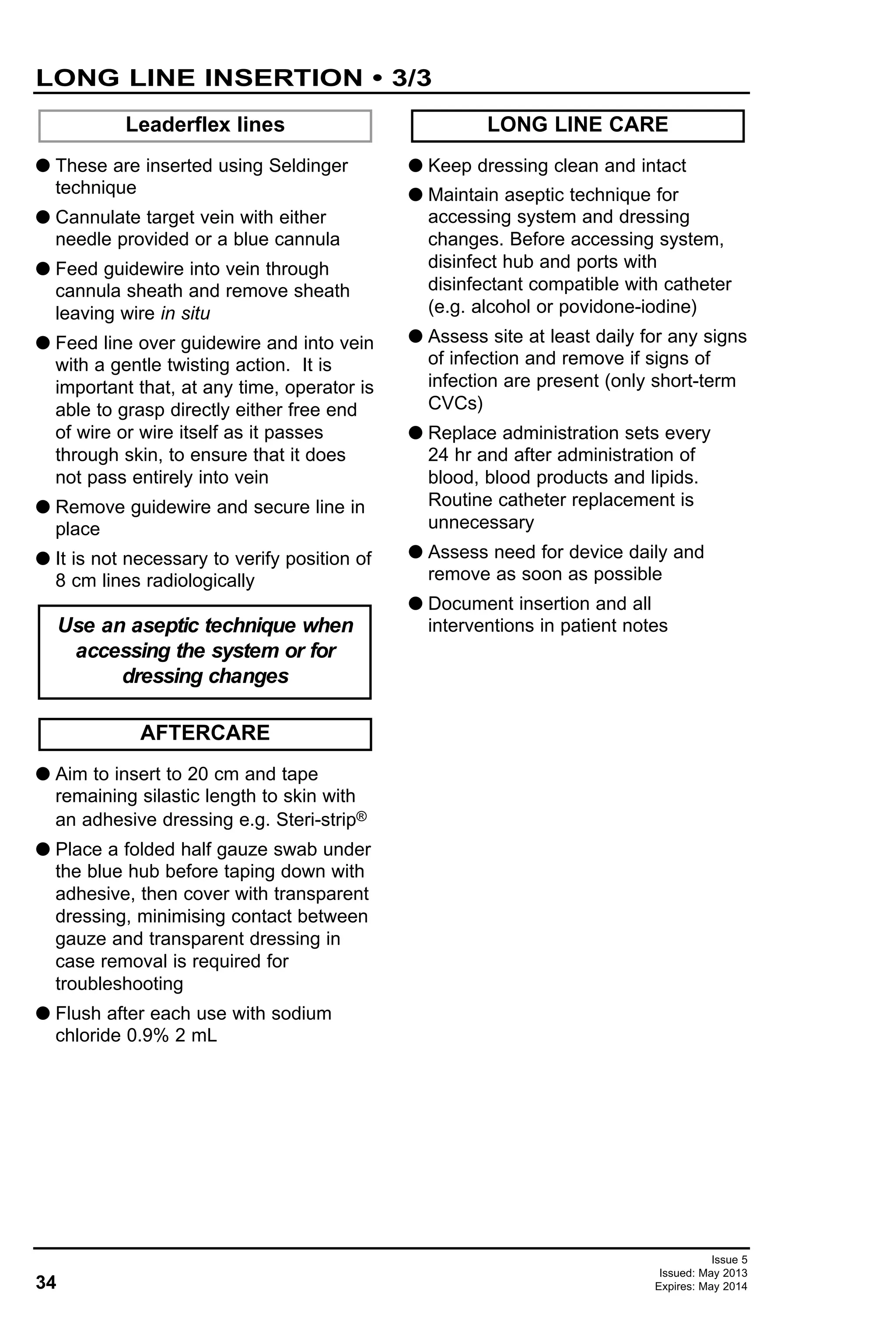
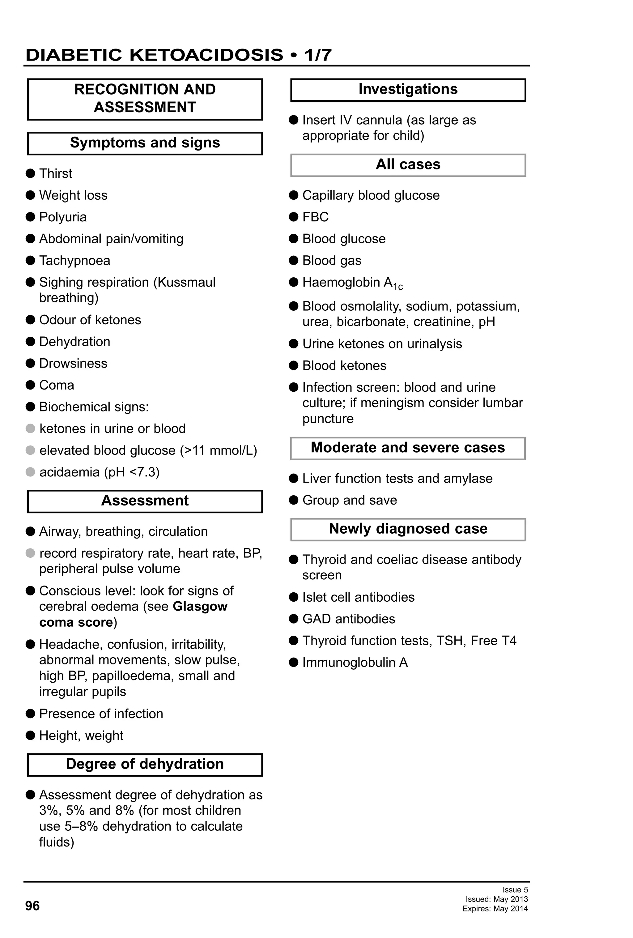
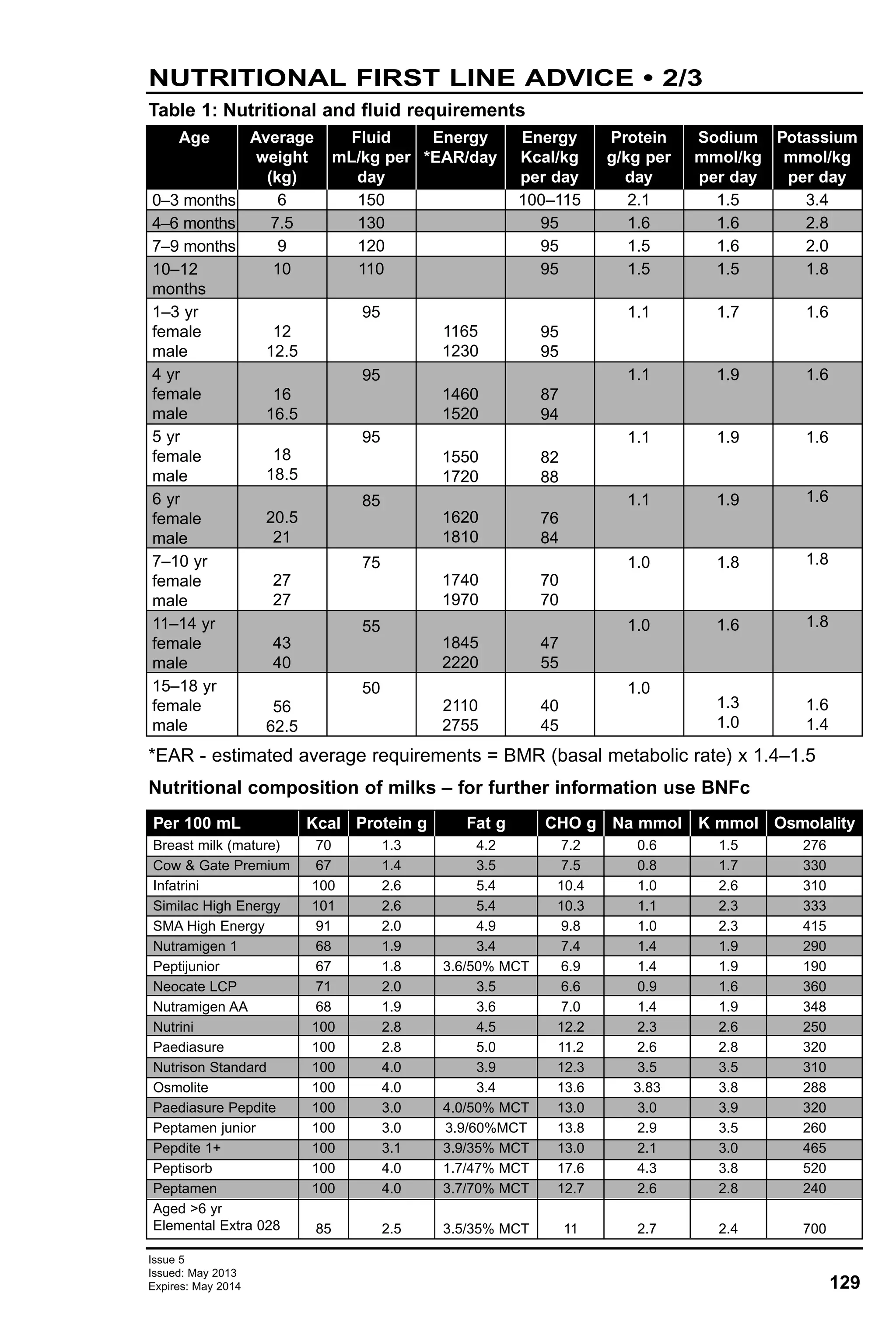
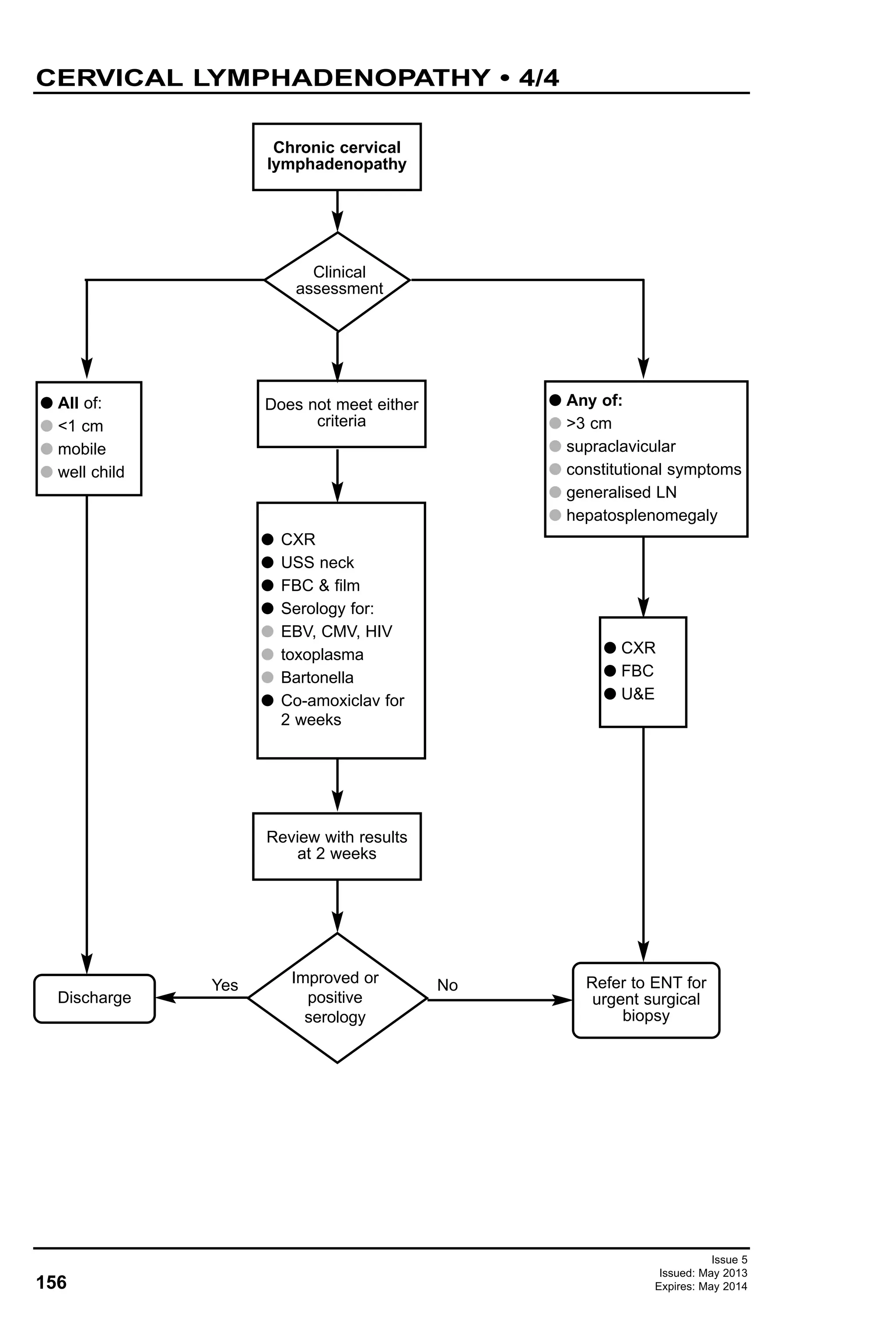
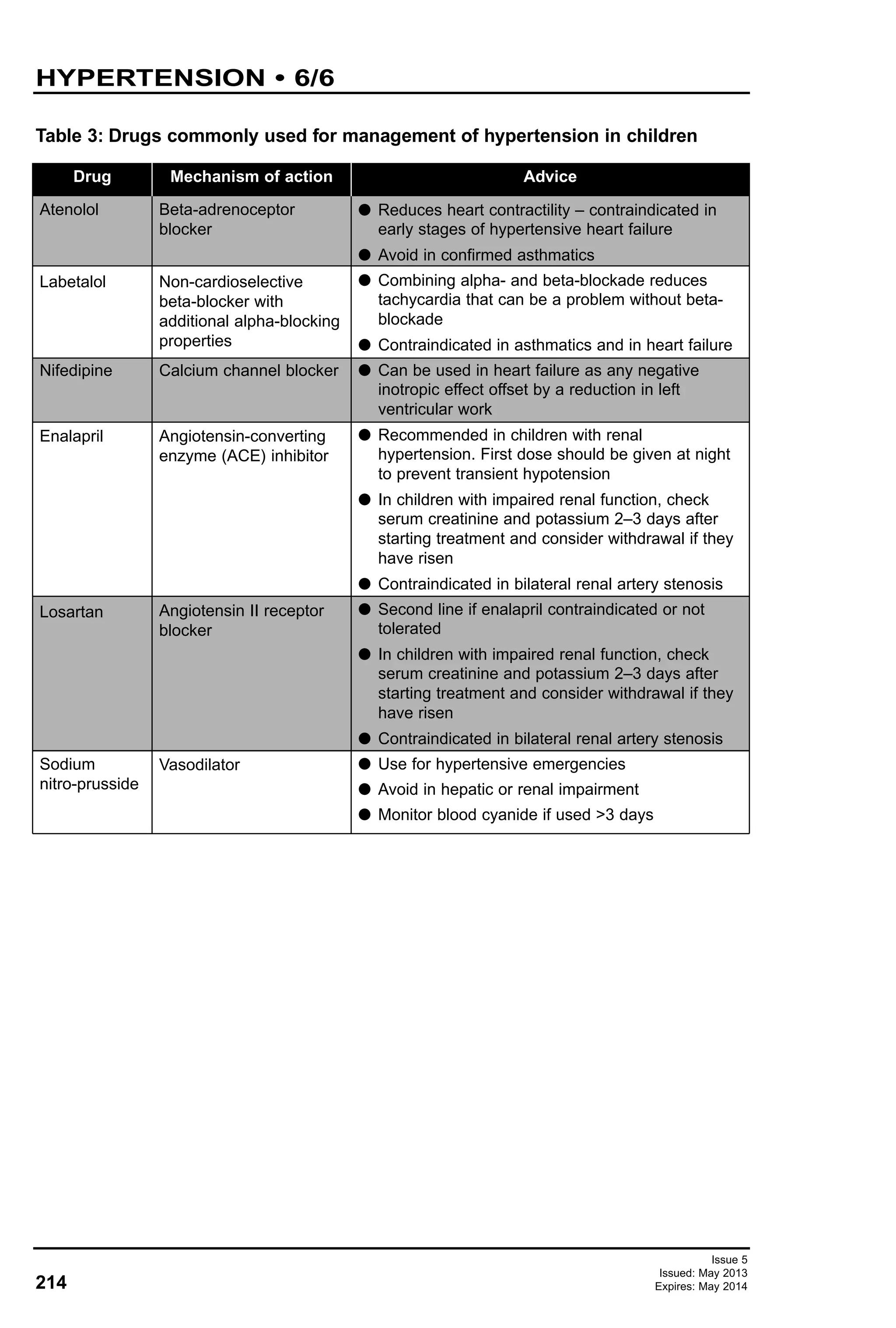
APLS - CARDIORESPIRATORY ARREST • 1/3
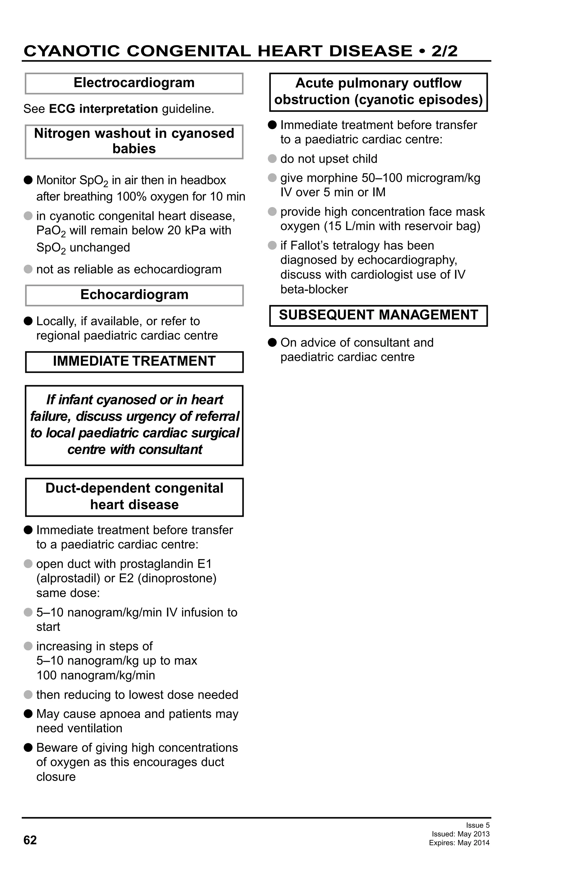
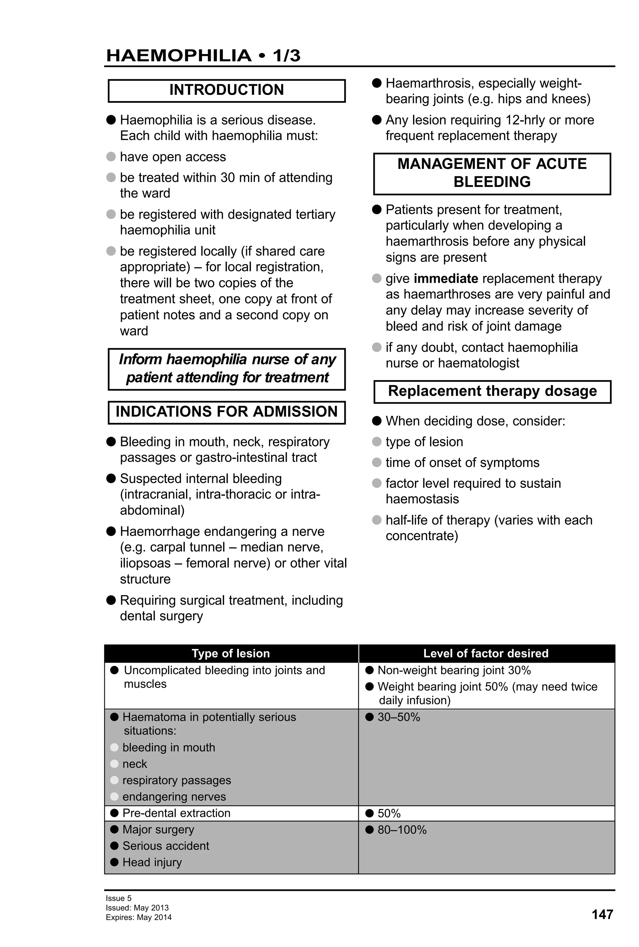
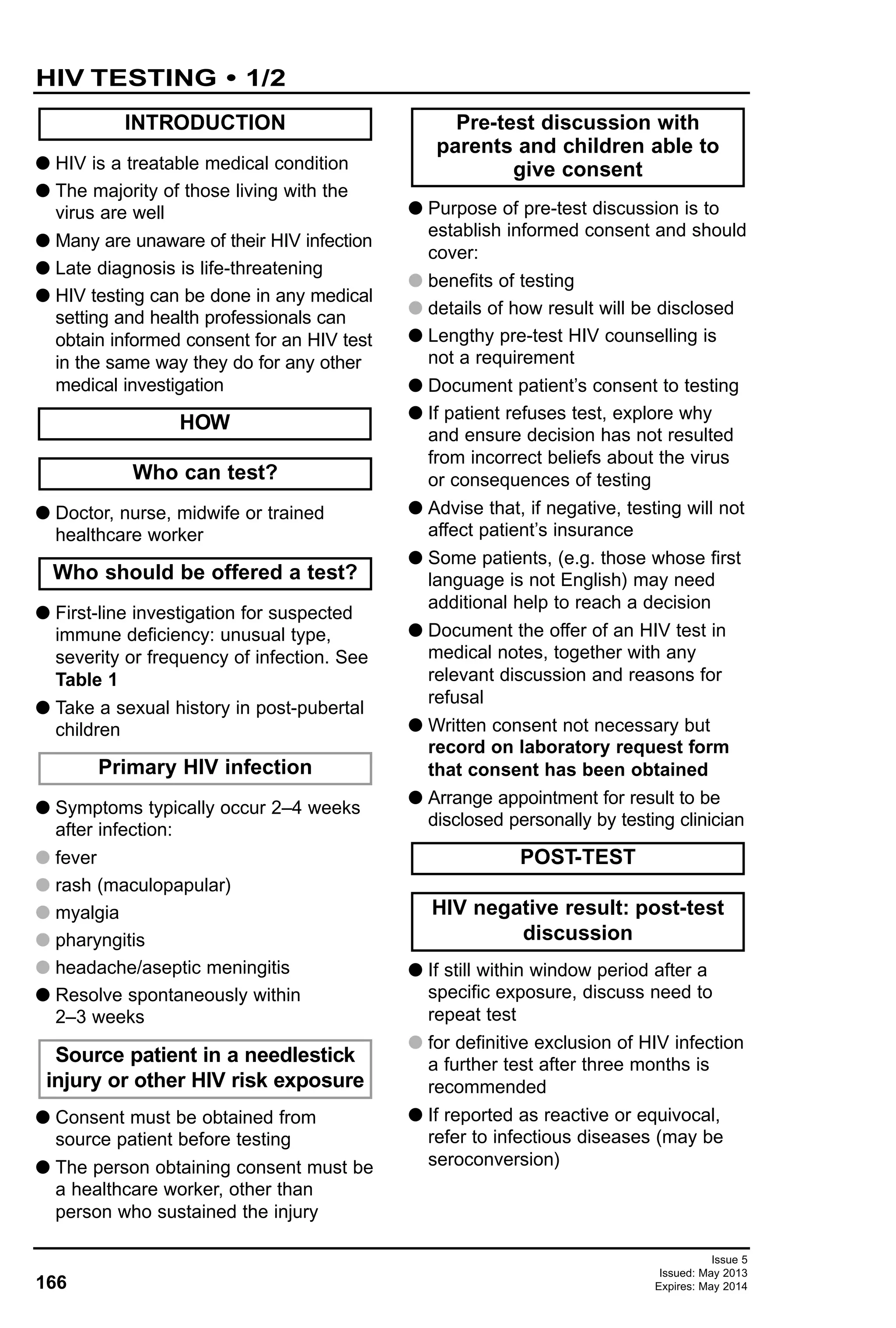
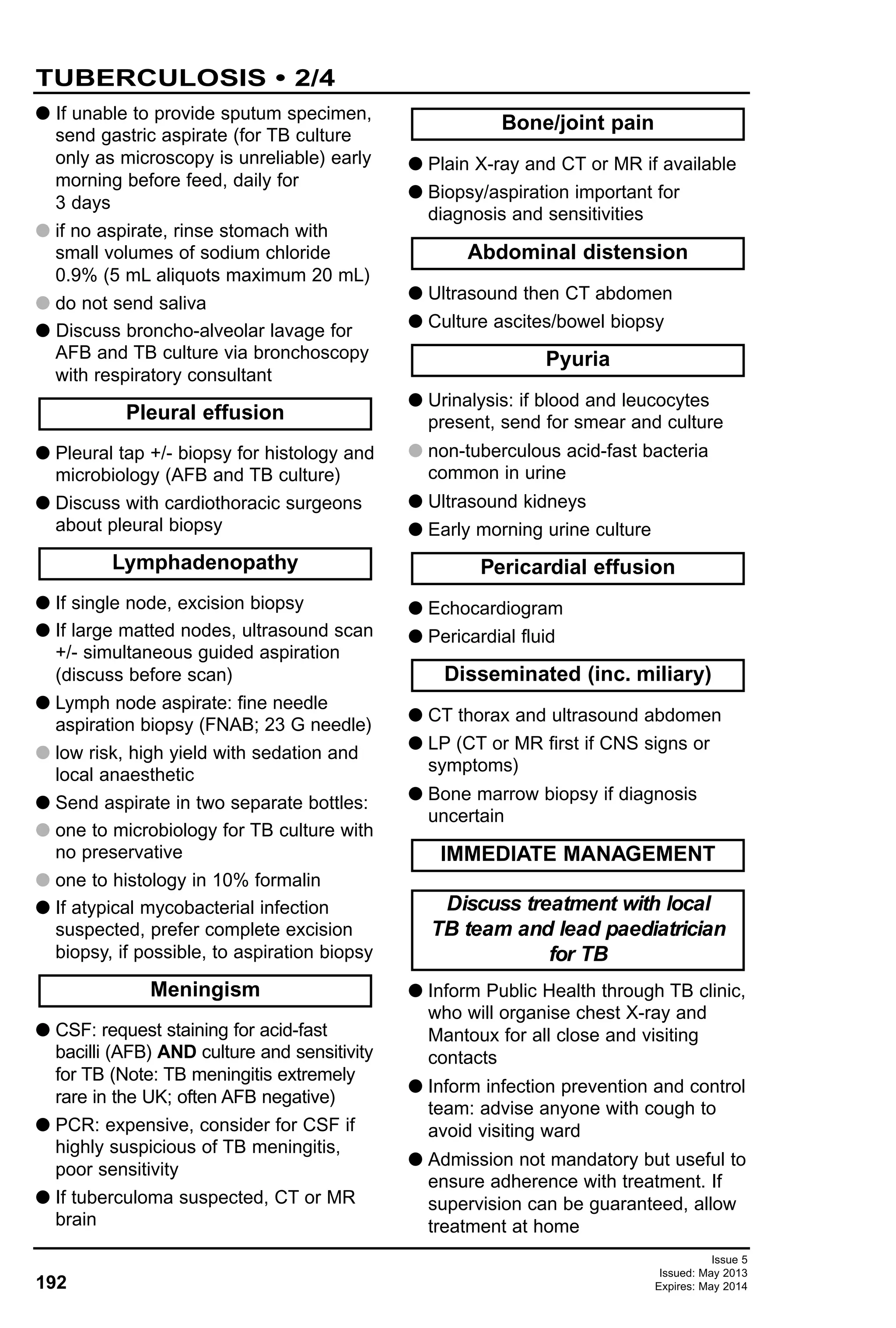
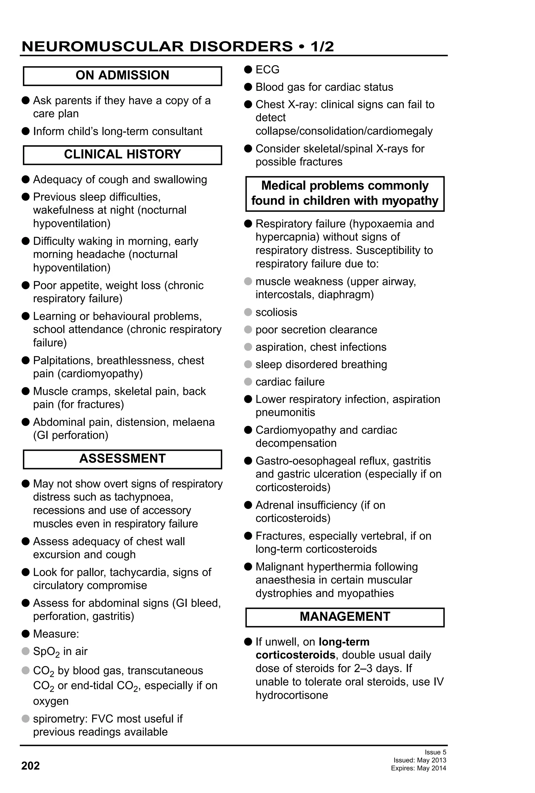
Route Aged <12 yr Aged 12 yr – adult Notes
IV rapid bolus/
intraosseous
Endotracheal
tube (ETT)
10 microgram/kg
(0.1 mL/kg
of 1:10,000)
100 microgram/kg
(0.1 mL/kg of 1:1000
or
1 mL/kg of 1:10,000)
100 microgram/kg
(0.1 mL/kg of 1:1000
or
1 mL/kg of 1:10,000)
1 mg
(10 mL of
1:10,000)
5 mg
(5 mL of
1:10,000)
5 mg
(5 mL of 1:1000)
Initial and
usual
subsequent
dose
Exceptional
circumstances
(e.g. beta-
blocker
overdose)
-
If given by
intraosseous route
flush with sodium
chloride 0.9%
Maximum dose
5 mL of 1:1000
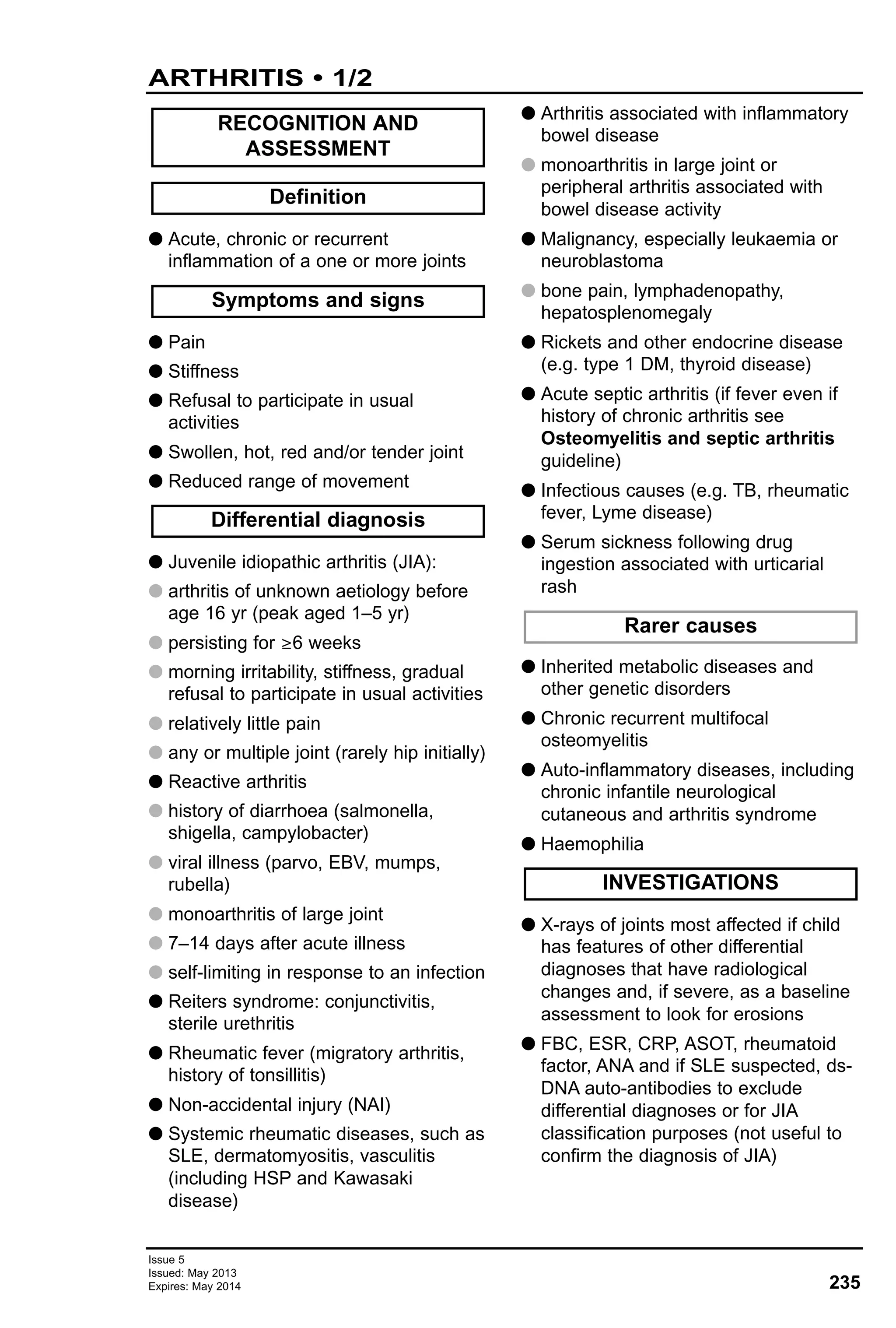
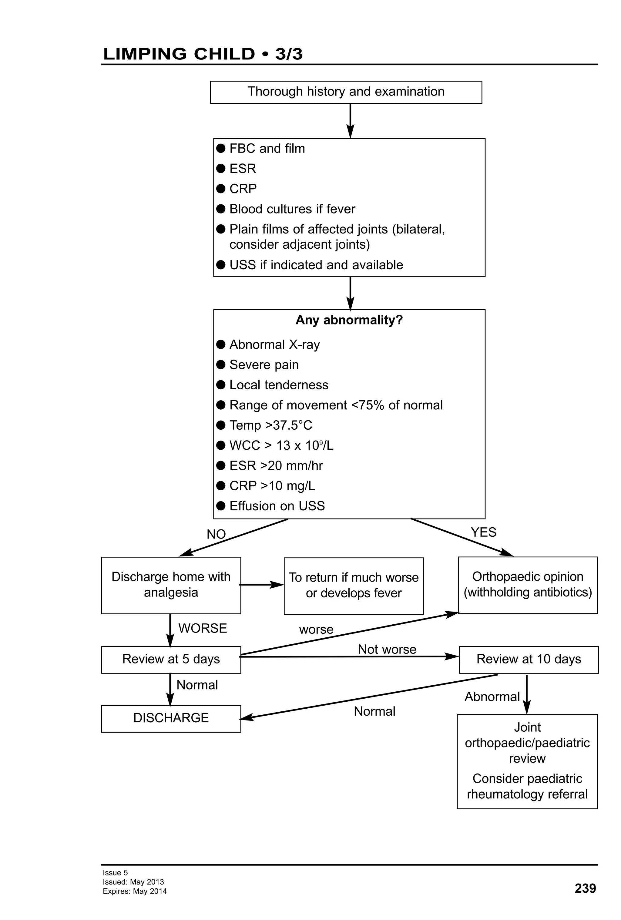
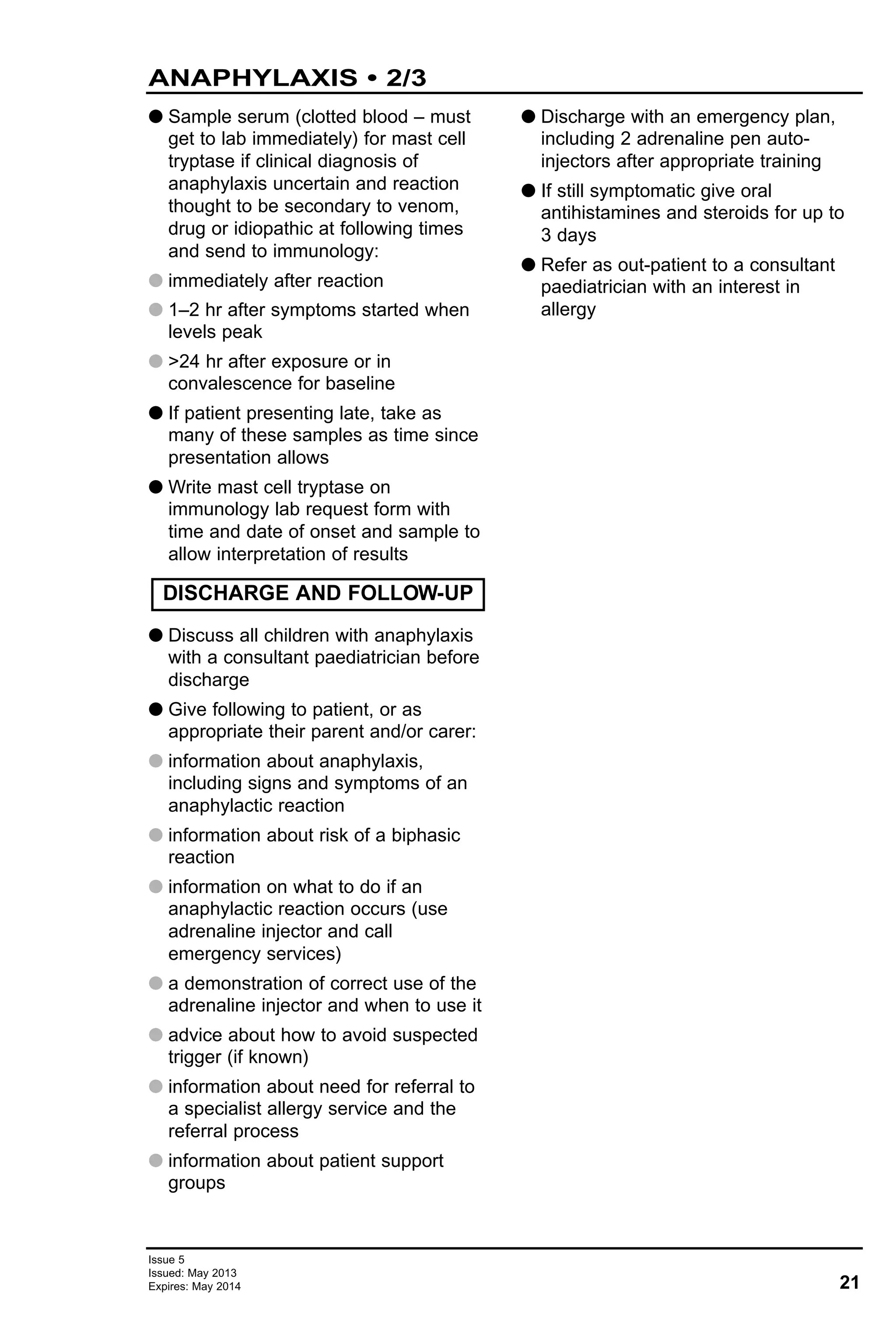
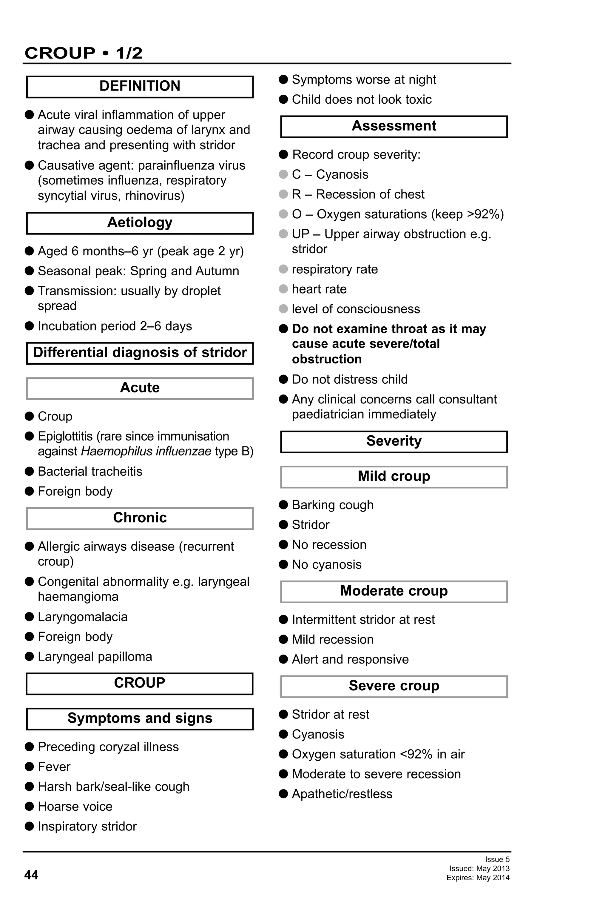
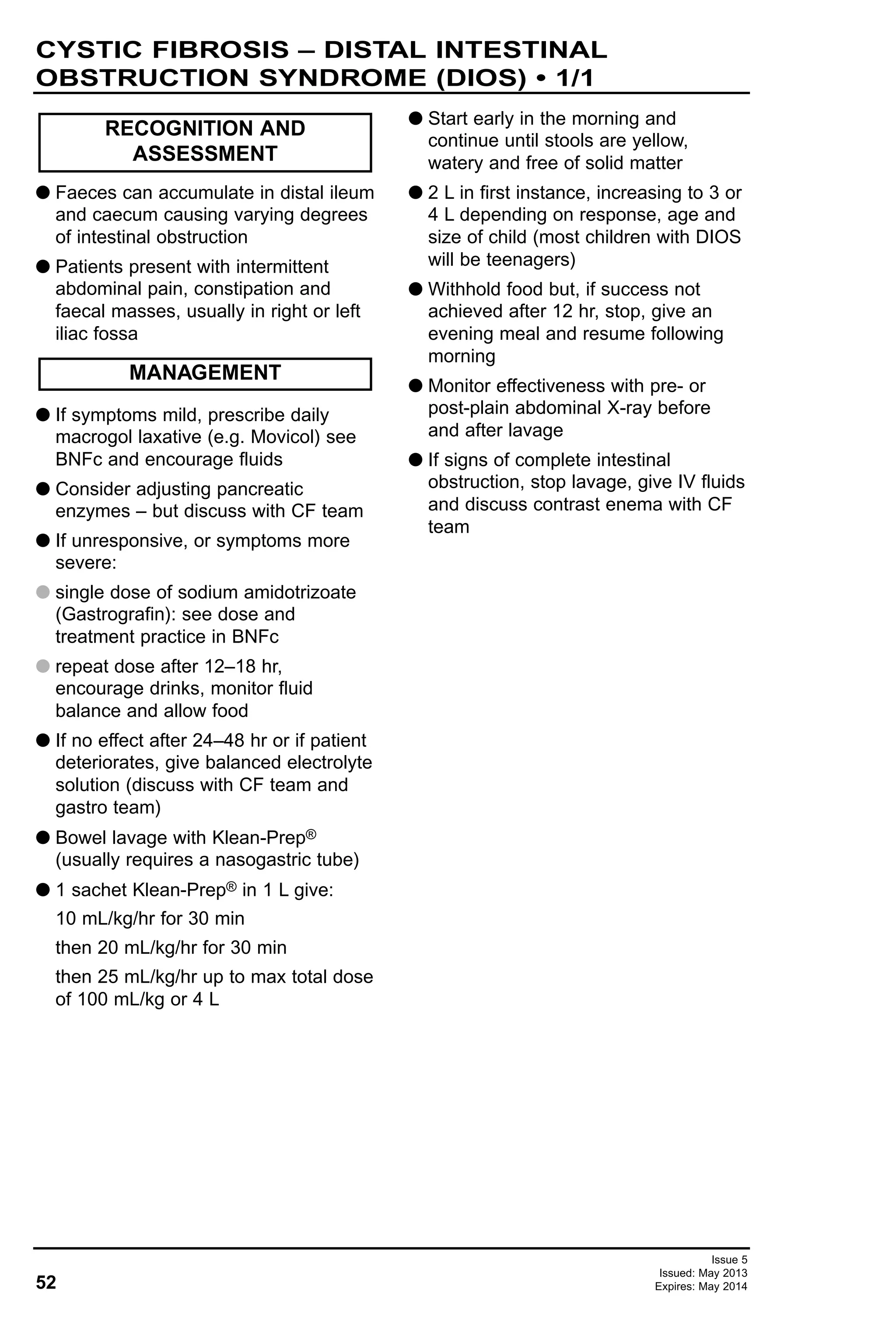
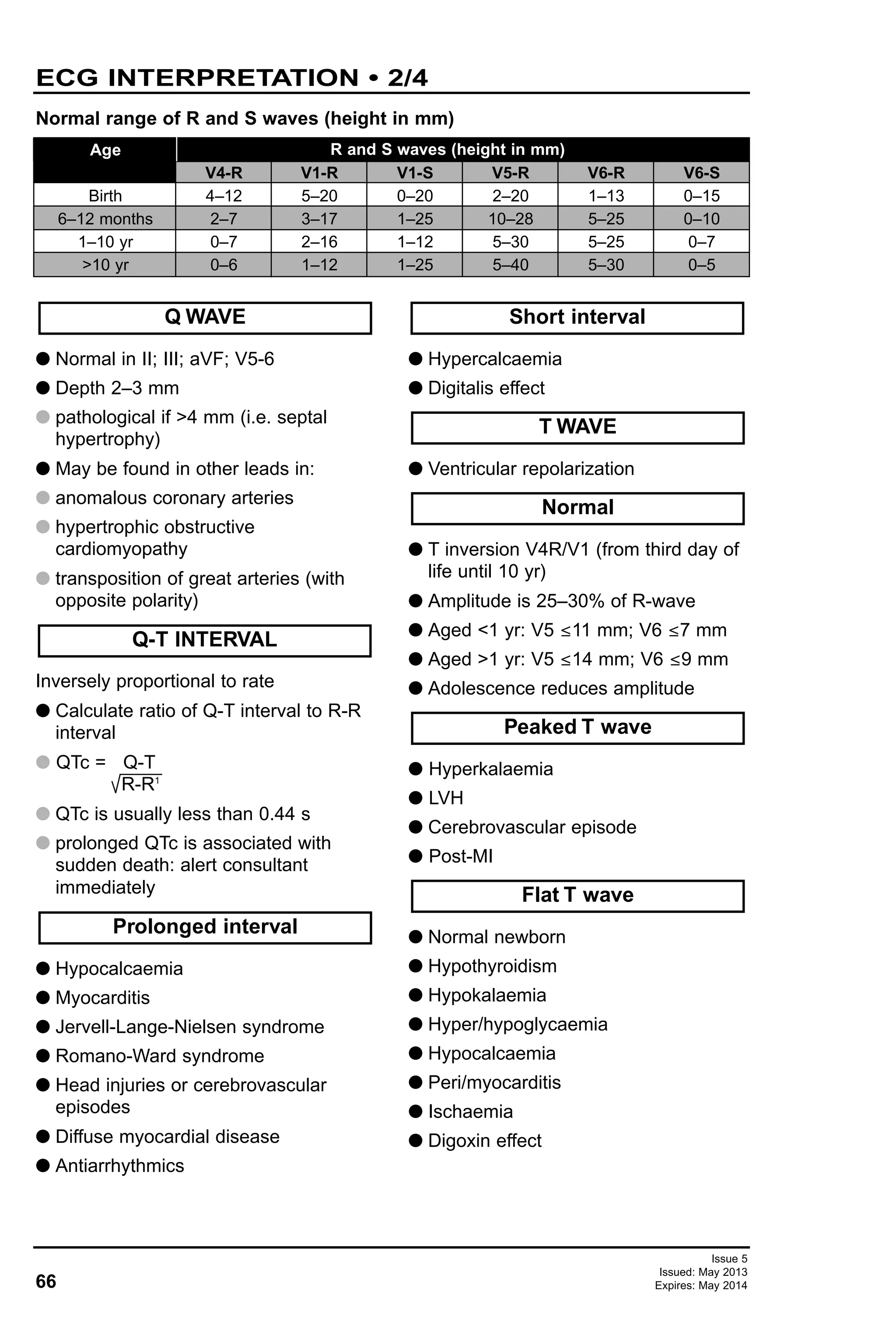
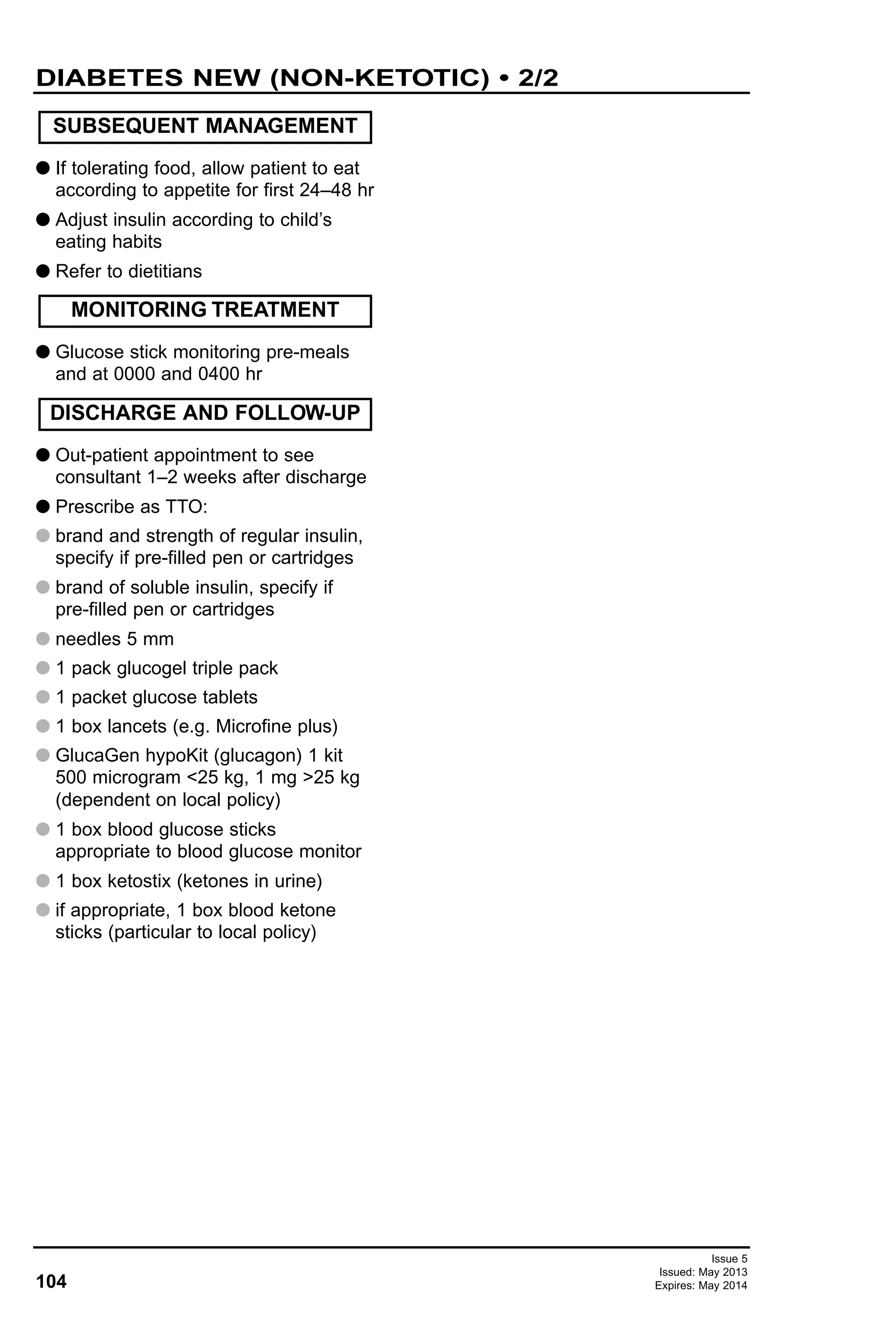
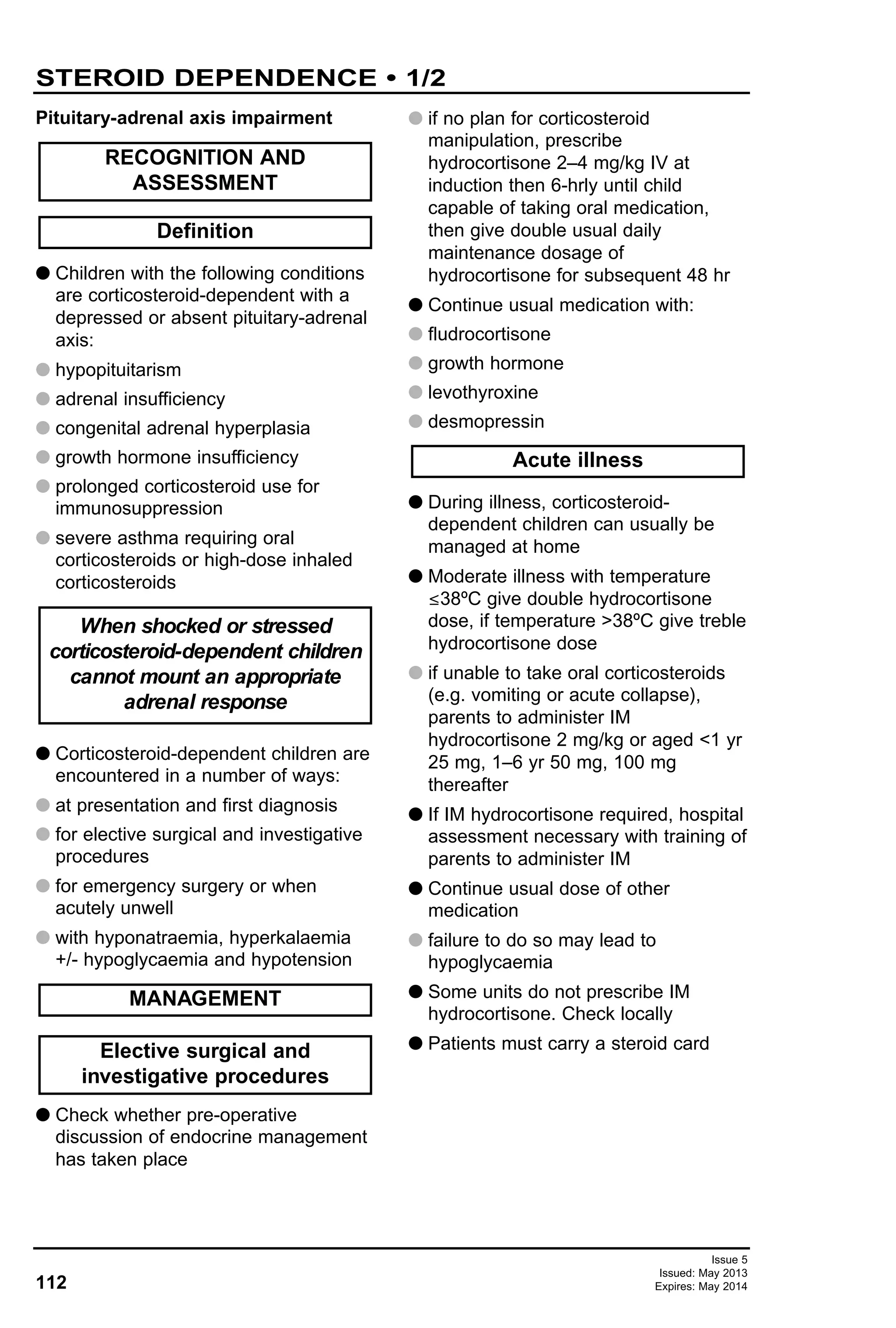
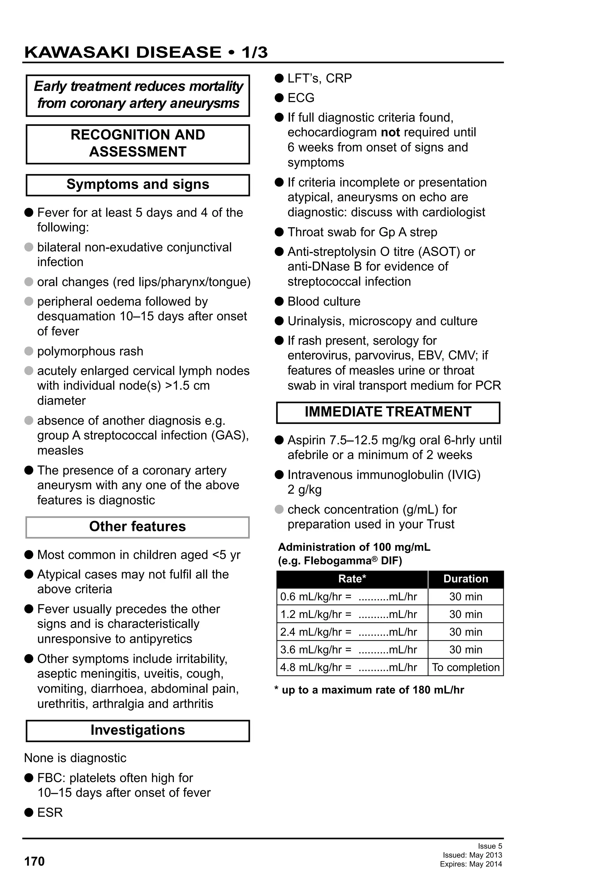
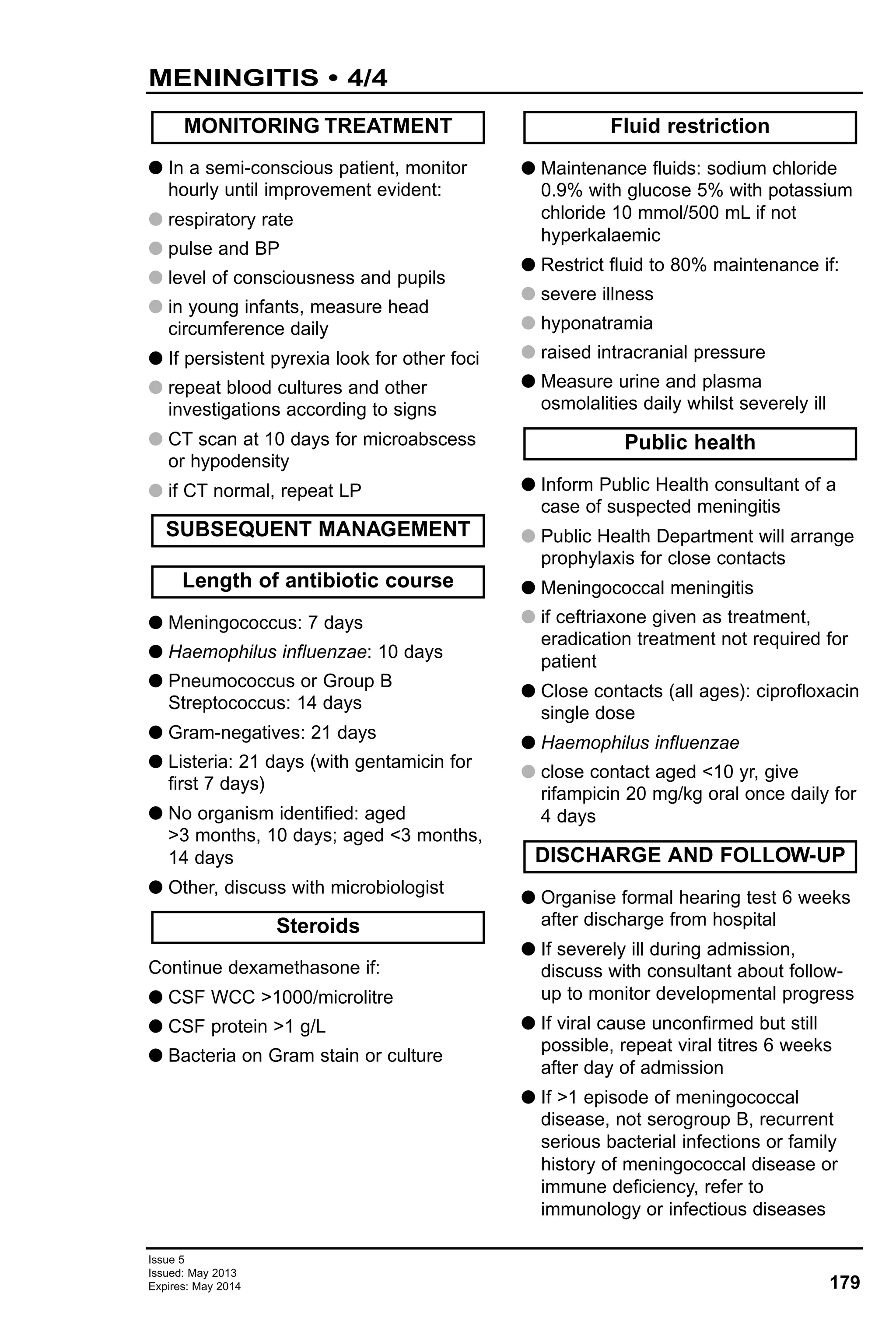
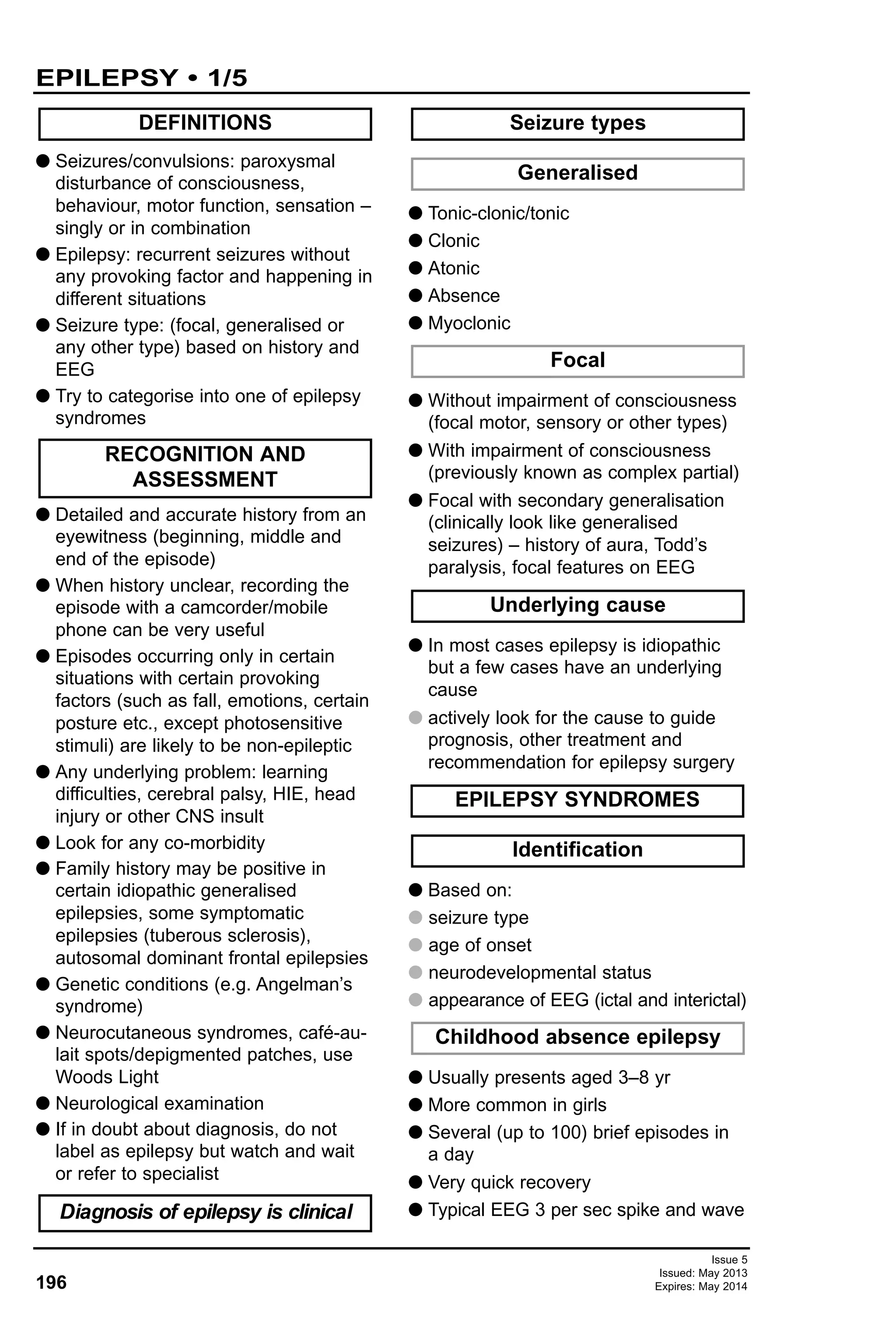
G Stimulate patient to assess for signs
of life and shout for help
G Establish basic life support: Airway –
Breathing – Circulation
G Connect ECG monitor: identify rhythm
and follow Algorithm
G Control airway and ventilation:
preferably intubate
G Obtain vascular access, peripheral or
intraosseous (IO)
G Change person performing chest
compressions every few minutes
G Inspect mouth: apply suction if
necessary
G Use either head tilt and chin lift or jaw
thrust
G Oro- or nasopharyngeal airway
G Intubation:
G endotracheal tube sizes
G term newborns 3–3.5 mm
G aged 1 yr 4.5 mm
G aged >1 yr: use formula [(age/4)
+ 4] mm for uncuffed tubes; 0.5
smaller for cuffed
G If airway cannot be achieved,
consider laryngeal mask or, failing
that, cricothyrotomy
G Self-inflating bag and mask with
100% oxygen
G Ventilation rate
G unintubated: 2 inflations for every 15
compressions
G intubated:10–12/min, with continuous
compressions
G Consider foreign body or
pneumothorax
G Cardiac compression rate:
100–120/min depressing lower half of
sternum by at least one third: push
hard, push fast
G Peripheral venous access: 1–2
attempts (<30 sec)
G Intraosseous access: 2–3 cm below
tibial tuberosity (see Intraosseous
infusion guideline)
G Use ECG monitor to decide between:
G a non-shockable rhythm: asystole or
pulseless electrical activity (PEA) i.e.
electromechanical dissociation
OR
G a shockable rhythm: ventricular
fibrillation or pulseless ventricular
tachycardia
Algorithm for managing these rhythms
follows:
G If arrest rhythm changes, restart
Algorithm
G If organised electrical activity seen,
check pulse and for signs of circulation
Circulation (C)
Breathing (B)
Airway (A)
MANAGEMENT
Adrenaline doses for asystole](https://image.slidesharecdn.com/paediatricguidelines2013-14withlinks-180801135506/75/Paediatric-guidelines-2013-14-with-links-9-2048.jpg)
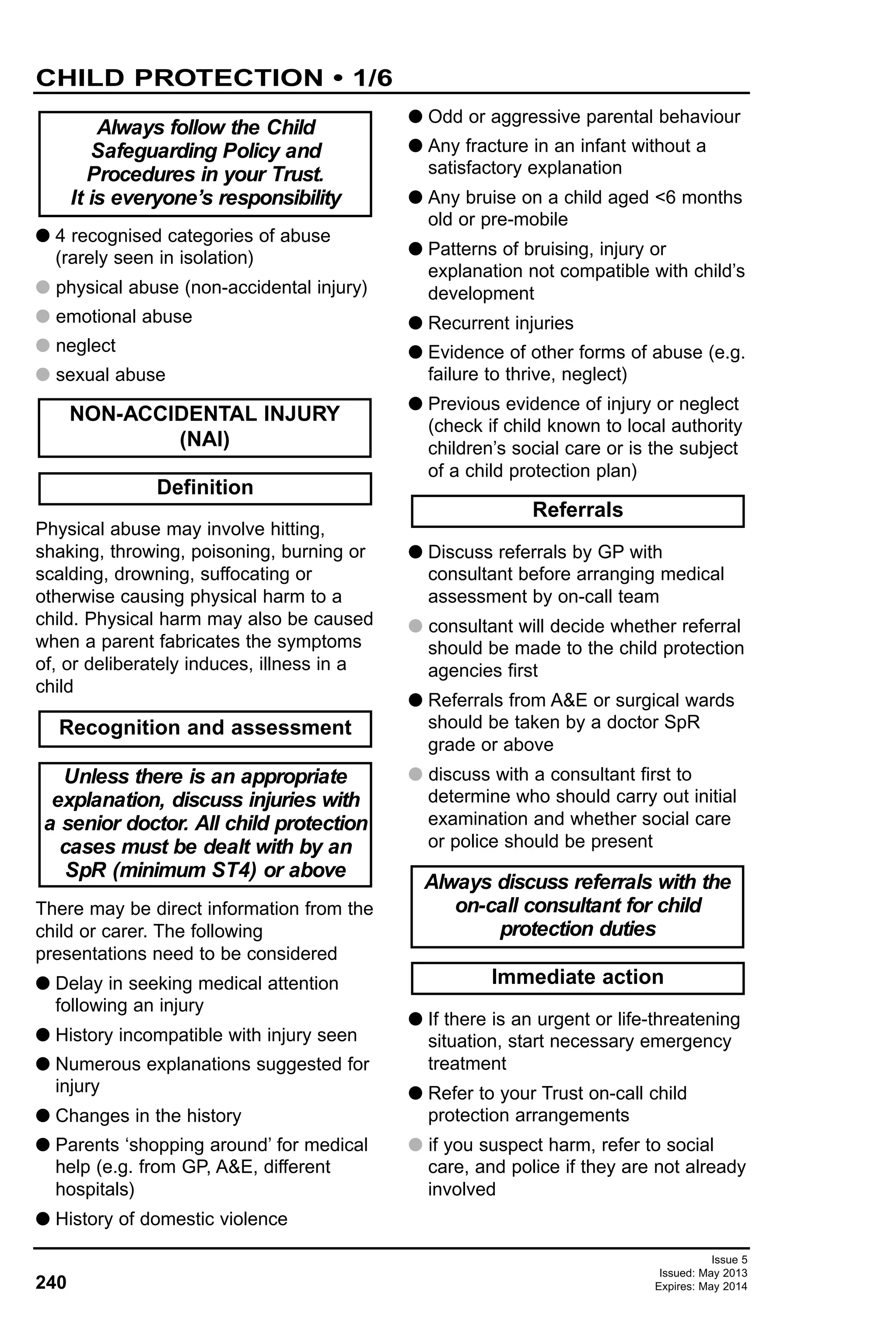
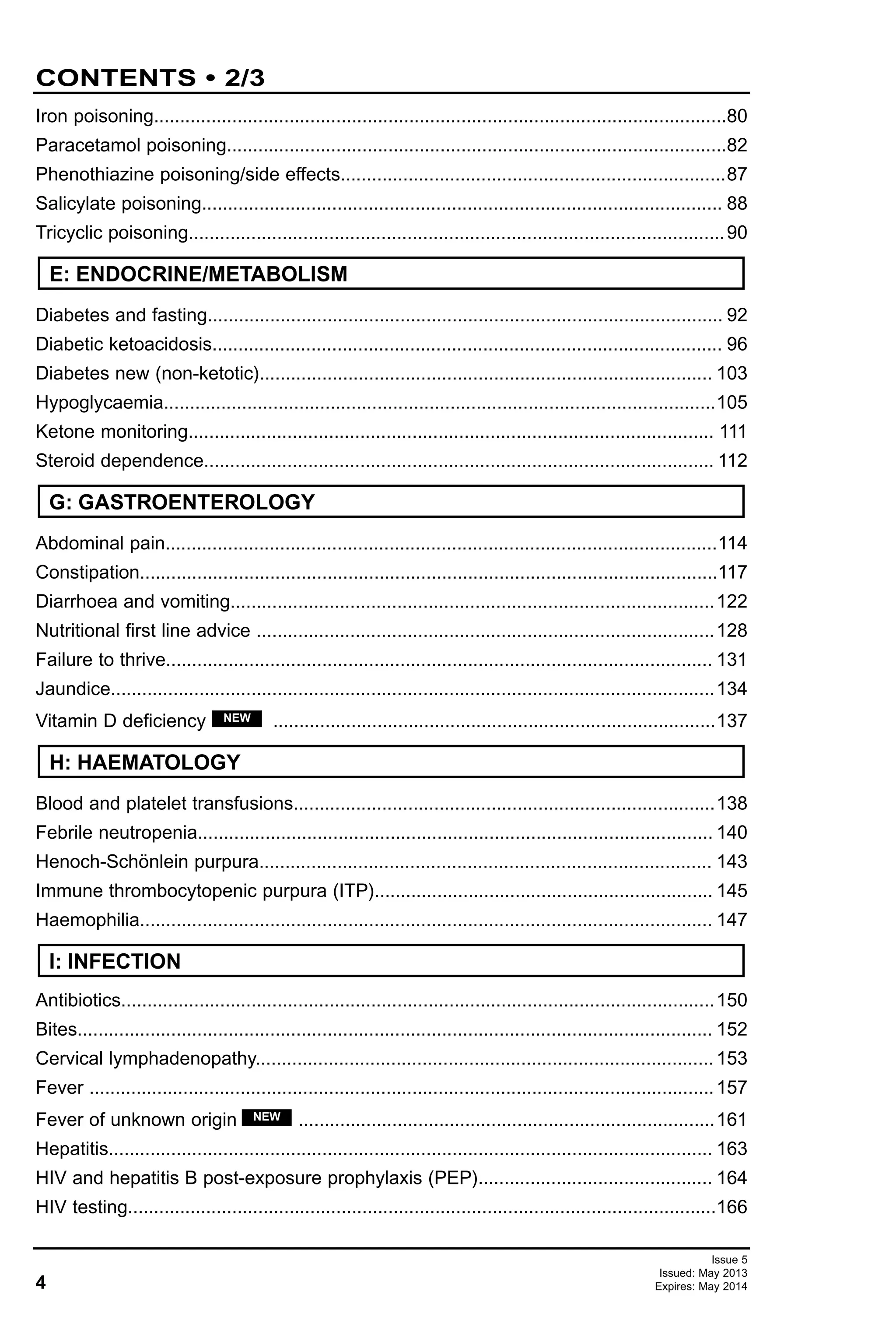
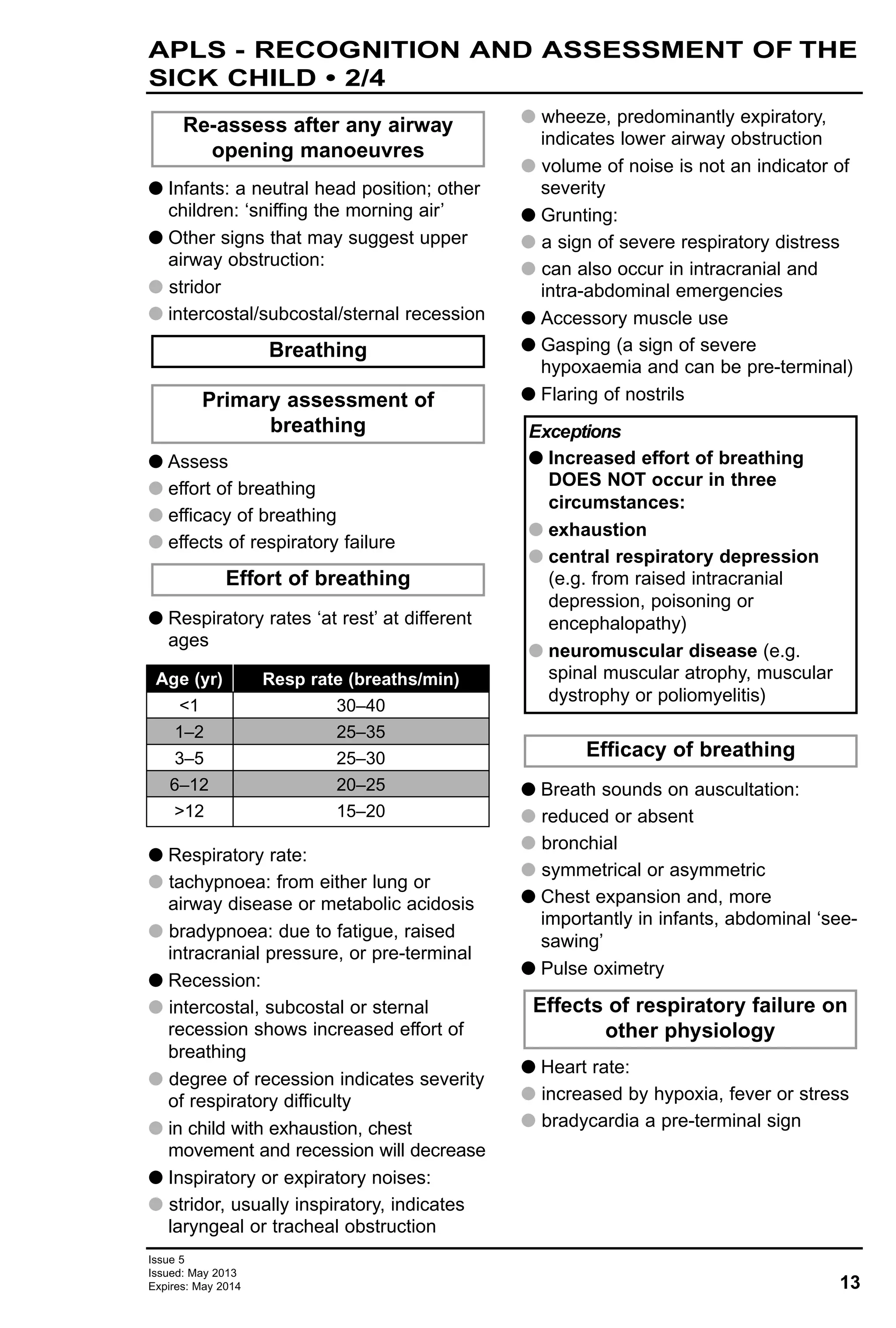
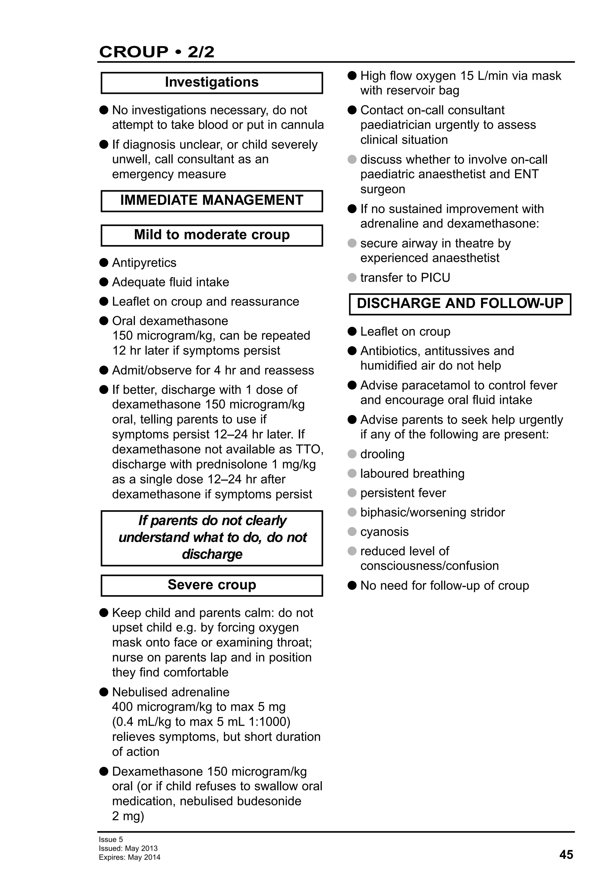
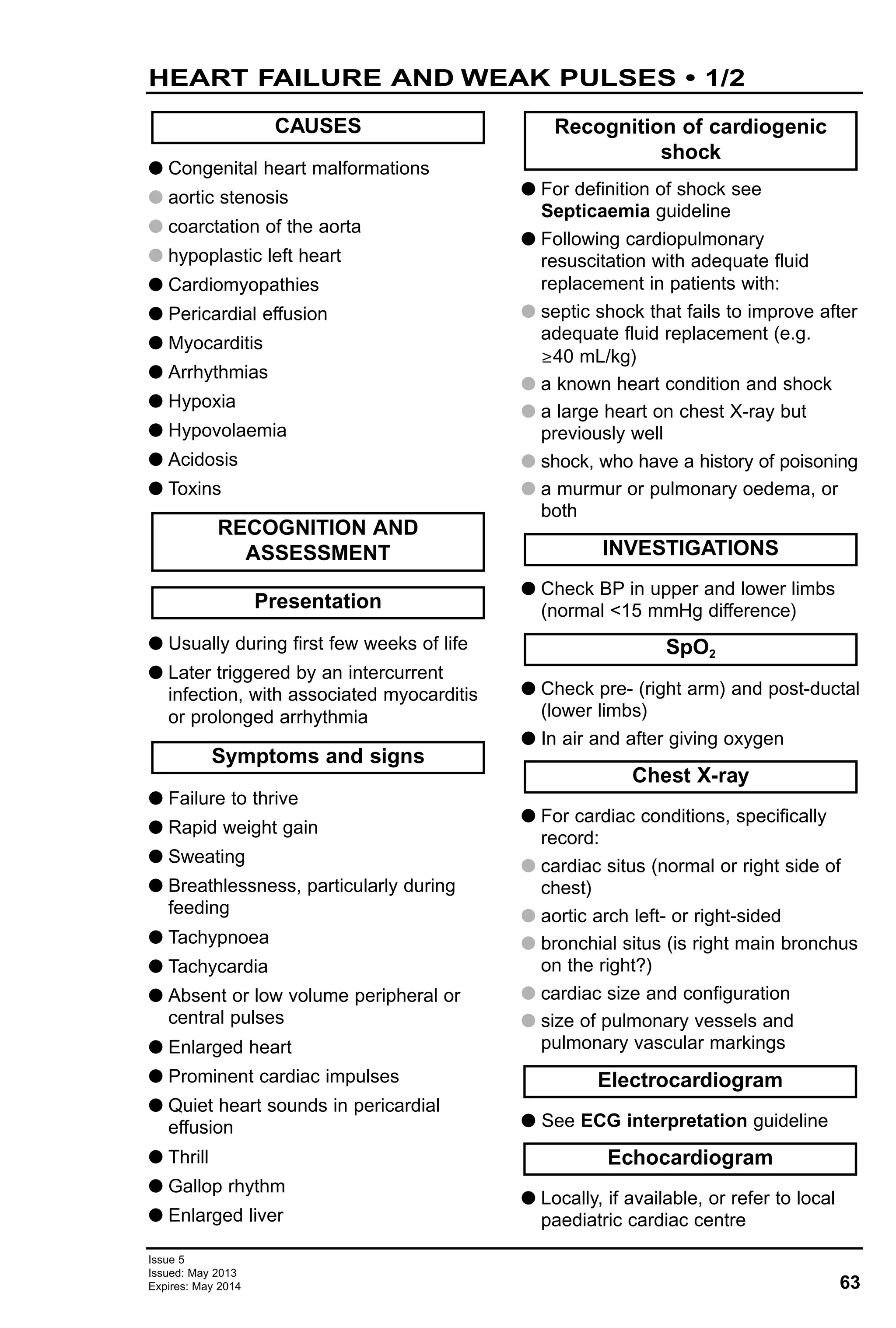
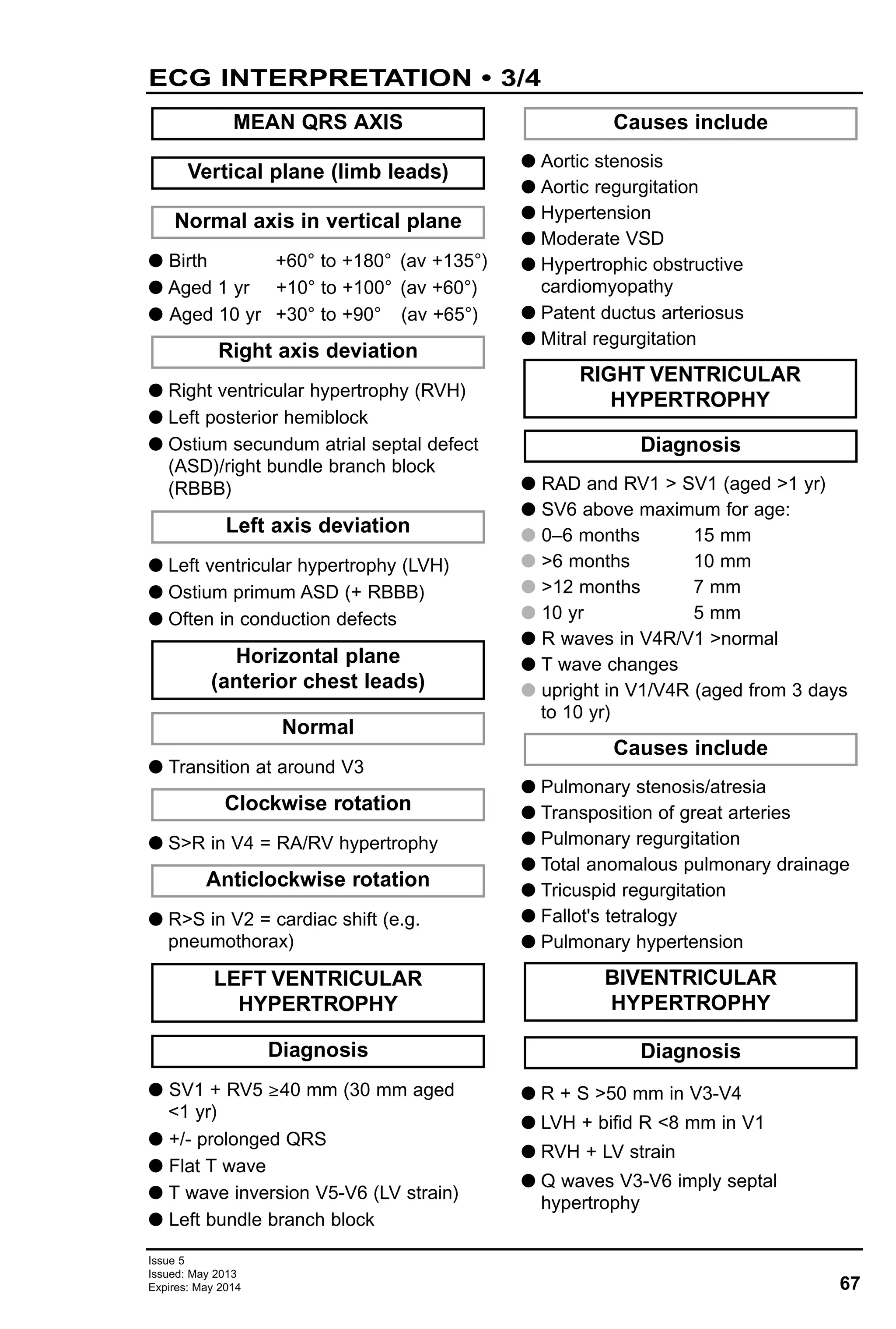
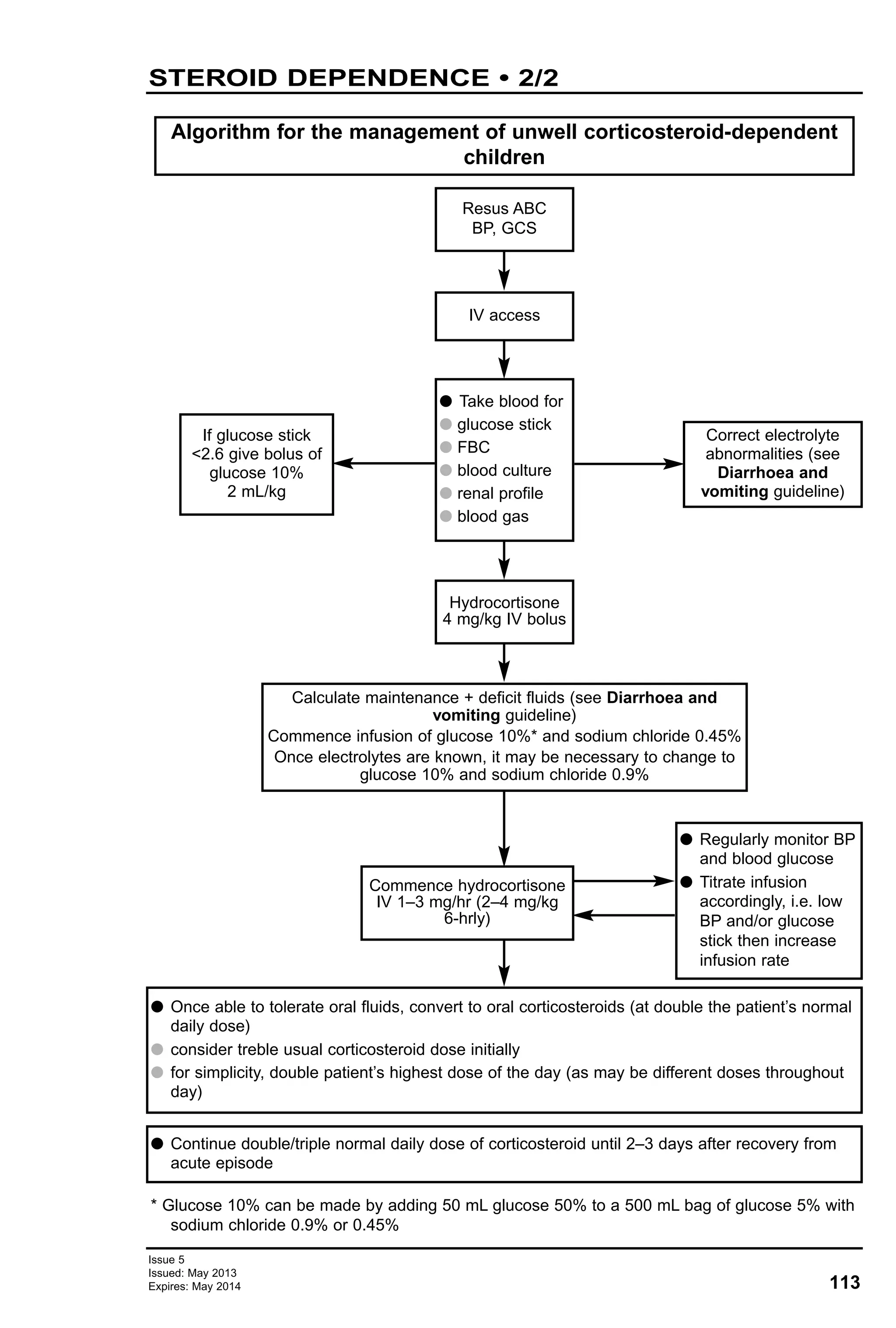
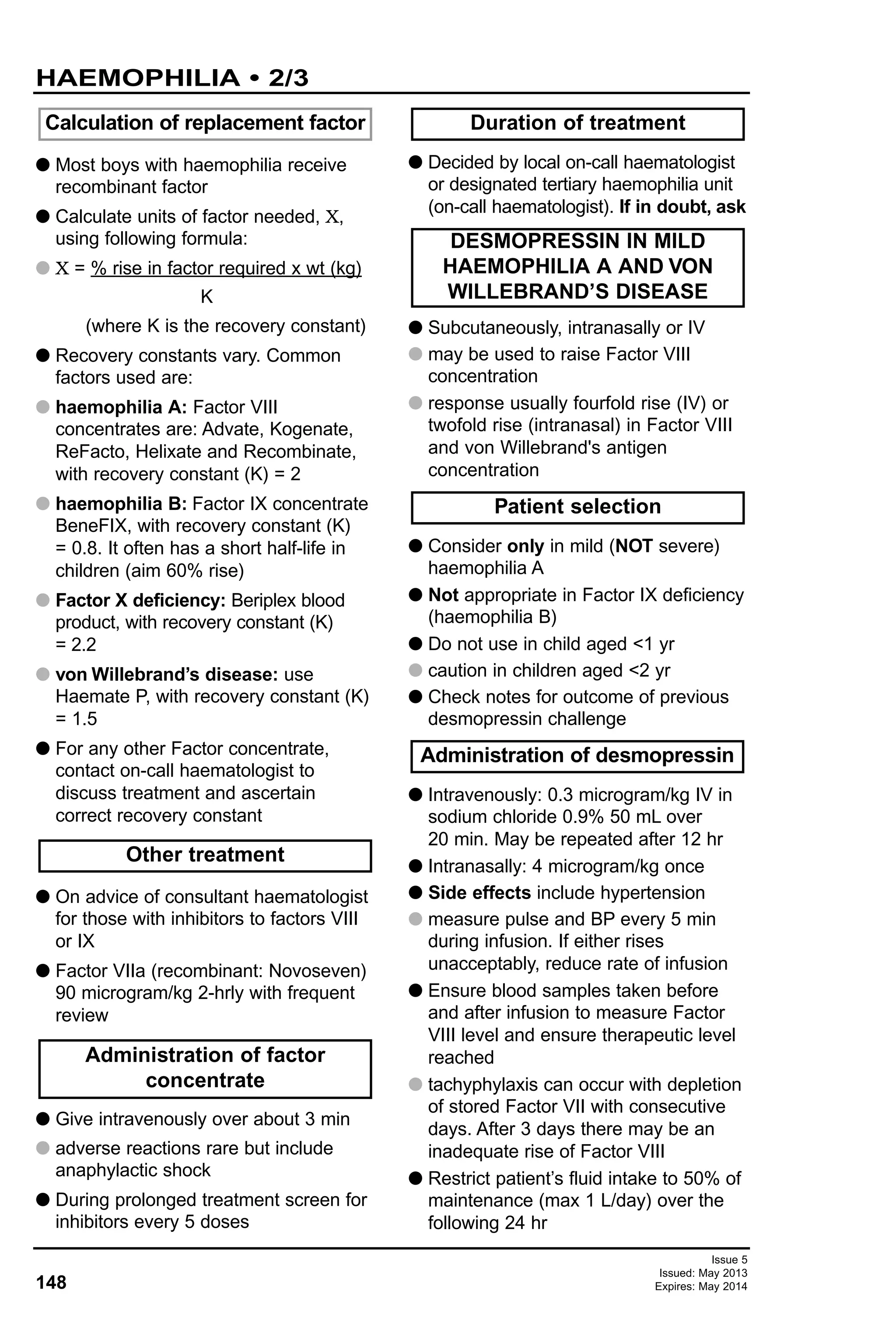
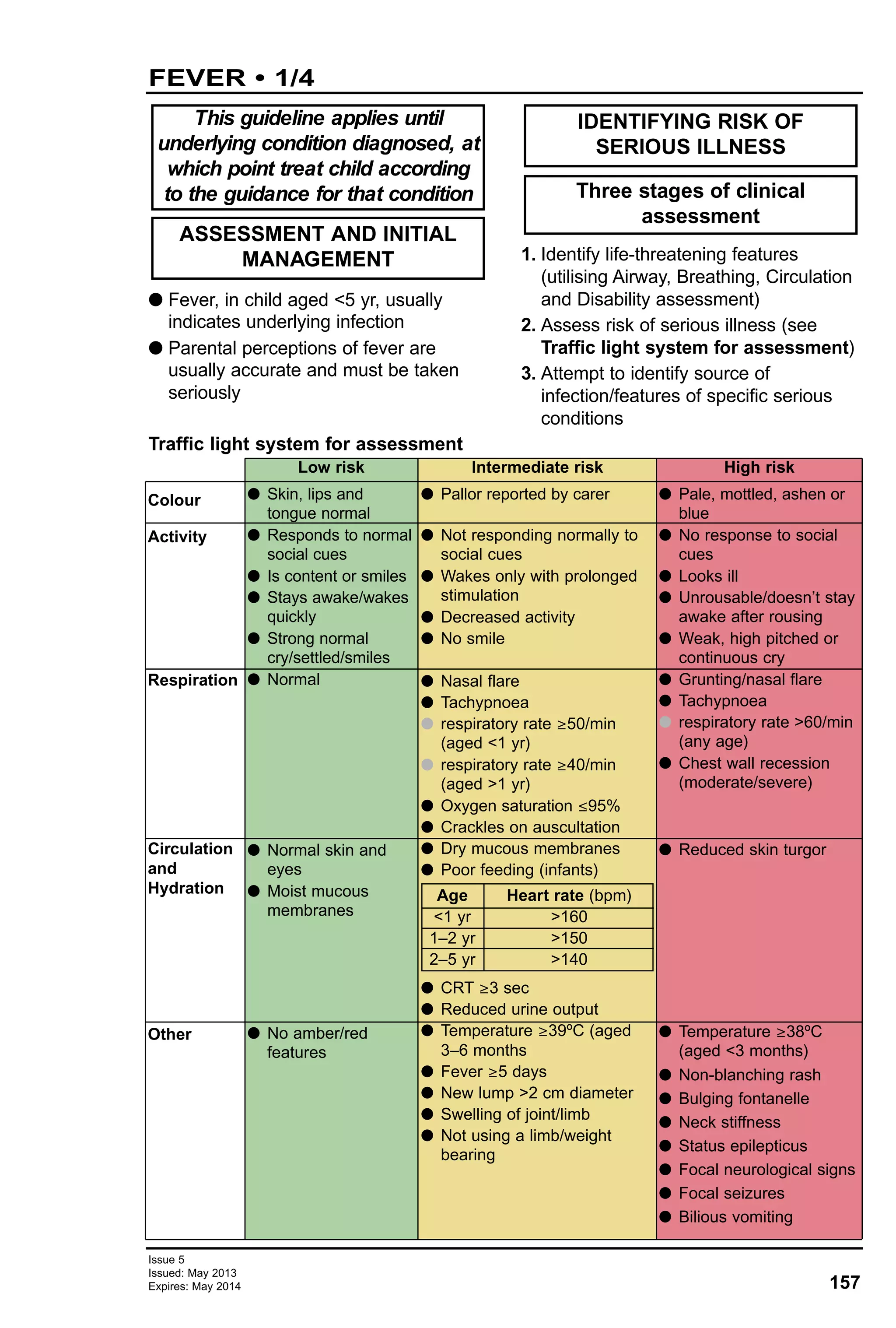
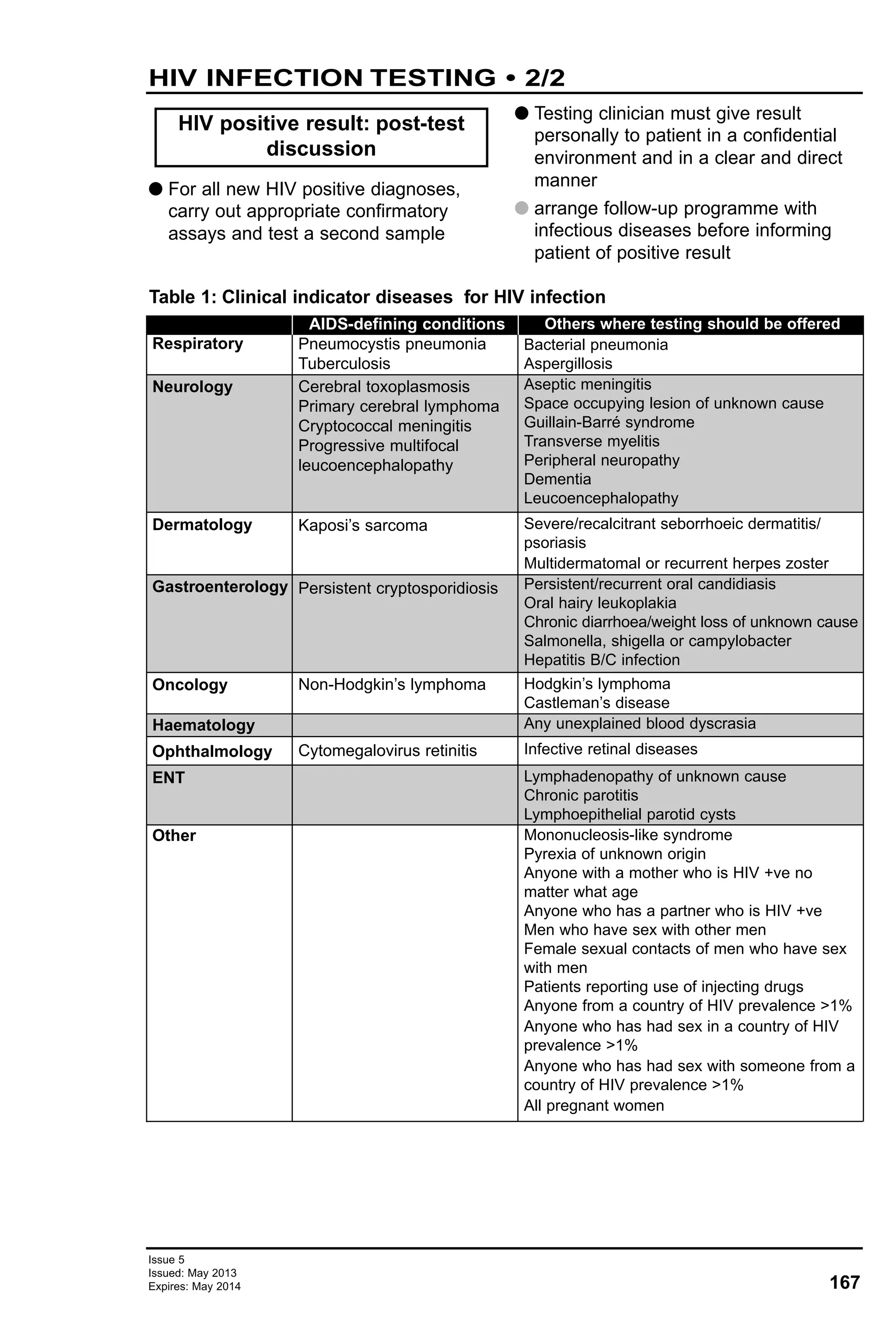
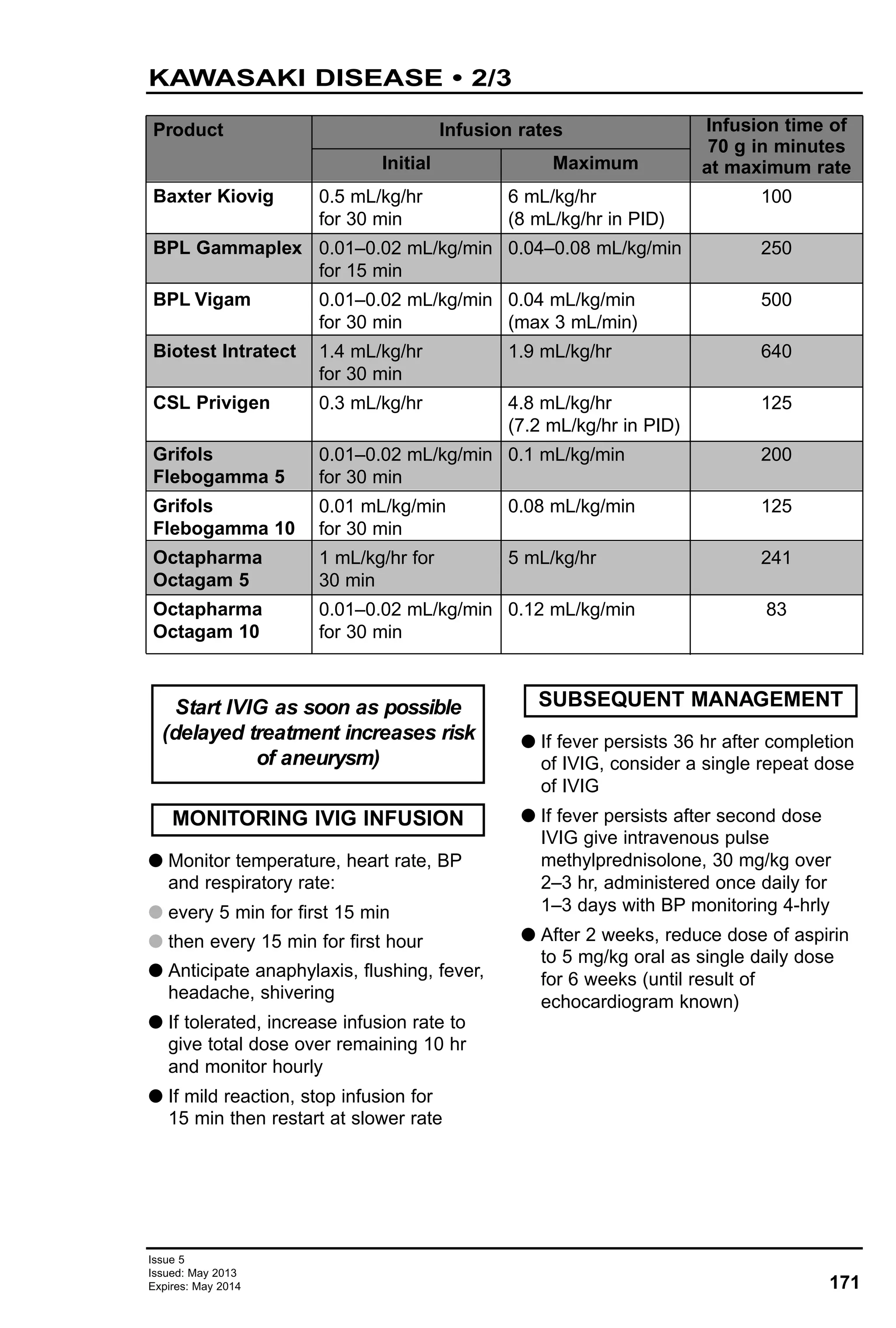
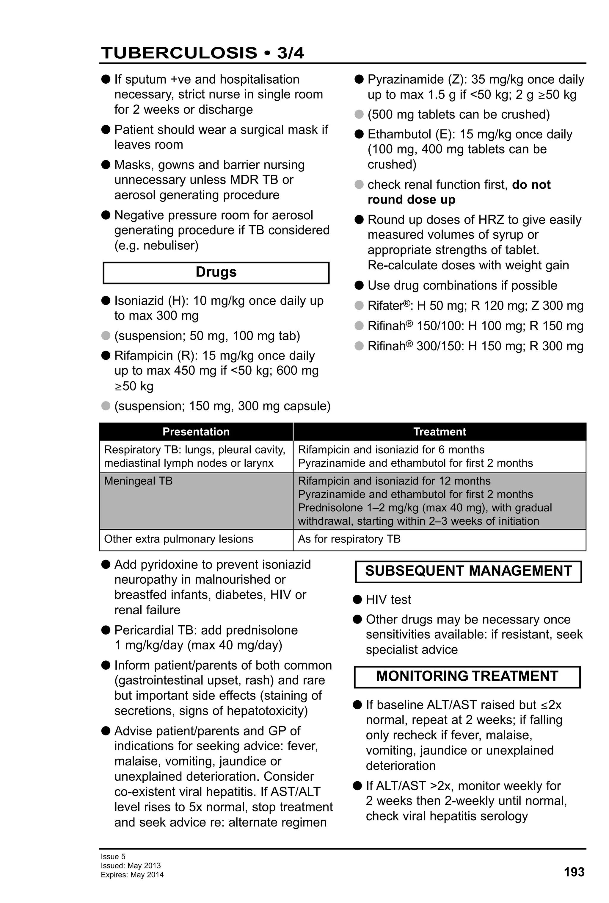
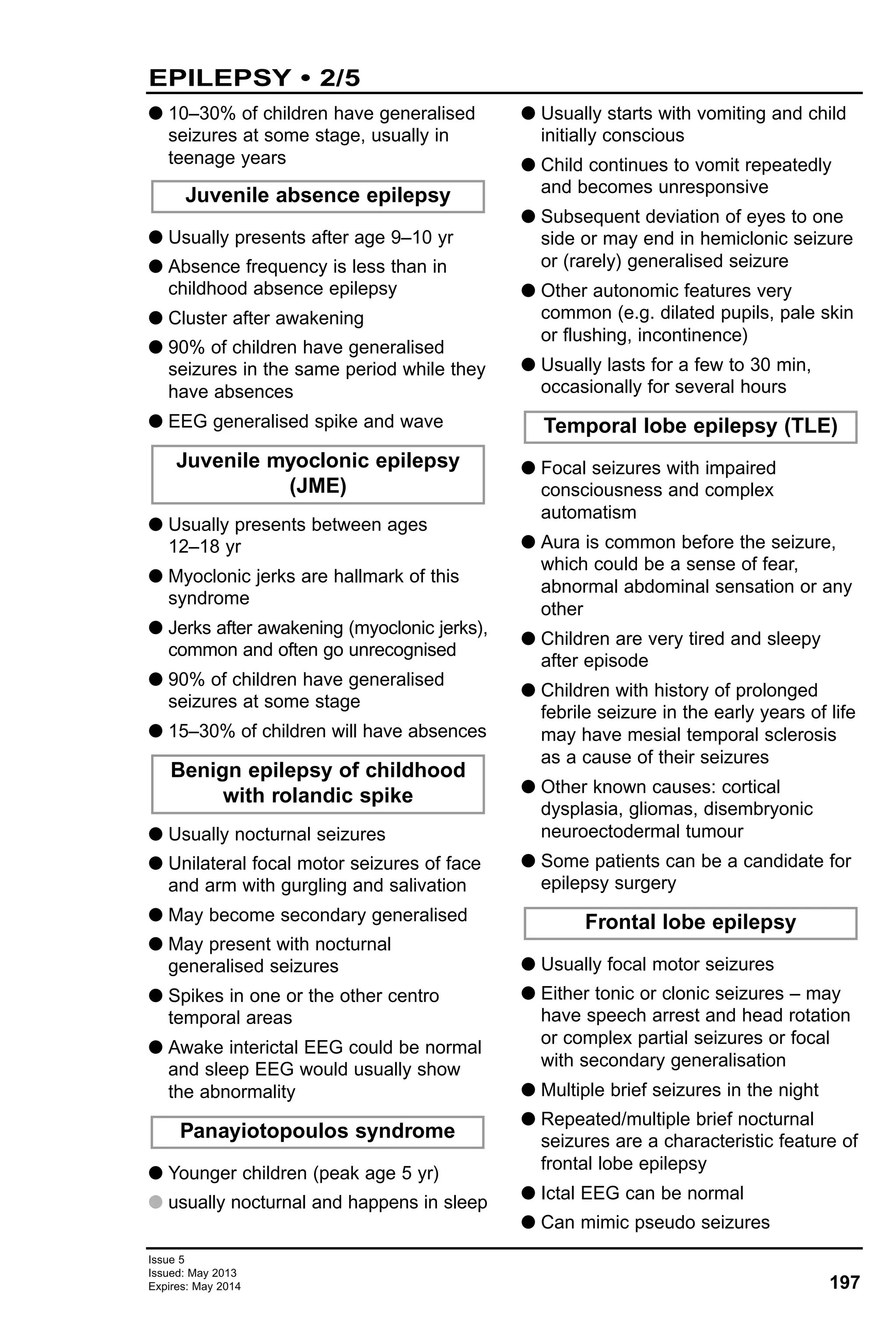
![G Effort of breathing
G respiratory rate
G recession
G use of accessory muscles
G additional sounds: stridor, wheeze,
grunting
G flaring of nostrils
G Efficacy of breathing
G chest movement and symmetry
G breath sounds
G SpO2 in air
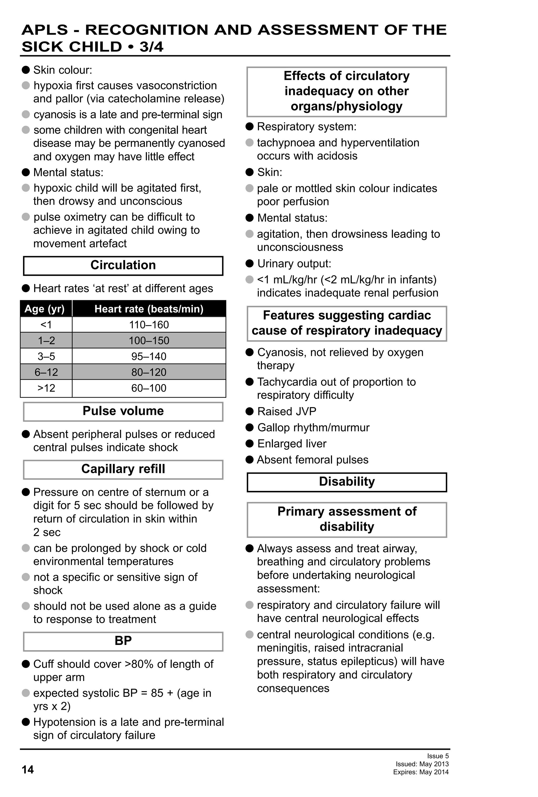
G Heart rate
G Pulse volume
G peripheral
G central (carotid/femoral)
G Blood pressure
G Capillary refill time
G Skin colour and temperature
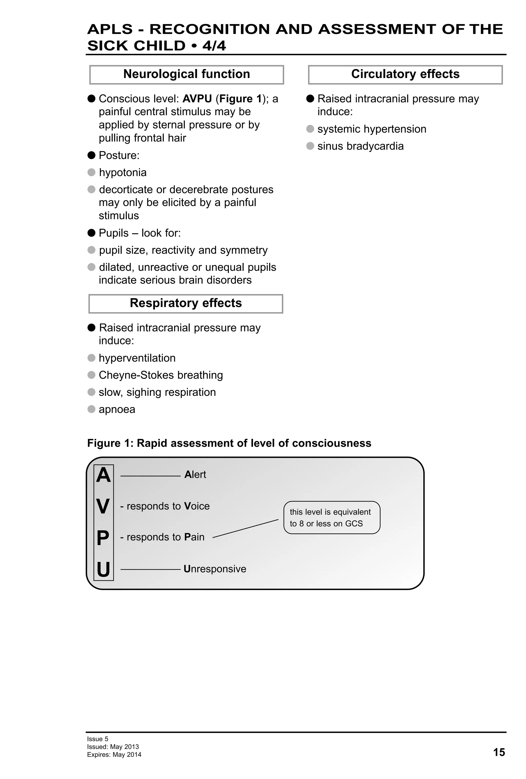
G Conscious level
G Posture
G Pupils
G Fever
G Skin rashes, bruising
G Glucose stix
G Complete assessment should take
<1 min
G Treat as problems are found
G Once airway (A), breathing (B) and
circulation (C) are clearly recognised
as being stable or have been
stabilised, definitive management of
underlying condition can proceed
G Reassessment of ABCDE at frequent
intervals necessary to assess progress
and detect deterioration
G Hypoglycaemia: glucose 10% 2 mL/kg
followed by IV glucose infusion
G Give clear explanations to parents and
child
G Allow and encourage parents to
remain with child at all times
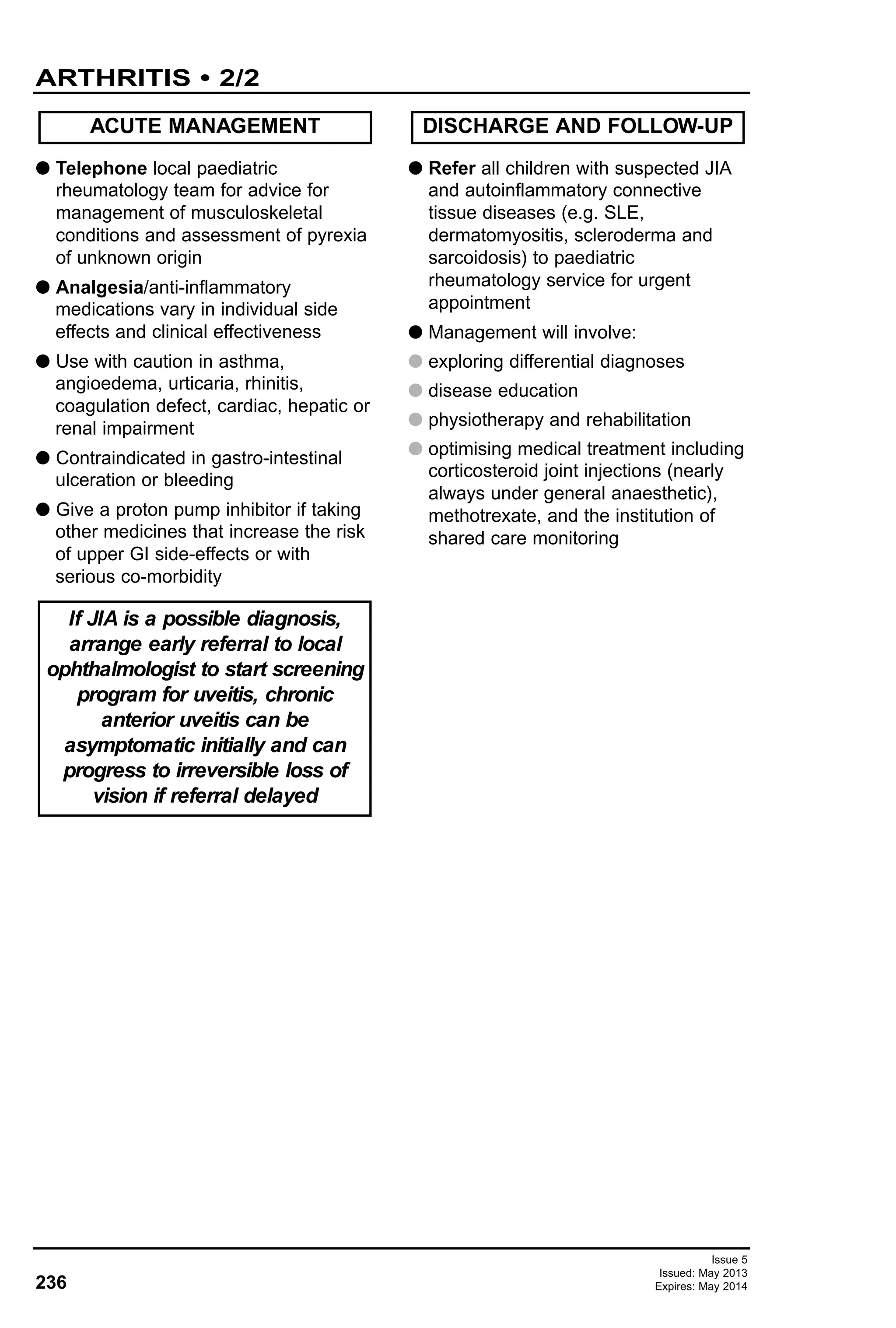
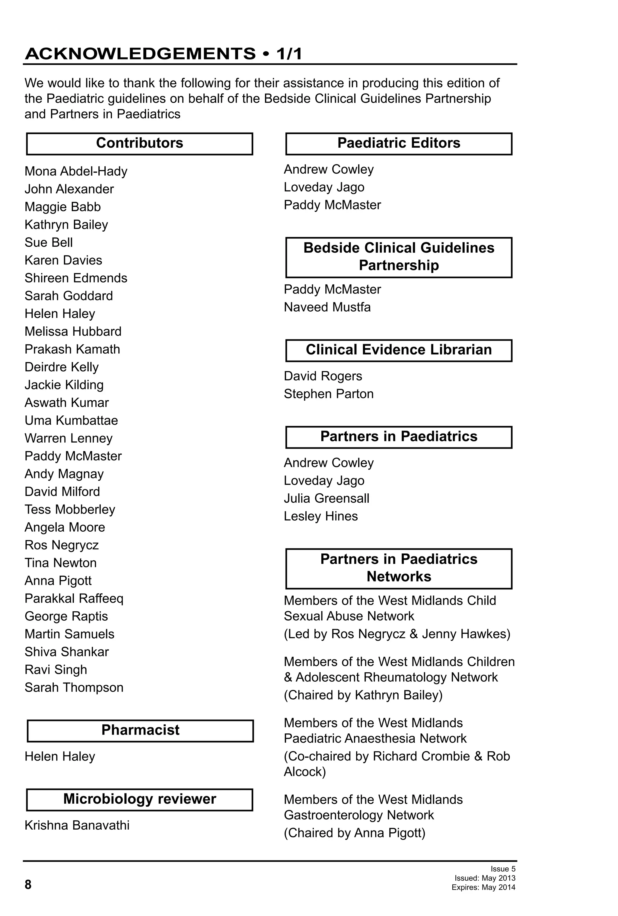
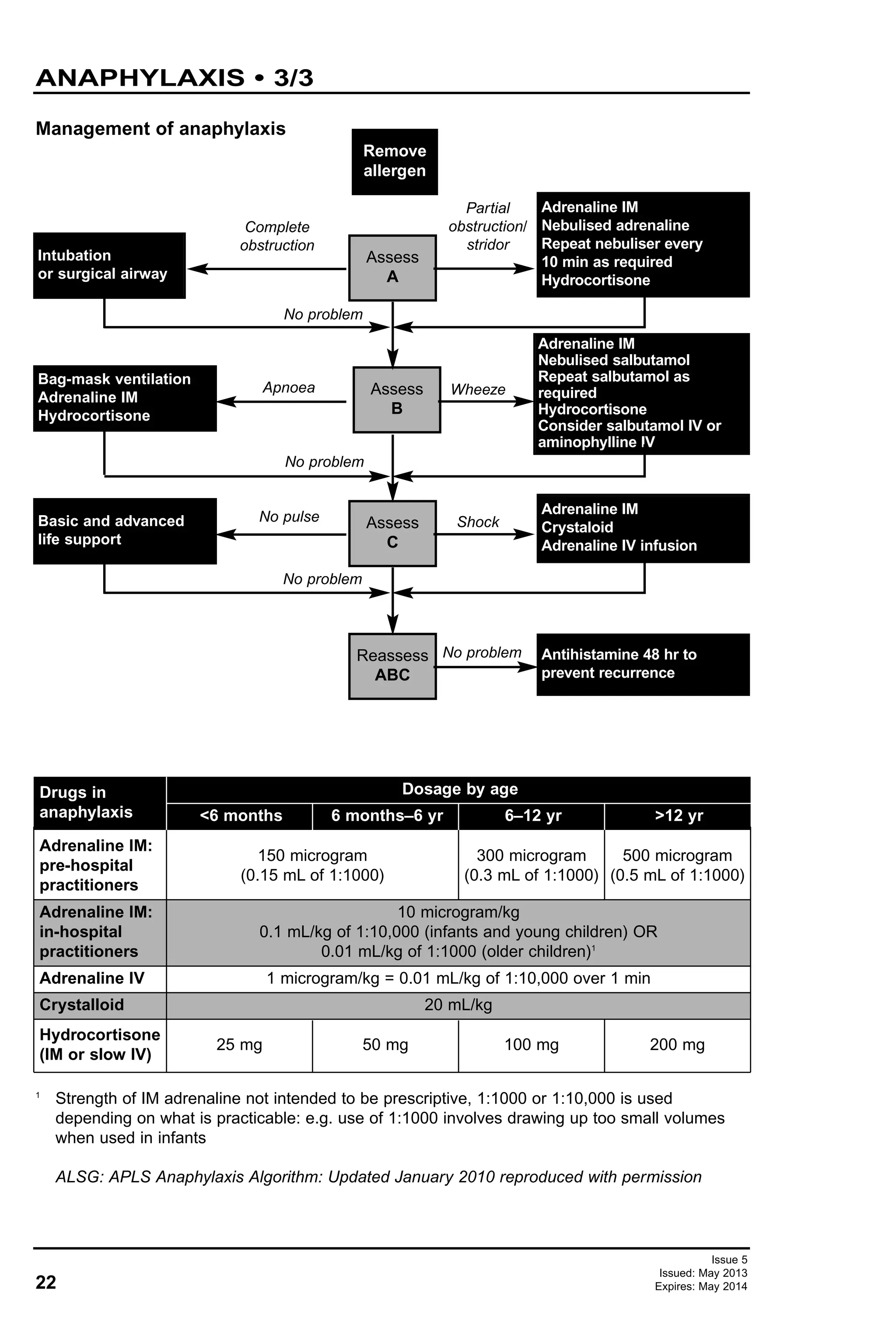
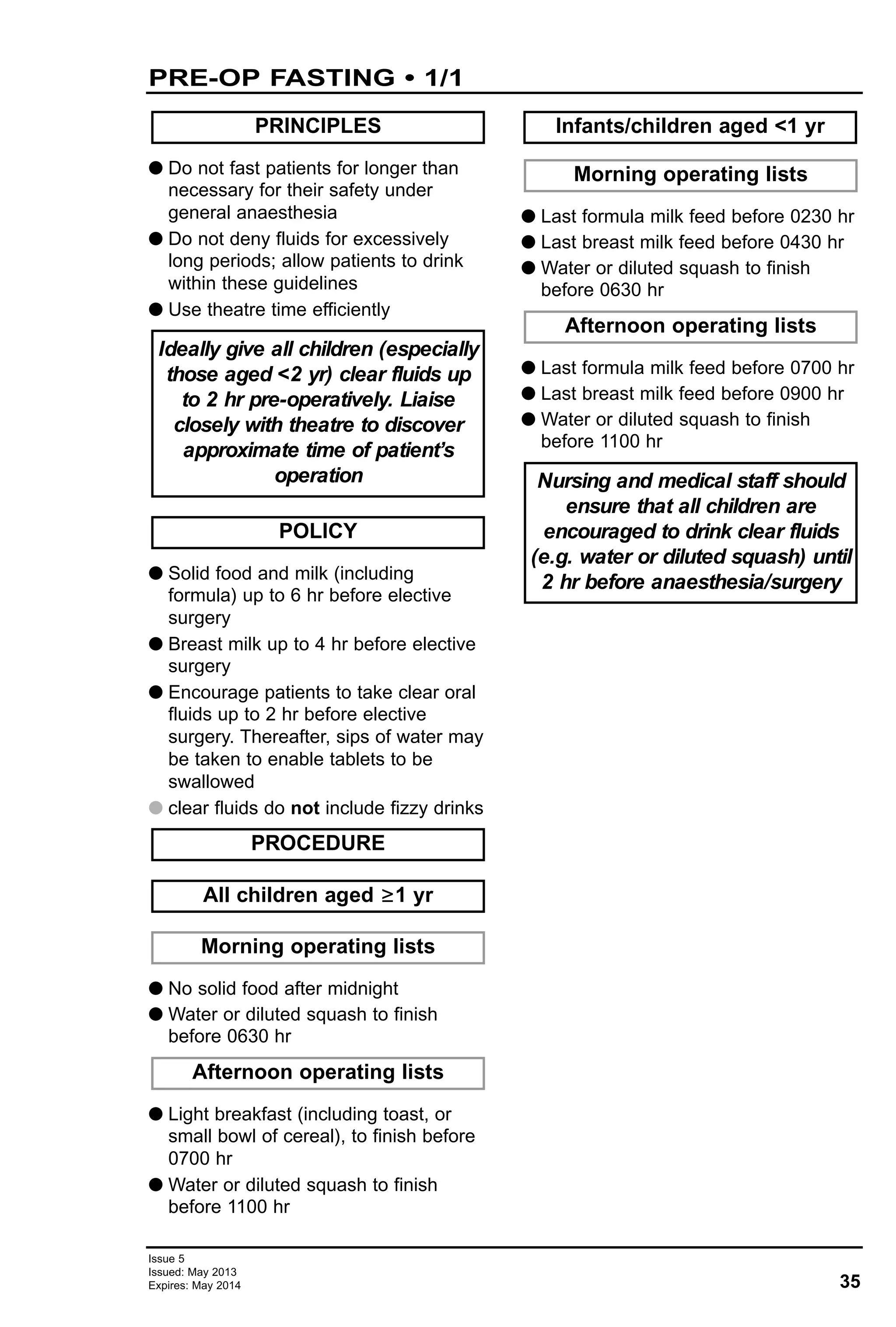
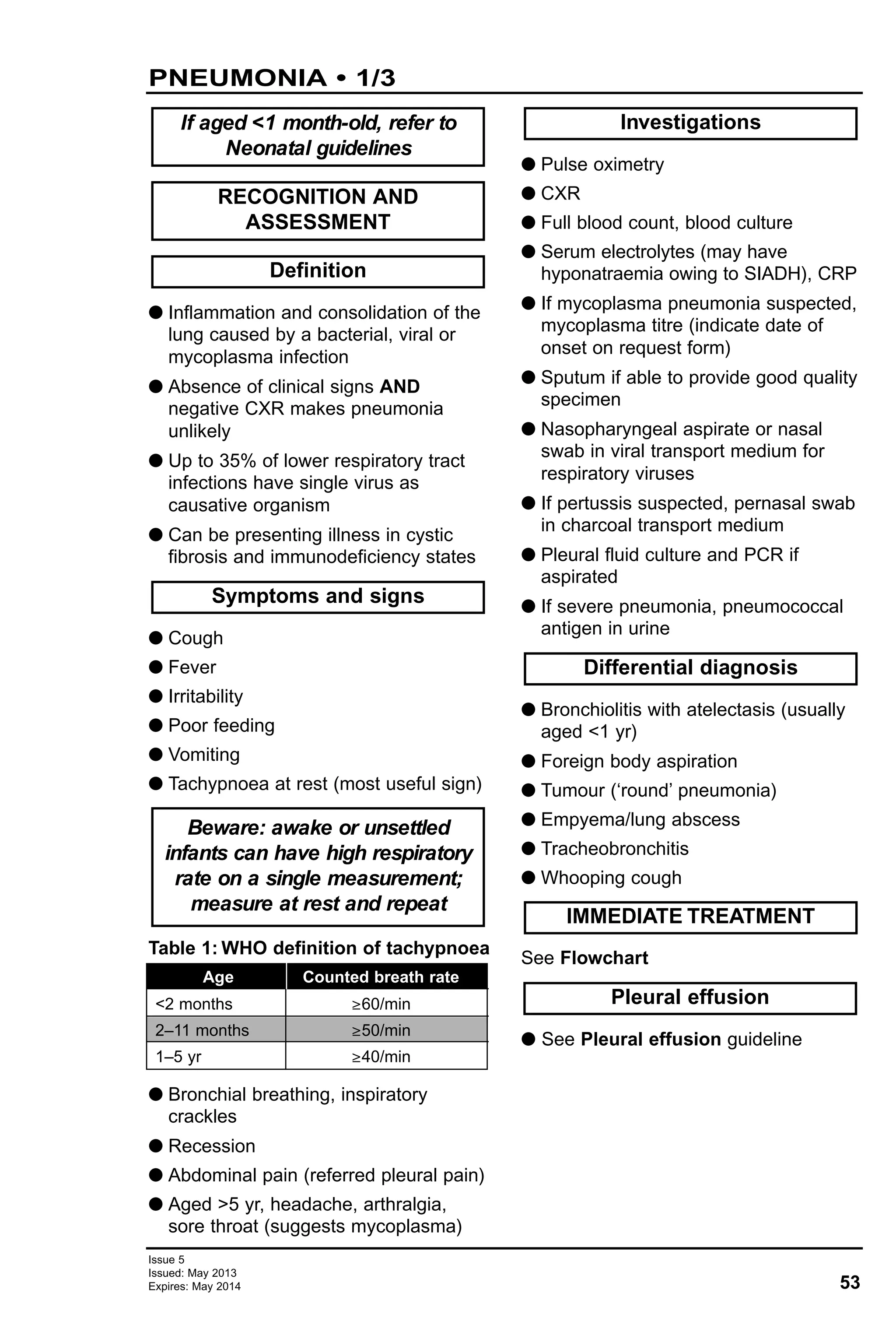
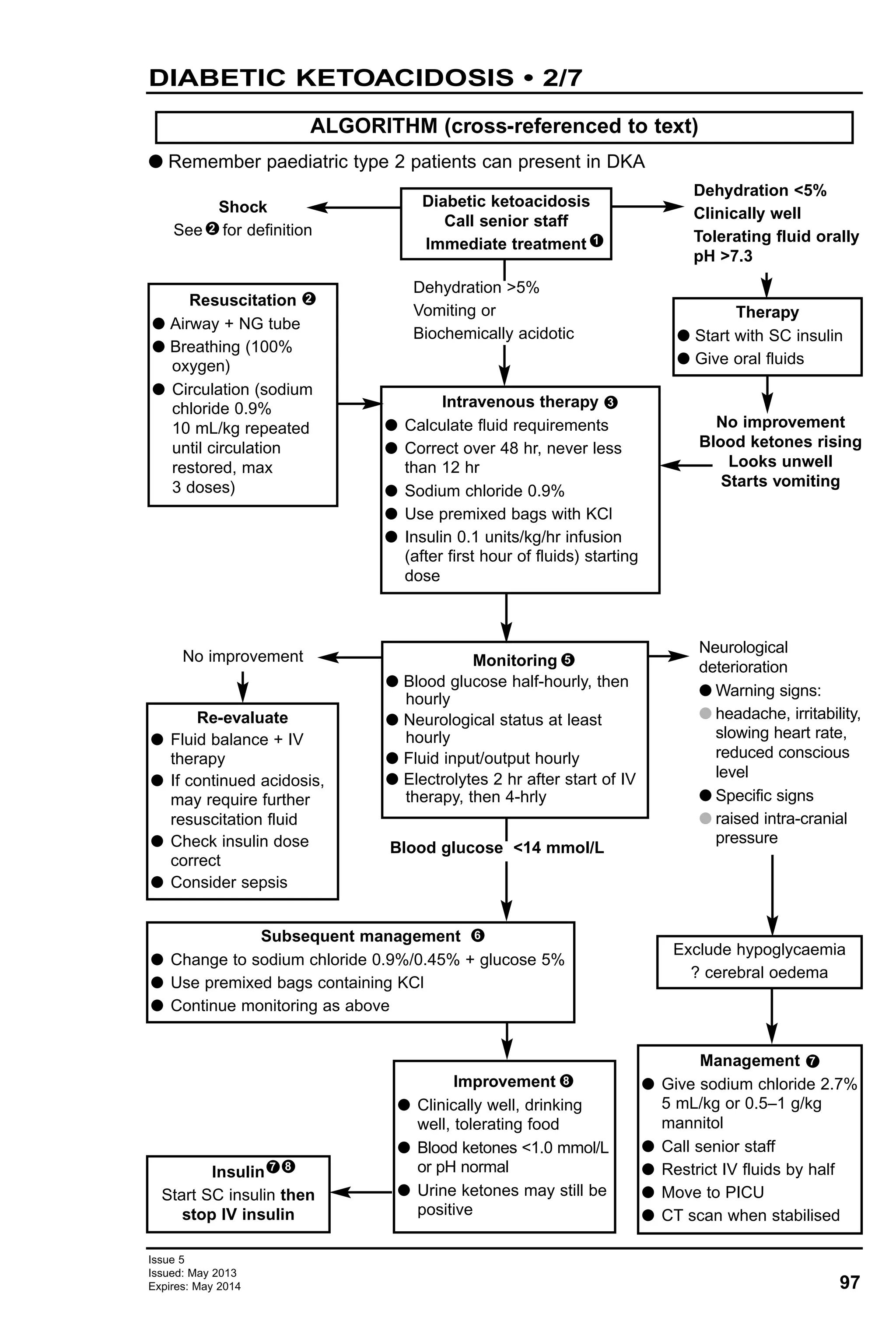
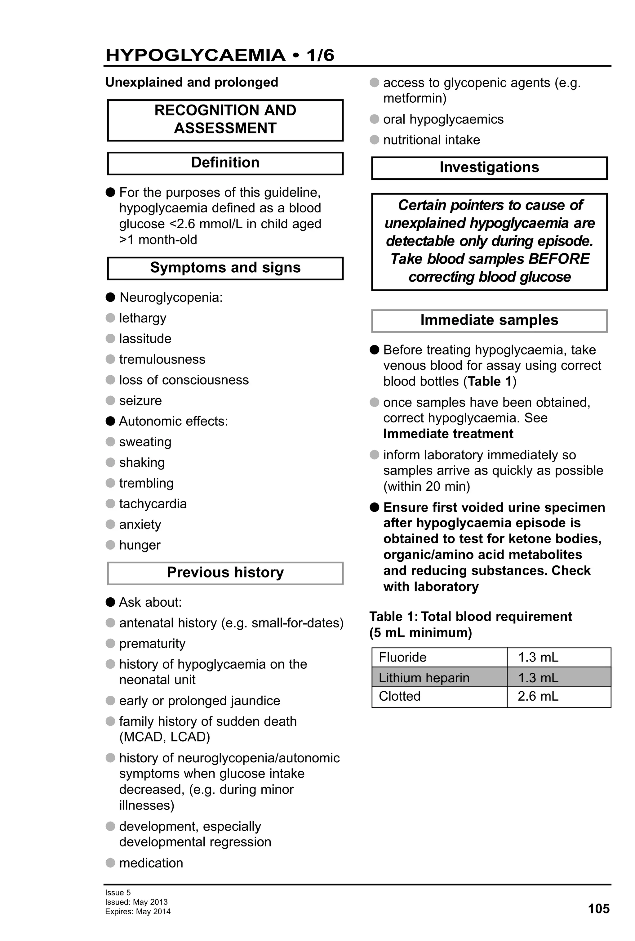
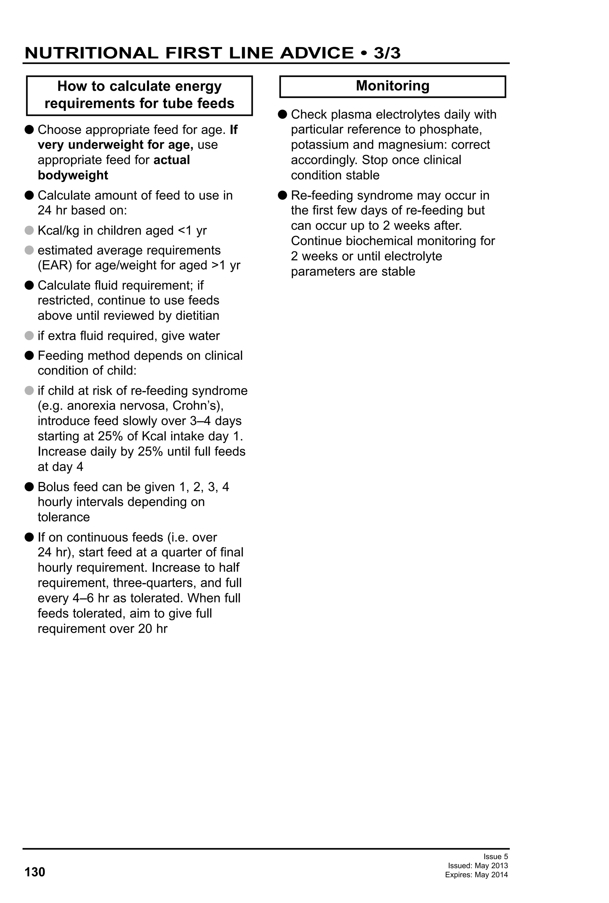
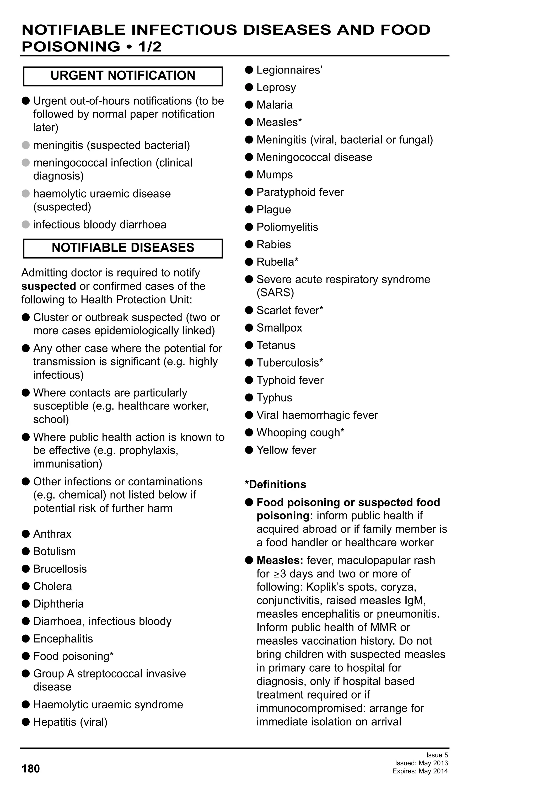
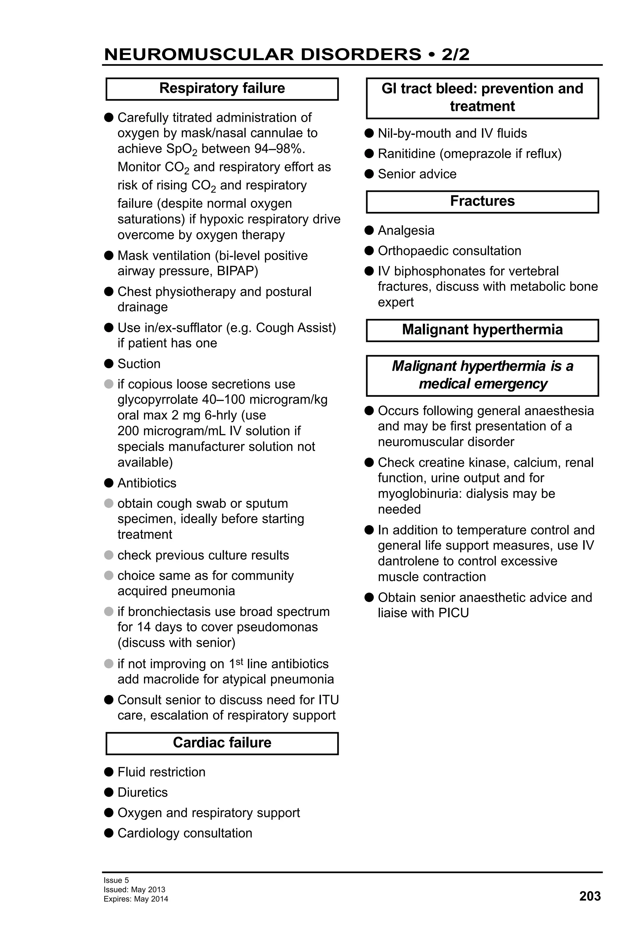
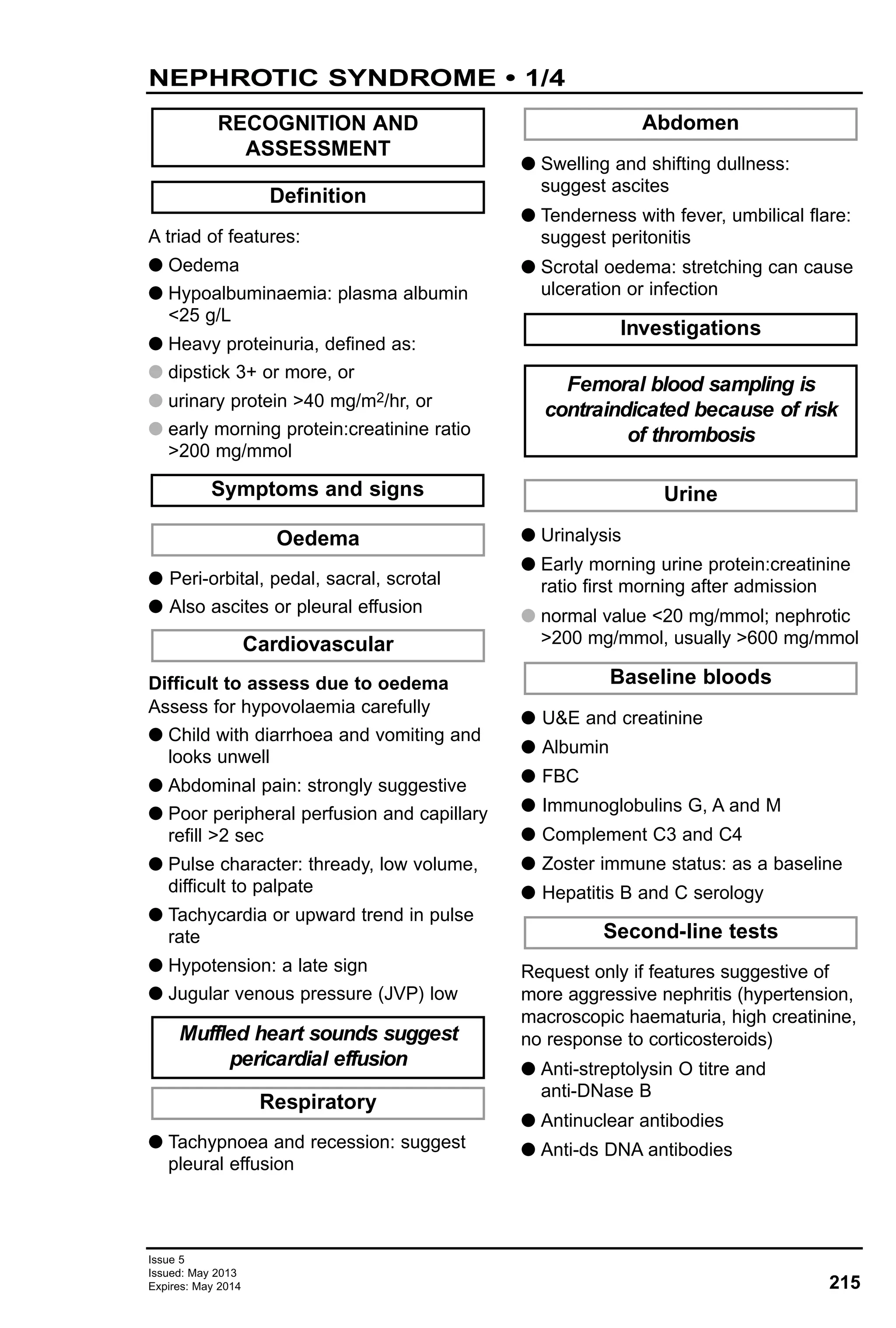
Anticipated weight in relation to age
Weight can be estimated using following
formulae:
G 0–12 months: wt (kg) = [age (m) / 2] + 4
G 1–6 years: wt (kg) = [age (y) + 4] x 2
G 7–14 years: wt (kg) = [age (y) x 3] + 7
G Vocalisations (e.g. crying or talking)
indicate ventilation and some degree
of airway patency
G Assess patency by:
G looking for chest and/or abdominal
movement
G listening for breath sounds
G feeling for expired air
Primary assessment of airway
Airway
Weight
RECOGNITION AND
ASSESSMENT OF THE SICK
CHILD
CHILD AND PARENTS
Assessment
Actions
Don’t Ever Forget Glucose
(DEFG)
Exposure (E)
Disability (D)
Circulation (C)
Airway (A) and Breathing (B)
SUMMARY OF RAPID CLINICAL
ASSESSMENT
12
APLS - RECOGNITION AND ASSESSMENT OF THE
SICK CHILD • 1/4
Issue 5
Issued: May 2013
Expires: May 2014
Age Weight (kg)
Birth 3.5
5 months 7
1 yr 10](https://image.slidesharecdn.com/paediatricguidelines2013-14withlinks-180801135506/75/Paediatric-guidelines-2013-14-with-links-12-2048.jpg)
![24
Issue 5
Issued: May 2013
Expires: May 2014
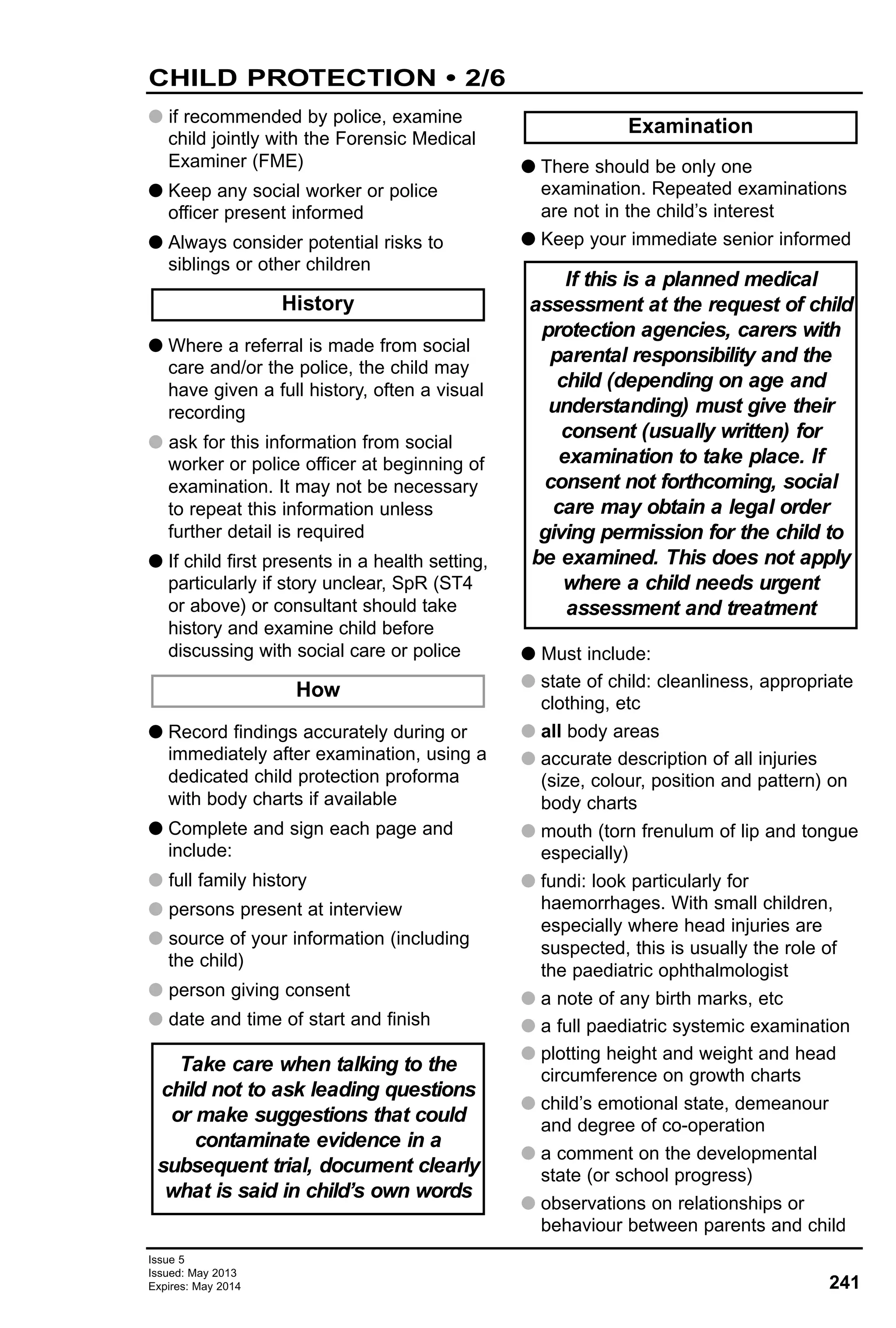
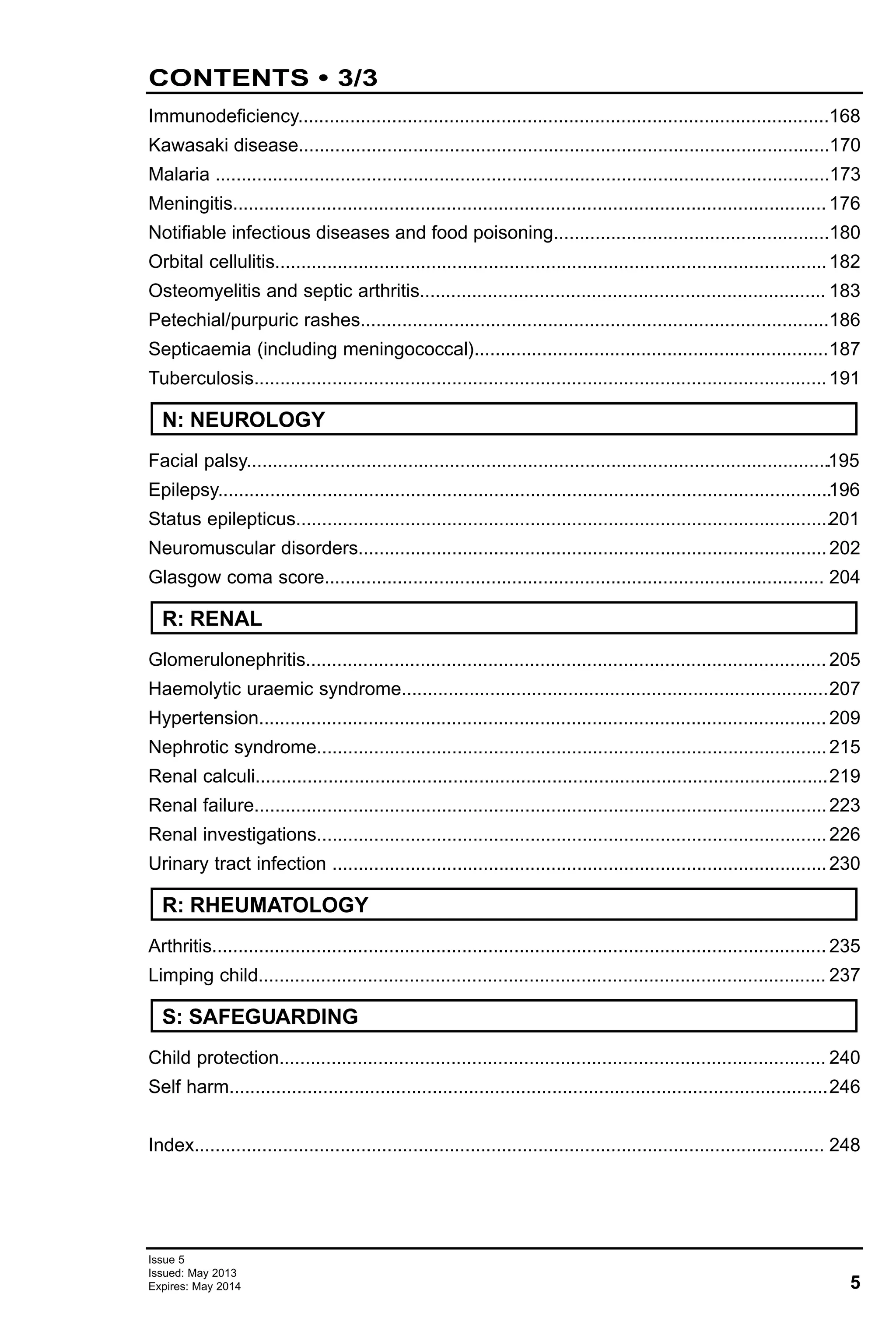
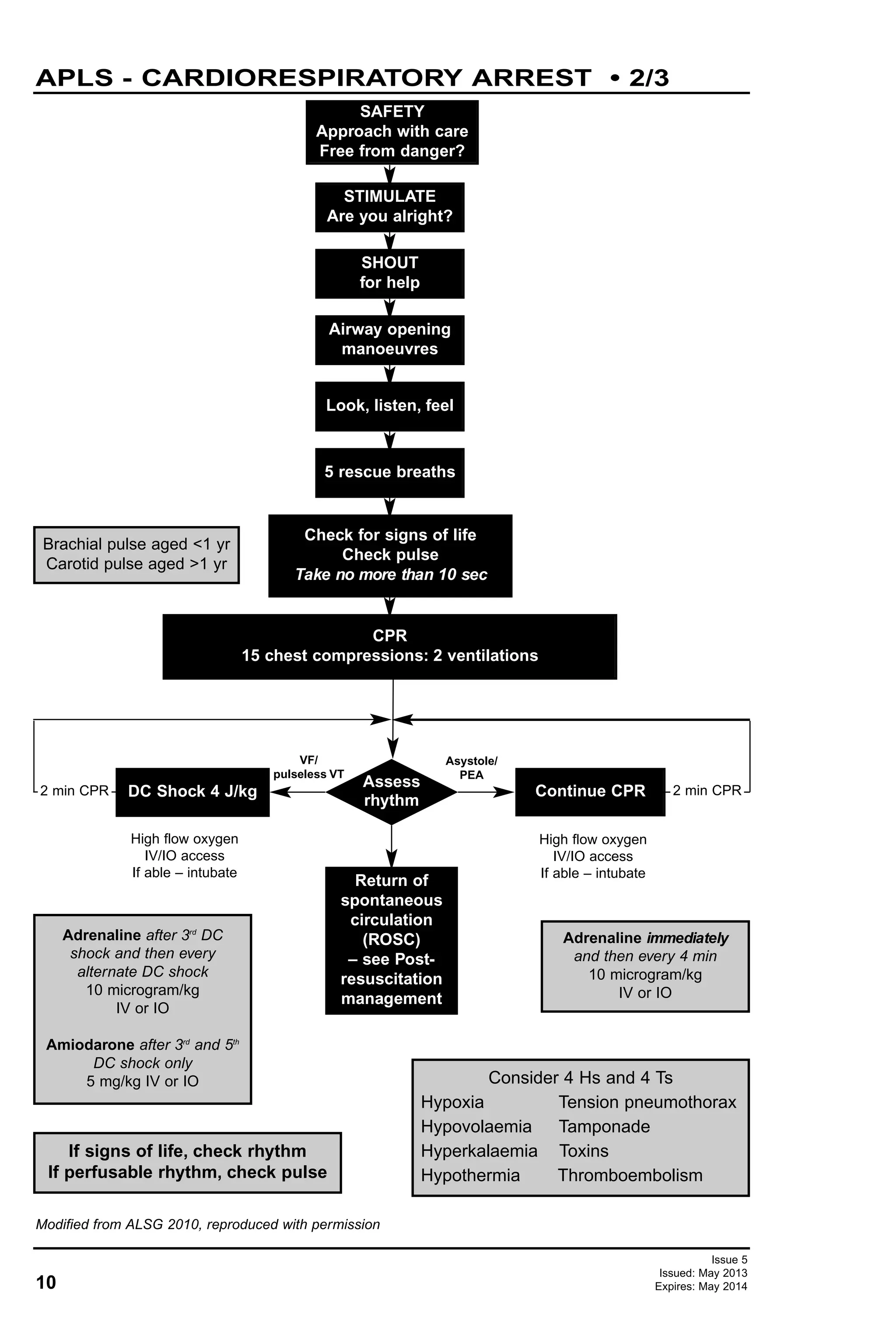
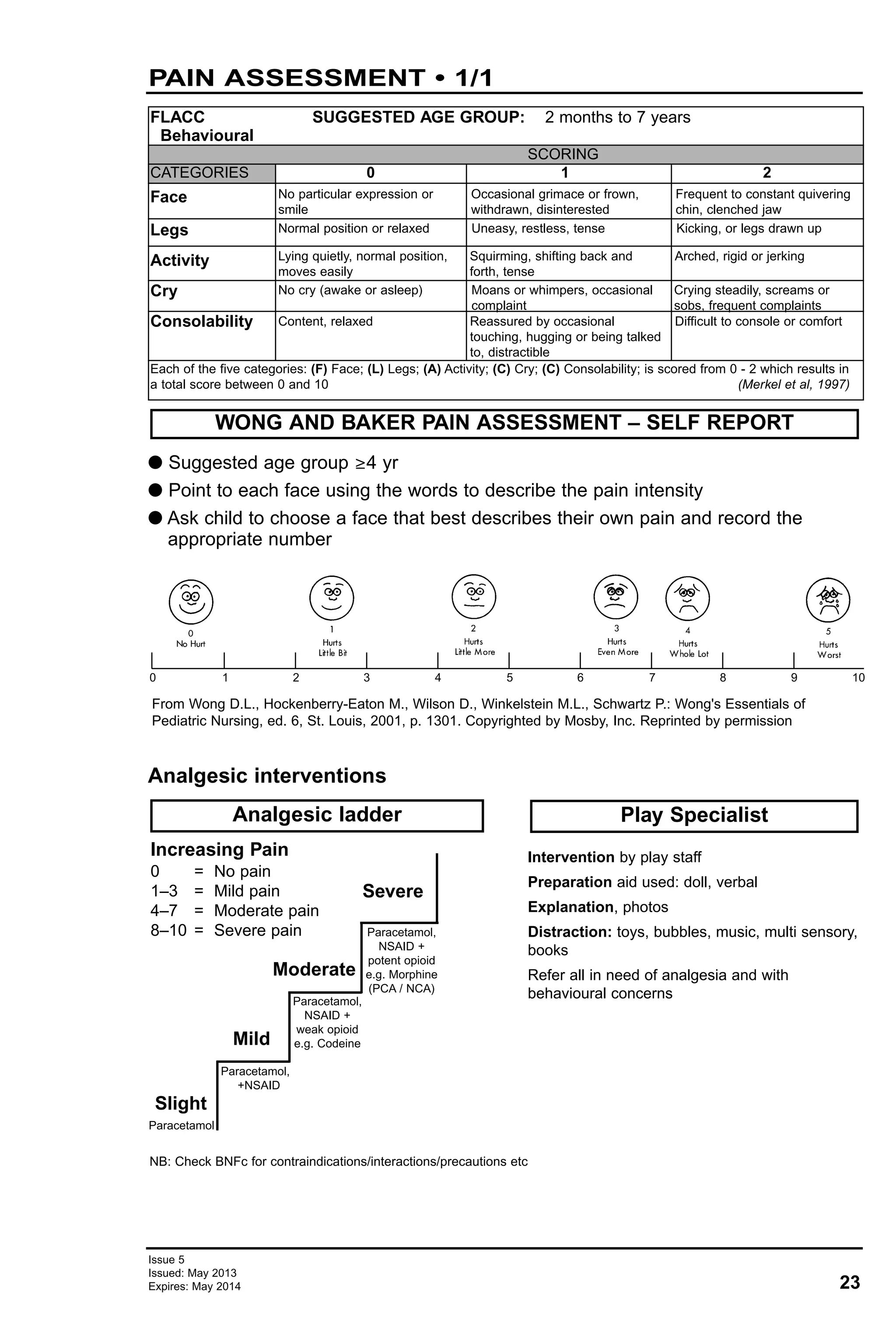
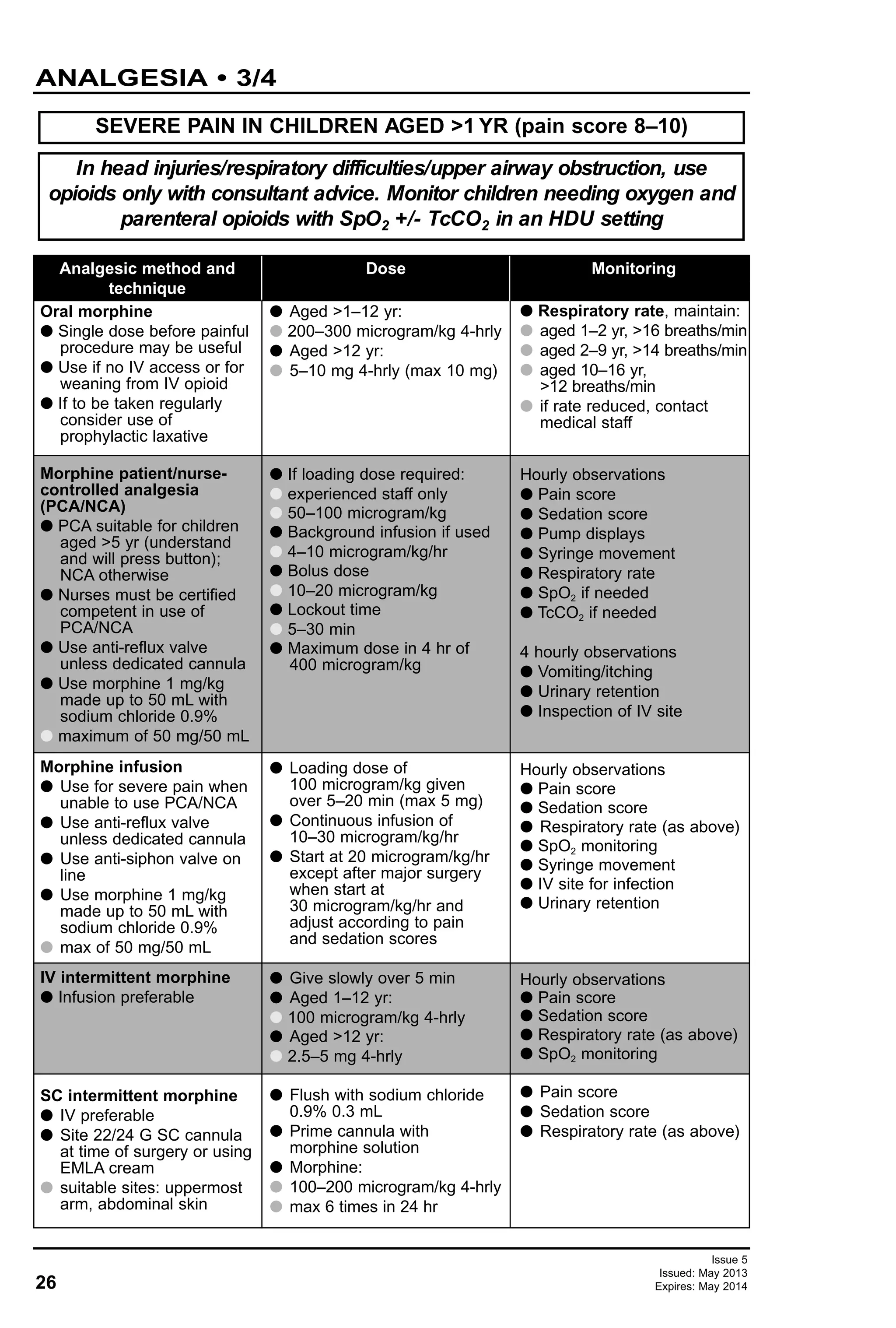
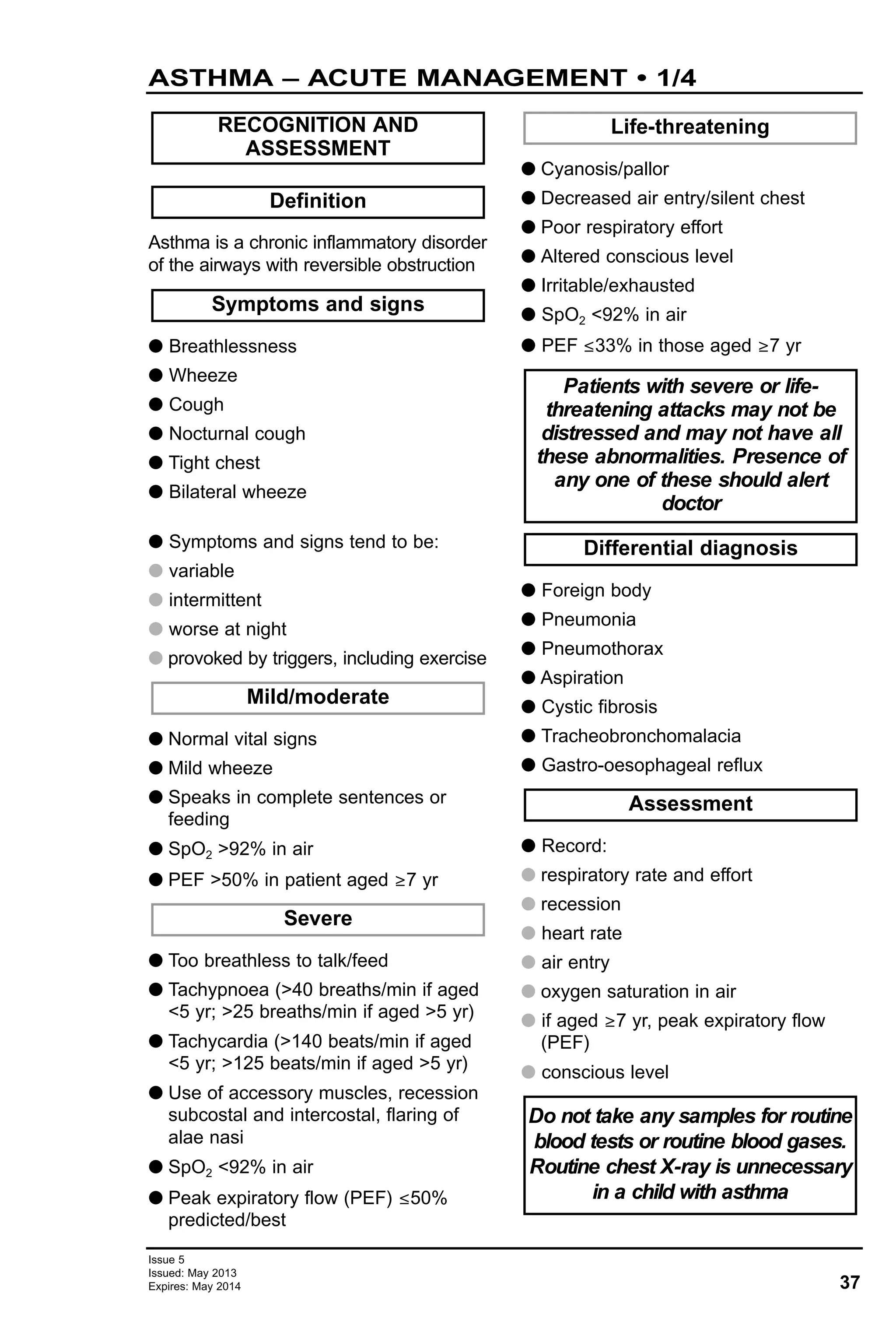
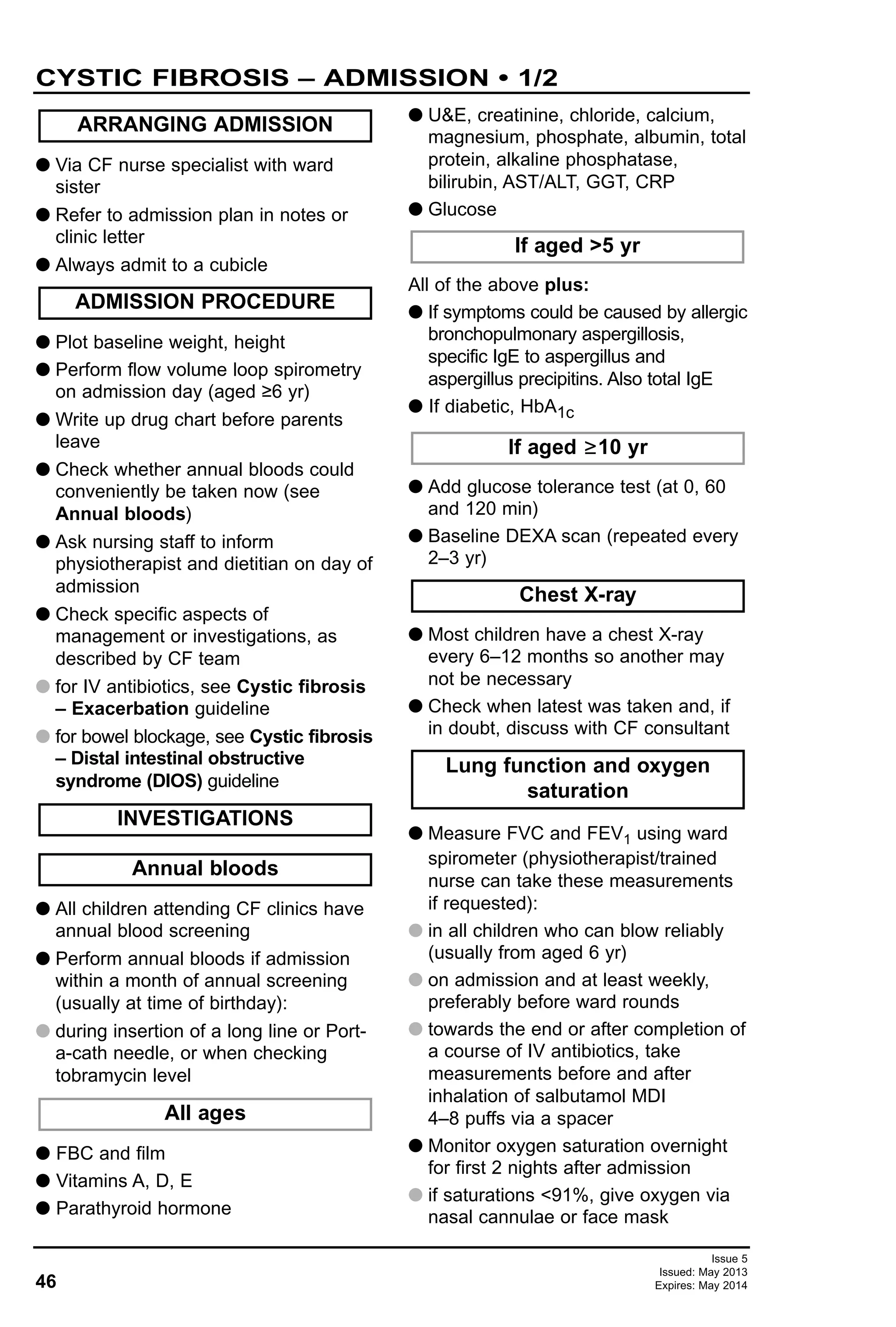
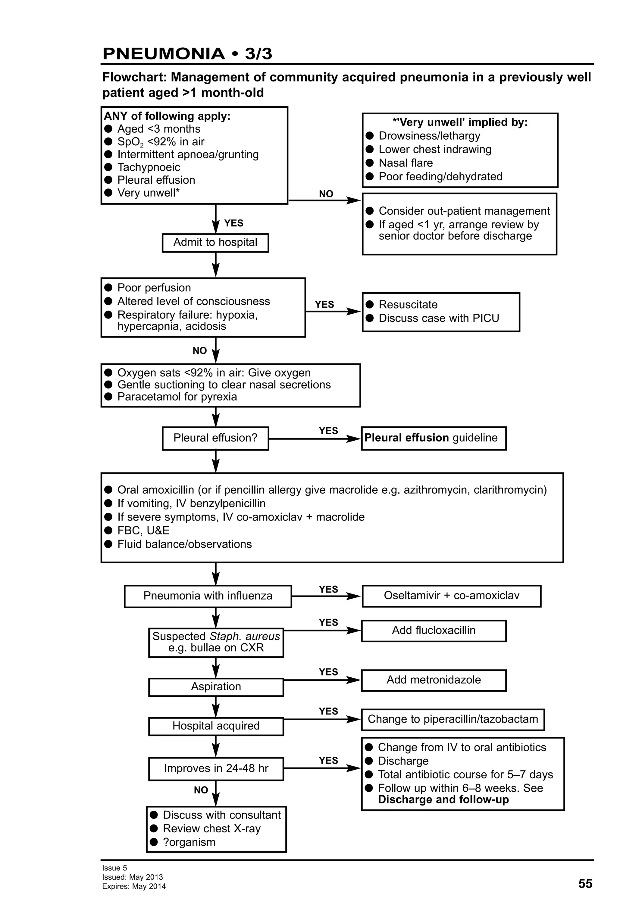
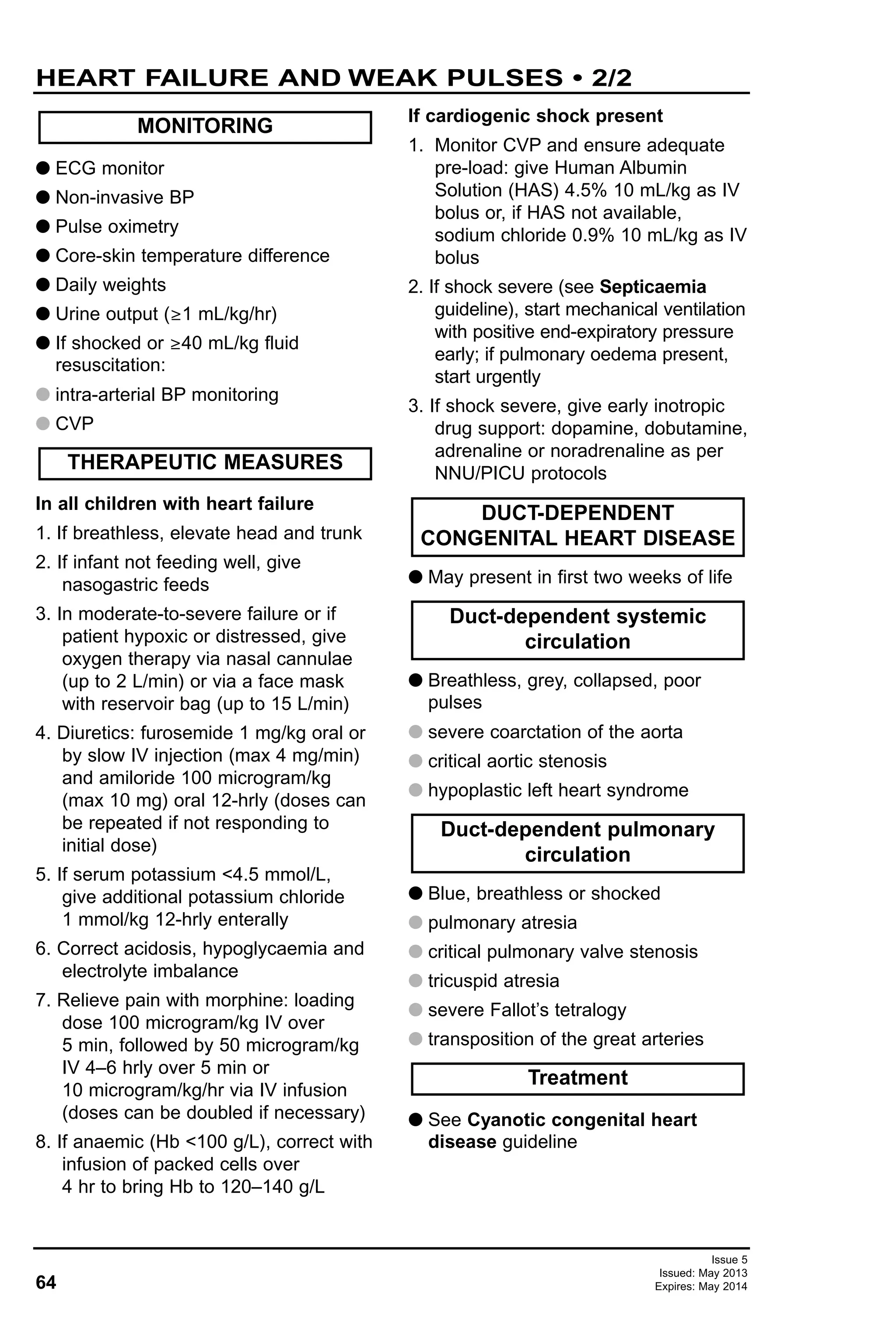
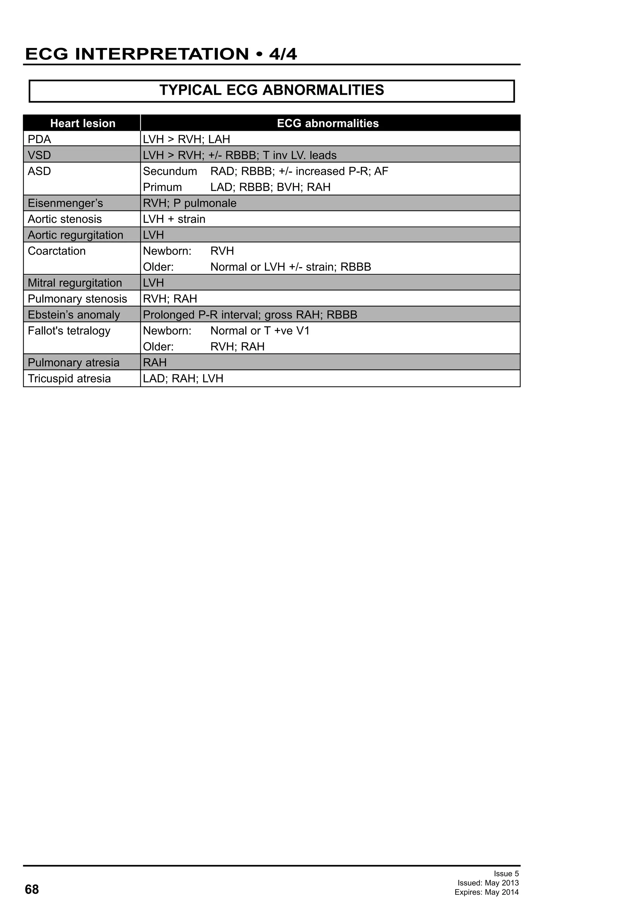
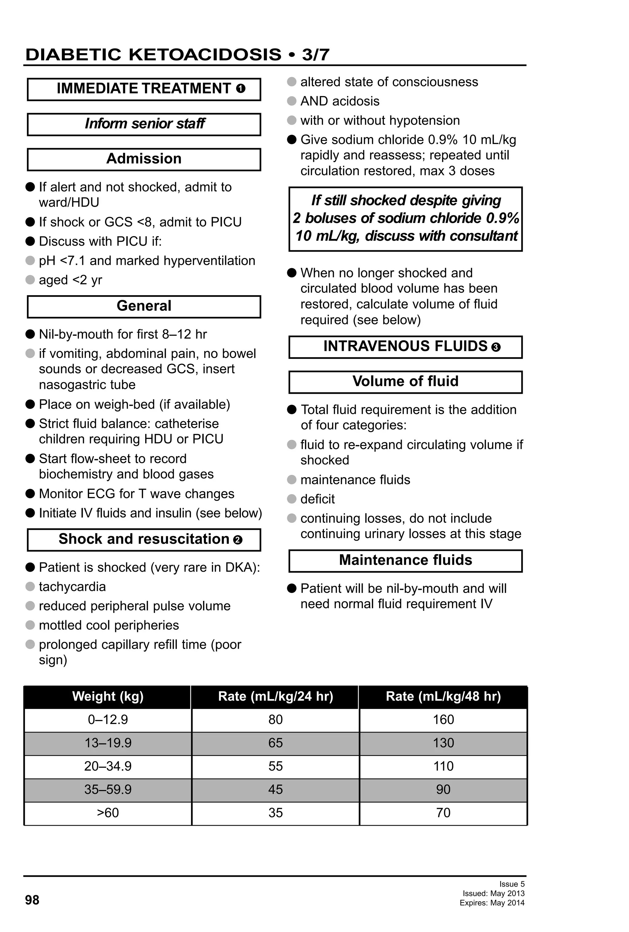
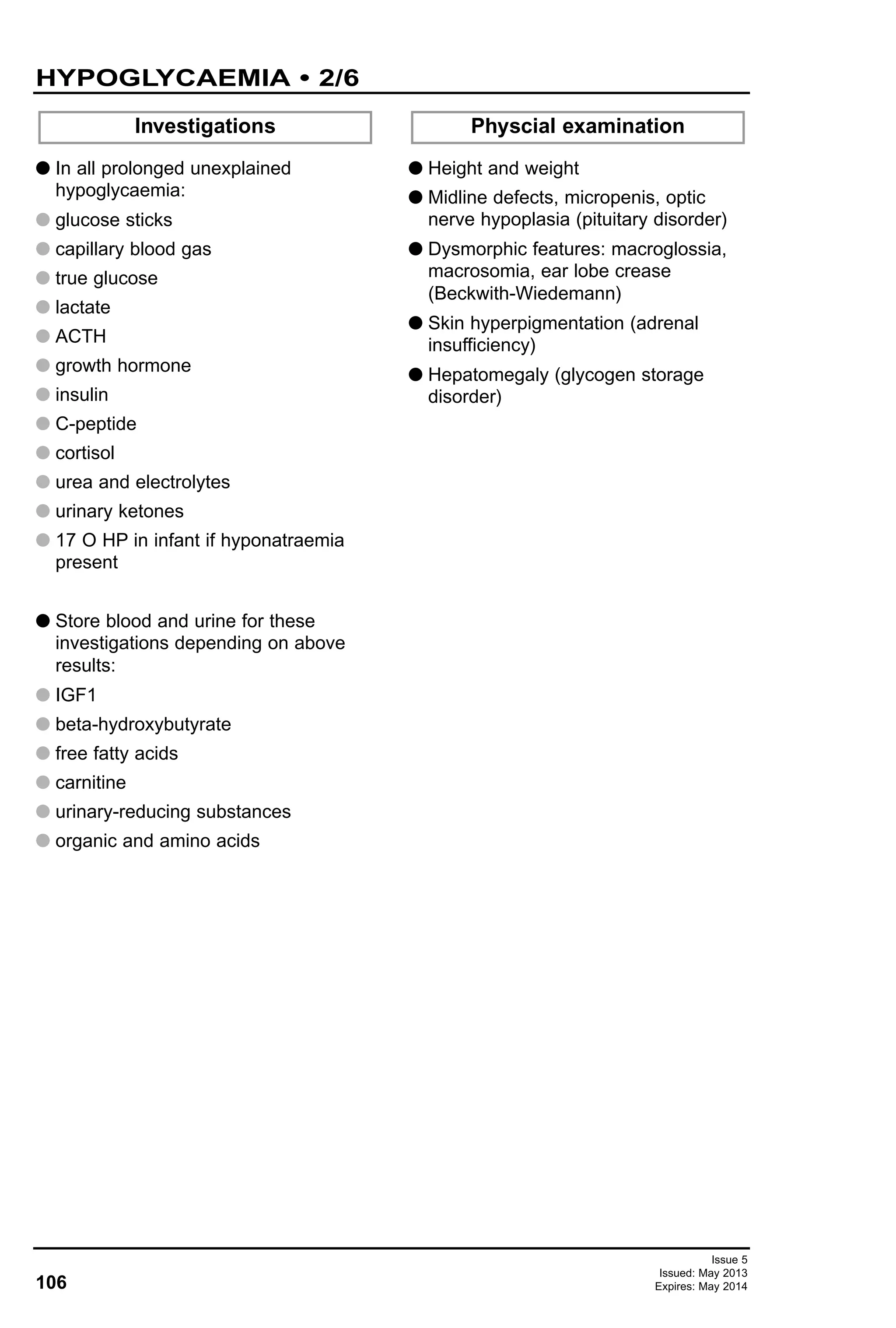
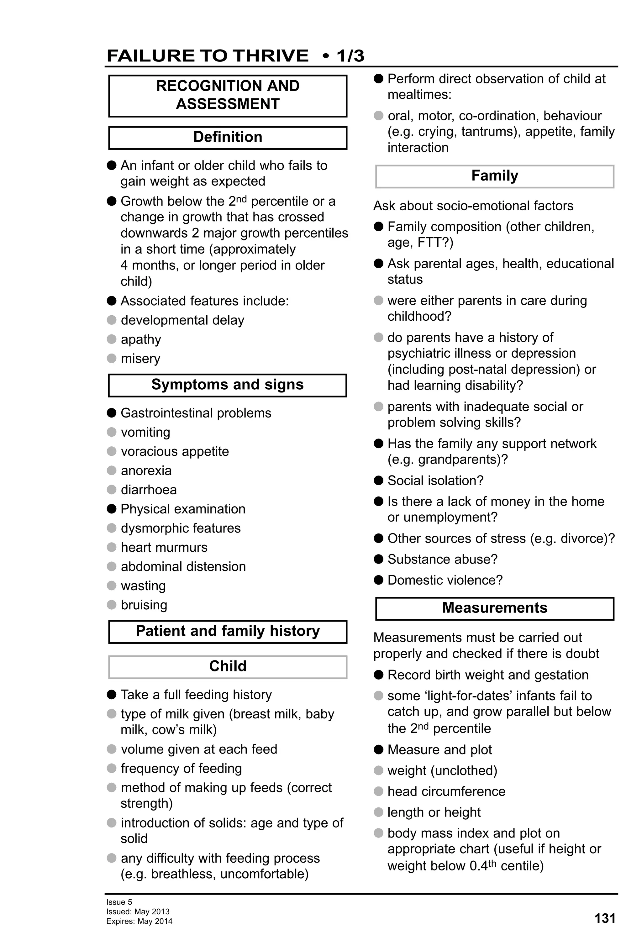
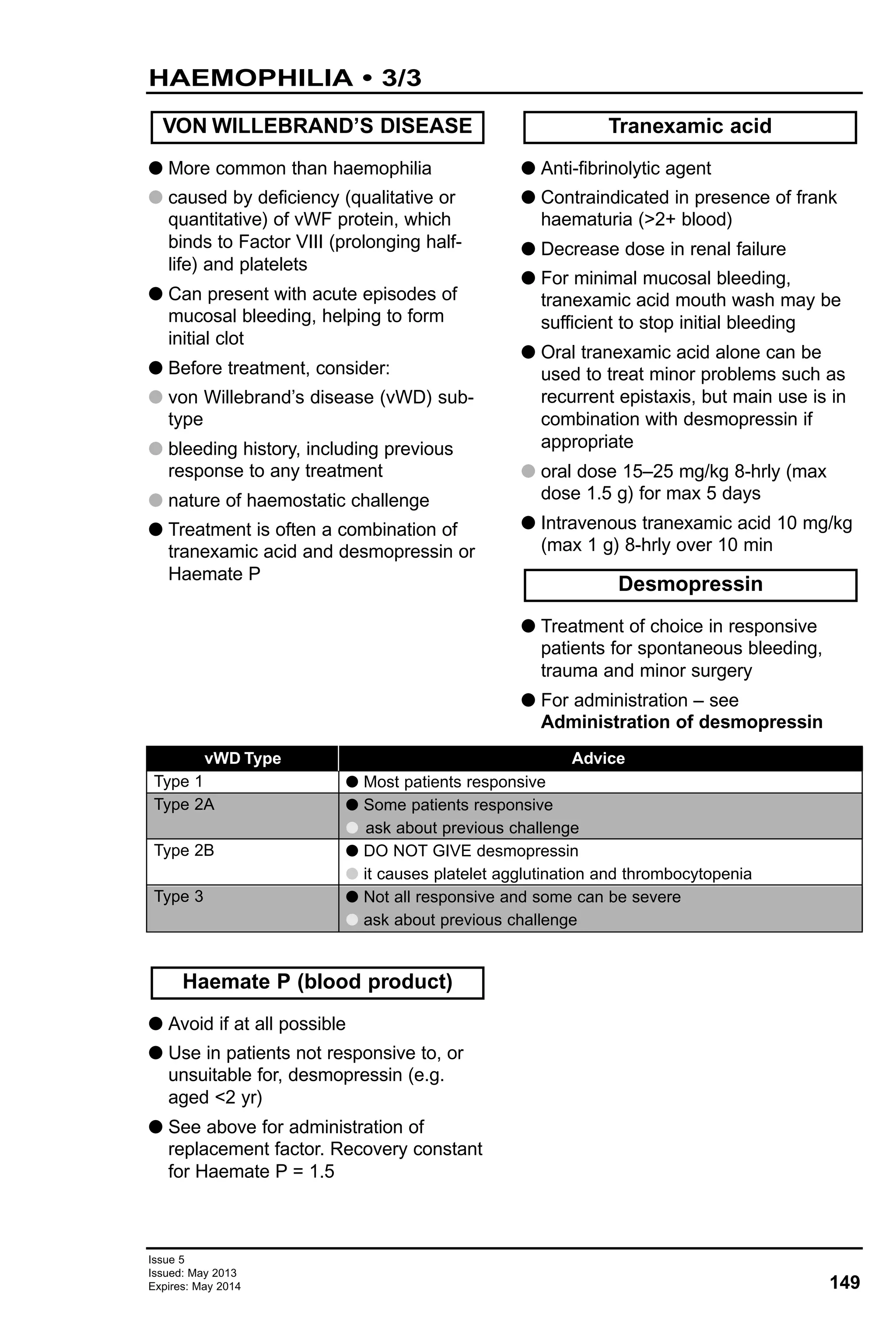
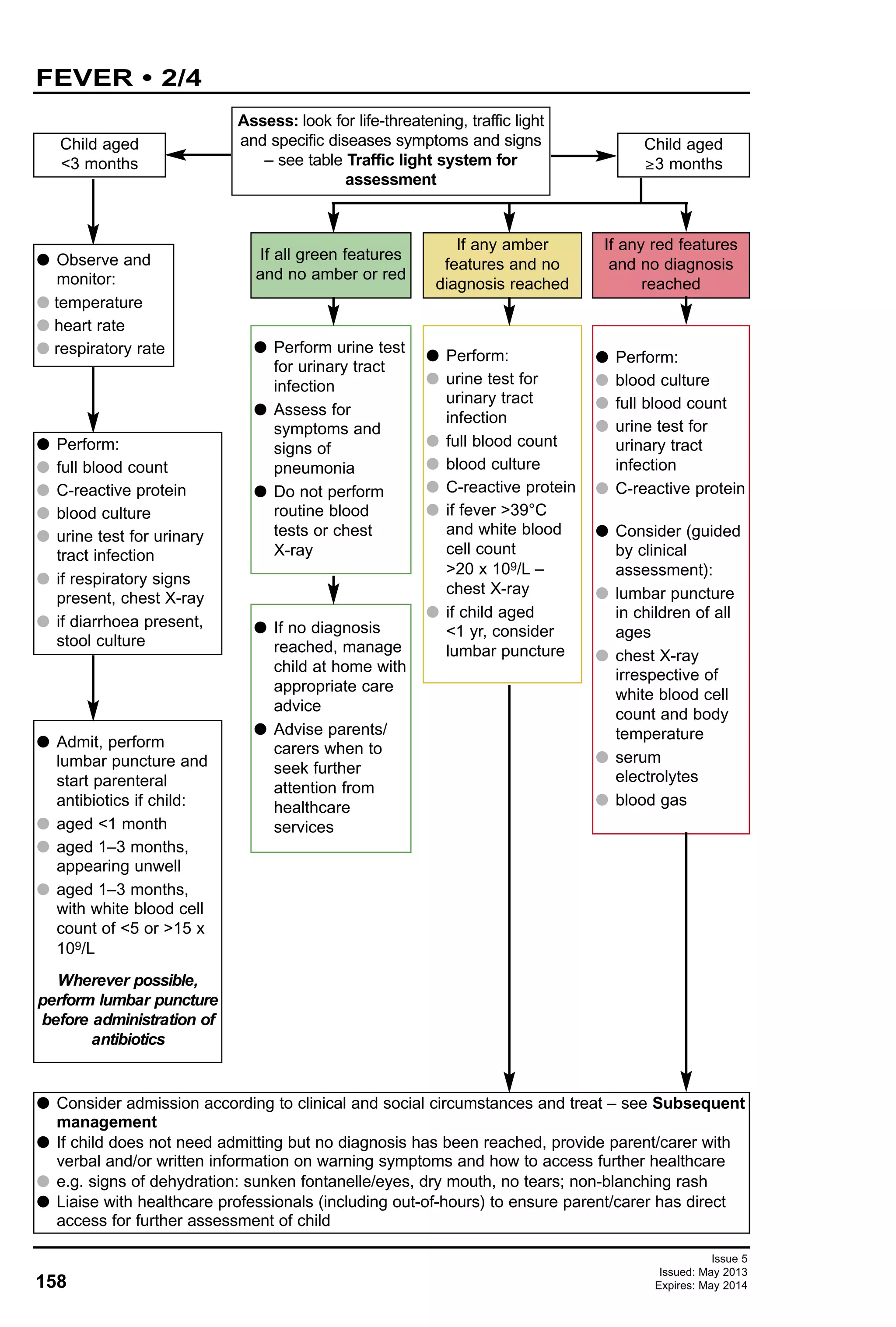
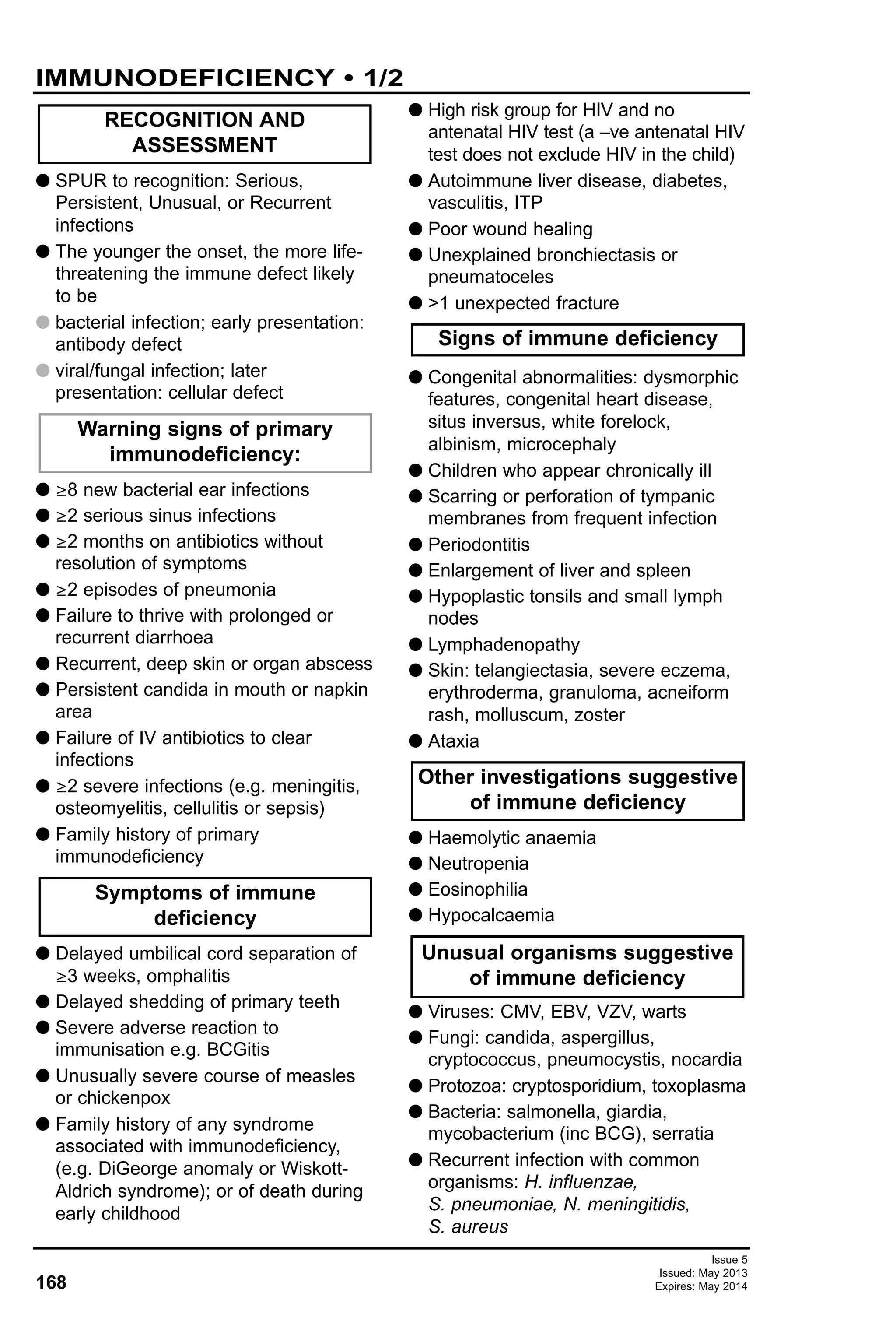
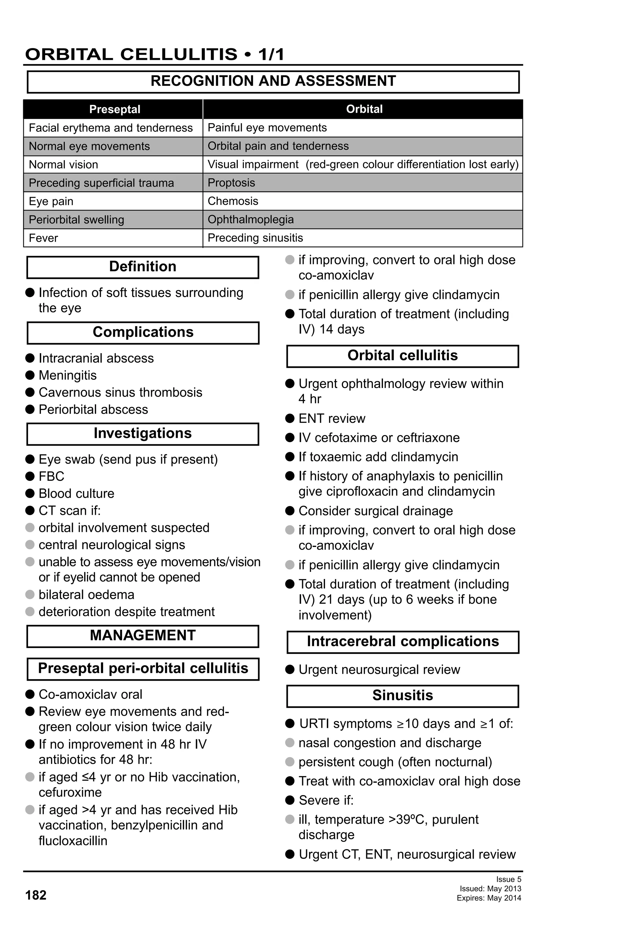
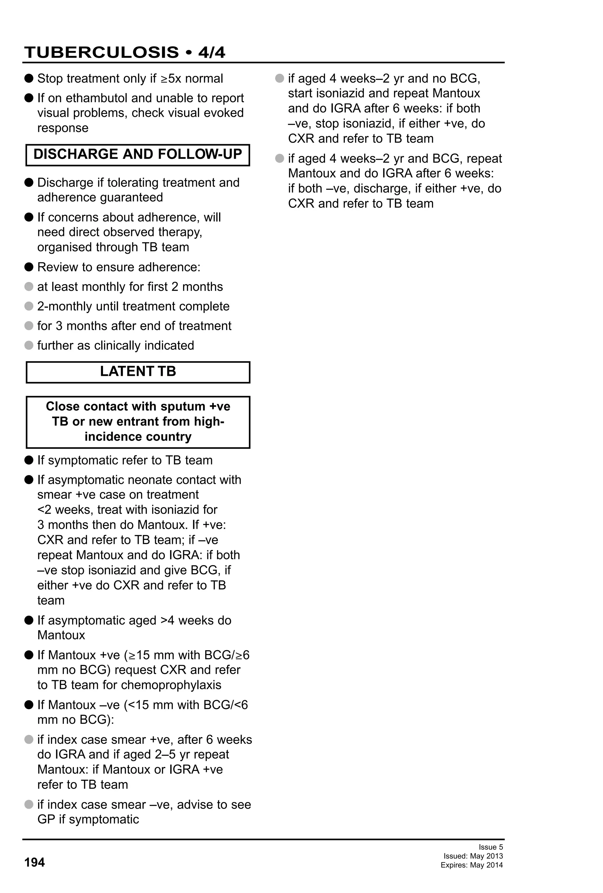
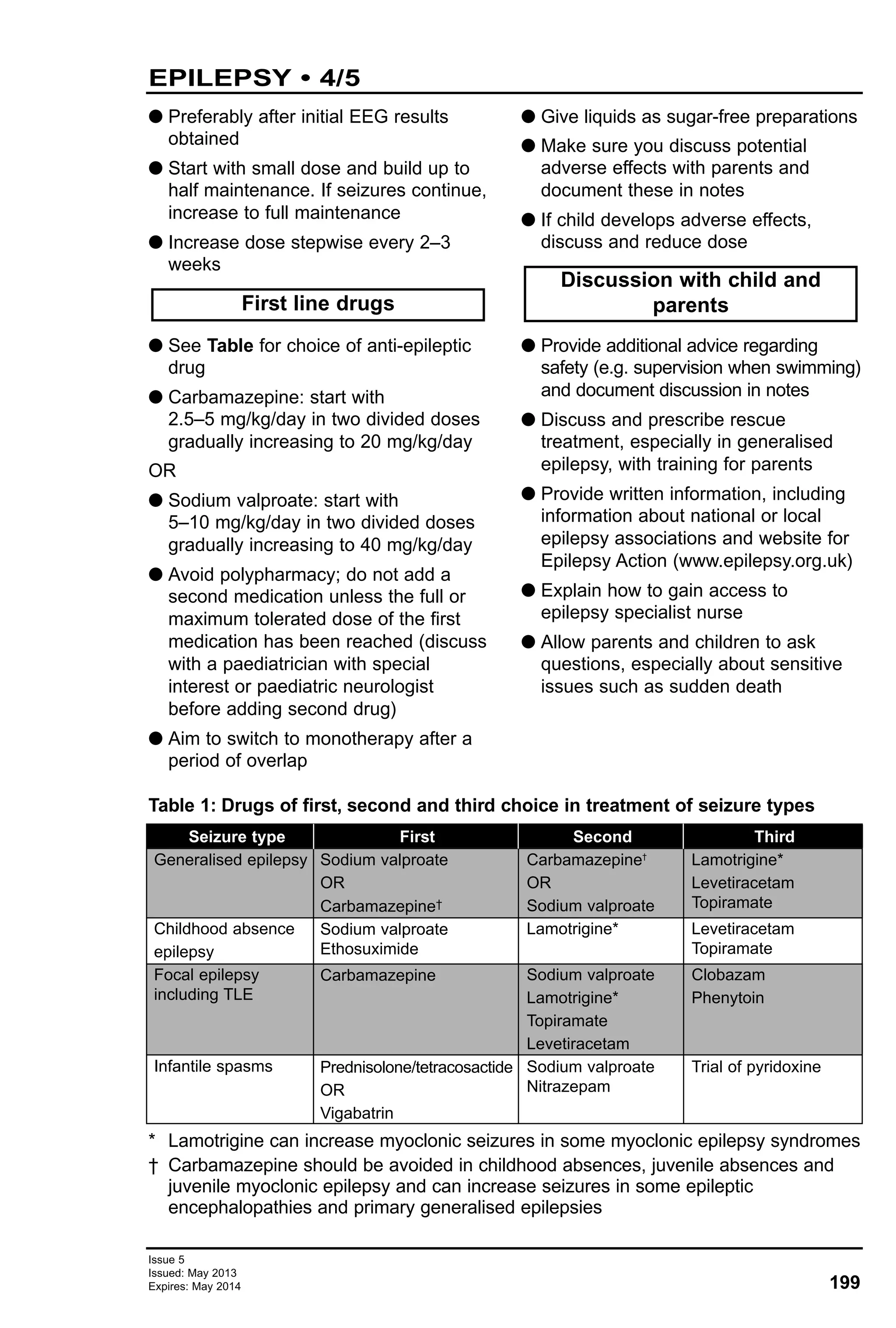
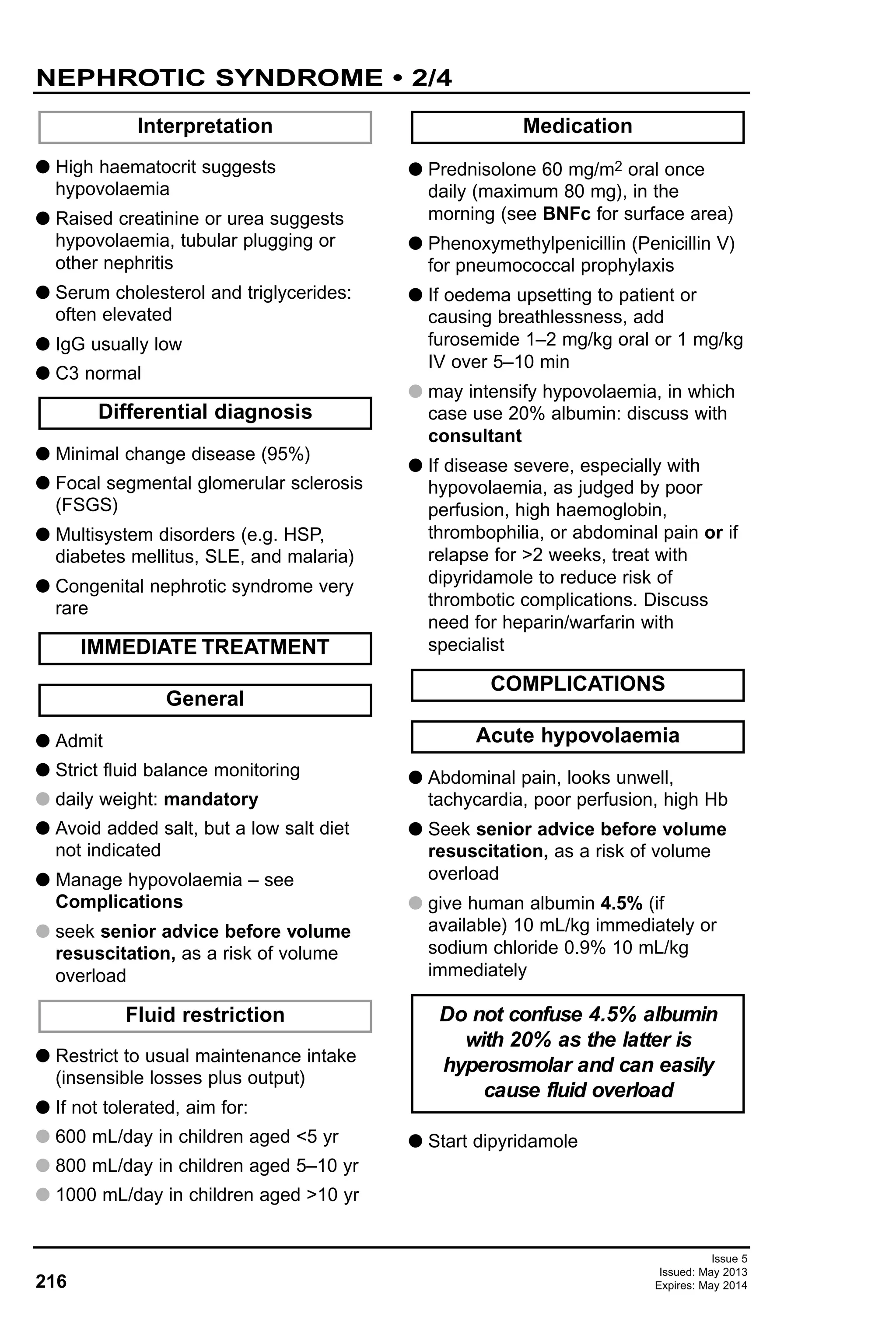
ANALGESIA • 1/4
G For combination of analgesics to use, see Analgesic ladder in Pain assessment
guideline
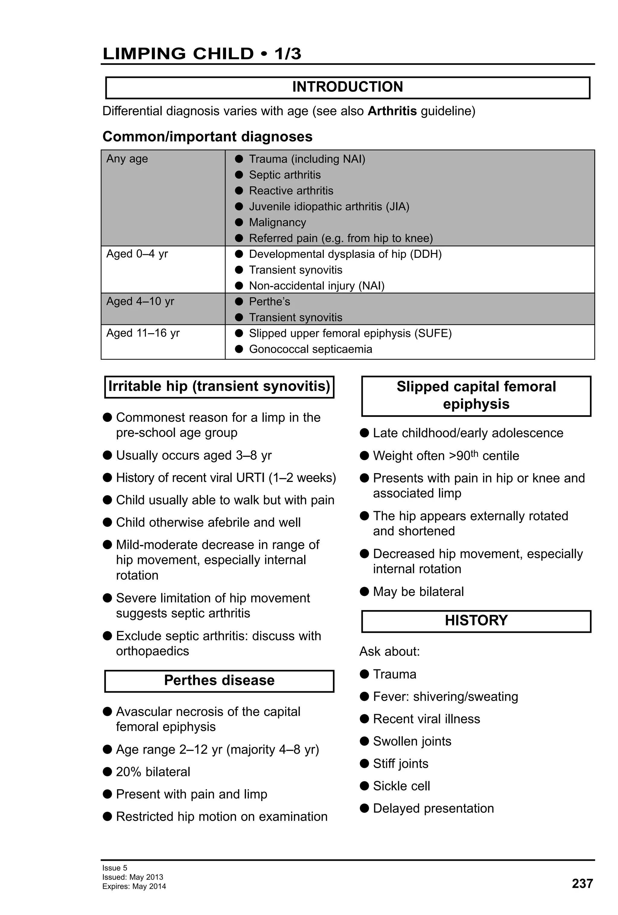
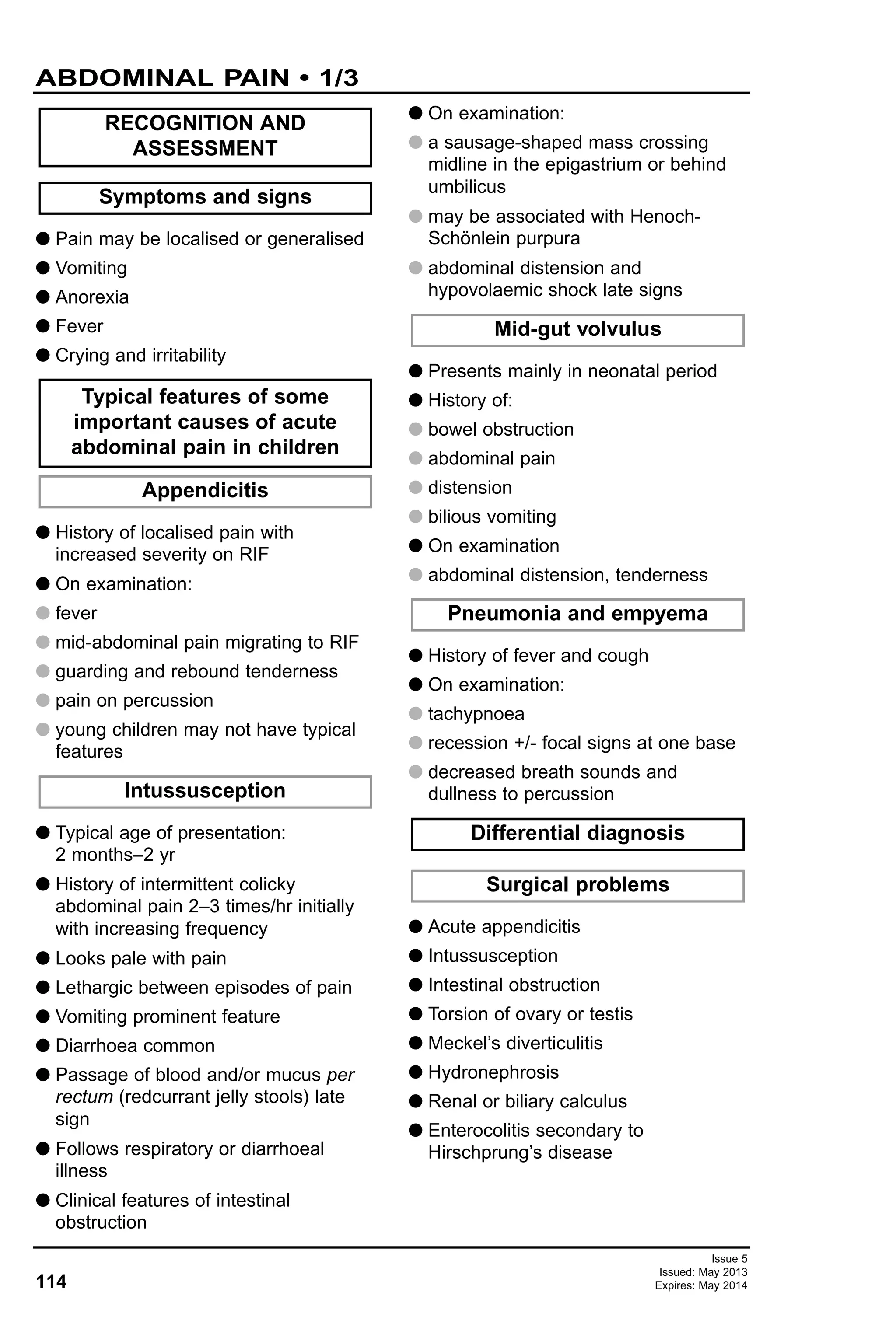
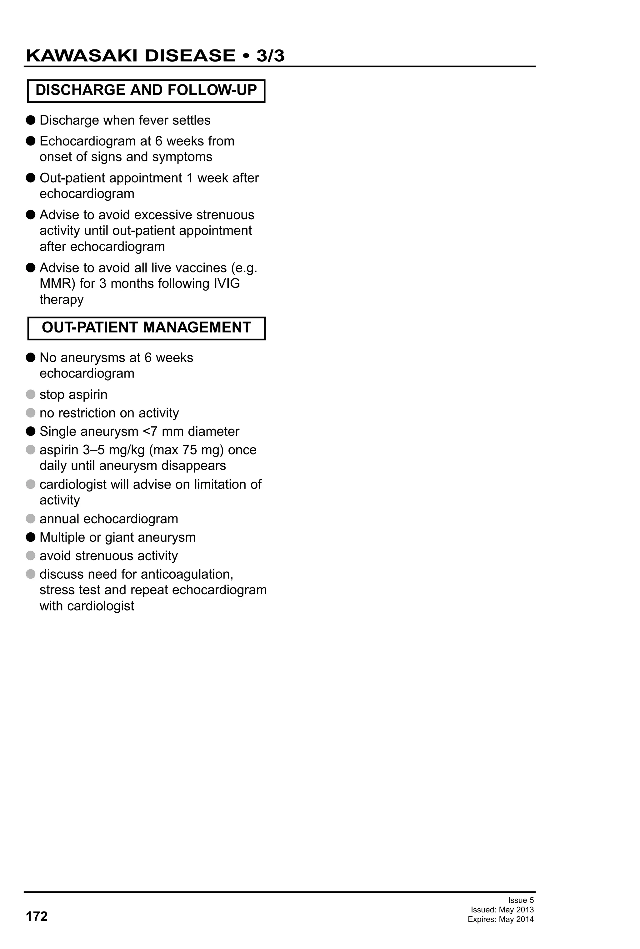
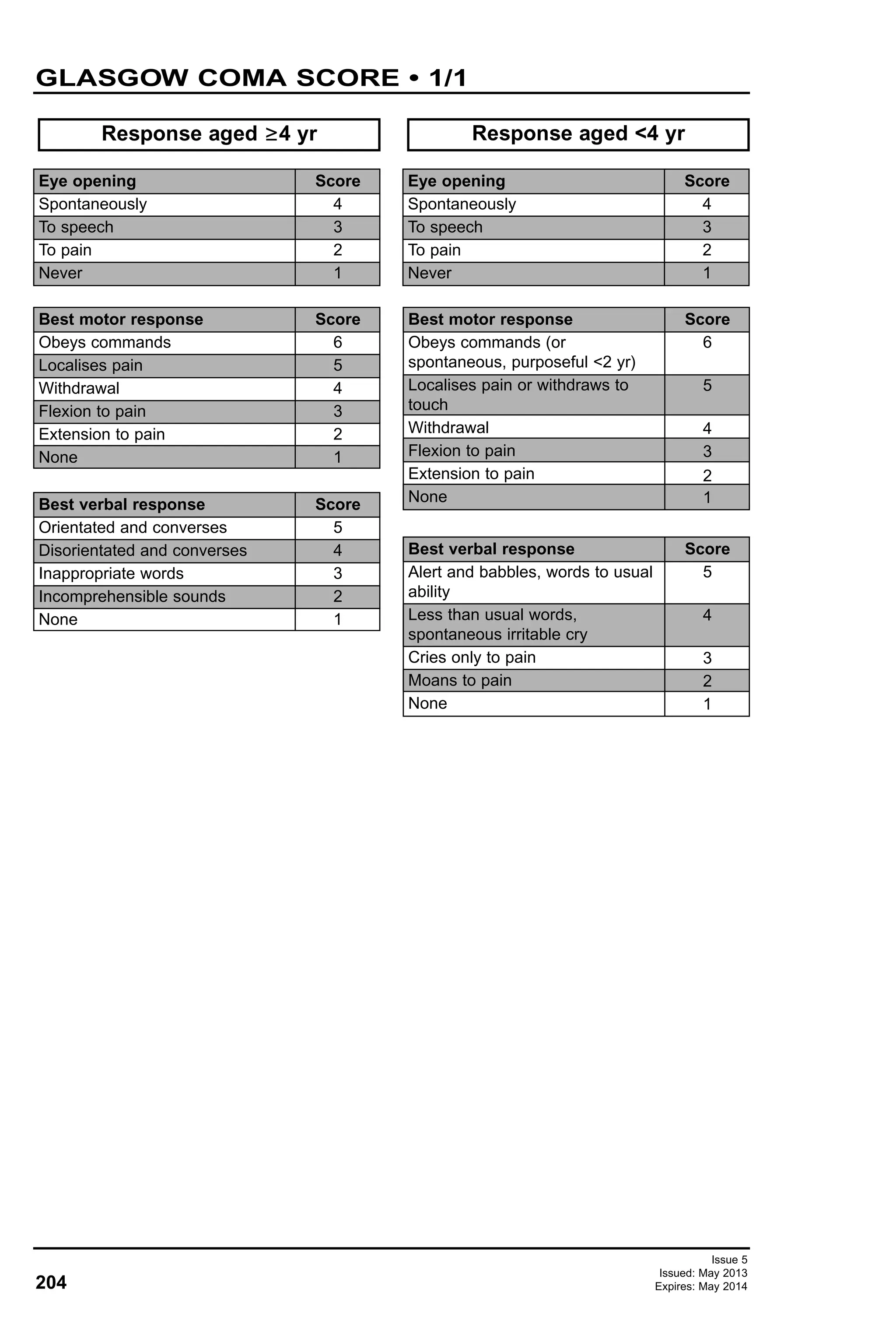
TOPICAL
Age group
<1 month
>1 month
>5 yr
Preparation
Glucose syrup on pacifier
(available as a tootsweet)
Ametop
LMX4
EMLA
Ethyl chloride
Time to onset
During procedure
30 min
30 min
1 hr
Immediately
Comments
For venepuncture or
cannulation
Causes itch, lasts 4 hr
Wait 5 min after removing
cream before cannulation
Remove after 1 hr
If cannot wait for cream
Drug and
preparation
Paracetamol
[oral/nasogastric
(NG)]
G Suspensions:
G 120 mg/5 mL
G 250 mg/5 mL
G Tablets/soluble
500 mg
Paracetamol
(rectal)
G Suppositories
G 60 mg
G 125 mg
G 250 mg
G 500 mg
G 1 g
Paracetamol (IV)
10 mg/mL
(<33 kg use
50 mL vial
via burette or in
syringe)
Prescribe in mg
(not mL)
Dose
G First dose 20 mg/kg
THEN
G aged 1–3 months: 30–60 mg
8-hrly
G aged 3–6 months: 60 mg
G aged 6–24 months: 120 mg
G aged 2–4 yr: 180 mg
G aged 4–6 yr: 240 mg
G aged 6–8 yr: 250 mg
G aged 8–10 yr: 375 mg
G aged 10–12 yr: 500 mg
G aged 12–16 yr: 750 mg
G aged >16 yr: 500 mg –1 g
G First dose 30 mg/kg
THEN
G birth–3 months: 20 mg/kg
8-hrly
G aged 3 months–12 yr:
20 mg/kg 4-hrly
G aged >12 yr: 500 mg–1 g
4-hrly
G <10 kg: 10 mg/kg 6-hrly
G 10–50 kg: 15 mg/kg 6-hrly
G >50 kg: 1 g 6-hrly
Maximum dose
Max total dose in 24 hr
G Aged <1 month:
60 mg/kg/day
G Aged ≥1 month–18 yr:
90 mg/kg (max 4 g)
G Max total dose in 24 hr:
G aged <3 months:
60 mg/kg
G aged
≥3 months: 90 mg/kg
for 48 hr then 60 mg/kg
G aged >12 yr: 4 g
G Aged <1 month:
30 mg/kg/day
G <50 kg:
60 mg/kg/day
G >50 kg:
60 mg/kg/day
G Up to 4 g daily
Comments
G For mild
pain
G Increase
dose
interval in
renal
impairment
G Avoid large
doses in
dehydration,
malnutrition,
hepatic
impairment
G As for oral
paracetamol
G For mild
pain when
oral/NG
route not
possible
G Suspension
can be
given
rectally
G As for oral
paracetamol
G For mild
pain when
oral/NG/PR
route not
possible
G Give over
15 min
MILD PAIN (pain score 1–3)](https://image.slidesharecdn.com/paediatricguidelines2013-14withlinks-180801135506/75/Paediatric-guidelines-2013-14-with-links-24-2048.jpg)
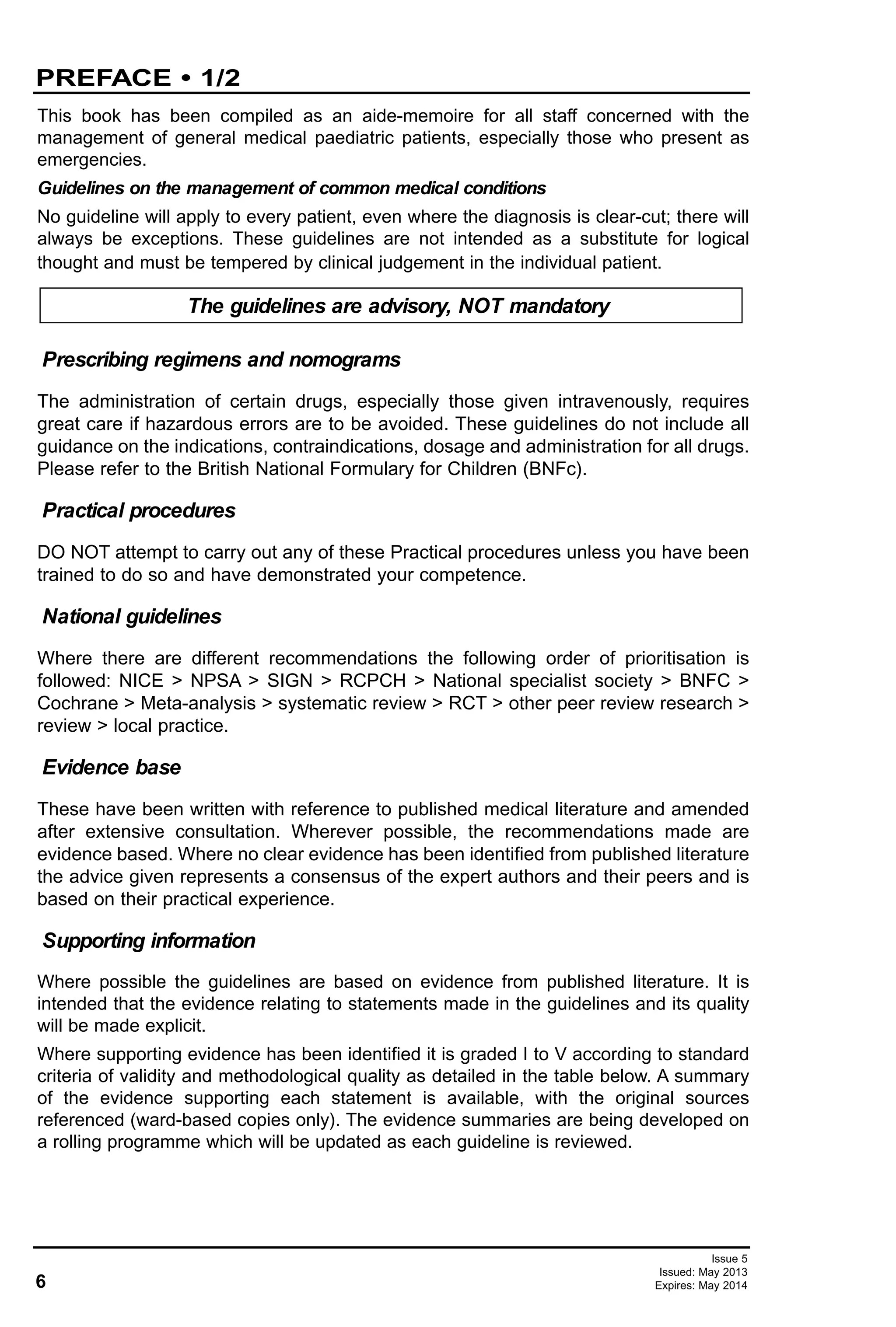
![36
Issue 5
Issued: May 2013
Expires: May 2014
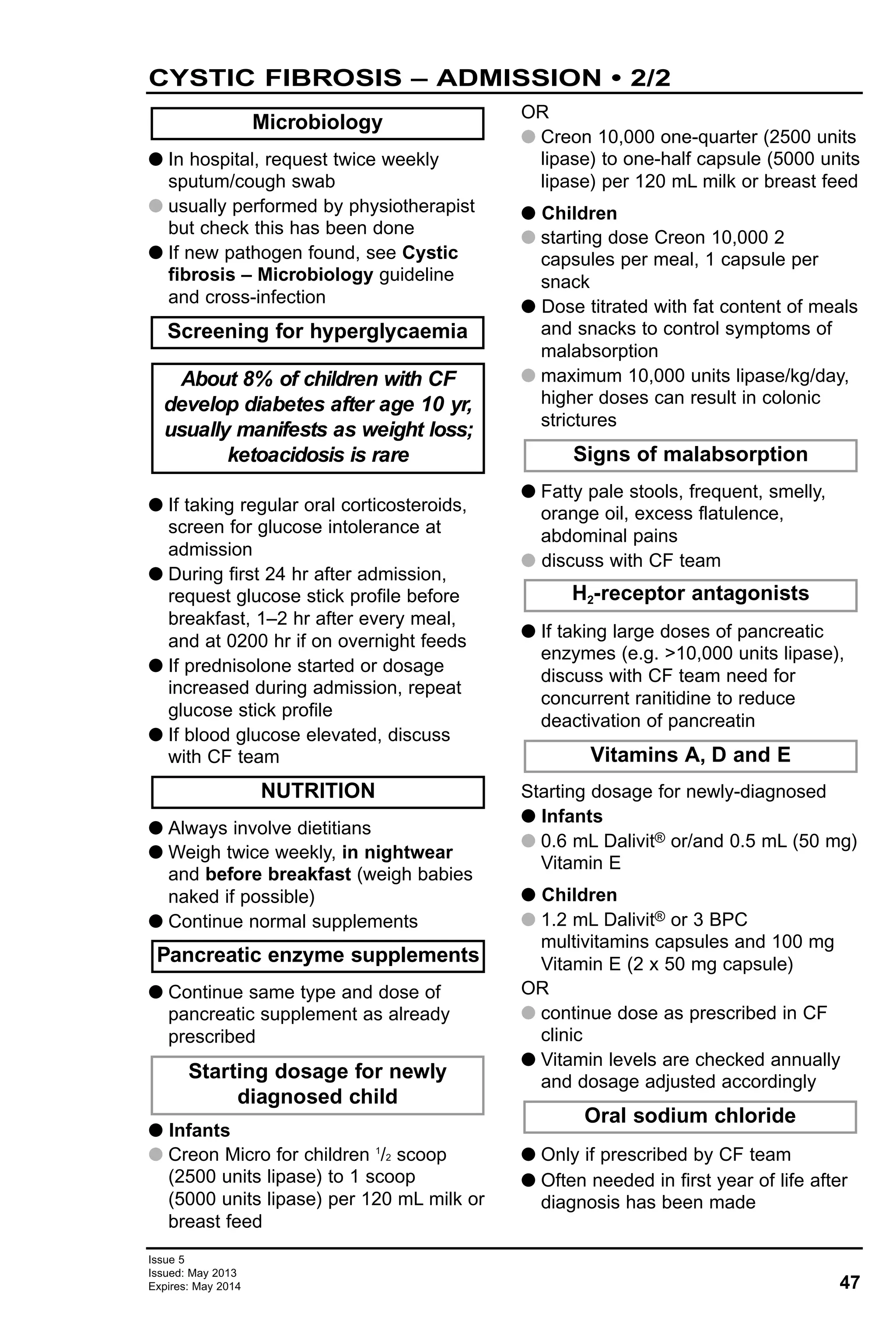
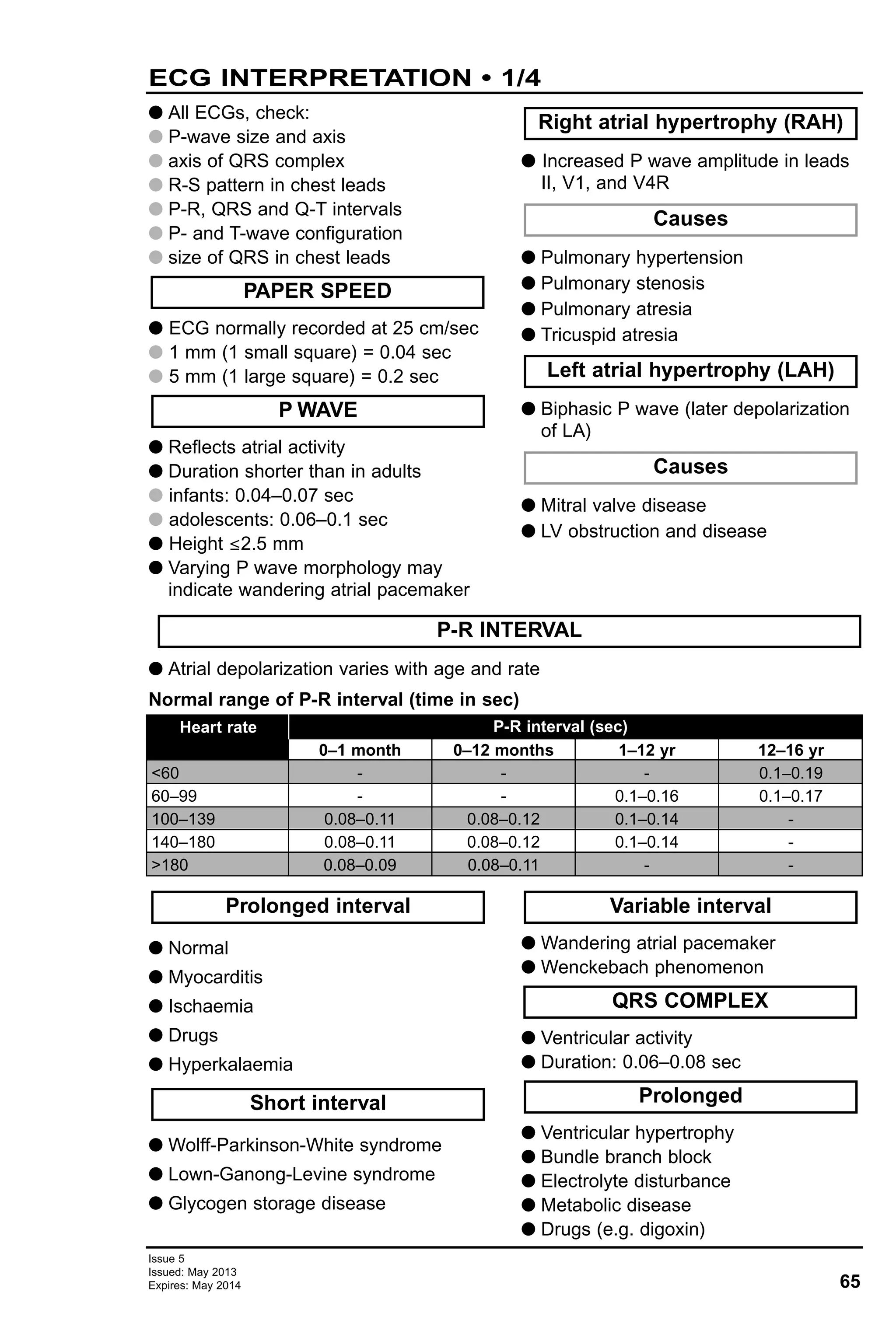
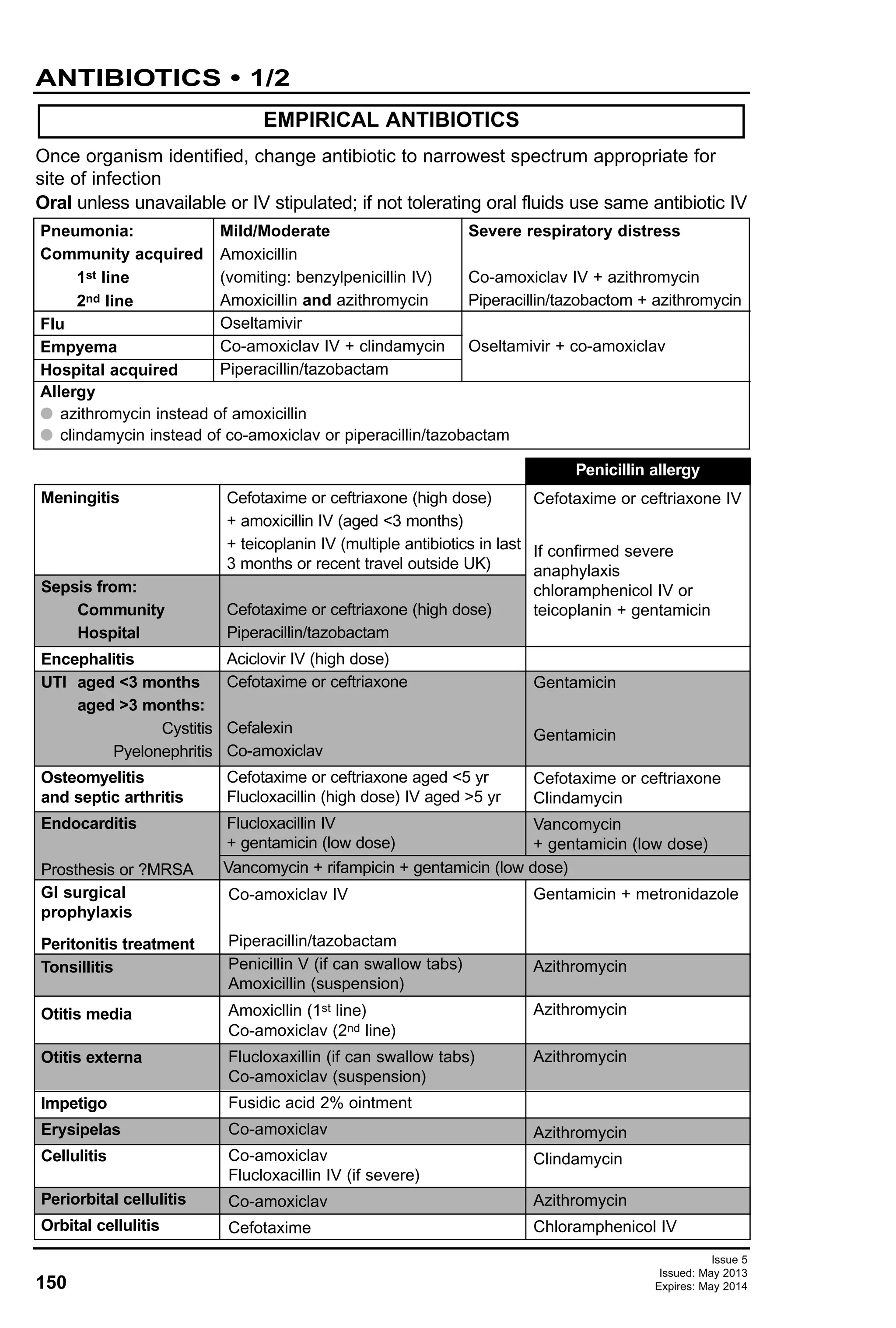
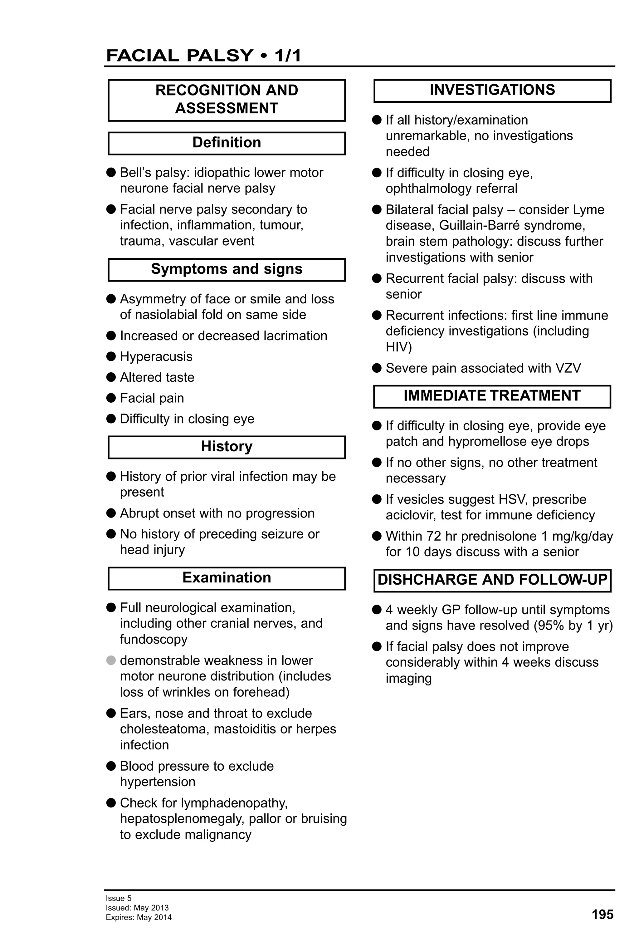
POST GA MONITORING EX-PREMATURE INFANTS
• 1/1
G Risk of apnoea after general
anaesthetic (GA)
G increased if anaemic
G with chronic lung disease who have
required oxygen treatment within last
6 months
G Check haemoglobin
G if Hb <90 g/L, arrange transfusion
G Arrange overnight stay for post-
operative monitoring if age (weeks)
<[3 x (38 – gestational age in weeks)]
e.g. baby born at 30 weeks gestation
would be kept overnight after GA if
<24 weeks old. A 36 week baby
would be allowed home after GA if
>6 weeks old
G Transfer patient with oxygen supply,
continuous SpO2 monitoring and full
resuscitative equipment
G Admit patient to a designated HDU
ward area
G High dependency nursing care
G Monitoring to include:
G continuous pulse oximetry
G continuous ECG
G continuous respiratory rate
G transcutaneous CO2
G If apnoea >15 sec:
G immediate respiratory support by
nurse (airway manoeuvres, bag and
mask ventilation)
G contact on-call SpR
G liaise with anaesthetist responsible for
patient
G review period of HDU care
G Discharge patient home same day or
next day as calculated by above
formula providing there have been no
apnoeic episodes
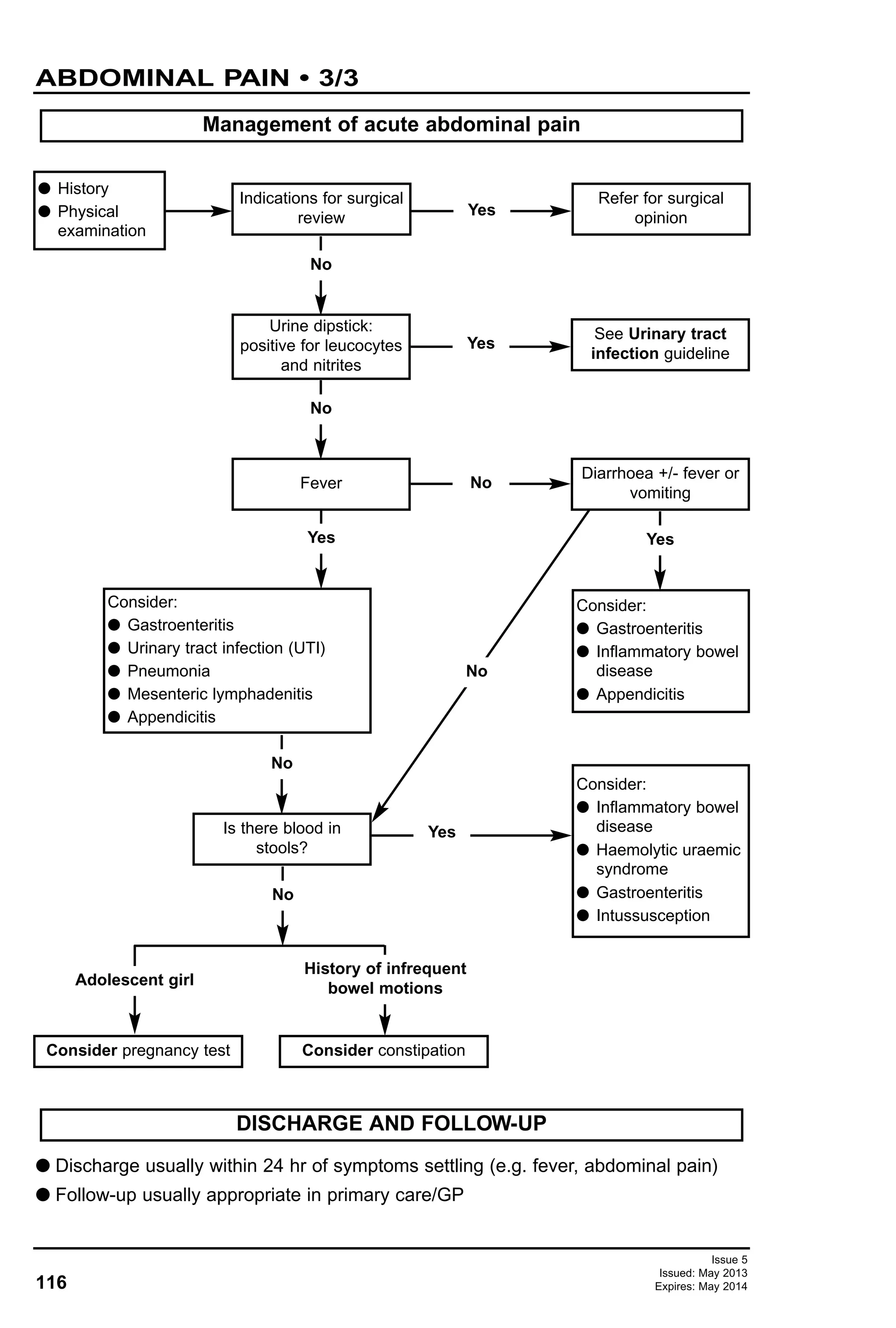
DISCHARGE AND FOLLOW-UP
Subsequent post-GA
management
Immediate post-GA period
Pre-operative
MANAGEMENT](https://image.slidesharecdn.com/paediatricguidelines2013-14withlinks-180801135506/75/Paediatric-guidelines-2013-14-with-links-36-2048.jpg)
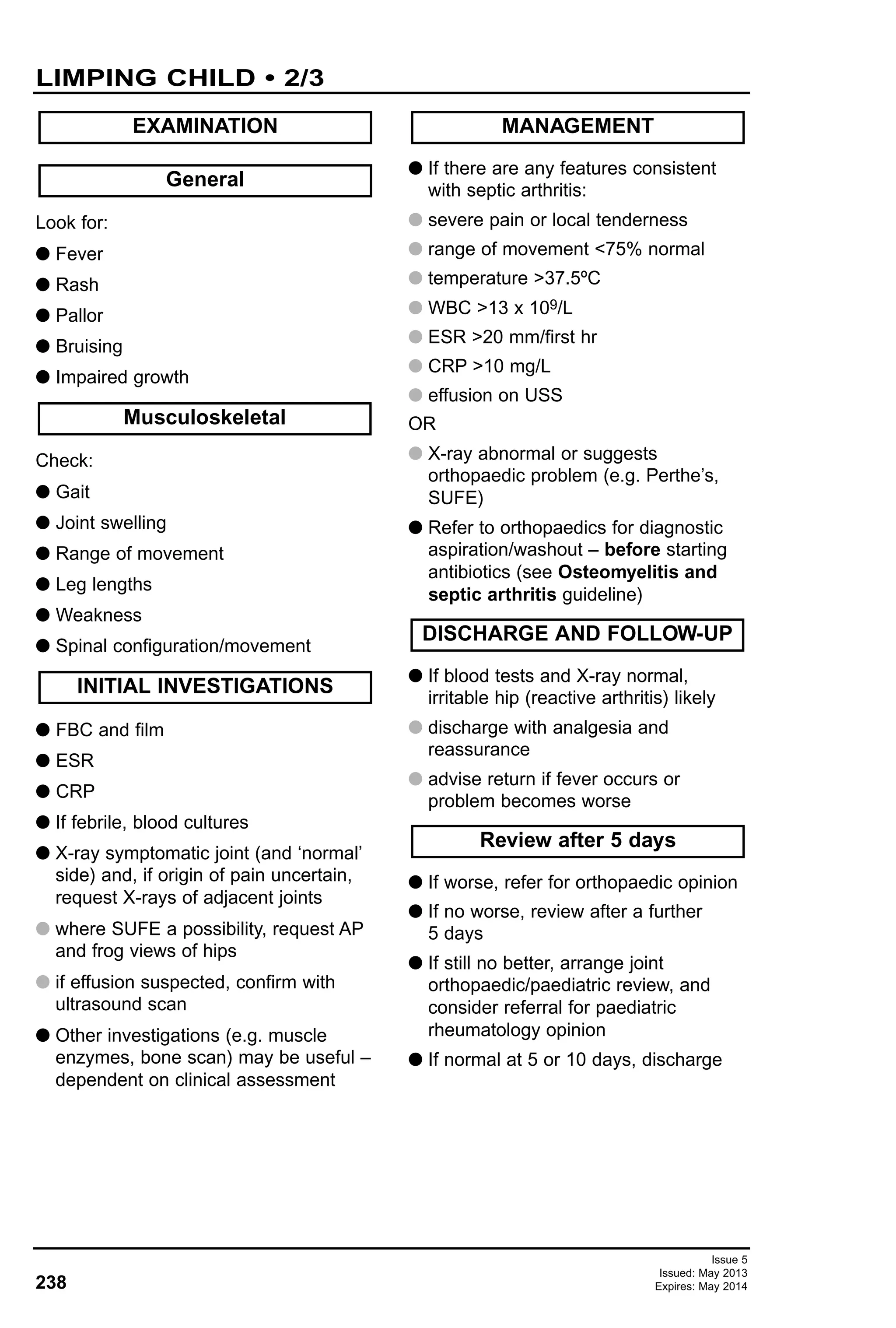
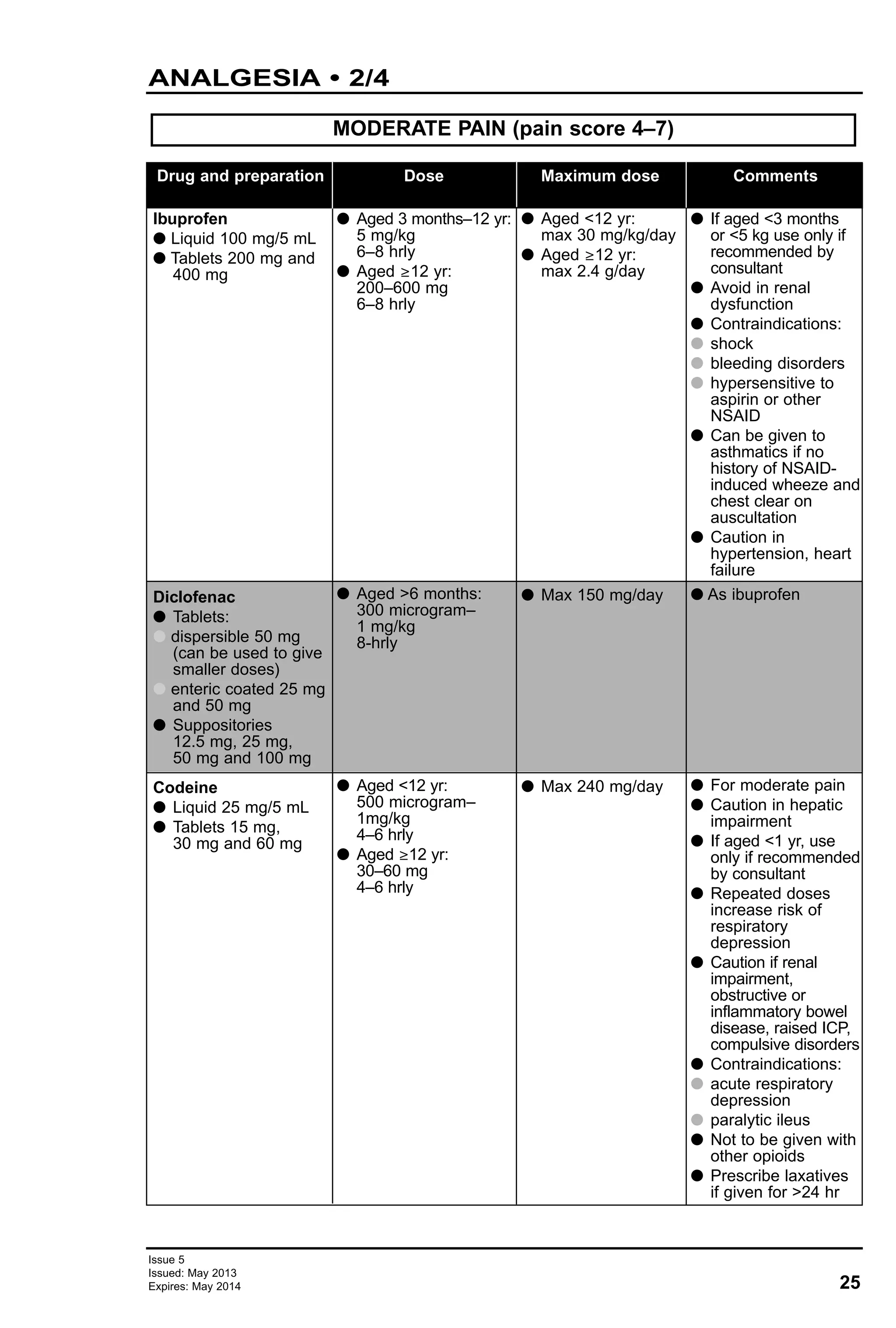
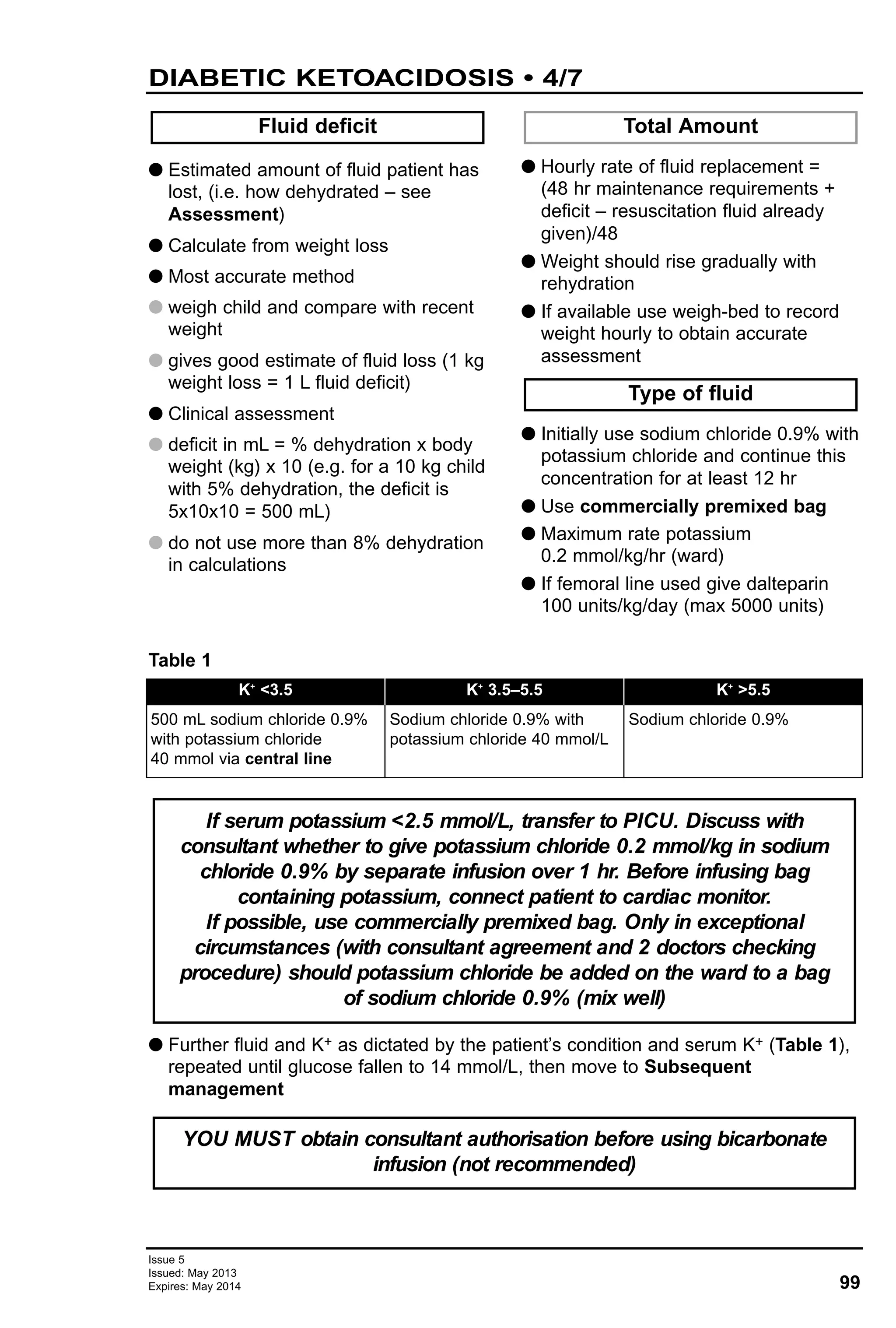
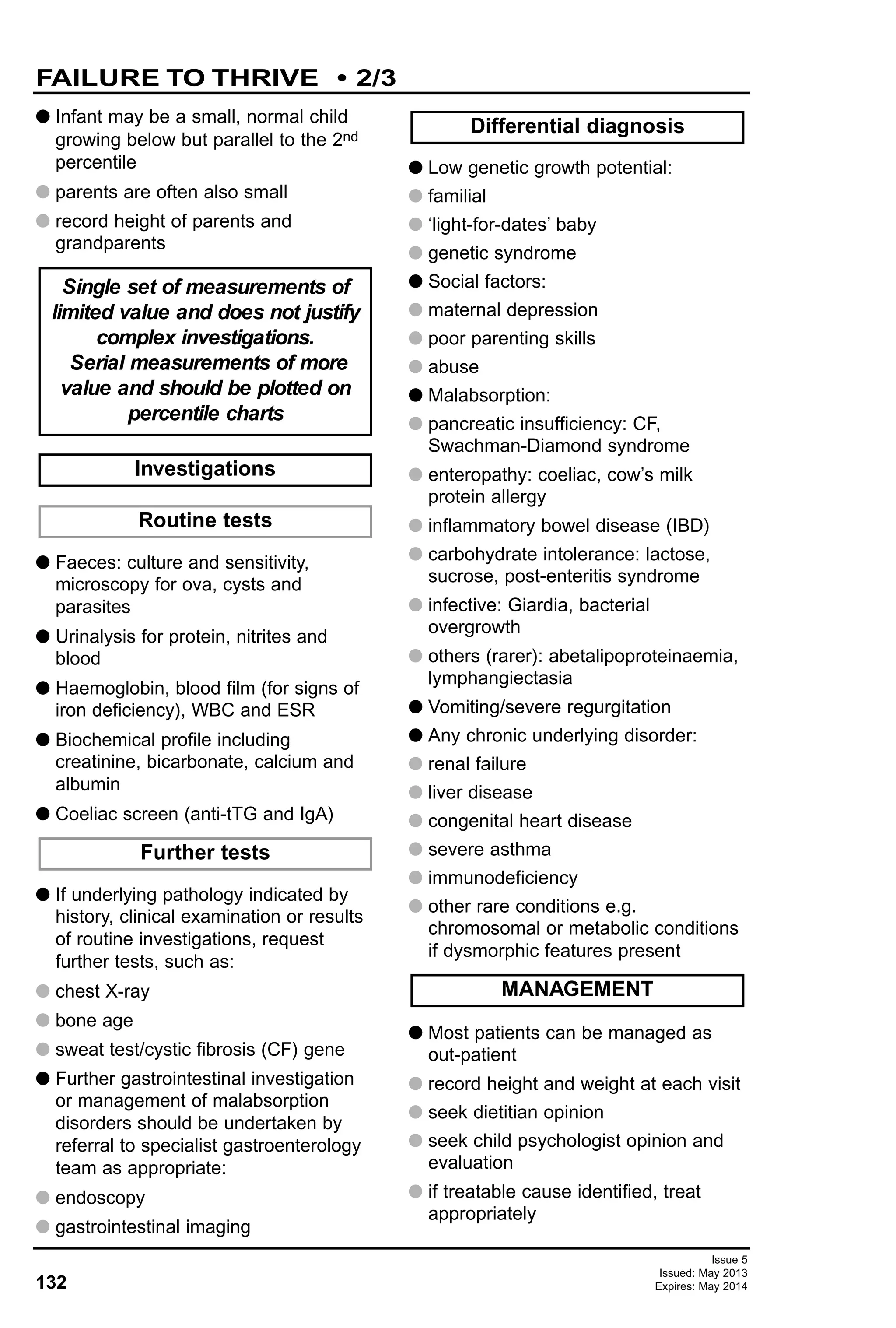
![40
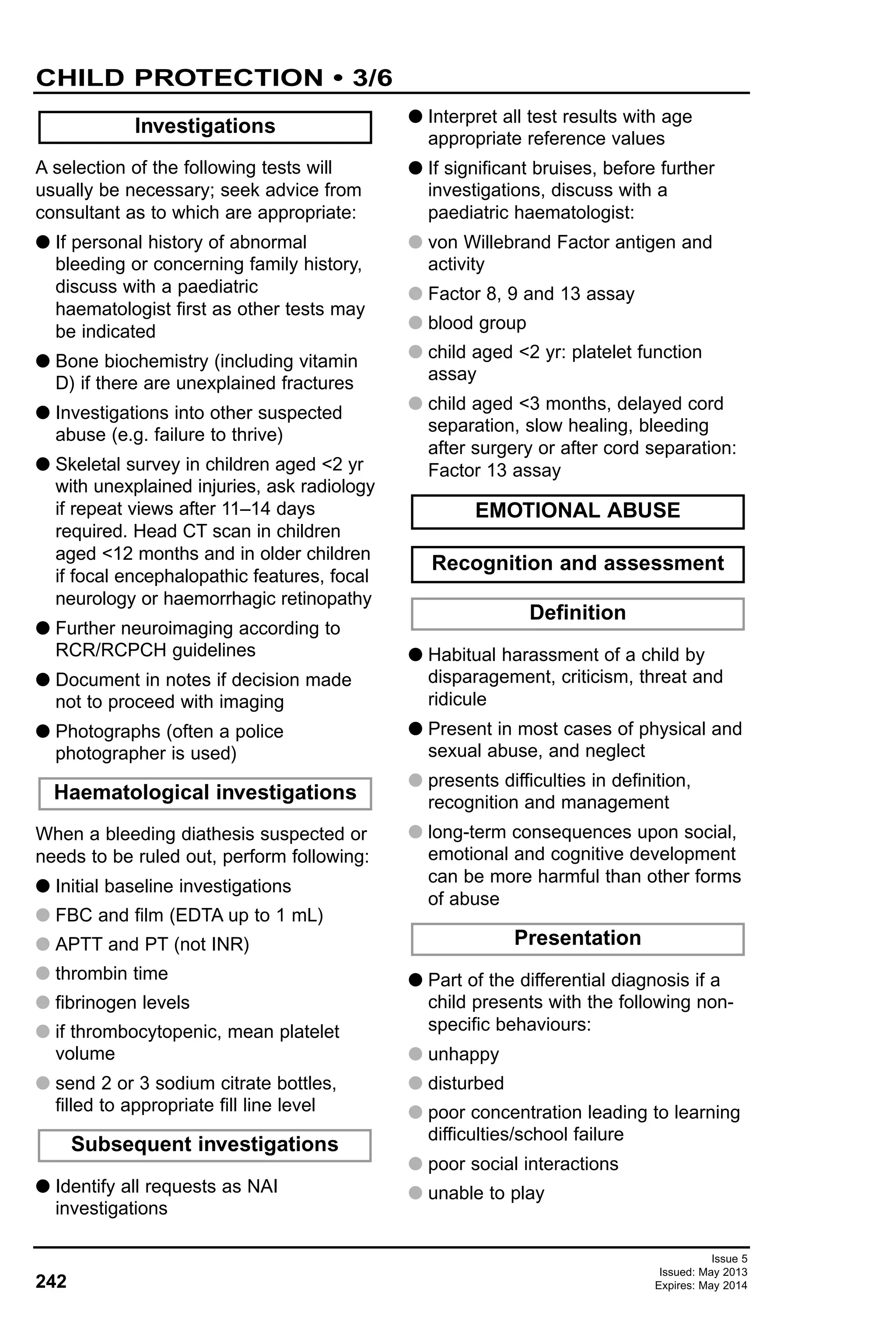
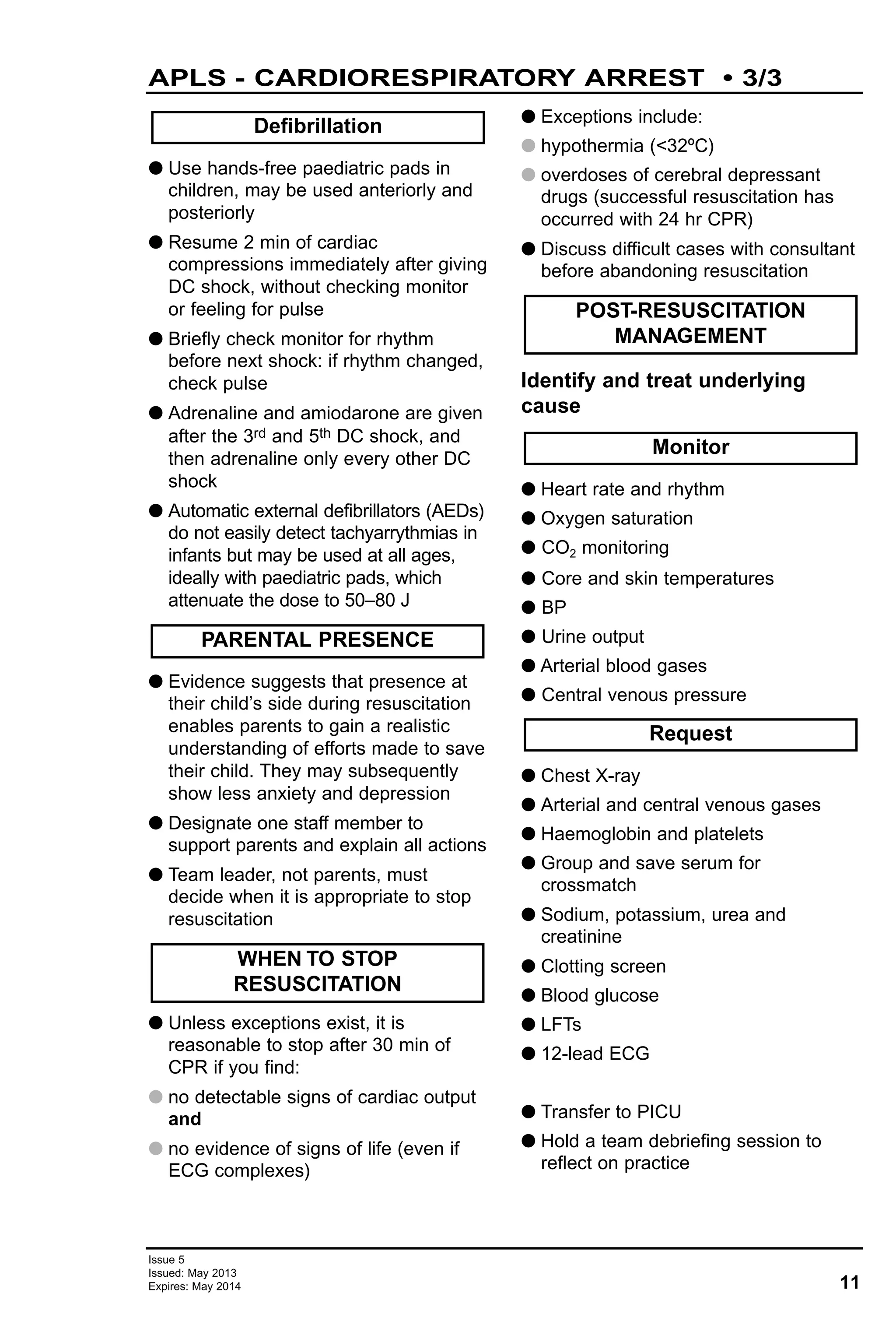
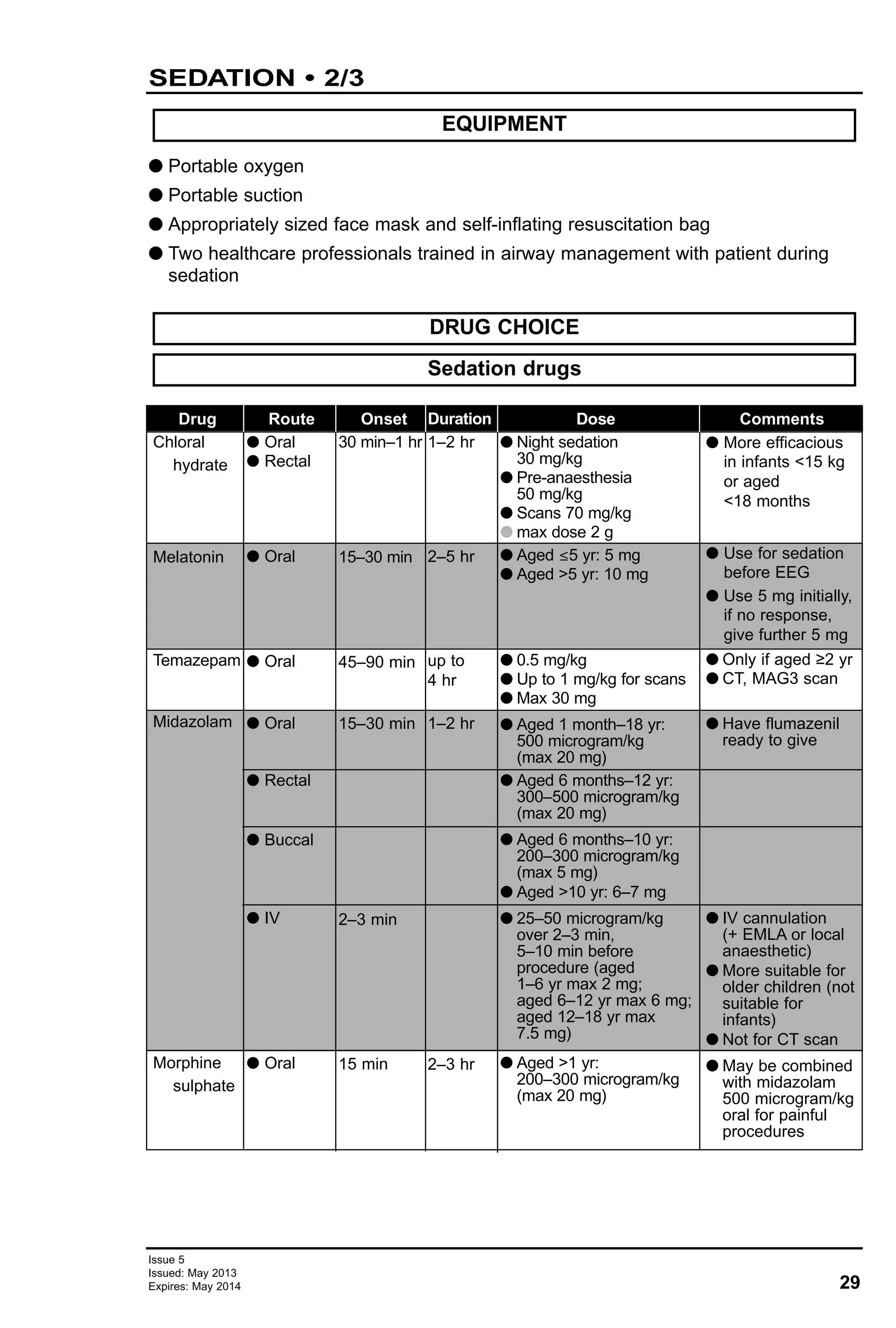
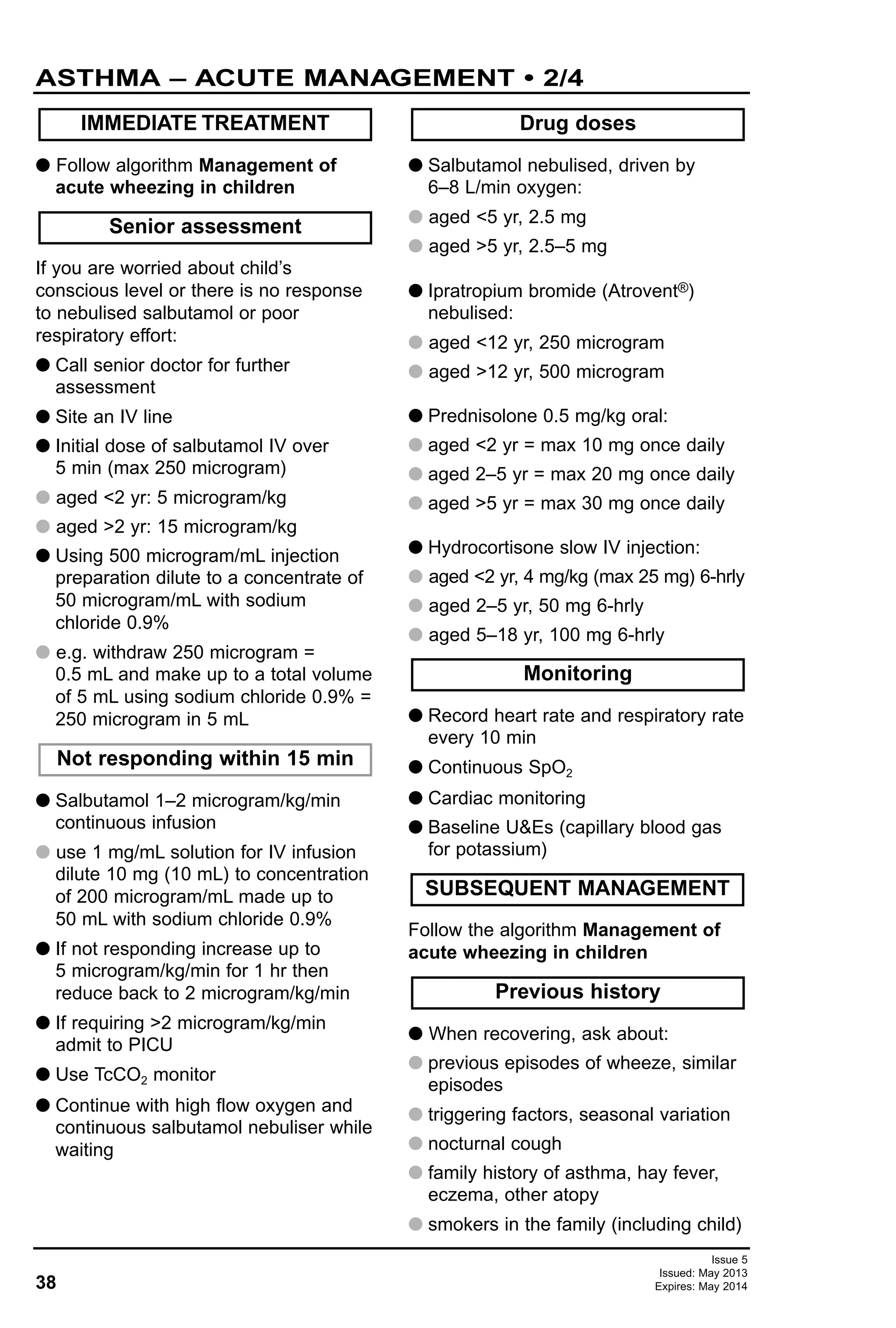
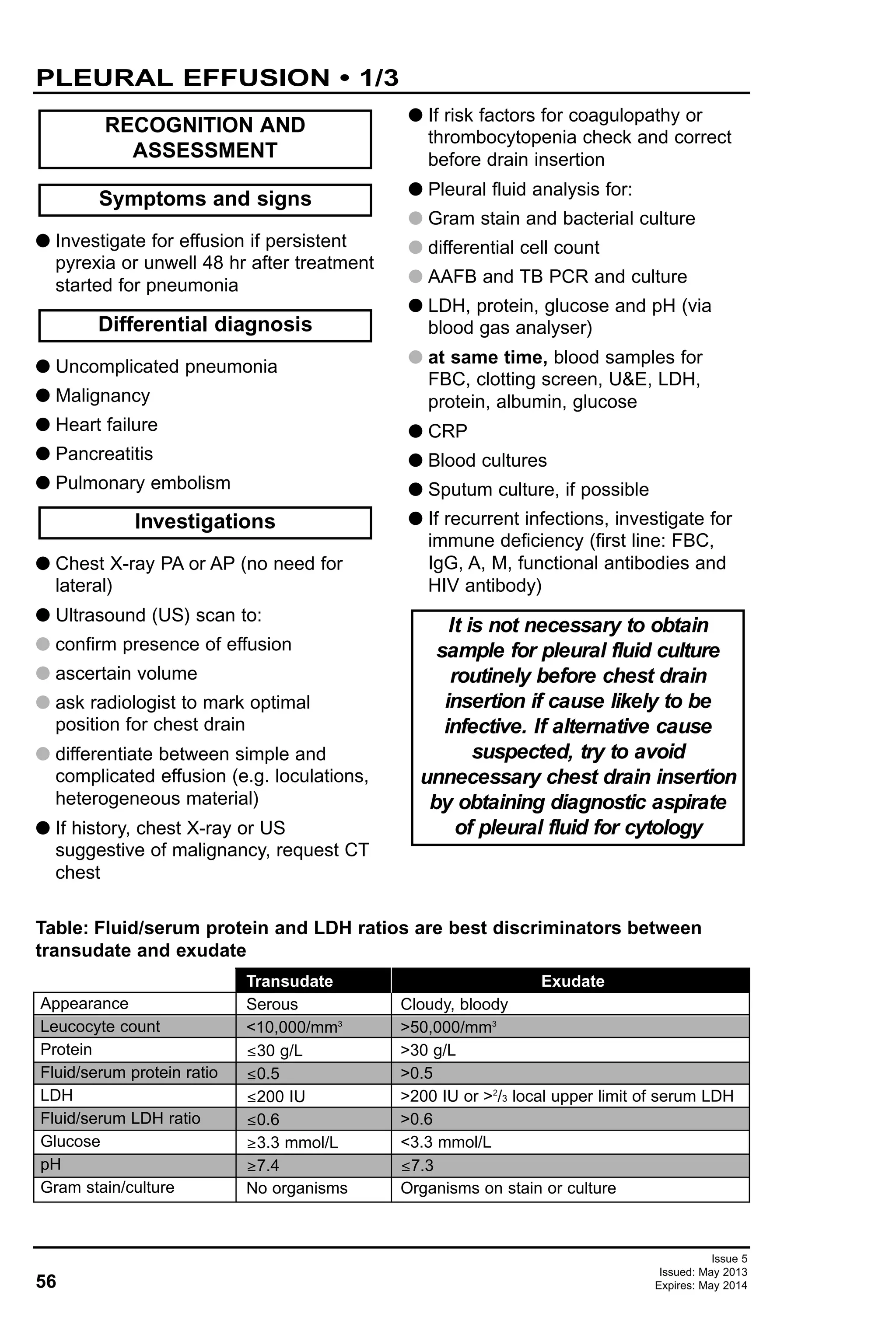
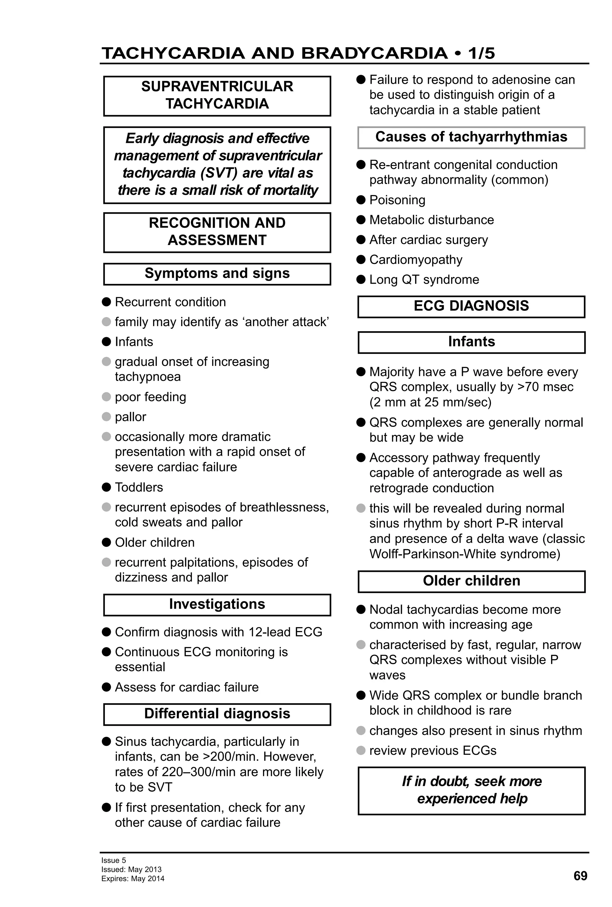
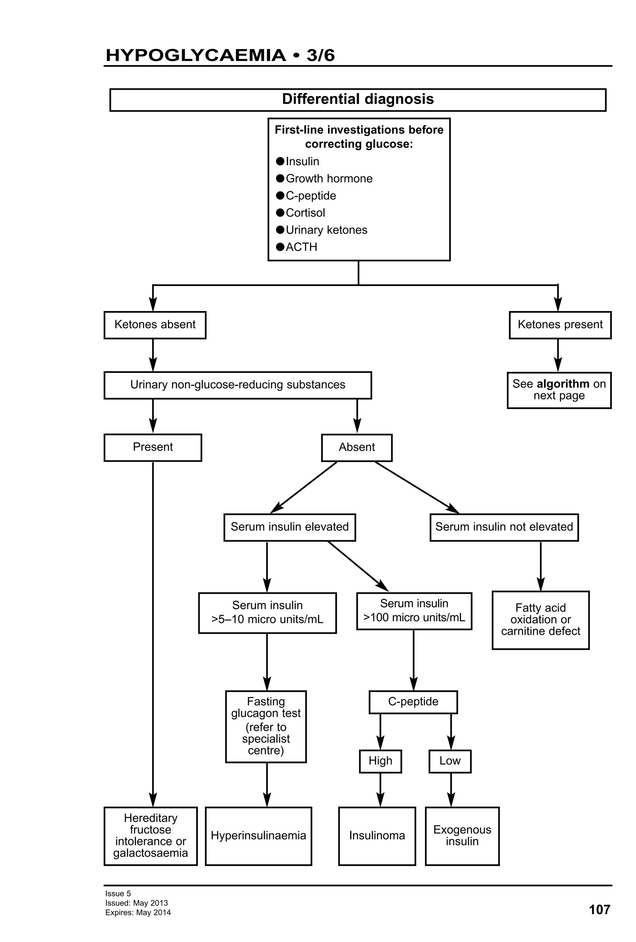
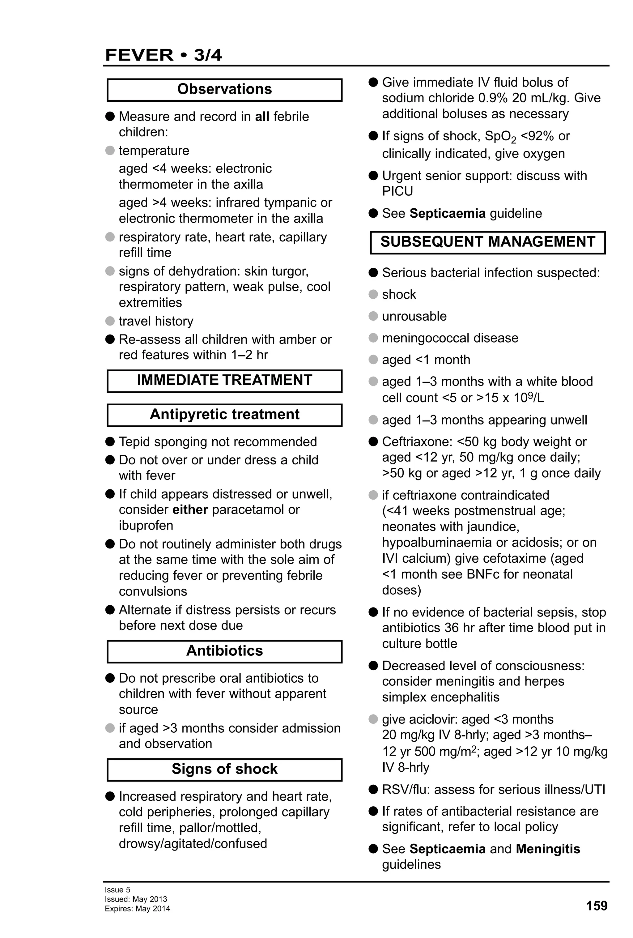
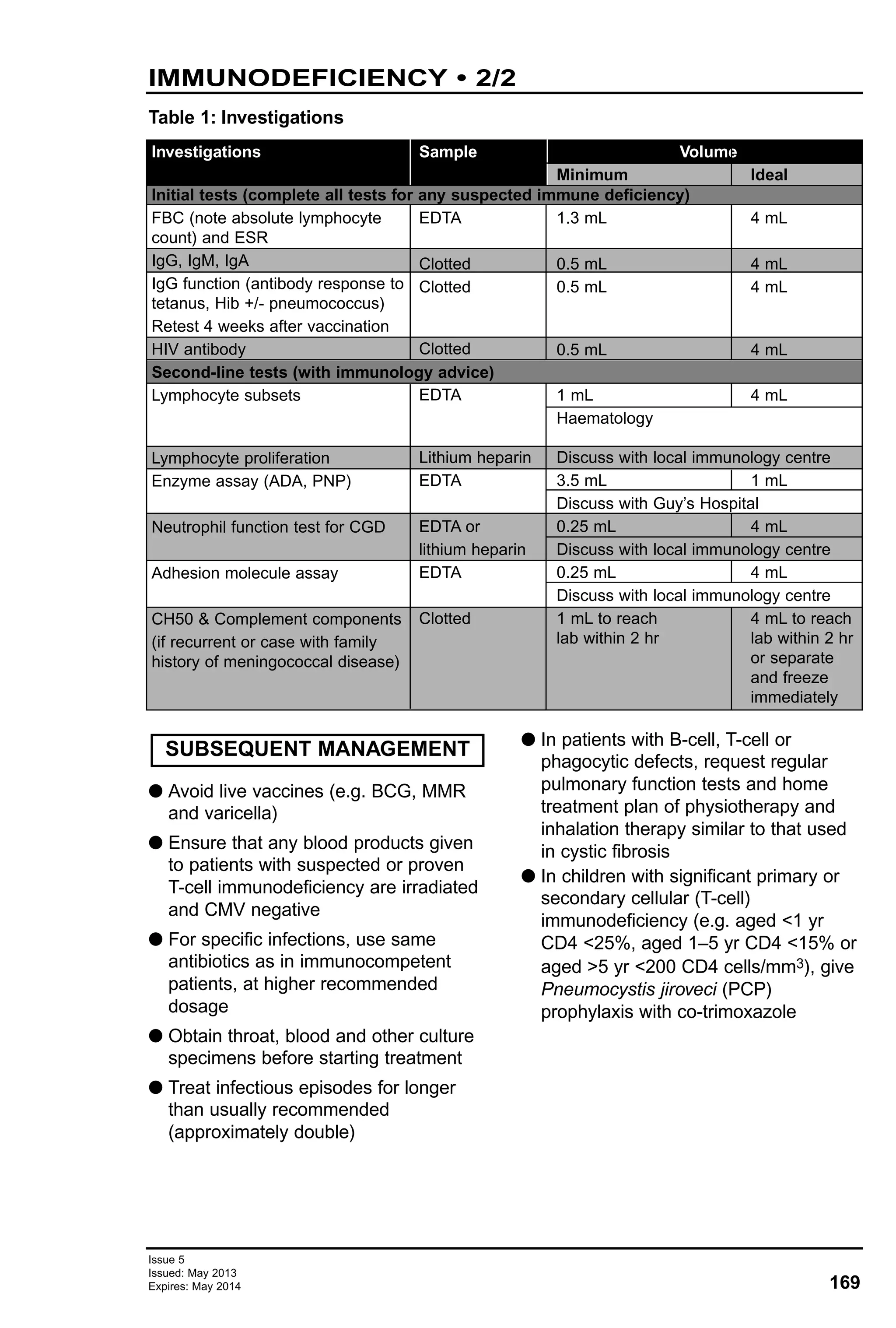
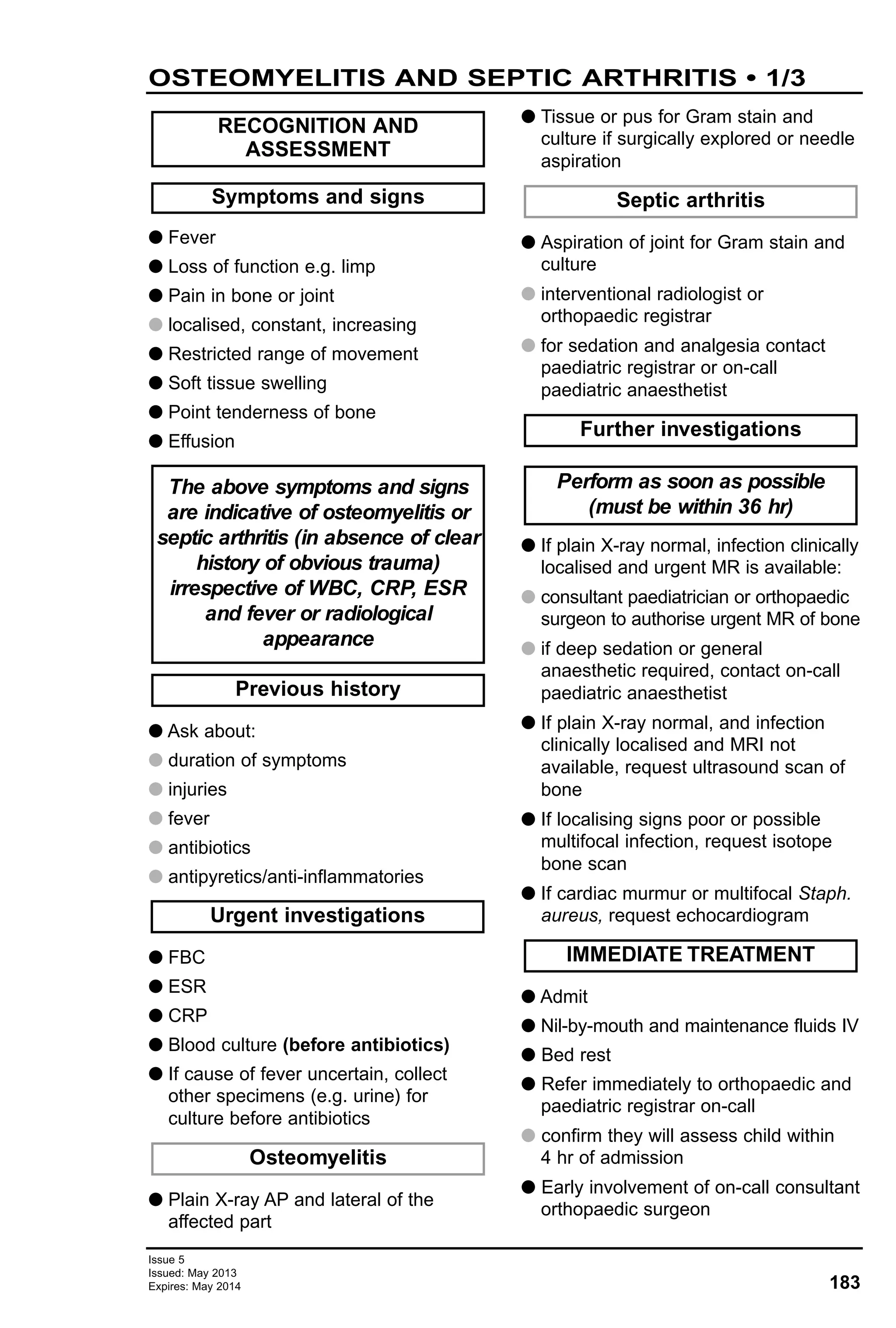
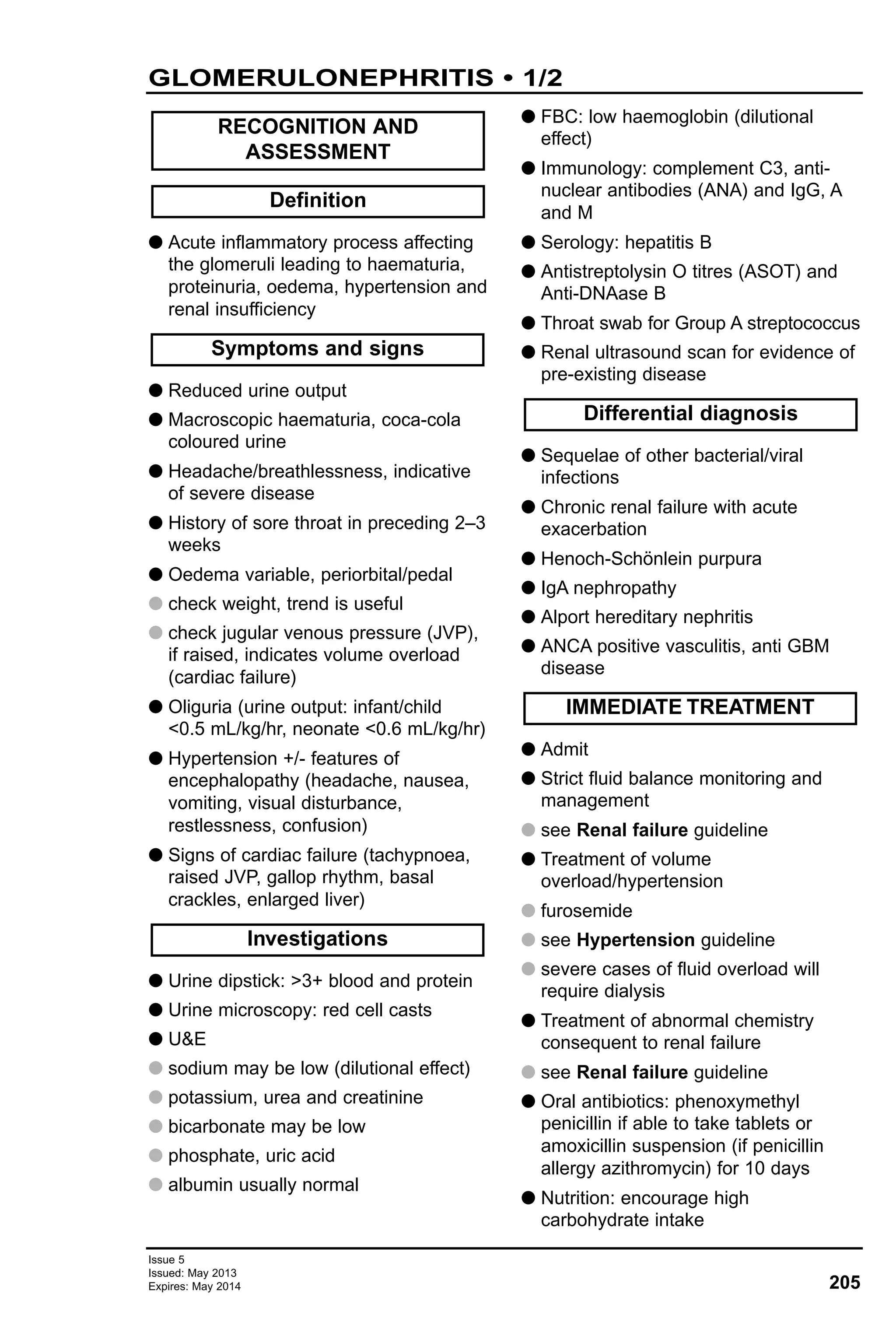
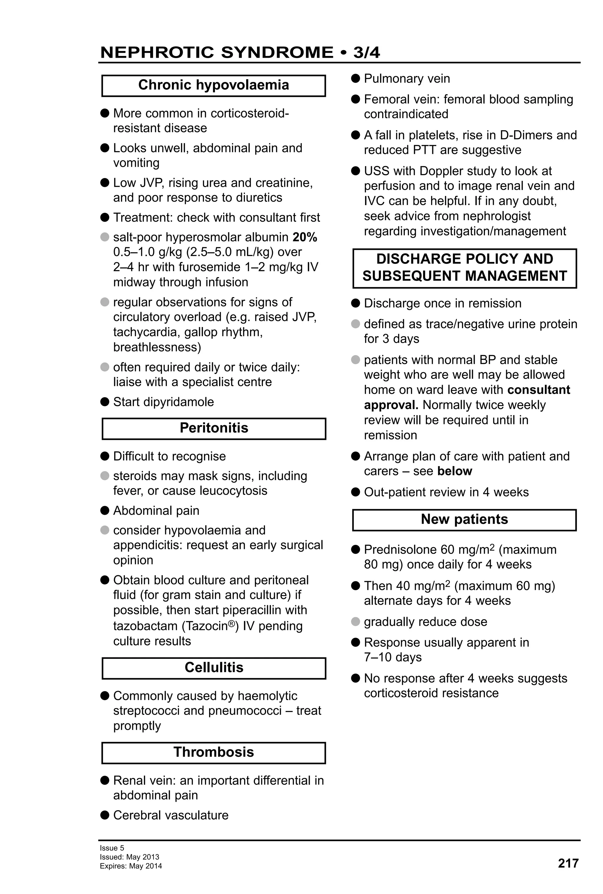
ASTHMA – ACUTE MANAGEMENT • 4/4
Issue 5
Issued: May 2013
Expires: May 2014
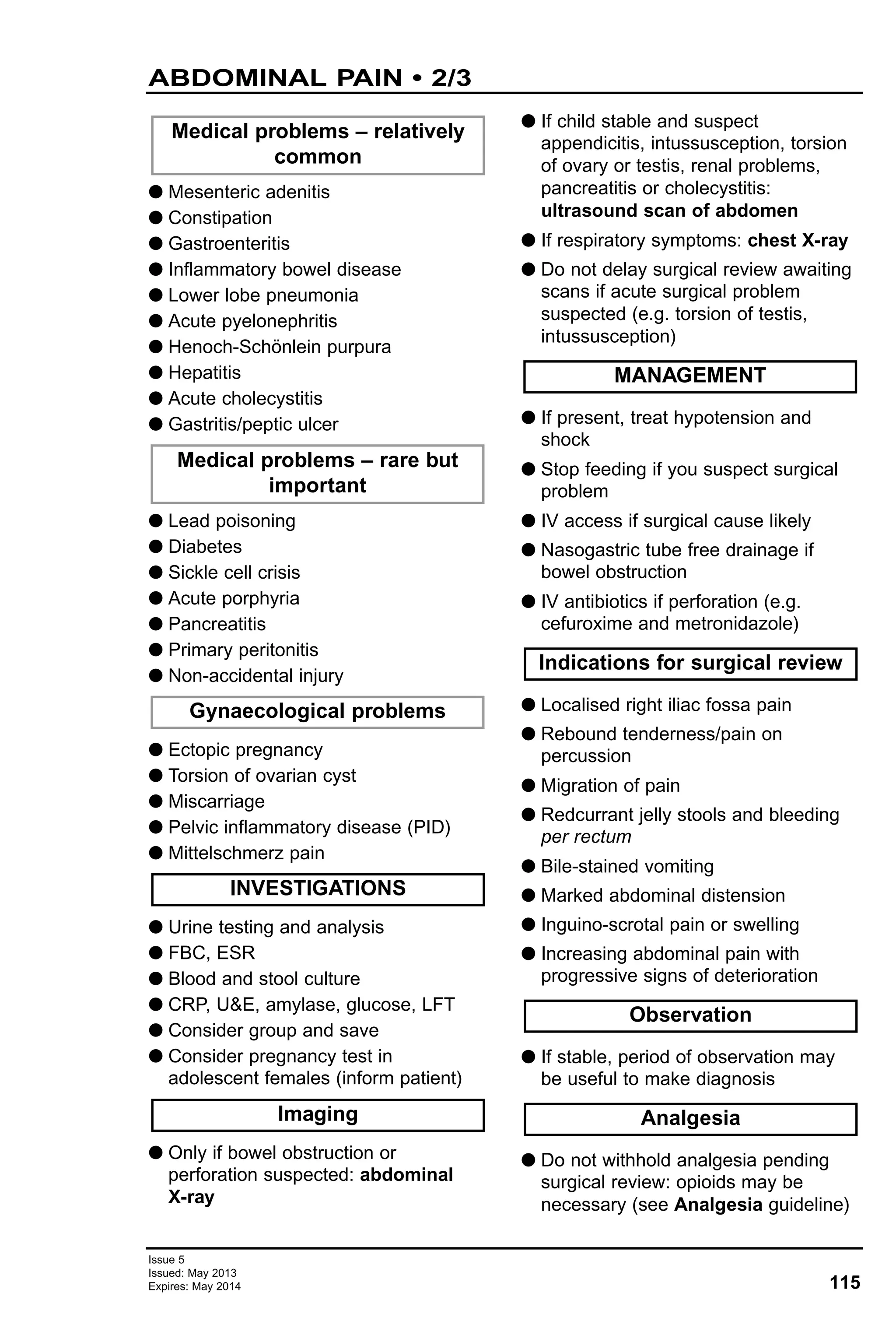
AssessmentCheck
MILD/MODERATE
Toobreathlesstotalk/feed
SpO2<92%inair
Useofaccessorymuscles
Respiratoryrate:>40breaths/minaged<5yr
>25breaths/minaged>5yr
Heartrate:>140beats/minaged<5yr
>125beats/minaged>5yr
Peakflow50%predicted/best
AssessABC
Cyanosis/pallor
Silentchest
Poorrespiratoryeffort
Alteredconsciousness
SpO2<92%inair
Irritable/exhausted
PEF33%inthoseaged7yr
Highflowoxygenviamaskornasalcannula
SalbutamolMDI10puffs(1000microgram)viaLVS
+/-facemask,or:
Salbutamolnebulised,drivenby68L/minoxygen:
Aged<5yr=2.5mg
Aged>5yr=2.55mg
Ifpoorresponse,giveipratropiumbromidenebulised:
Aged<12yr=250microgram
Aged>12yr=500microgram
Oncedailyoralprednisolone0.5mg/kg(ifalreadyon
maintenancetherapy,speaktorespiratory
consultant/nurse)
Aged<2yr=max10mgoncedaily
Aged25yr=max20mgoncedaily
Aged>5yr=max30mgoncedaily
Iforalsteroidsnottoleratedgivehydrocortisoneby
slowIVinjection
Aged<2yr=4mg/kg(max25mg)6-hrly
Aged25yr=50mg6-hrly
Aged518yr=100mg6-hrly
Haspatientreceived:
Continuoussalbutamolnebulised?
Ipratropiumbromidenebulised?
HydrocortisoneIV?
SALBUTAMOLBOLUS
Aged1month2yr=5microgram/kg(max
250microgram)
Aged218yr=15microgram/kg(max250microgram)
Using500microgram/mLinjectionpreparation,dilutetoa
concentrationof50microgram/mLwithsodiumchloride
0.9%(e.g.withdraw250microgram=0.5mLandmakeup
toatotalvolumeof5mLusingsodiumchloride0.9%=
250microgramin5mL).Calculatedoseperkgasabove
andadministerasaslowbolusover5min
MONITORING
Recordheartrateandrespiratoryrateevery10min
ContinuousSpO2andCO2monitoring
ECGmonitoring
BaselineU&E(capillarybloodgasforpotassium)
SalbutamolMDI210puffs(2001000
microgram)vialargevolumespacer(LVS)
+/-facemask
OxygenifSpO2<94%inair
Oncedailyoralprednisolone0.5mg/kg(if
alreadyonmaintenancetherapy,speakto
respiratoryconsultant/nurse)
Aged<2yr=max10mgoncedaily
Aged25yr=max20mgoncedaily
Aged>5yr=max30mgoncedaily
ADMIT
Continuehighflowoxygenviamask/nasalcannula
Nebulisedsalbutamol¼4hrly
Repeatipratropiumbromide.Ifpoorresponse,give
every2030minforfirst2hr
Informon-callconsultantandPICU
Highflowoxygenviamask/nasalcannula
Continuoussalbutamolnebulised,drivenby
68L/minoxygen
Ipratropiumbromidenebulised:
Aged<12yr=250microgram
Aged>12yr=500microgram
HydrocortisonebyslowIVinjection
Aged<2yr=4mg/kg(max25mg)6-hrly
Aged25yr=50mg6-hrly
Aged518yr=100mg6-hrly
Ifsignsofshock,sodiumchloride0.9%
20mL/kgIVbolus
Consideranaphylaxis
DISCHARGEHOME
Continueoncedailyoralprednisolone,
completea3daycourse
Reviewlong-termasthmacontrol+treatment
Checkinhalertechnique
Providepersonalasthmaactionplan
Agreefollow-upplan
Completerespiratorydischargeletter
DISCHARGECRITERIAMET
SpO2>94%inair
Respiratoryrate:<40breaths/minaged<5yr
<30breaths/minaged>5yr
Heartrate:<140beats/minaged<5yr
<125beats/minaged>5yr
Peakflow75%predicted/best
Stableon4-hrlyinhaledtreatment
Continuousnebulisedsalbutamol
Repeatipratropiumbromide.Ifpoor
response,giveevery2030minforfirst2hr
SalbutamolIV(seeSalbutamol
infusion)
ConsidermagnesiumsulphateIV
Bloodgas
ChestX-ray
SALBUTAMOLINFUSION
Using1mg/mLsolutionforIVinfusiondilutetoa
concentrationof200microgram/mLwithsodiumchloride
0.9%[e.g.take10mg(10mL)of1mg/mLsolutionforIV
infusionandmakeupto50mLwithsodiumchloride0.9%
=200microgram/mLsolution]
Infuseat60300microgram/kg/hr
=0.3mL/kg/hr1.5mL/kg/hrwhenusing200microgram/mL
solution
MONITORING
ContinuousSpO2andCO2monitoring
ECGmonitoring
Repeatbloodsat2hr,4hr,then4-hrly
Normalvitalsigns
Mildwheeze
Speakingincompletesentencesorfeeding
SpO2>92%inair
PEF>50%inthoseaged7yr
SEVERELIFE-THREATENING
RE-ASSESSEVERY1530MIN
YES
DISCHARGE
DISCHARGECRITERIAMETYES
NO
REASSESSFREQUENCYOF
BRONCHODILATORTHERAPY
SYMPTOMSIMPROVING
IMPROVING
REASSESS
REASSESS
SYMPTOMSIMPROVING
Ispatientstillnot
improving/worseningandmeets
severe/life-threateningcriteria?
NO
SYMPTOMS
IMPROVING
NOIMPROVEMENT
NOCHANGE
WORSENING
YES
NO
YES
YES
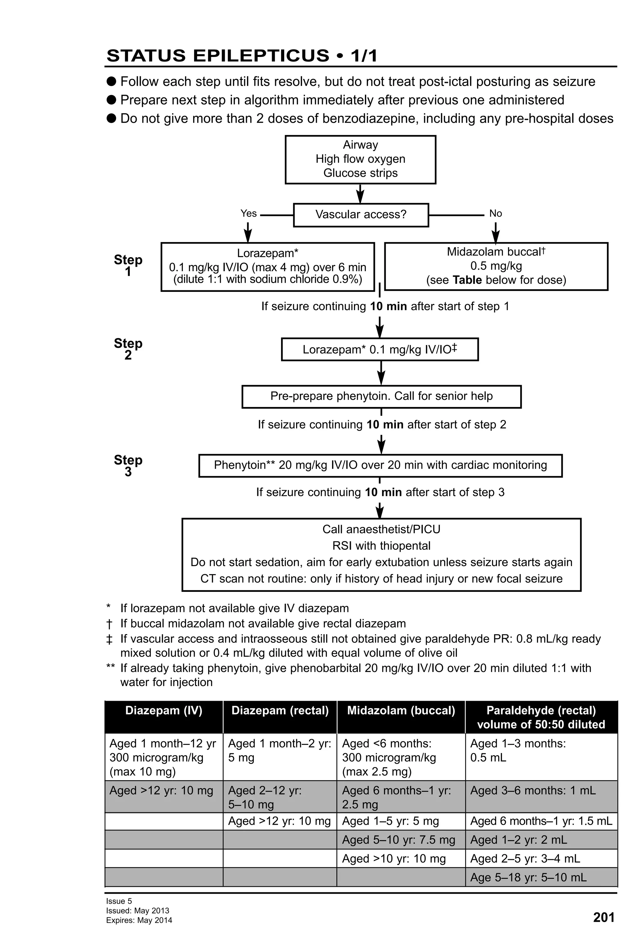
Algorithm: Management of acute wheezing in children](https://image.slidesharecdn.com/paediatricguidelines2013-14withlinks-180801135506/75/Paediatric-guidelines-2013-14-with-links-40-2048.jpg)


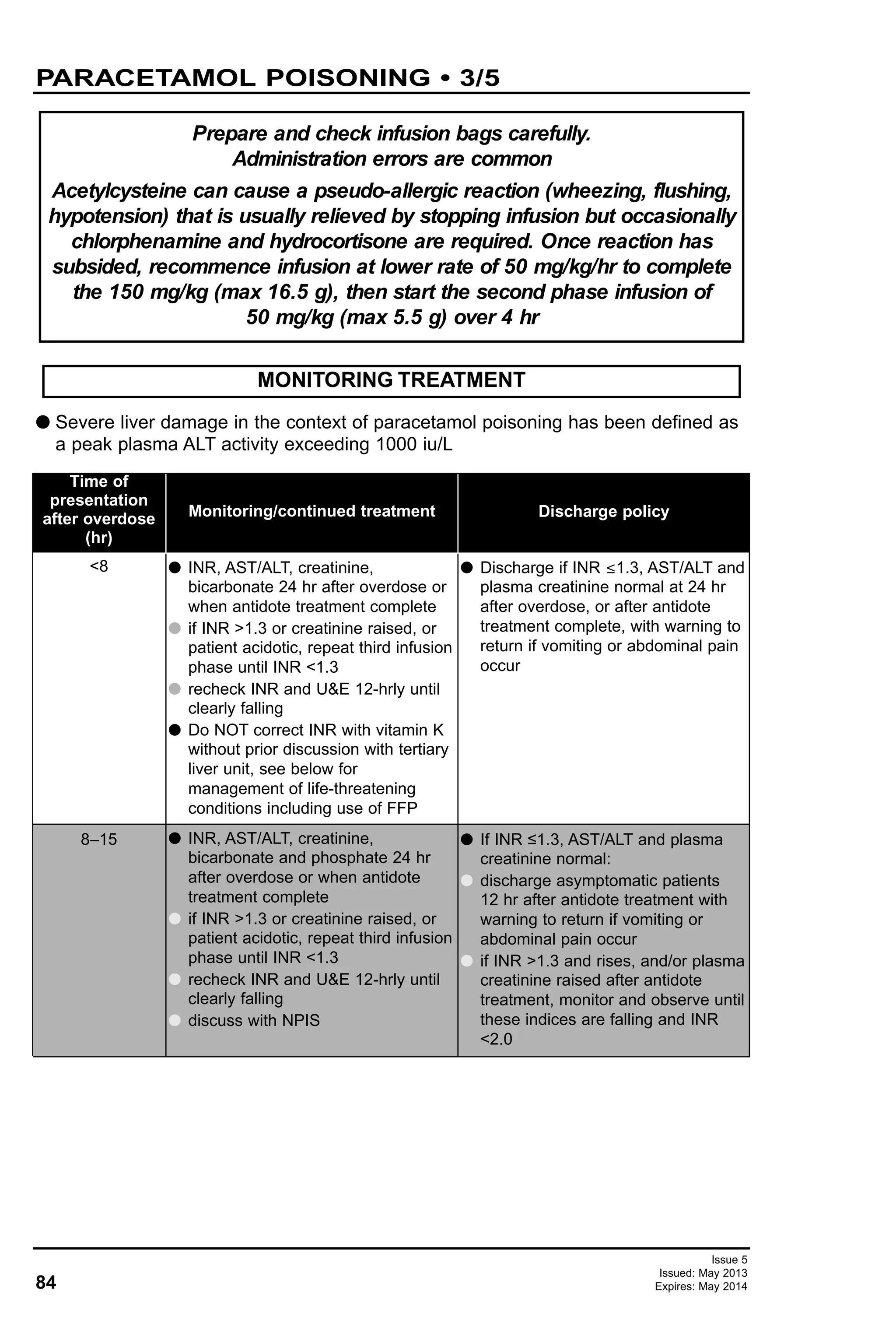
![82
Issue 5
Issued: May 2013
Expires: May 2014
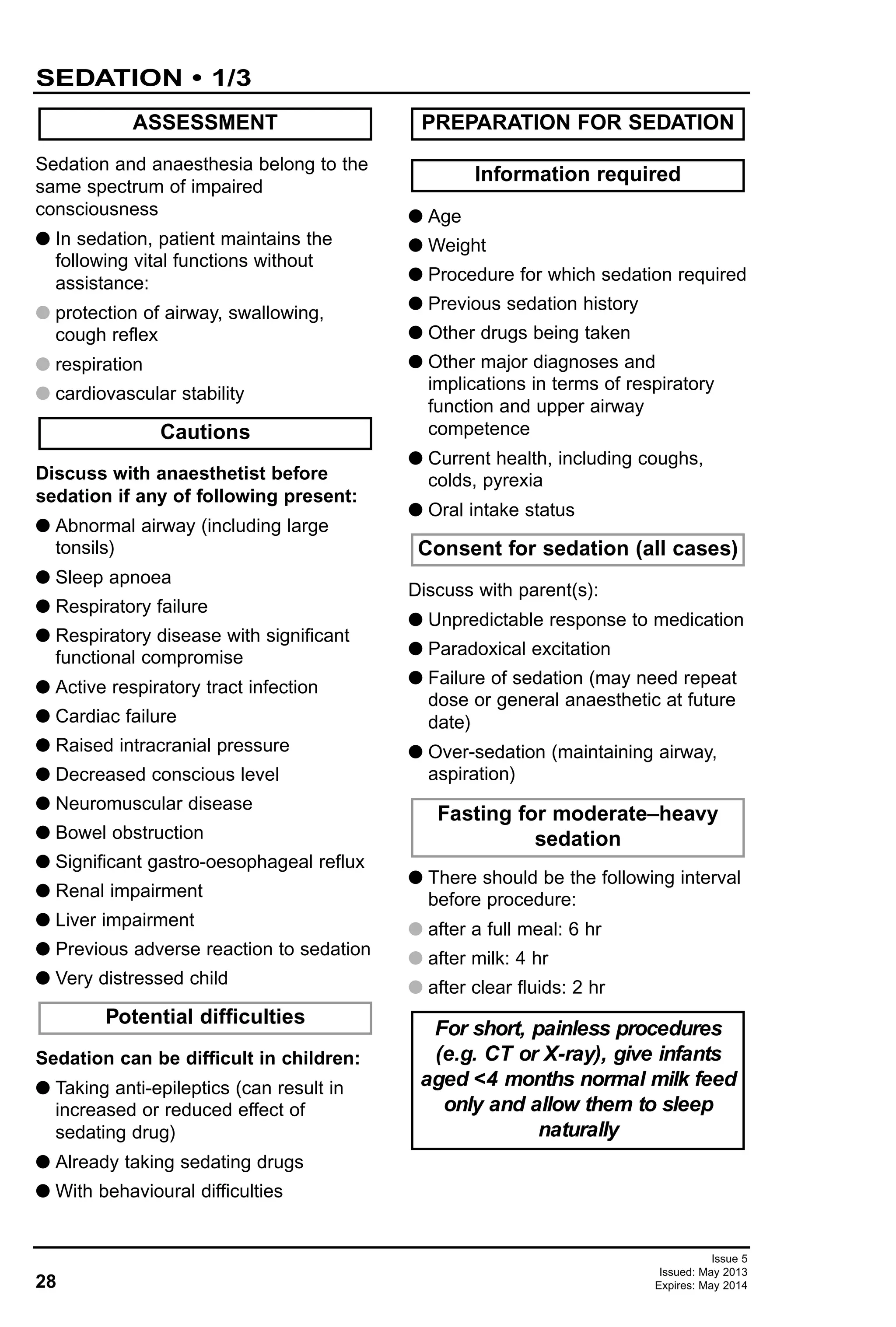
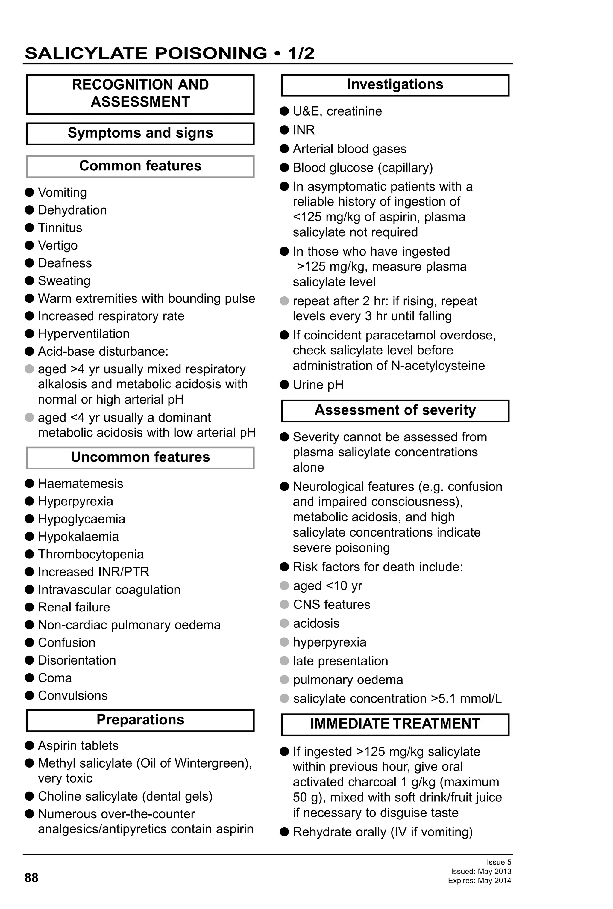
PARACETAMOL POISONING • 1/5
G Common: nausea and vomiting
G Rare: coma and metabolic acidosis
G Late: abdominal pain
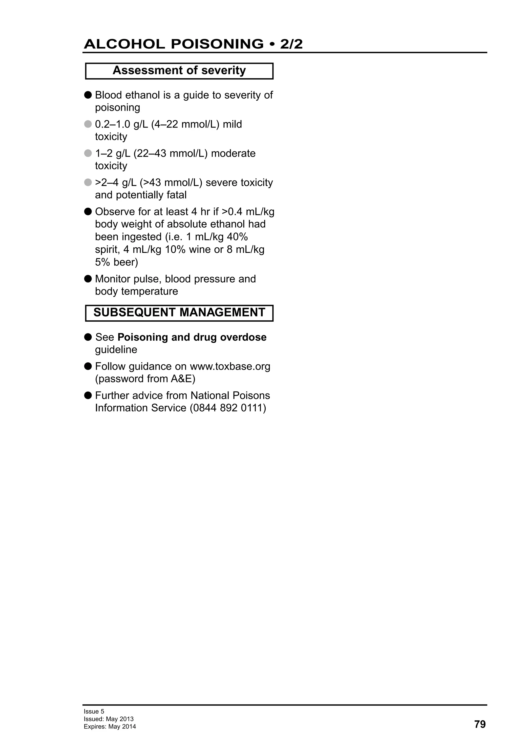
G Paracetamol dose >6 g or >75 mg/kg
G Staggered overdose [including
chronic therapeutic excess
>75 mg/kg/d (>60 mg/kg in neonate)]
G Symptomatic
OR
G INR >1.3 or ALT >upper limit of
normal, or abnormal acid/base or
bicarbonate
G Follow guidance on www.toxbase.org
(password from A&E)
G Further advice from National Poisons
Information Service (NPIS) 0844 892
0111
G If there is absolute certainty that a
single dose of paracetamol of <6 g
and <75 mg/kg has been ingested,
plasma paracetamol need not be
measured and child requires no
antidote
G Plasma paracetamol 4–16 hr (but not
outside this interval) is a reliable
guide to the need for treatment after
single overdose ingested of <60 min
G If patient presents >8 hr after single
overdose; or after staggered
overdose; request baseline:
G FBC, INR
G U&E, liver function, phosphate
G acid-base (venous sample)
G Compare plasma paracetamol with
treatment graph (Figure 1)
G if above, or on, the ‘treatment line’,
give IV acetylcysteine in glucose 5%
G Time interval is critical in assessing
need for treatment. Detailed
questioning essential
If there is doubt about timing or
need for treatment, treat
IMMEDIATE TREATMENT
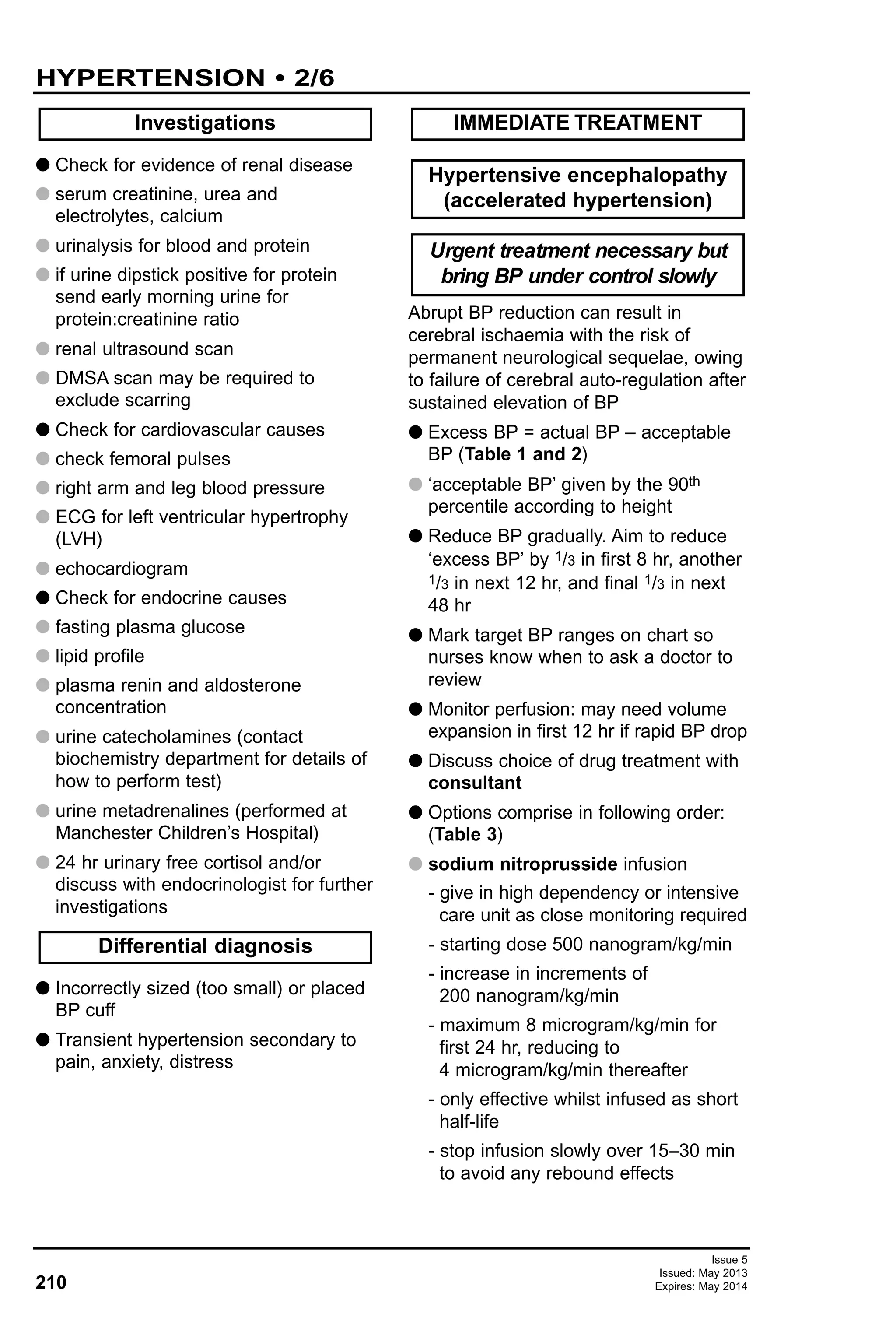
Investigations
Management for
Symptoms and signs
RECOGNITION AND
ASSESSMENT
<1
4–7
8–14
15–24
Multiple/
staggered
overdose
>24
Give activated charcoal 1 g/kg (max 50 g) oral or via nasogastric tube (gastric
lavage is not indicated)
Await paracetamol level if available <8 hr from ingestion. Treat if level ≥
‘treatment line’ OR if biochemical tests (INR, ALT) suggest acute liver injury
Give at once while awaiting paracetamol concentration result. Cease if concentration
well below appropriate ‘treatment line’ and ALT within normal limit, INR ≤1.3
Give at once. Cease at 24 hr after ingestion if patient asymptomatic, and INR
≤1.3, and ALT <upper limit of normal. Otherwise complete antidote course
Plasma paracetamol will confirm ingestion but cannot be related to nomogram.
Start acetylcysteine and discuss with NPIS
Give if paracetamol still detectable in the blood (>5 mg/L), or INR >1.3 or ALT
>twice upper limit of normal, or symptomatic.
If patient has, or is at risk of developing, fulminant hepatic failure (see life-
threatening features below), continue to give 50 mg/kg in 500 mL every 8 hr
Discuss with NPIS and follow Toxbase guidance
Time from
overdose (hr)
Guidance on use of acetylcysteine](https://image.slidesharecdn.com/paediatricguidelines2013-14withlinks-180801135506/75/Paediatric-guidelines-2013-14-with-links-82-2048.jpg)
![119
Issue 5
Issued: May 2013
Expires: May 2014
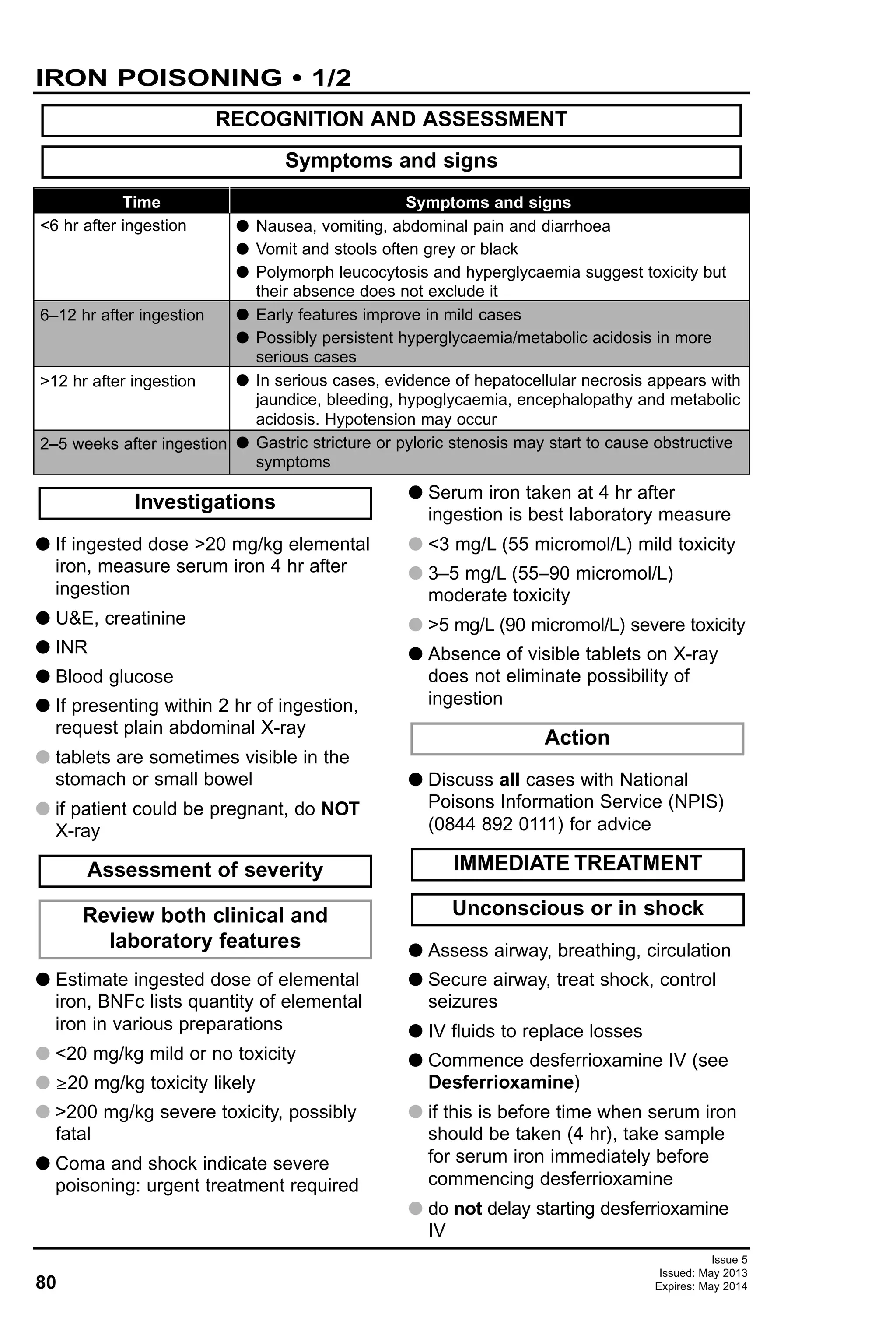
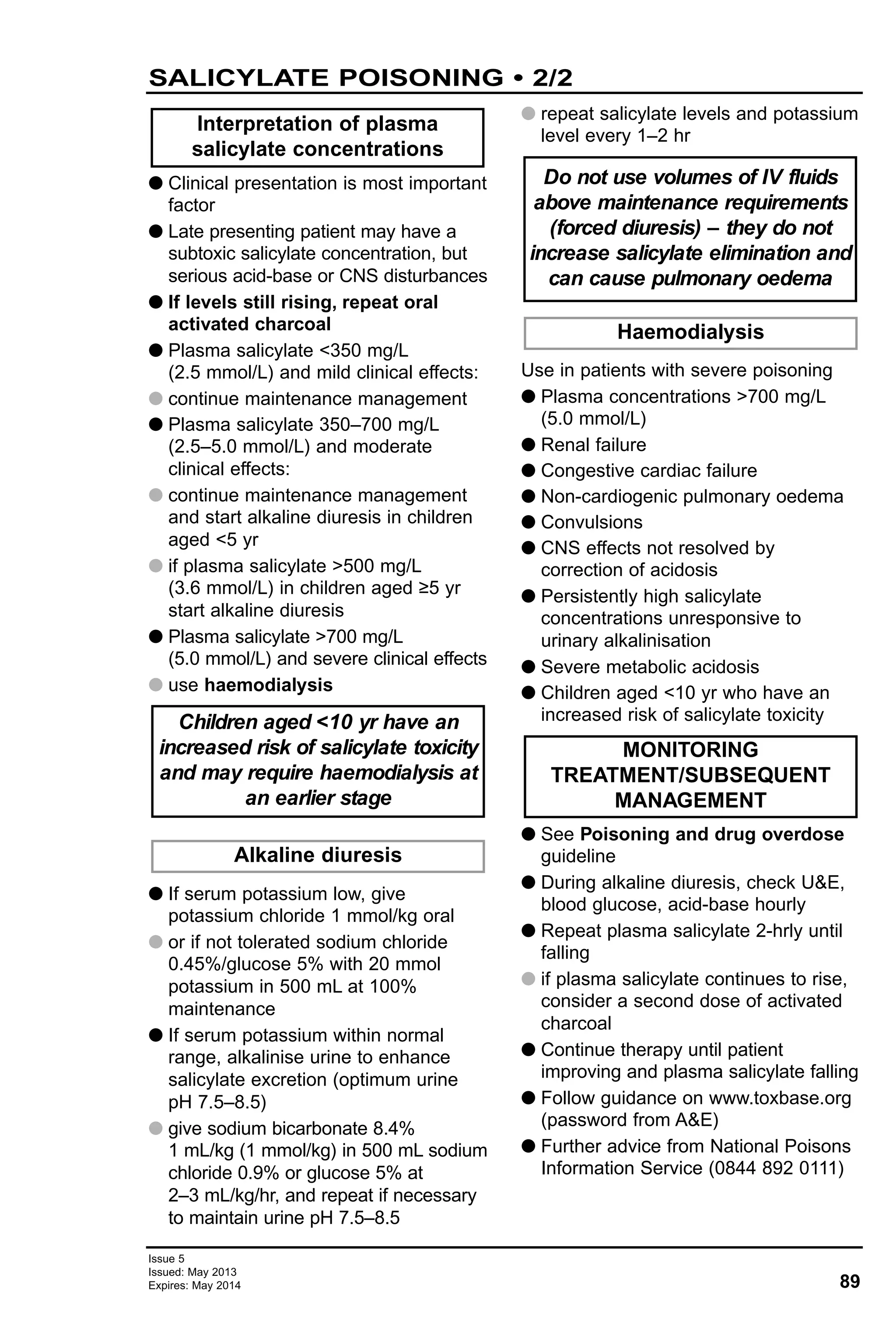
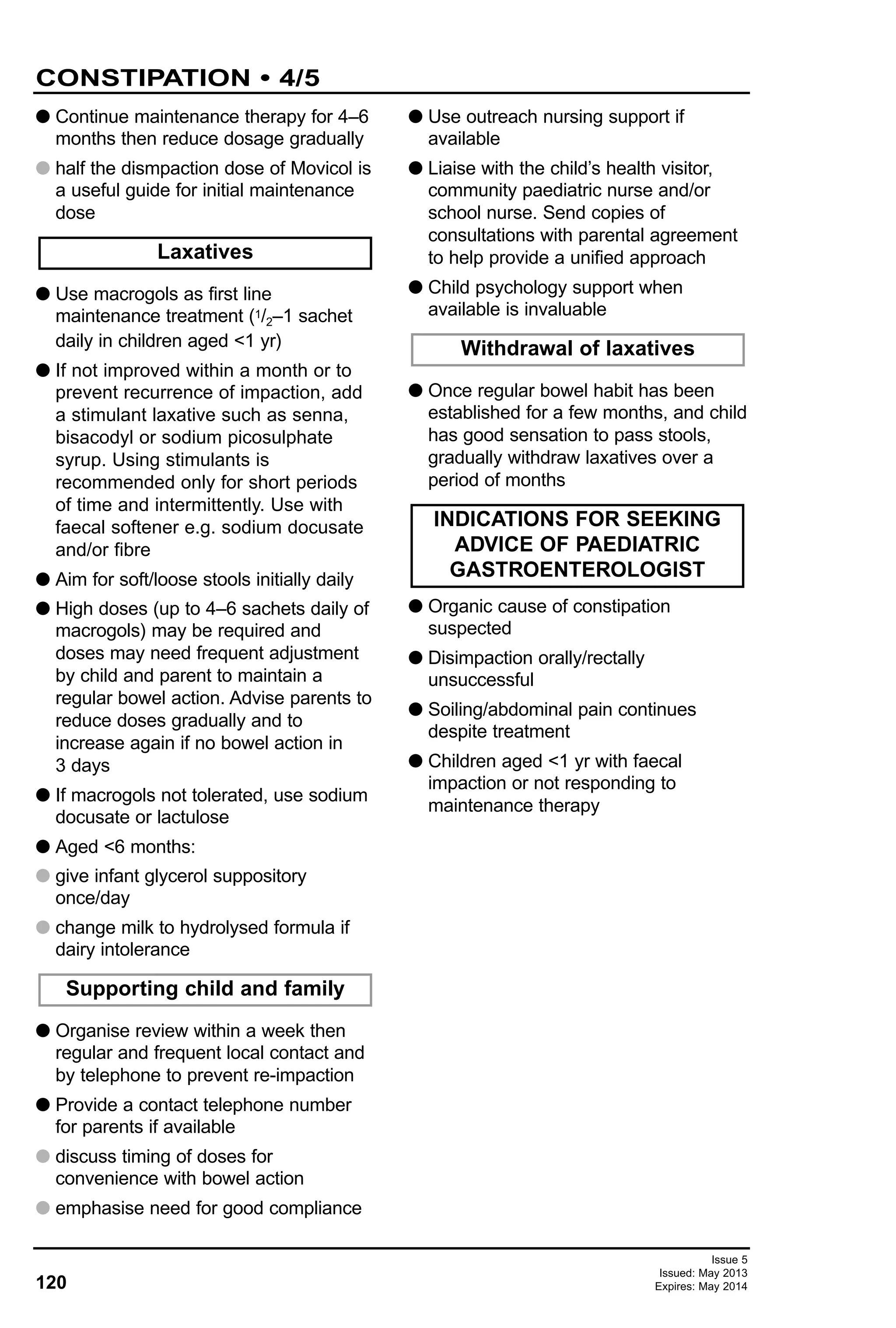
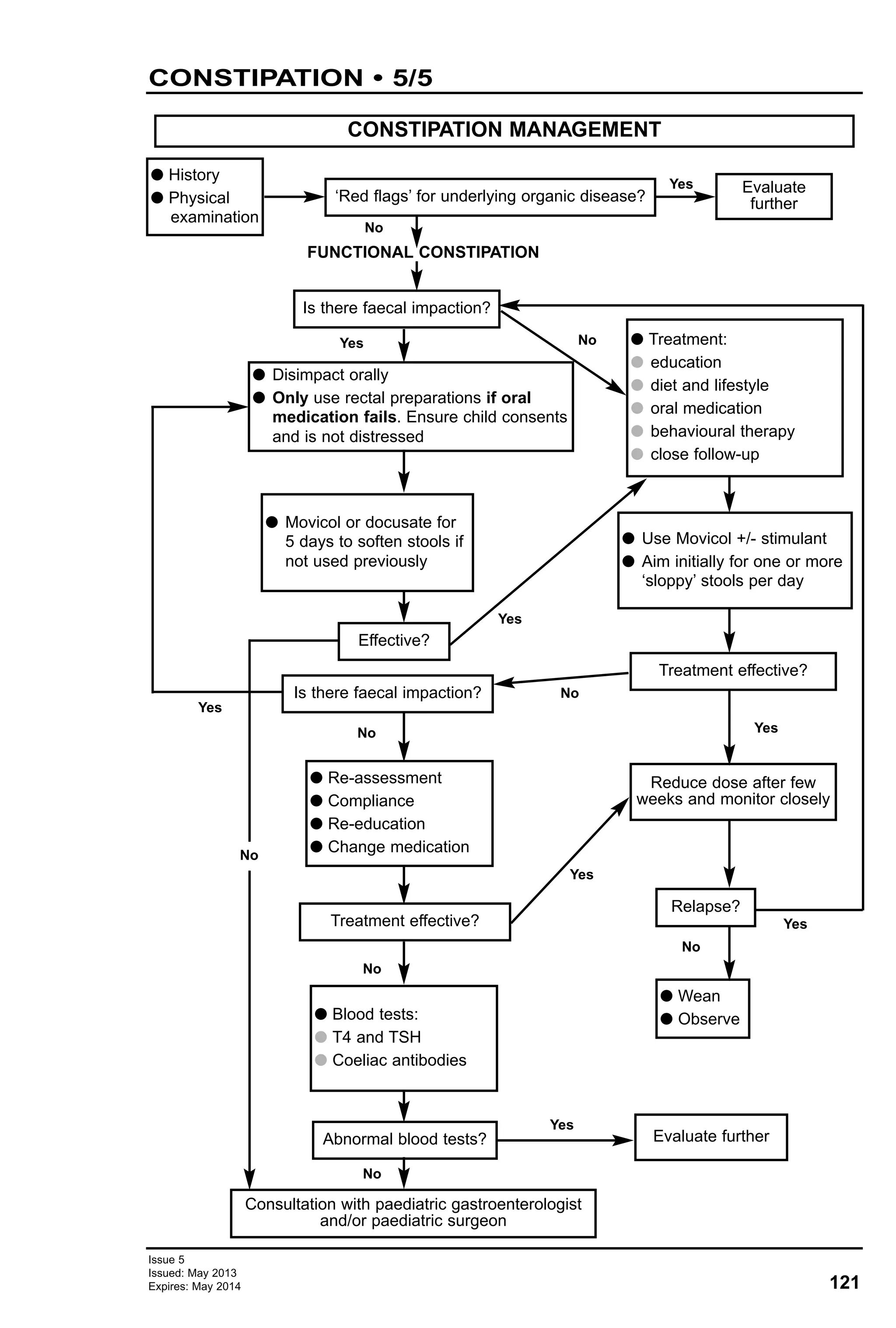
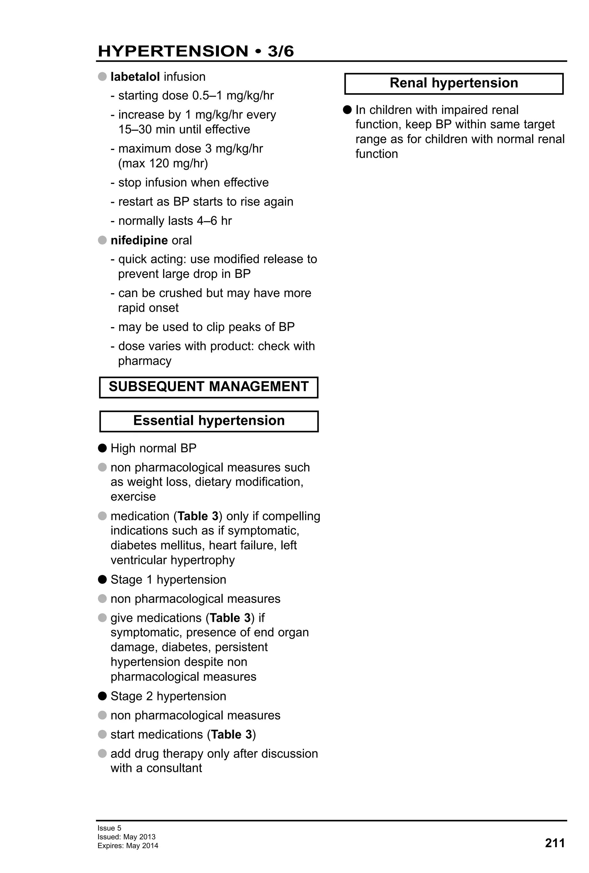
Disimpaction dosage
CONSTIPATION • 3/5
1–5
5–12
2
4
4
6
4
8
6
10
6
12
8
12
8
12
>12–18 4 6 8 8 8 8 8
G Use of behavioural management in
combination with medications
decreases time to remission
G regular toileting: unhurried time on the
toilet after meals
G correct toilet position
G maintain diaries of stool frequency
combined with reward system
G regular review and positive
reinforcement
G discourage negative responses to
soiling from family
G encourage older children to take
responsibility
G May need counselling or a
psychology referral in case of
motivational or behavioural problems
G Disimpaction in the presence of
impacted stools
1. A macrogol laxative [polyethylene
glycol (e.g. Movicol paediatric plain)];
faecal impaction dose, see below up
to a maximum of 7 days
2. Use stimulant laxative, senna or
sodium picosulphate (Picolax) if no
result with macrogol or if not tolerated
3. Review all children within/after one
week of disimpaction (in hospital or
by GP)
Behavioural management Medication
DISIMPACTION
Aged <1 yr, refer to paediatric
gastroenterologist
G Sodium citrate micro-enemas (Relaxit)
G Small volume sodium citrate enemas
(Microlax) is preferable to large volume
phosphate enemas
G Phosphate enemas (only if oral
medications and Microlax enemas
failed). Use only under specialist
supervision. Consider sedation if child
is distressed
G If all above have failed, consider
manual evacuation under general
anaesthetic. Consult with paediatric
gastroenterologist or paediatric
surgeon
G After disimpaction, or if child had no
impaction, focus treatment on
prevention of recurrence and
establishment of a regular bowel habit
to allow bowel to regain normal tone
and sensation
MAINTENANCE THERAPY
Rectal disimpaction
(only if oral disimpaction fails)
Manual evacuation
Age (yr) Day 1 Day 2 Day 3 Day 4 Day 5 Day 6 Day 7
Number of Movicol paediatric plain sachets daily divided into 2–3 doses
Number of adult Movicol preparation for children aged >12 yr](https://image.slidesharecdn.com/paediatricguidelines2013-14withlinks-180801135506/75/Paediatric-guidelines-2013-14-with-links-119-2048.jpg)
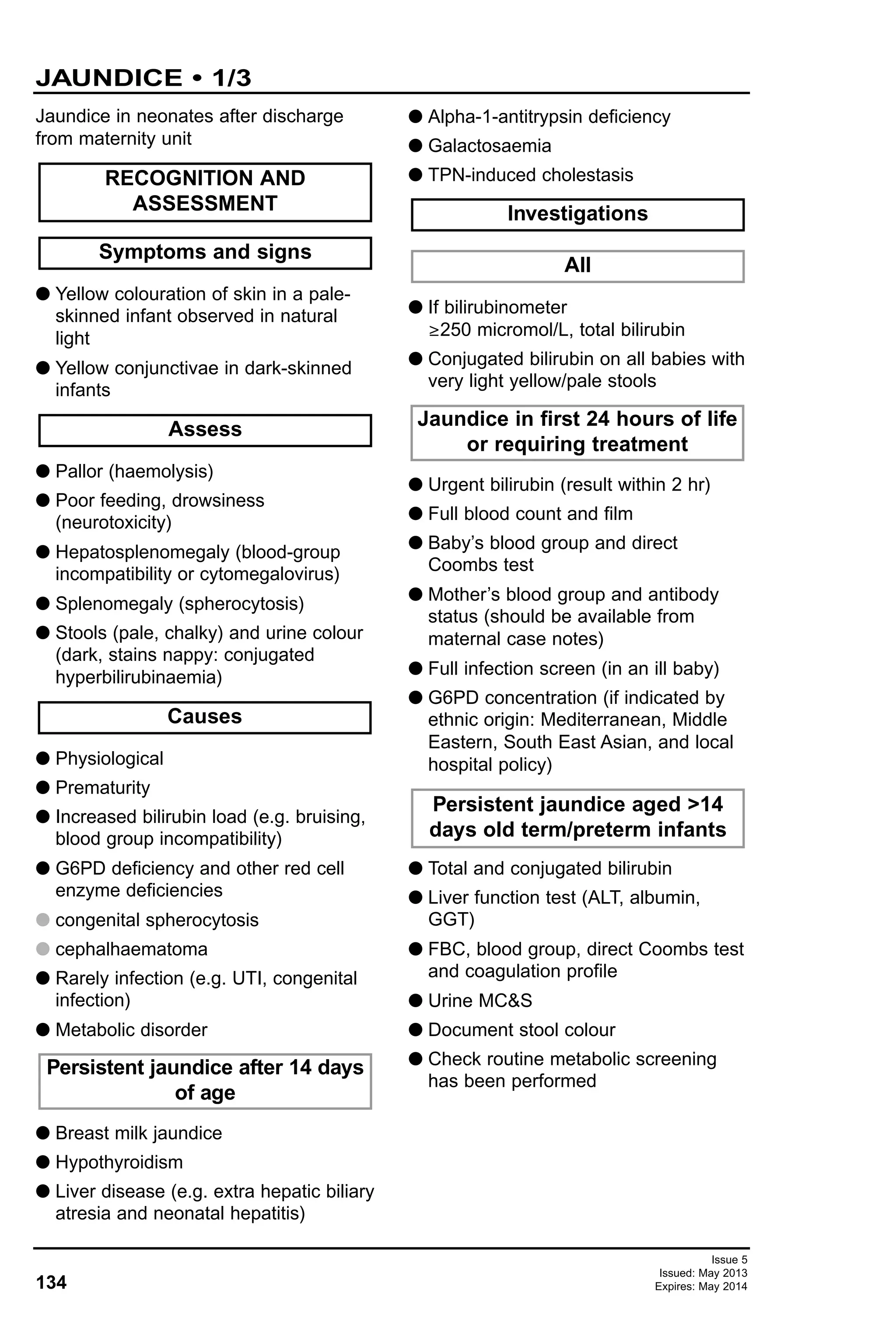
![136
Issue 5
Issued: May 2013
Expires: May 2014
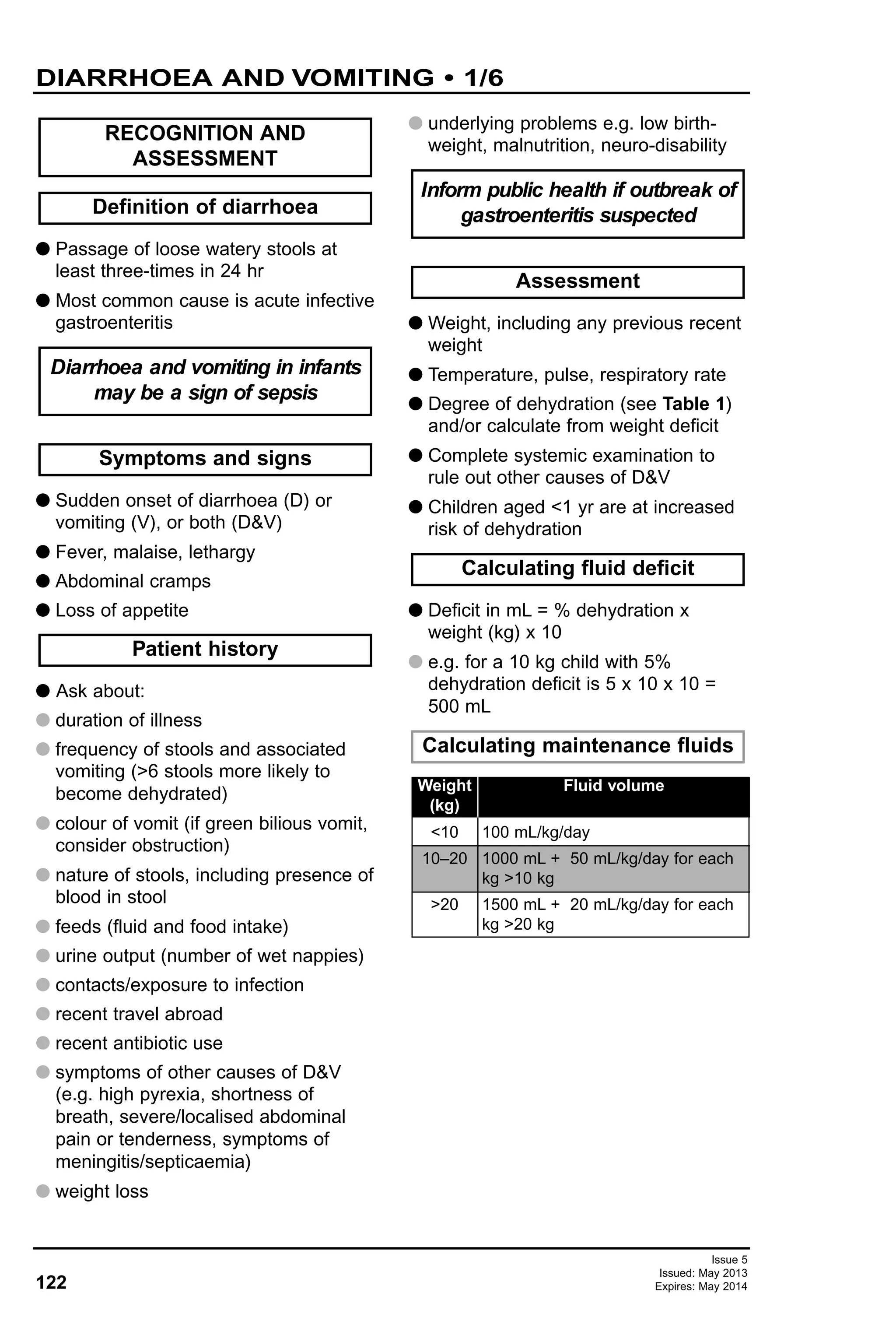
G Adequate fluid and energy intake
G Phototherapy
G Visible jaundice can be treated with
phototherapy after sample taken for
bilirubin measurement
G Bilirubin >100 micromol/L: repeat in
6–12 hr
G Commence phototherapy according to
following equation:
G for infants <37 weeks, start if serum
bilirubin (micromol/L) ≥ phototherapy
level [(gestational age in completed
weeks x 10) – 100]
G for infants ≥37 weeks, start if serum
bilirubin >340 micromol/L
(phototherapy level)
G If bilirubin near exchange threshold or
still rising:
G increase power number of lights
G increase area exposed (e.g.
biliblanket and overhead)
G See Exchange transfusion in
Neonatal guidelines
G Use as an adjunct to multiple
phototherapy in rhesus disease when
bilirubin continues to rise by
>8.5 micromol/L/hr
G If haemolysis present, check bilirubin
4–6 hrly until rate of rise flattens
G If bilirubin concentration approaching
threshold for exchange transfusion, or
rising rapidly (>10 micromol/hr), check
4-hrly
G When bilirubin concentration has
fallen below threshold for
phototherapy (see above), discontinue
phototherapy
G If jaundice persists after 14 days of
age, review and treat cause
G GP follow-up with routine examination
at 6–8 weeks
G If exchange transfusion necessary or
considered, request development
follow-up and hearing test
G In babies with positive Coombs test
who require phototherapy, check
haemoglobin at 2 and 4 weeks of age
because of risk of continuing
haemolysis and give folate
DISCHARGE AND FOLLOW-UP
SUBSEQUENT MANAGEMENT
IVIG
Exchange transfusion
After first 24 hours
MONITORING TREATMENT
Phototherapy
Jaundice presenting in first
24 hours of life
TREATMENT <7 DAYS
JAUNDICE • 3/3](https://image.slidesharecdn.com/paediatricguidelines2013-14withlinks-180801135506/75/Paediatric-guidelines-2013-14-with-links-136-2048.jpg)
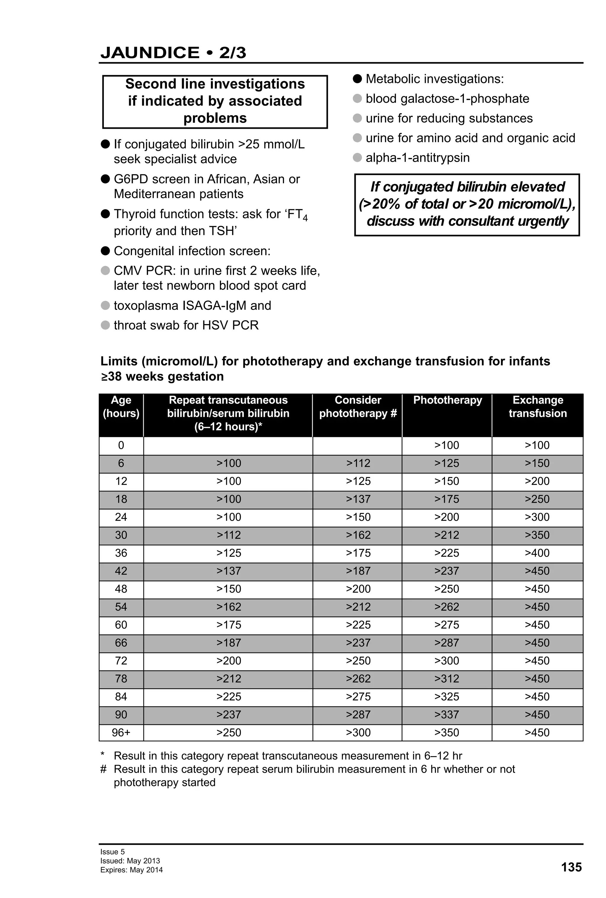
![138
Issue 5
Issued: May 2013
Expires: May 2014
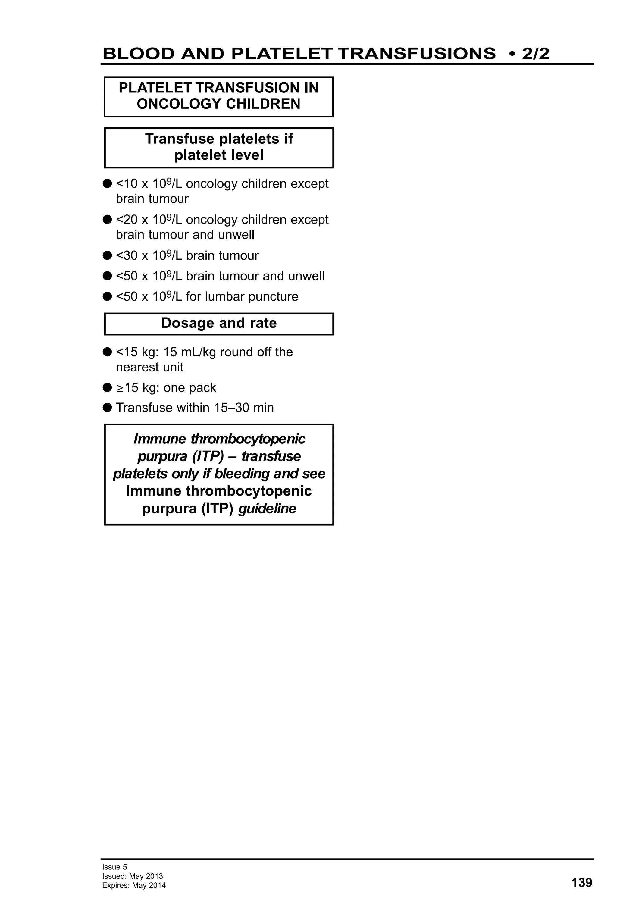
G Explain indications for blood products
to parents
G Document indications and verbal
consent
G If previous reactions to blood products
have occurred, pre-medicate with
chlorphenamine (oral or IV), if severe
with hydrocortisone 4 mg/kg IV
G If haemoglobin ≤80 g/L or if >80 g/L
and symptomatic, transfuse
G If having radiotherapy, transfuse if Hb
<100 g/L
G If haemoglobin <60 g/L or >60 g/L
and symptomatic
G Aim for target haemoglobin of 120 g/L
or for 100 g/L if initial haemoglobin
<60 g/L
G In newly diagnosed patients with
leukaemia/profound anaemia, aim for
target Hb 80–90 g/L
G Calculate volume to be given as:
(round to nearest unit)
[Target Hb – actual Hb (g/L)] x
weight (kg) x 0.4 mL
G Total volume should not exceed
20 mL/kg
G Give total over 3–4 hr. Max rate
5 mL/kg/hr
G If Hb <60 g/L, give blood over 4–8 hr
(each unit must be used within 4 hr
once removed from fridge)
G Give furosemide 1 mg/kg oral if
tolerated, or IV half-way through
G Allogenic bone marrow transplant
(BMT) from start of conditioning
regimen
G Allogenic BMT donors
G If <7 days pre-harvest for autologous
BMT and stem cell transplant patients
(e.g. stage IV neuroblastoma)
G Hodgkin’s disease or if patient has
received fludarabine
G Children with severe
immunodeficiency (e.g. SCID)
G HLA-matched platelets
G For high risk neonates e.g. post
intrauterine transfusion
G All packed cells are leucodepleted
G All the packed cells are leucodepleted
and therefore CMV negative
G For neonates aged <28 days post
expected date of delivery and for
intrauterine transfusions CMV
serology negative blood requested
CMV negative blood
Leucodepleted blood
Non-oncology children
Oncology children
When to transfuse
Before transfusion
Use irradiated blood if
Rate of infusion
Target Hb and volume to be
transfused
BLOOD TRANSFUSION
Always check front sheet in
patient notes before prescribing
any blood product
BLOOD AND PLATELET TRANSFUSIONS • 1/2](https://image.slidesharecdn.com/paediatricguidelines2013-14withlinks-180801135506/75/Paediatric-guidelines-2013-14-with-links-138-2048.jpg)
![152
Issue 5
Issued: May 2013
Expires: May 2014
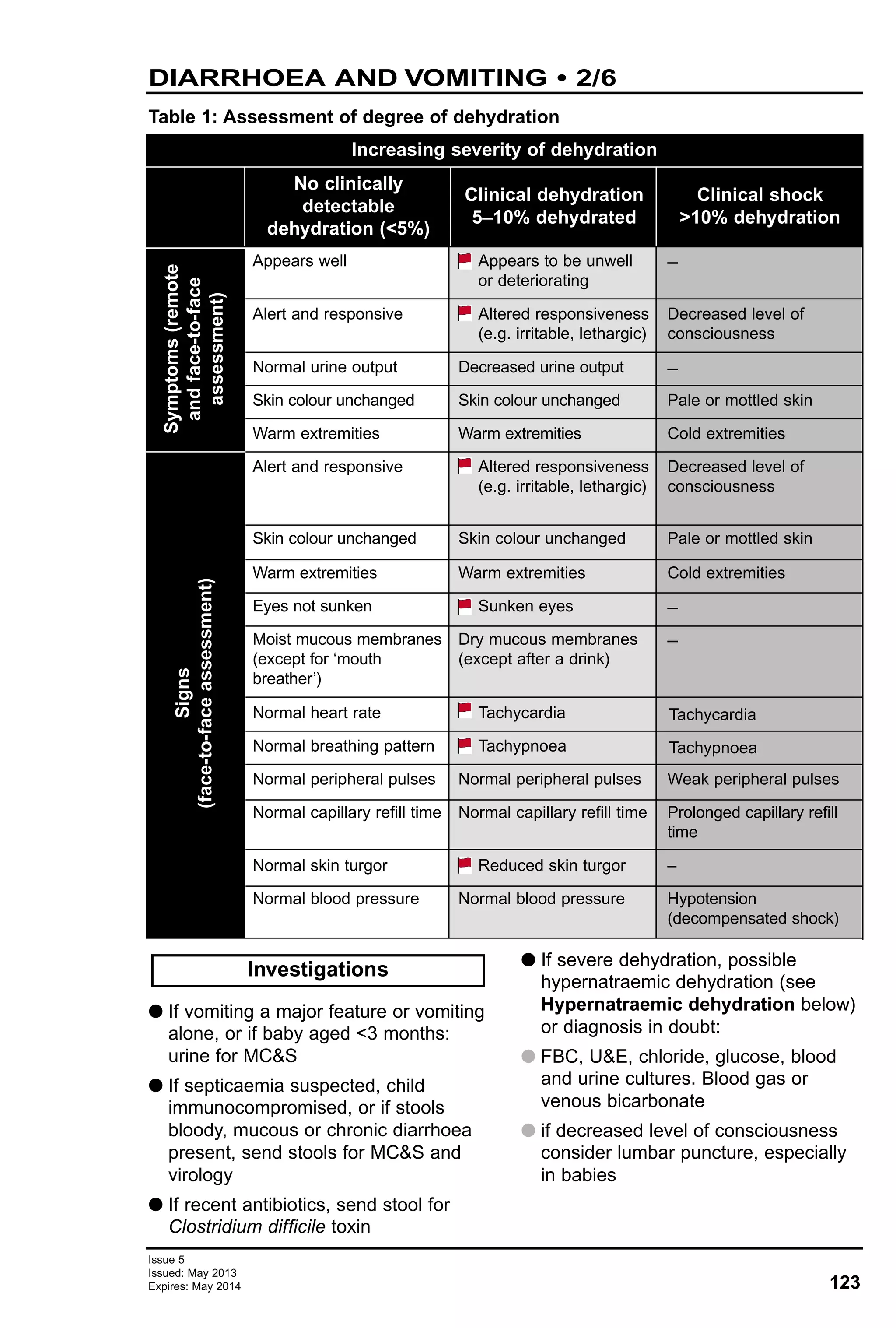
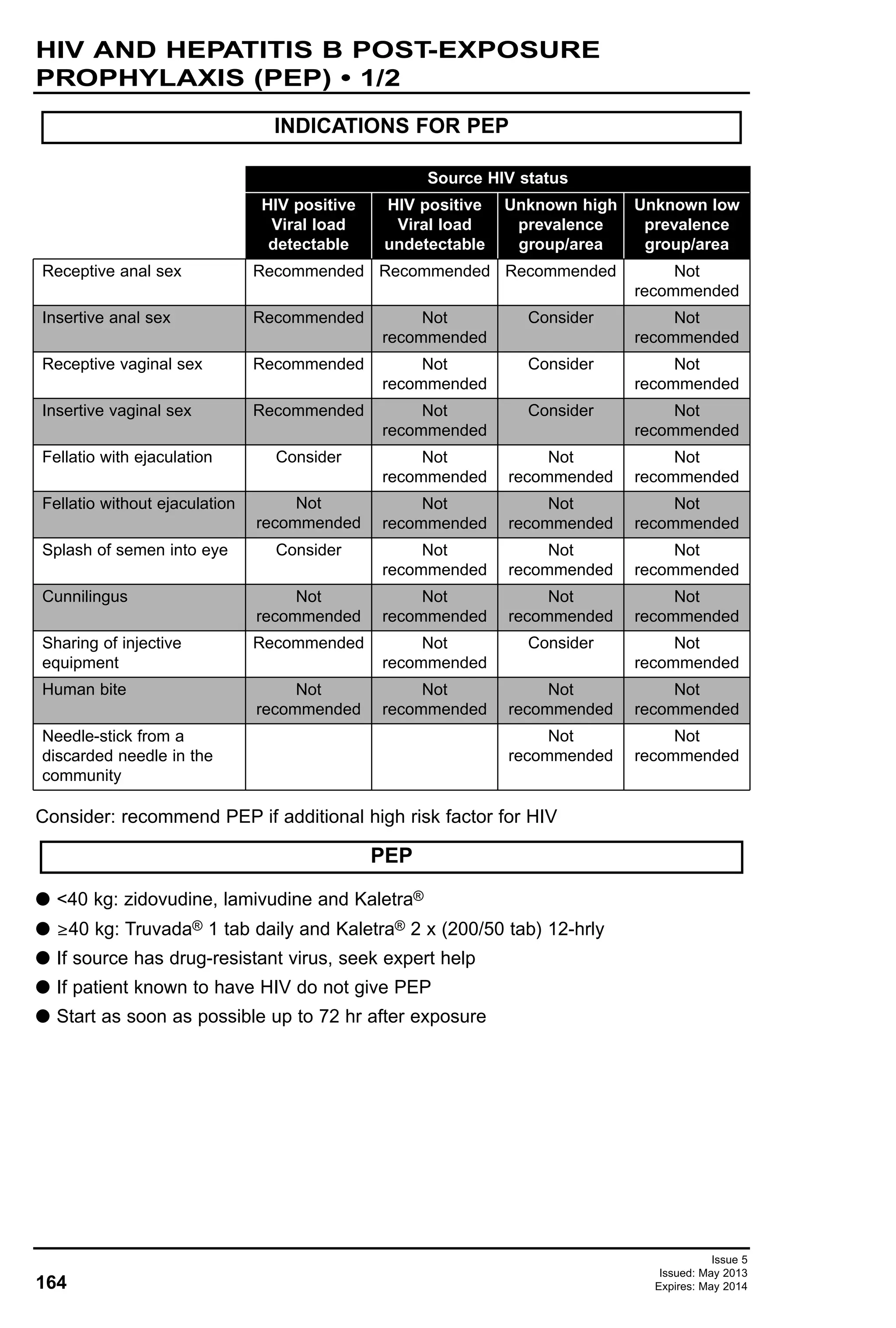
Prevention of infection after bites from
humans and other animals
G All human bite wounds ≤72 hr old,
even if no sign of infection
G Animal bite wounds if wound ≤48 hr
old and risk of infection high as follows:
G bites to hand, foot, and face; puncture
wounds; wounds requiring surgical
debridement; crush wounds with
devitalised tissue; wounds in genital
areas; wounds with associated
oedema; wounds involving joints,
tendons, ligaments, or suspected
fractures
G wounds that have undergone primary
closure
G patients at risk of serious wound
infection (e.g. immunosuppressed)
G asplenic patients, even after trivial
animal bites
G patients with prosthetic implants e.g.
heart valve, VP shunt
G antibiotics are not generally needed if
wound ≥2 days old and no sign of local
or systemic infection
G Advise patient and carers of signs of
developing infection and to attend
urgently for review should this happen
G Ask vaccination and HIV/Hep B and C
status of person bitten
G Ask if biter is willing to be tested
G Risk of HIV transmission is extremely
small. See HIV and hepatitis B post-
exposure prophylaxis (PEP) guideline
G Seek advice from consultant
microbiologist or consultant in
infectious diseases
G If significant risk of blood borne virus
transmission, offer to test person bitten:
Source is known or suspected
to be positive for HIV, hepatitis
B or C, or rabies
Give prophylactic antibiotics to
BITES • 1/1
Human, dog or cat†
If clinical infection present, send tissue, aspirate or swab for bacterial culture
If patient systemically unwell, blood cultures
Tetanus vaccine [e.g. combined diphtheria (low dose), tetanus, and
poliomyelitis] and immunoglobulin if indicated (see current BNFc)
First line Alternative (penicillin allergy)
Co-amoxiclav Clindamycin and cotrimoxazole
5 days
Antibiotic prophylaxis
† For bites from other mammals, contact consultant microbiologist or consultant in infectious
diseases
Type of bite
Specimen
Treatment
Duration
Time
clotted sample for archiving at time of incident
6 weeks
3 months
6 months
PCR
PCR and antibody
Antibody
Hepatitis C
HBsAg Antigen/antibody combined test
HBsAg Antigen/antibody combined test
HBsAg
Anti-HBc antibody
(anti-HBs antibody)*
Antigen/antibody combined test if PEP
was given
Hepatitis B HIV
* Anti-HBs only needed at 6 months if vaccination only started at injury](https://image.slidesharecdn.com/paediatricguidelines2013-14withlinks-180801135506/75/Paediatric-guidelines-2013-14-with-links-152-2048.jpg)
![G Type of thermometer used, site, user
(factitious)
G Duration, height
G Pattern:
G intermittent [pyogenic, TB, lymphoma,
juvenile idiopathic arthritis (JIA)]
G baseline raised (viral, endocarditis,
lymphoma)
G sustained (typhoid)
G days between (malaria, lymphoma)
G weeks between (metabolic, CNS,
cyclic neutropenia, hyperIgD)
G Circumstances when fever (e.g.
exercise)
G Appearance
G when fever: well (factitious)
G between fever: ill (serious)
G Response to paracetamol and or
NSAID (no response: dysautonomia)
G Red eyes (Kawasaki)
G Nasal discharge (sinusitis)
G Recurrent pharyngitis with ulcers
(periodic fever)
G GI: salmonella, intra-abdominal
abscess, inflammatory bowel disease
(IBD)
G Limb pain (leukaemia, osteomyelitis)
G Human illness
G Animals
G Years ago (histoplasmosis)
G Part of country
G Prophylaxis and immunisations
G Contaminated water/food
G Bites (tick: arbovirus, malaria)
G Meat: undercooked (brucella,
toxoplasma, hepatitis)
G Pica (visceral larva migrans,
toxoplasmosis)
G Operations
G All, including any non-prescription
G Sephardic Jew, Armenian, Turkish,
Arab (Familial Mediterranean Fever)
G Ashkenazi Jew (familial
dysautonomia)
G Sinuses
G Lymph nodes
G Chest: murmur, crackles
G Abdominal: hepato/spleno-megally
(salmonella, cat scratch, endocarditis,
malaria)
G Genito-urinary: girls – pelvic
tenderness (child sex abuse – STI)
G Rash only during fever (JIA)
G No sweat (familial dysautonomia)
G Petechiae (endocarditis, rickettsia)
G Papules (cat scratch)
G Eschar (tularaemia)
G Erythema migrans (Lyme)
G Malar (SLE)
G Palpable purpura [polyarteritis nodosa
(PAN)]
G Erythema nodosum (JIA, SLE,
malignancy, IBD, TB)
G Seborrheic (histiocytosis)
G Sparse hair (ectodermal dysplasia)
G Scars (dysautonomia)
Skin
Examination
Ethnic group
Drug history
Medical history
Travel
Contact
Symptoms
Fever
RECOGNITION AND
ASSESSMENT
161
Issue 5
Issued: May 2013
Expires: May 2014
FEVER OF UNKNOWN ORIGIN • 1/2](https://image.slidesharecdn.com/paediatricguidelines2013-14withlinks-180801135506/75/Paediatric-guidelines-2013-14-with-links-161-2048.jpg)
![G Hepatitis B vaccine accelerated course
(0, 1, 2 and 12 months)
G Hepatitis B immunoglobulin only if
source known to be HBsAg +ve
G Before discharge, provide families
embarking on HIV PEP with:
G appointment to see a paediatrician
with experience in antiretroviral drugs
or member of ID/GUM team the same
day or next working day
G contact telephone number in case of
concerns about any aspect of HIV
PEP
G enough antiretroviral medication to last
until clinic appointment
G letter for GP
G After sexual exposure consider
emergency contraception and screen
for other sexually transmitted infections
G Arrange HBV, HCV and HIV antibody
test baseline and 3 months after
exposure
G Check FBC, U&Es and LFTs if starting
PEP
G Check need for tetanus immunisation
G If source is HCV RNA PCR positive,
arrange the following enhanced HCV
follow-up:
G at 6 weeks: EDTA blood for HCV PCR
G at 12 weeks: EDTA blood for HCV
PCR and clotted blood for anti-HCV
antibodies
G at 24 weeks: clotted blood for
anti-HCV antibodies
FOLLOW-UP
If HIV PEP indicated give
HEPATITIS PEP
165
Issue 5
Issued: May 2013
Expires: May 2014
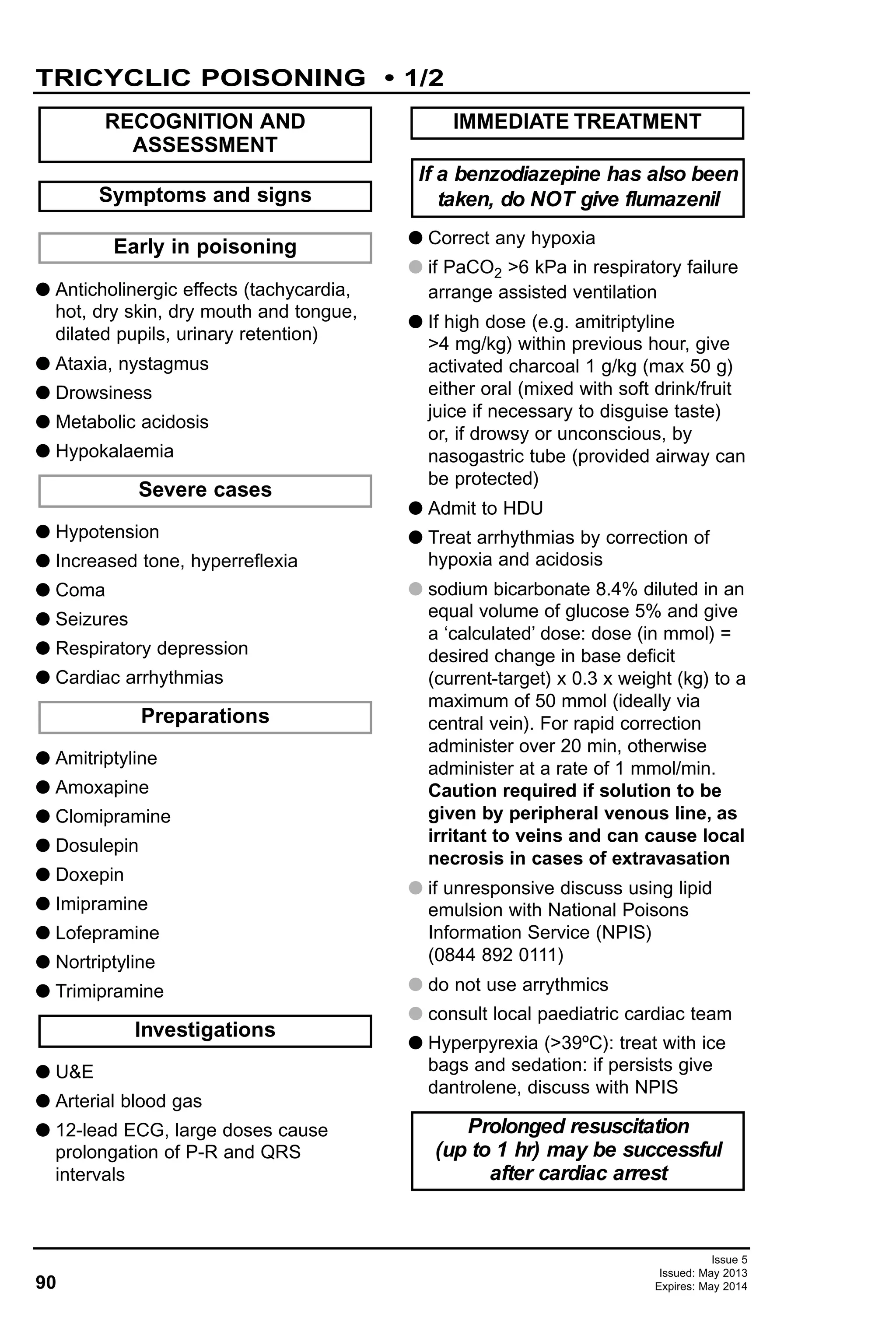
HIV AND HEPATITIS B POST-EXPOSURE
PROPHYLAXIS (PEP) • 2/2
HIV PEP drugs
Drug
Zidovudine
(ZDV or AZT)
Lamivudine (3TC)
Truvada®
(TDF and FTC)
Kaletra®
[lopinavir (LPV)/
ritonavir (RTV)]
Dose
180 mg/m2/dose
(max 250 mg)
12-hrly
4 mg/kg/dose
12-hrly; max
dose 150 mg
12-hrly
>40 kg
1 tab daily
300 mg LPV/m2 +
75 mg RTV/m2
12-hrly
15–25 kg: 2 x
100/25 tab 12-hrly
25–35 kg: 3 x
100/25 tab 12-hrly
>35 kg: 2 x
200/50 tab 12-hrly
Formulation
Caps.
100, 250 mg
Susp. 10 mg/mL
Tab.
100, 150 mg;
Susp. 10 mg/mL;
5 mg/mL (room
temp)
Tab. 300 mg
Tenofovir (TDF)
200 mg
Emticitabine (FTC)
Tab 200 mg LPV/
50 mg RTV
Paed tab 100 mg
LPV/25 mg RTV
Liq 5 mL = 400 mg
LPV/100 mg RTV
Side effects
Neutropenia +/-
anaemia, nausea,
headache,
hepatitis
myopathy,
neuropathy
Peripheral
neuropathy,
nausea,
diarrhoea,
headache
Headache,
diarrhoea,
nausea,
renal tubular
dysfunction
Diarrhoea,
headache,
nausea, vomiting
Caution in liver
disease
Intake
recommendation
Can be given
with food;
capsules can be
opened and
dissolved in
water
Can be given
with food
Can be given
with or without
food
Give with or after
food](https://image.slidesharecdn.com/paediatricguidelines2013-14withlinks-180801135506/75/Paediatric-guidelines-2013-14-with-links-165-2048.jpg)
![174
Issue 5
Issued: May 2013
Expires: May 2014
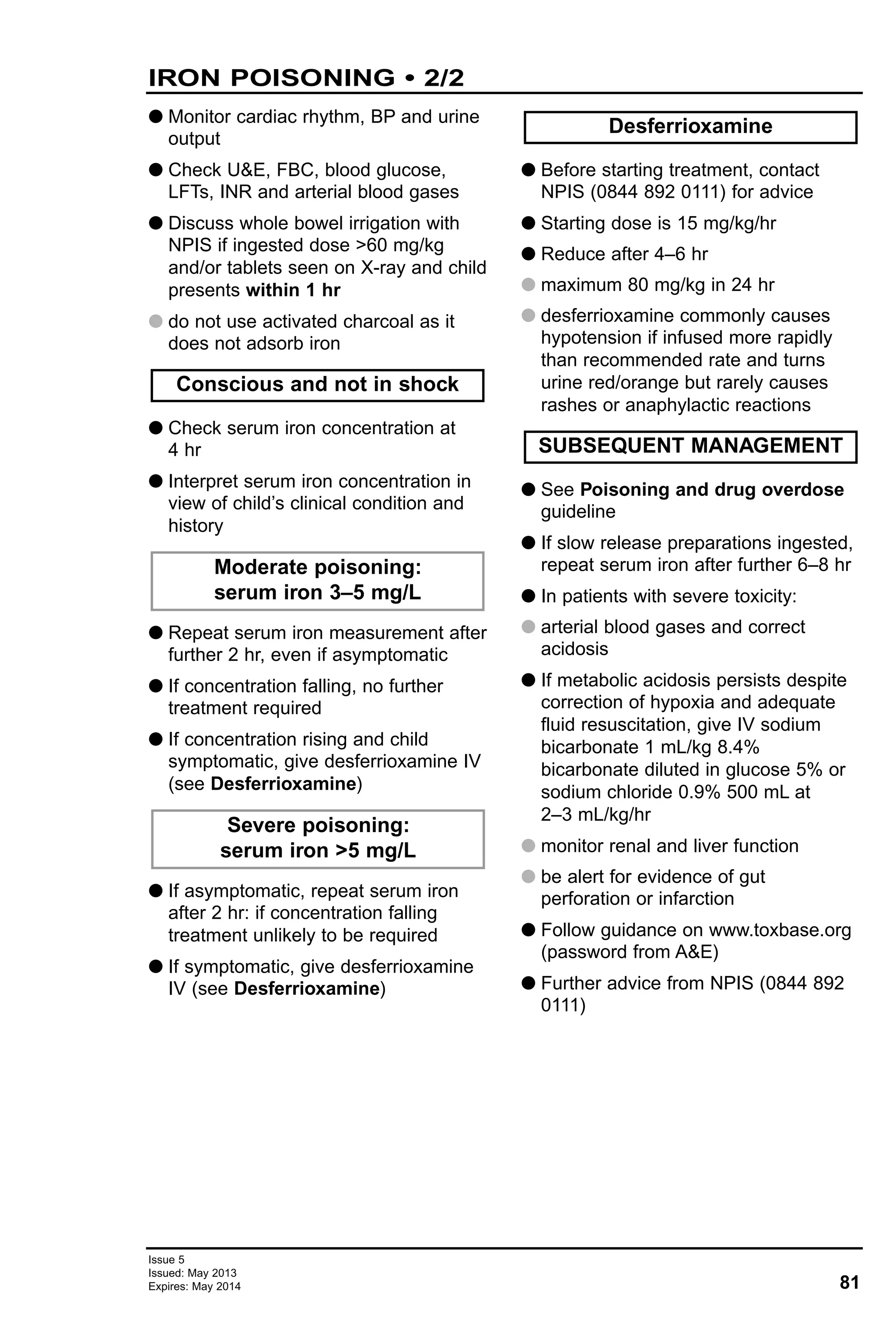
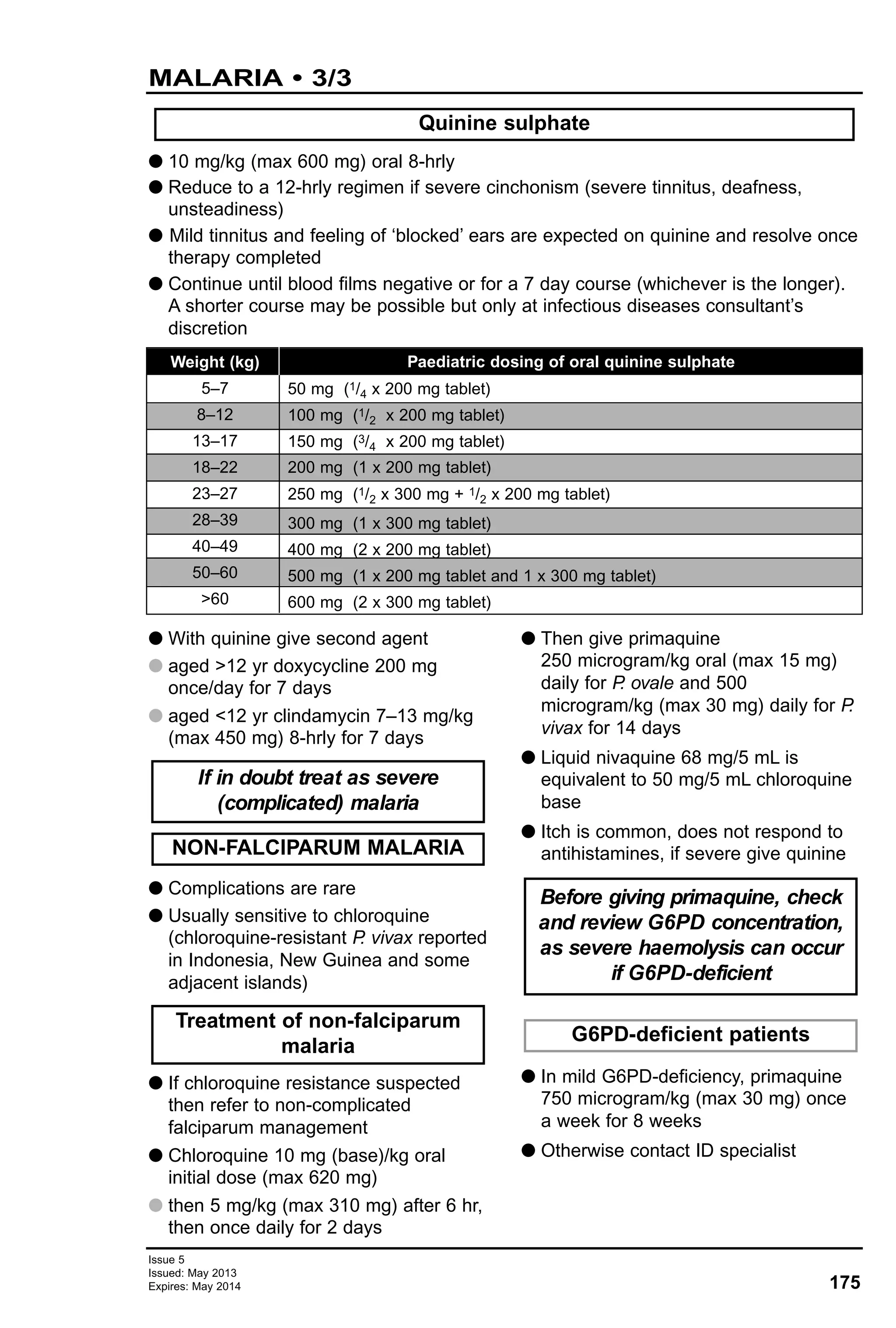
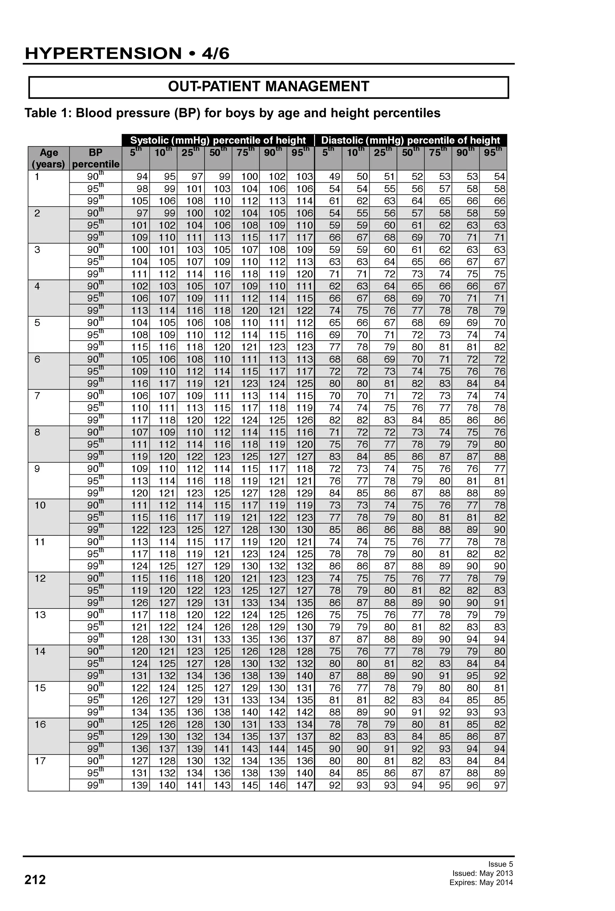
MALARIA • 2/3
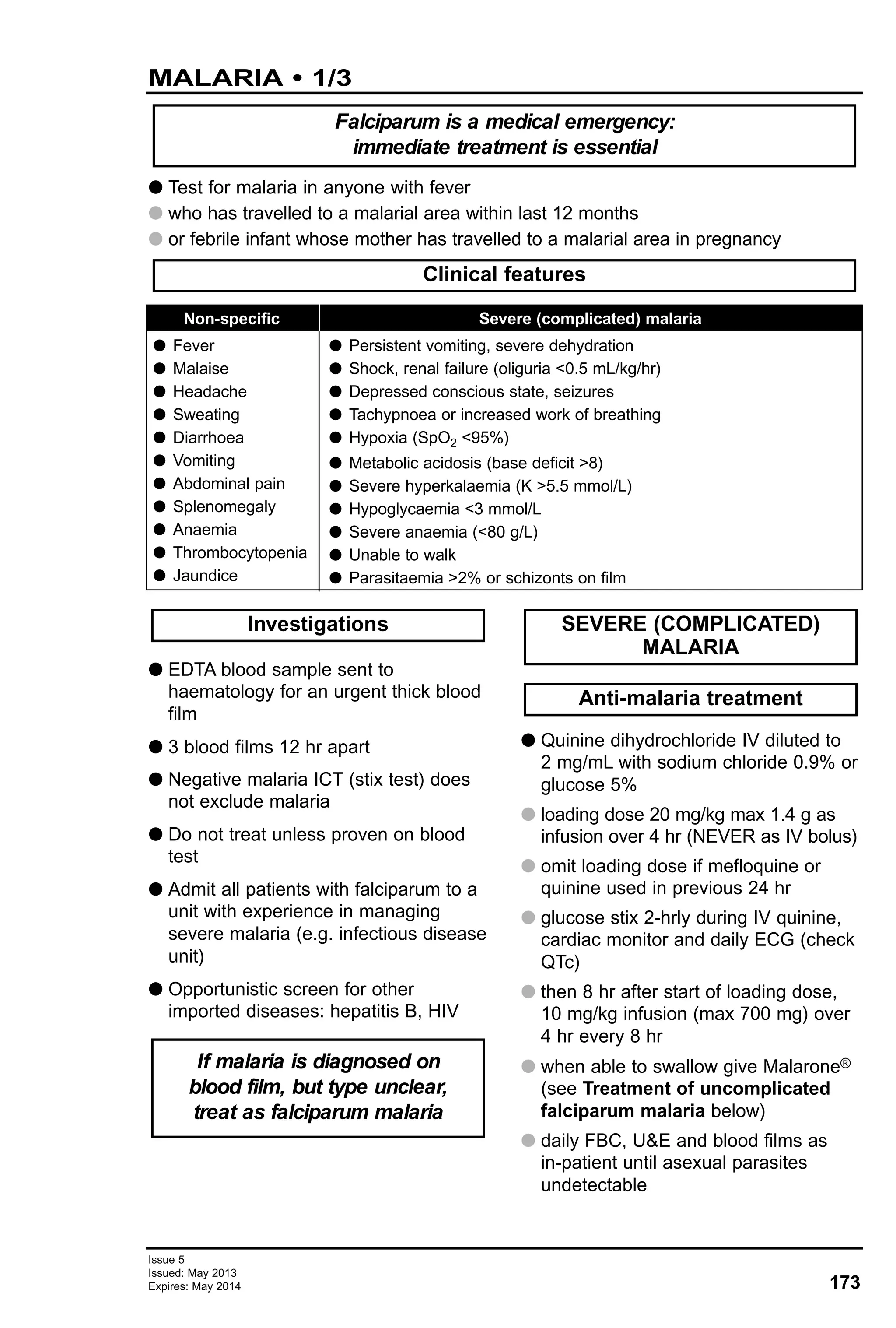
G If child can tolerate oral intake (can be crushed):
G Malarone® (proguanil with atovaquone) once a day for 3 days
TREATMENT OF UNCOMPLICATED FALCIPARUM MALARIA
(no clinical features of severe malaria)
Paediatric tablet contains proguanil 25 mg and adult tablet 100 mg
No second agent required
Or
Riamet® (artemether with lumefantrine)
No second agent required
Or
Weight (kg)
Dose
5–8
2 paed
tablets
9–10
3 paed
tablets
11–20
1 adult
tablet
21–30
2 adult
tablets
31–40
3 adult
tablets
>40
4 adult
tablets
G If parasitaemia >15% or from area of
quinine resistance (Thai/Cambodia
border, Papua New Guinea) or history
of arrhythmias, discuss with ID
specialist about artesunate instead of
quinine
G Artesunate 2.4 mg/kg IV [in 1 mL
sodium bicarbonate (vial provided with
drug), dilute further in 5 mL glucose 5%
and inject over approximately 2 min] at
0, 12 and 24 hrs and then daily
G When parasitaemia resolving and
patient improving, switch to oral agent:
G Malarone®, or if resistance suspected
Riamet®, or oral quinine (if neither
other agent available)
G Renal failure: discuss early
filtration/dialysis with PICU
G Hypovolaemia: cautious rehydration
(high risk pulmonary oedema)
G Shock: add cefotaxime
G Hypoglycaemia: common, give
glucose 10% 2 mL/kg IV bolus then
glucose 10% 5 mL/kg/hr with sodium
chloride 0.45%
G Anaemia: common, transfuse if Hb
<80 g/L
G Thrombocytopenia: expected,
transfuse only if bleeding and platelets
<20 x 109/L
Impaired level of consciousness
G Correct hypoglycaemia
G Monitor GCS, reflexes, pupils
G Plan for intubation and transfer to
PICU if:
G signs of raised ICP
G persisting shock after 40 mL/kg fluid
G or pulmonary oedema
Complications
CEREBRAL MALARIA
Weight (kg)
5–15
16–25
26–35
>35
(12–18 yr)
Dose
1 tablet initially followed by 5 further doses of 1 tablet each given at 8, 24, 36,
48 and 60 hrs (total 6 tablets over 60 hr)
2 tablets initially followed by 5 further doses of 2 tablets each given at 8, 24,
36, 48 and 60 hrs (total 12 tablets over 60 hr)
3 tablets initially followed by 5 further doses of 3 tablets each given at 8, 24,
36, 48 and 60 hrs (total 18 tablets over 60 hr)
4 tablets initially followed by 5 further doses of 4 tablets each given at 8, 24,
36, 48 and 60 hrs (total 24 tablets over 60 hr)](https://image.slidesharecdn.com/paediatricguidelines2013-14withlinks-180801135506/75/Paediatric-guidelines-2013-14-with-links-174-2048.jpg)

![G Epileptic encephalopathy in newborns
(myoclonic or Ohtohara syndrome)
G West’s syndrome (infantile spasms)
G Severe myoclonic epilepsy of infancy
(Dravet’s syndrome)
G Lennox-Gastaut syndrome
G Laundau-Kleffner syndrome
G For other epilepsy syndromes see
International League against Epilepsy
website (www.ilae-epilepsy.org)
G Clinically diagnosed epilepsy
G After an episode of status epilepticus
G Unexplained coma or encephalopathy
G Suspicion of non-convulsive status in
children with learning difficulties and
epilepsy
G Acquired regression of speech or
language function
G Developmental regression suspected
to have neurodegenerative condition
G To monitor progress in West’s
syndrome and non-convulsive status
G Funny turns, apnoeic attacks, dizzy
spells, strange behaviour
G Non-convulsive episodes [e.g. syncope,
reflex anoxic seizures, breath-holding
episodes (ECG more appropriate)]
G Febrile seizures
G Single uncomplicated generalised
tonic-clonic seizures
G To monitor progress in well-controlled
epilepsy
G Before stopping treatment
G Focal epilepsy (including TLE) except
rolandic seizures
G Epilepsy in children aged <2 yr
G Myoclonic epilepsy
G Intractable seizures
G Loss of previous good control
G Seizures continuing in spite of first line
medication
G Associated neurological deficits or
appearance of new neurological signs
G Developmental regression in children
with epilepsy
G Infantile spasms (West’s syndrome)
G Sleep or sleep-deprived EEG useful in
all children in whom there is a high
clinical suspicion but awake EEG
normal
G sleep EEG useful to pick up some
focal/generalised epilepsies and
sleep-deprived EEG useful in
generalised epilepsies in young adults
including JME. Perform sleep EEG
with melatonin
G Video telemetry useful if diagnostic
dilemma, pseudo seizures or before
surgery
G Drug levels: phenytoin, phenobarbitone
(other anticonvulsants only if concerns
about compliance and overdose)
G Biochemistry: glucose, calcium, LFT,
lactate, ammonia; metabolic and
genetic investigations where suspicion
of metabolic disorder (e.g. progressive
developmental delay)
G Epileptic encephalopathies, such as
West's Syndrome, need a series of
investigations (discuss with paediatric
neurologist)
G Discuss treatment with a consultant
before starting
G Start anti-epileptic only if diagnosis
certain (two or more unprovoked
seizures)
General guidelines
TREATMENT
Other investigations
Other epilepsy syndromes
INVESTIGATIONS
Indications for MRI of brain
EEG not indicated
Indications for EEG
Issue 5
Issued: May 2013
Expires: May 2014198
EPILEPSY • 3/5](https://image.slidesharecdn.com/paediatricguidelines2013-14withlinks-180801135506/75/Paediatric-guidelines-2013-14-with-links-198-2048.jpg)

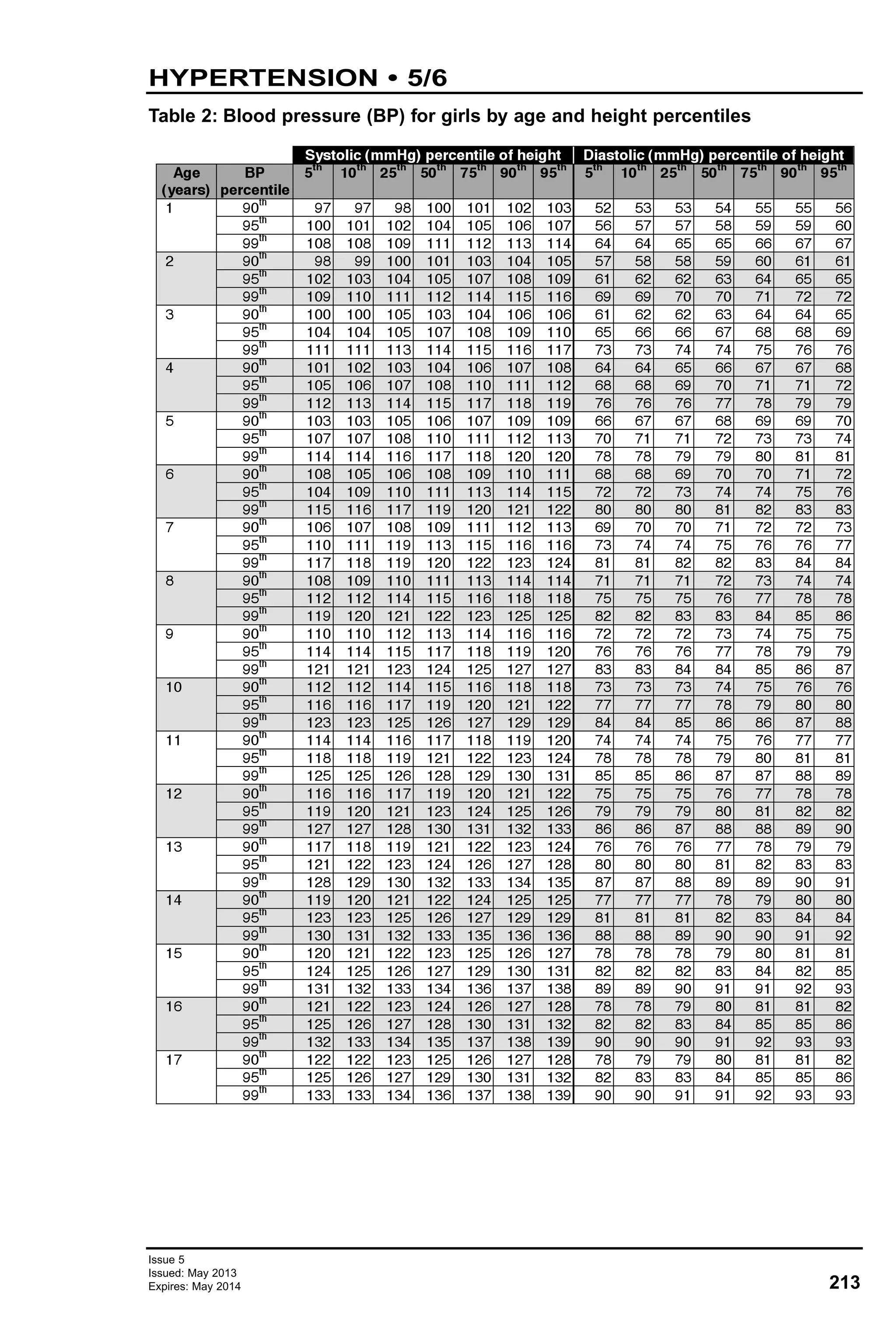
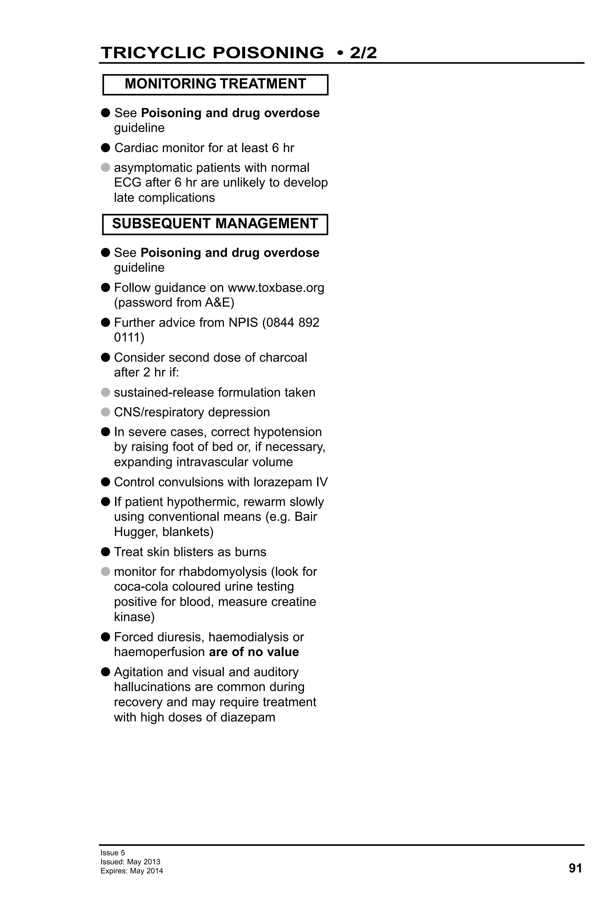
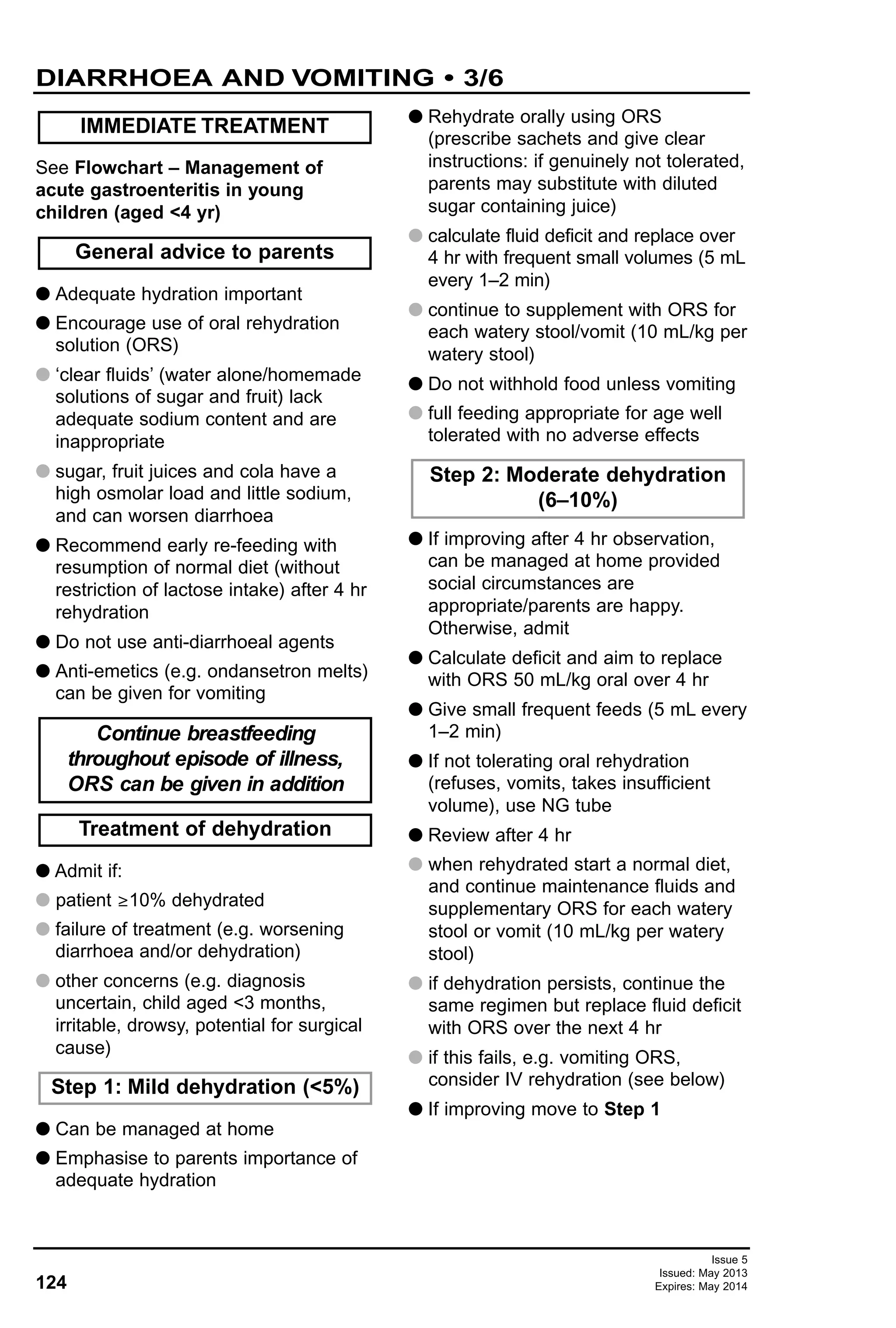
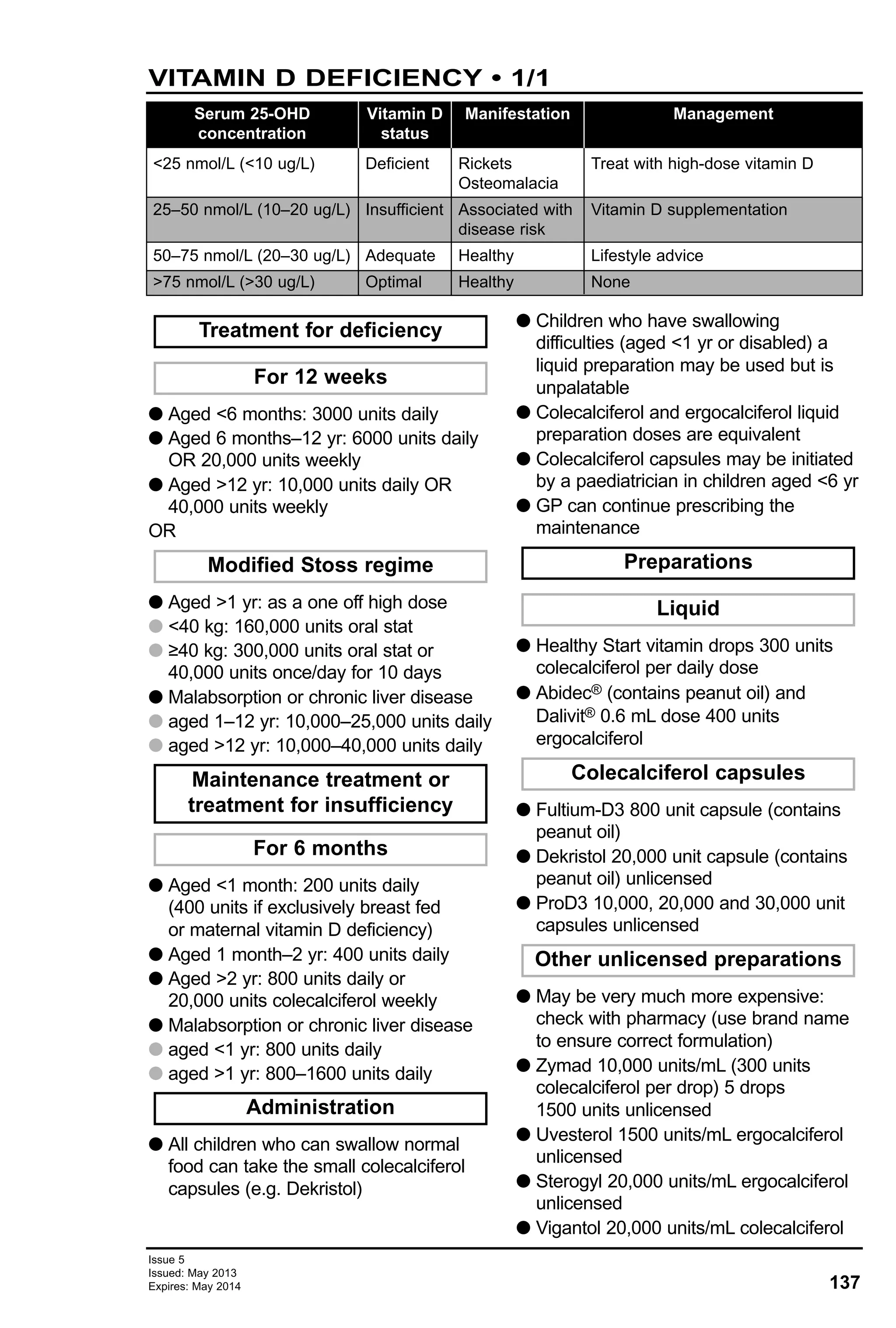
![222
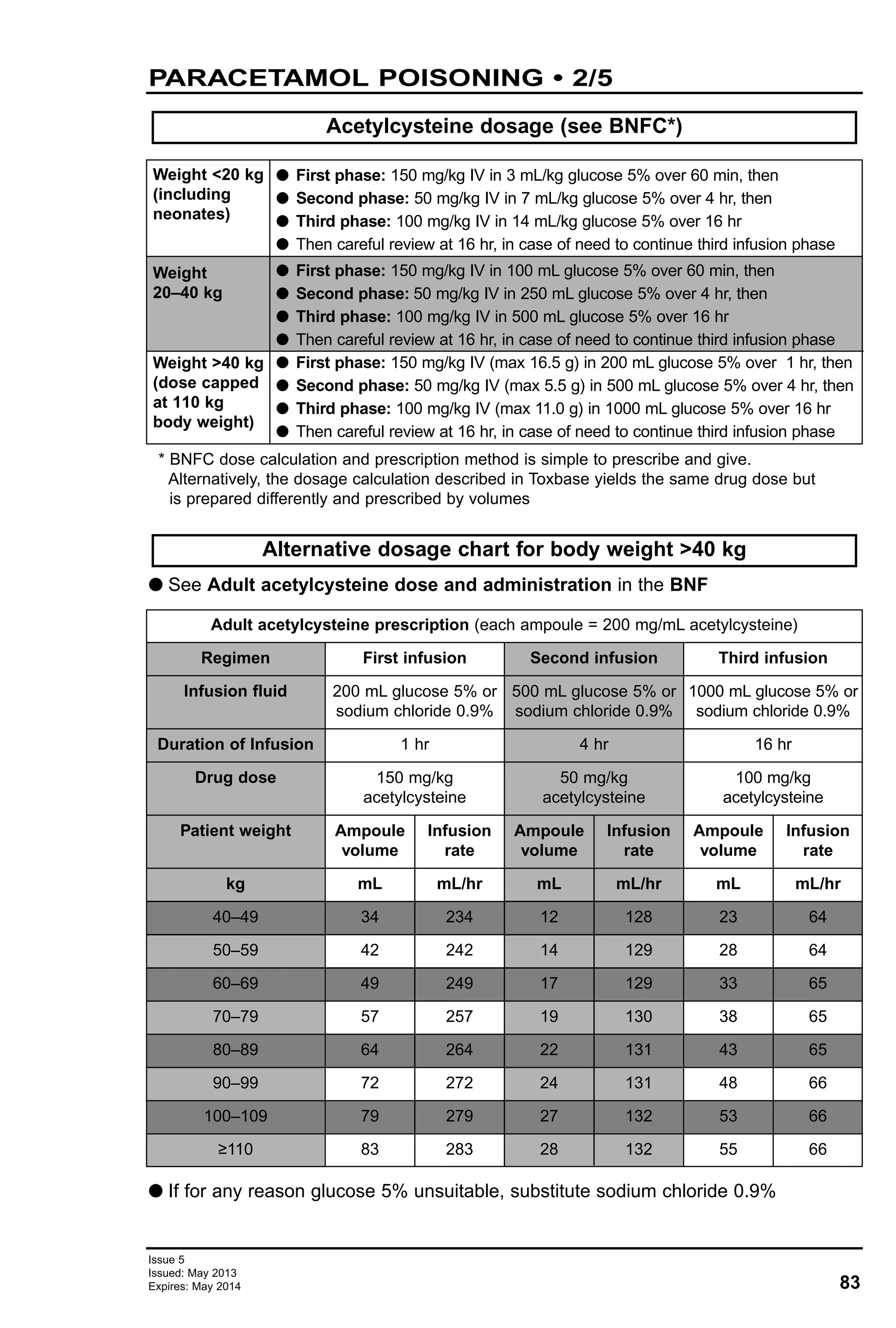
RENAL CALCULI • 4/4
Issue 5
Issued: May 2013
Expires: May 2014
Paediatric stone patient
Stone analysis
Mg Ammonium
phosphate (struvite)
Total elimination of
stone (surgery/SWL)
antibiotics
Hyperparathyroidism
Alkali replacement
– potassium citrate
Allopurinol
Low purine diet
High fluid intake
Potassium citrate
Alpha-mercaptopropionylglycine
Penicillamine
Uric acid
stone
Cystine
Calcium stones
CaOX-CaPO
Elimination of stones by spontaneous passage or active removal
[extracorporeal shockwave lithotripsy (SWL), surgery]
Urine culture
Possibly urease
producing bacteria
Urine pH
Urine and serum
Uric acid levels
Urine-blood pH
Acidic urine
Hyperuricosuria
Hyperuricaemia
Urine pH
Urine cystine
level
Cystinuria
Algorithm for metabolic investigations
K-citrate
Diet (normal calcium low
sodium intake)
Bendroflumethiazide diuretic
Diet low in
oxalate
K-citrate
Pyridoxine
Alkali
replacement
(k-citrate)
Allopurinol
Citrate
replacement
K-citrate
Hypercalciuria Hyperoxaluria
Urine pH <5.5
Urine pH >5.5
Hyperuricosiuria Hypocitraturia
Hypercalcaemia
Serum parathyroid
hormone (PTH) Urine – blood Ca – uric acid levels,
Mg, phosphate urine Ca – oxalate –
citrate – Mg, uric acid – phosphate
Further investigation for renal tubular acidosis](https://image.slidesharecdn.com/paediatricguidelines2013-14withlinks-180801135506/75/Paediatric-guidelines-2013-14-with-links-222-2048.jpg)