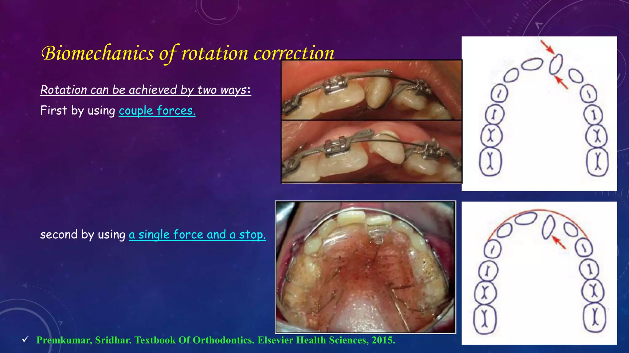
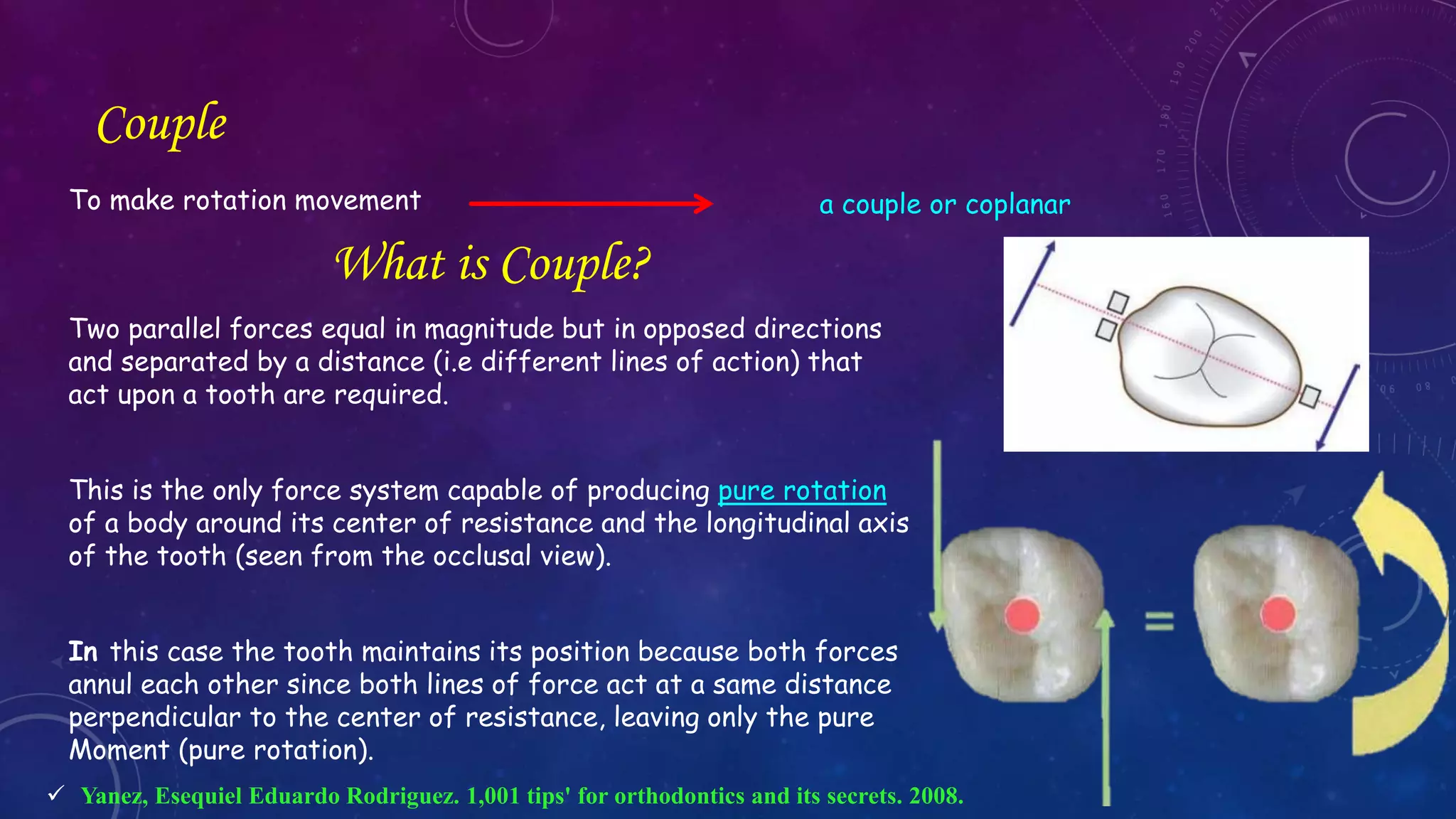
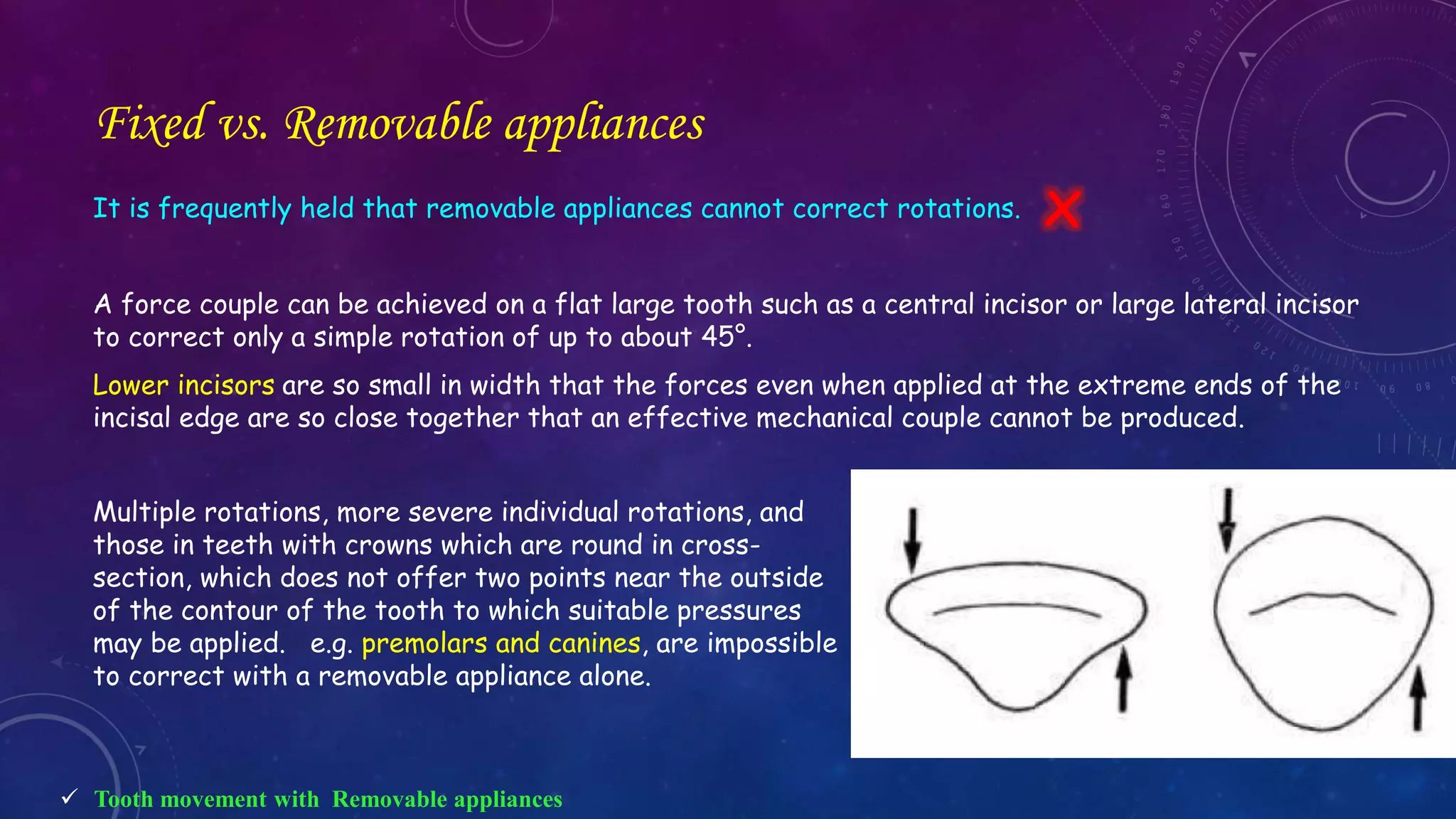
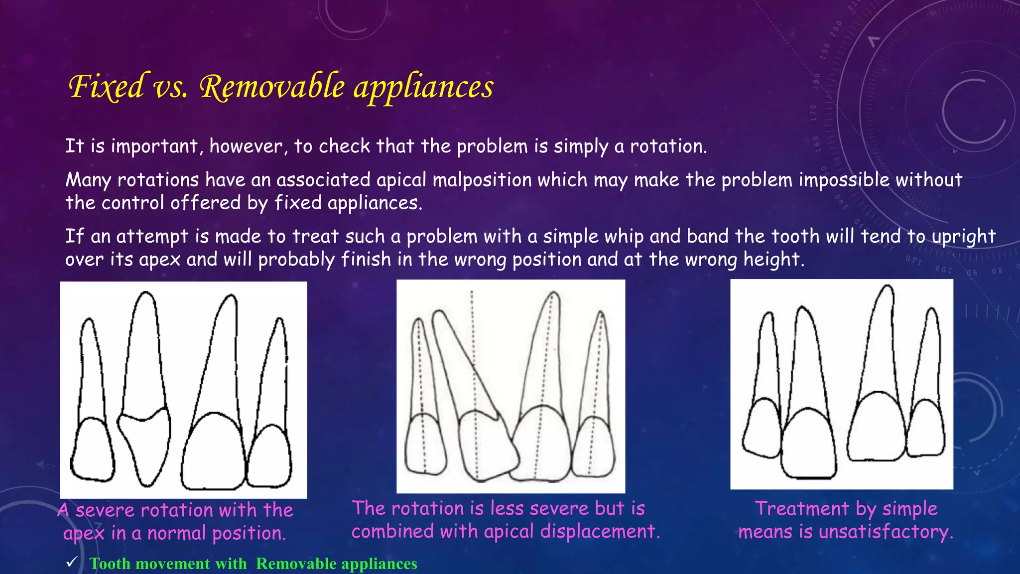
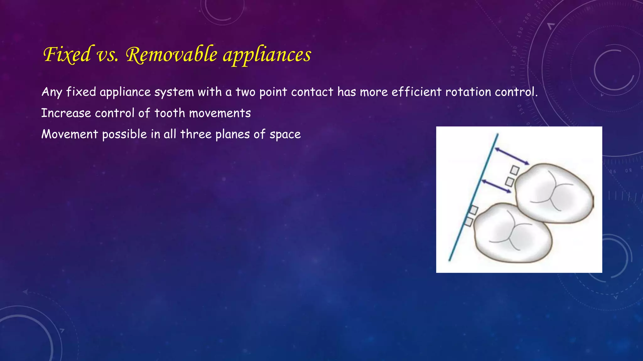
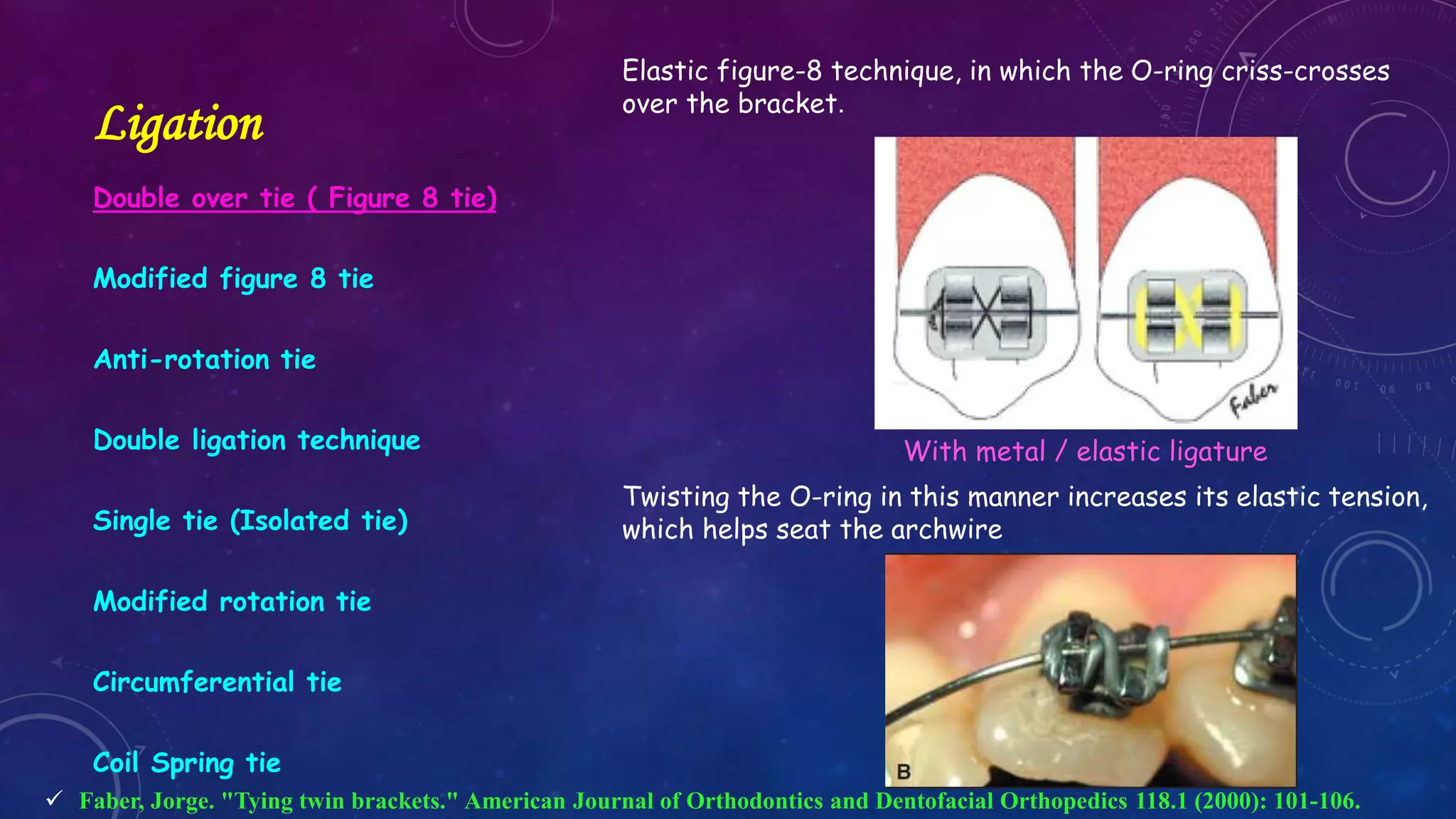
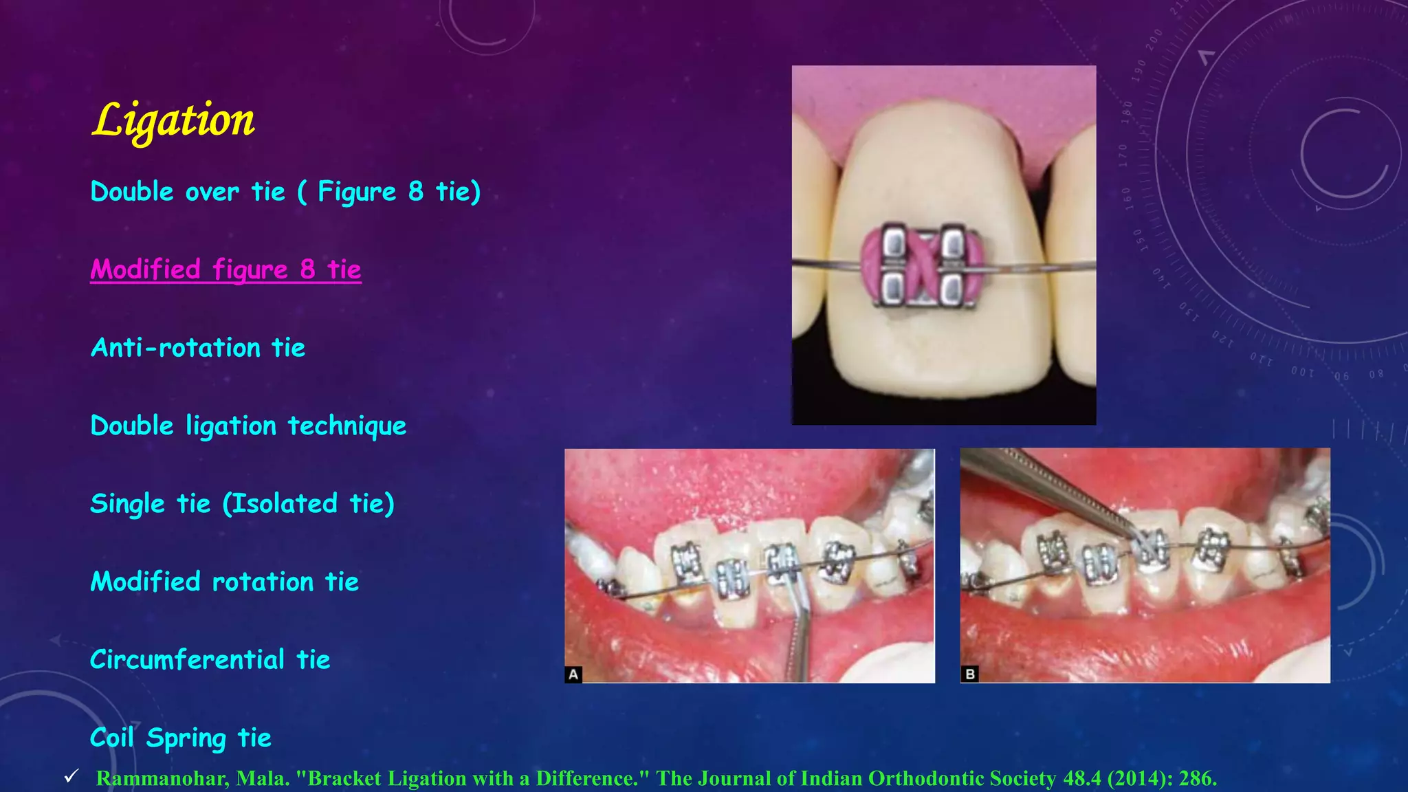
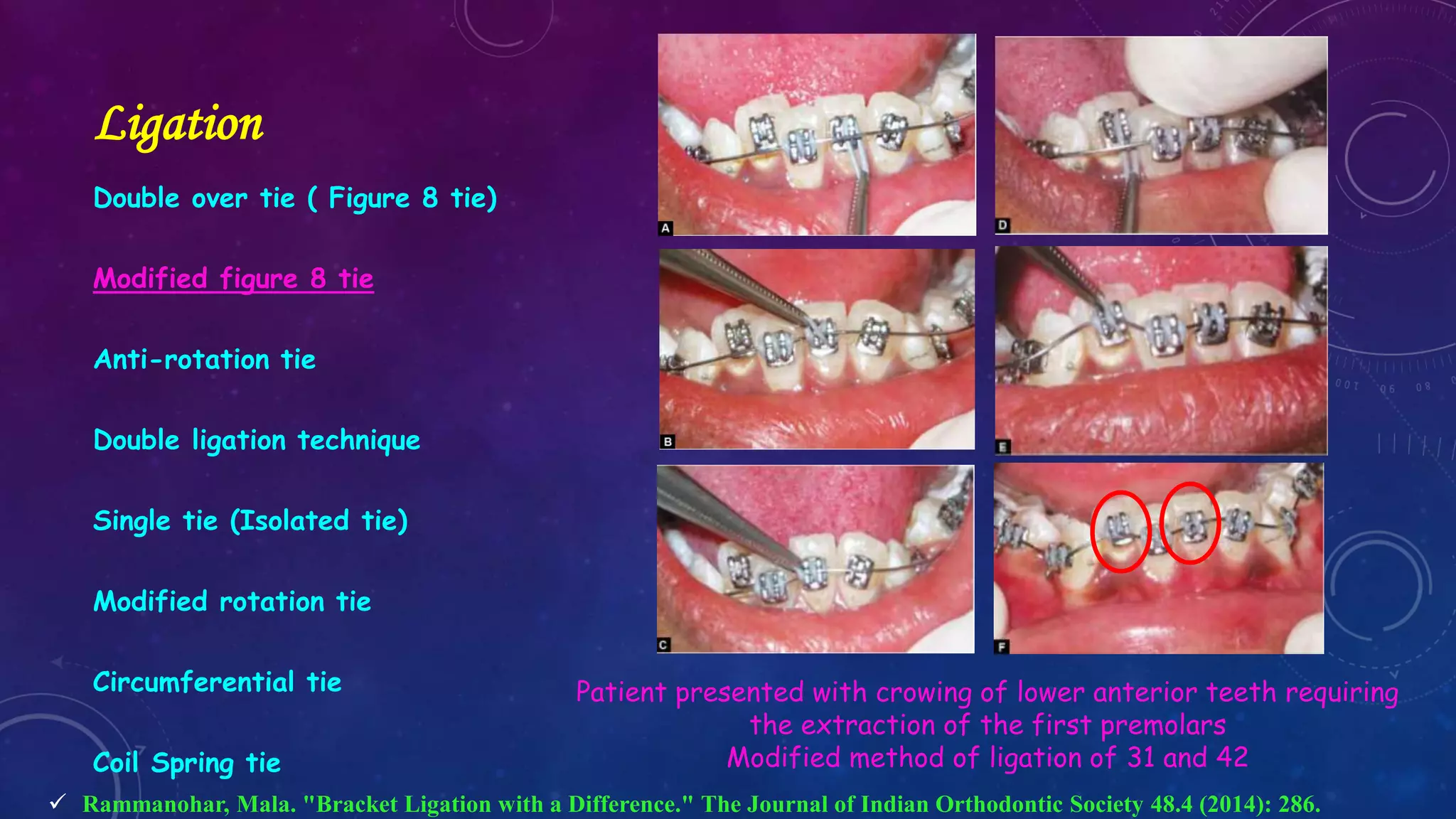
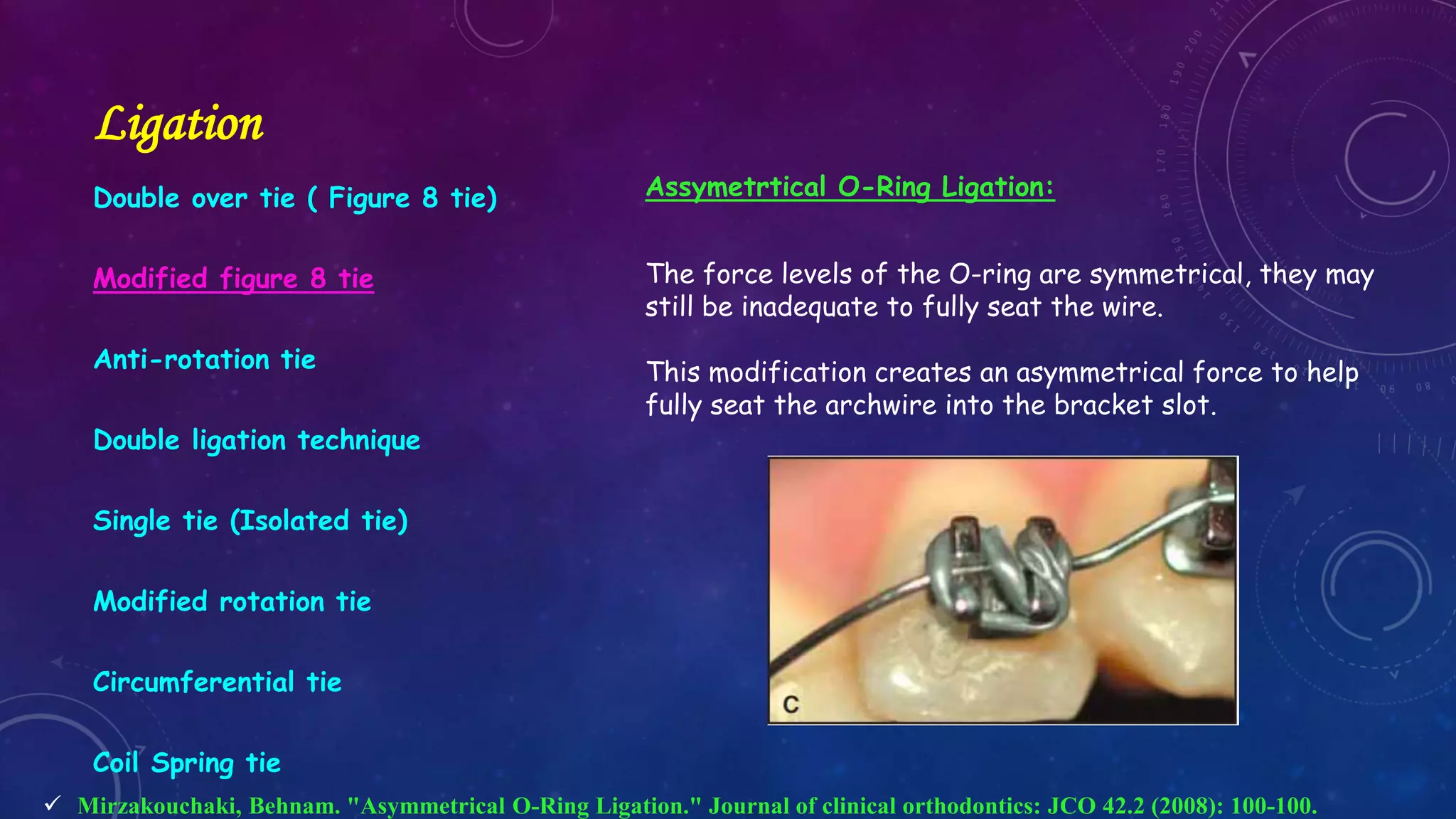
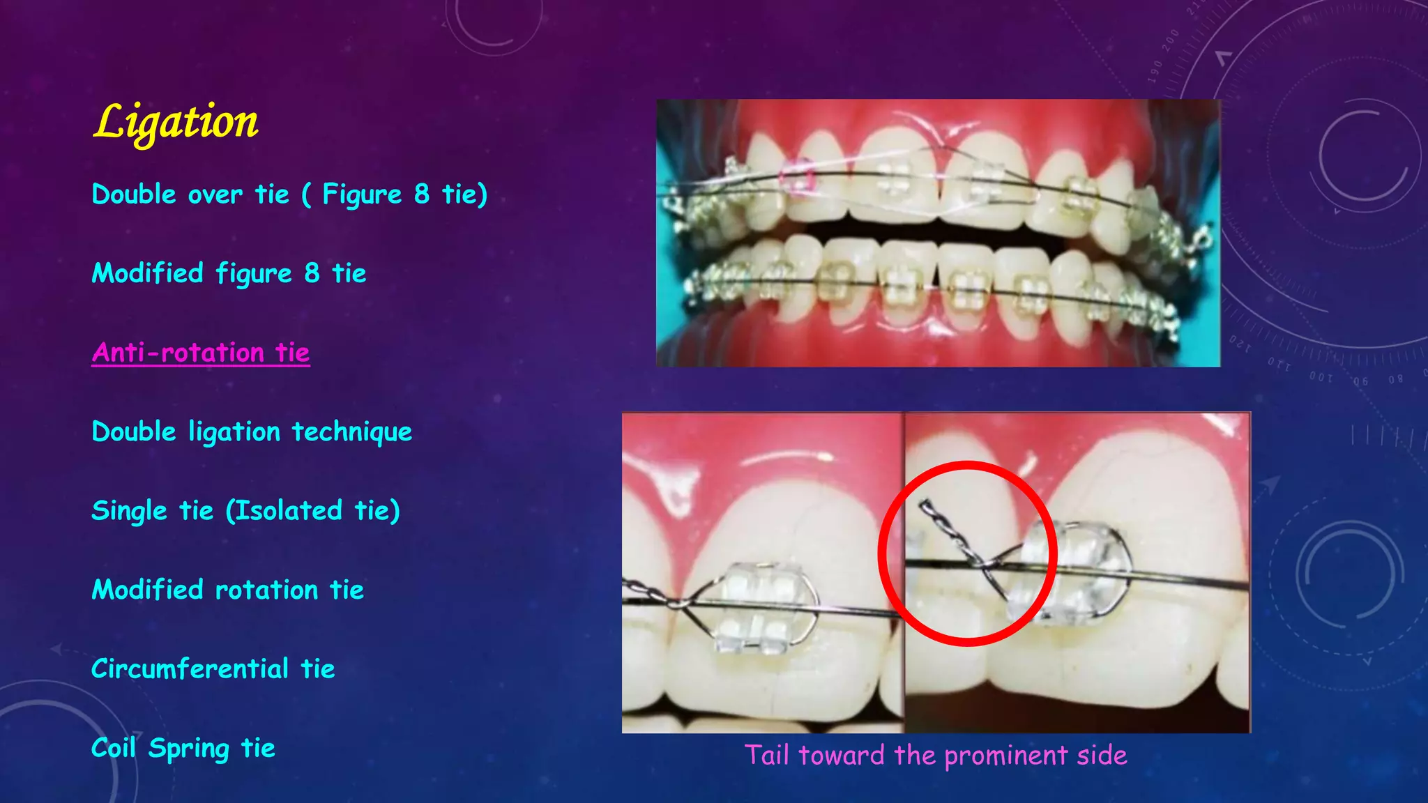
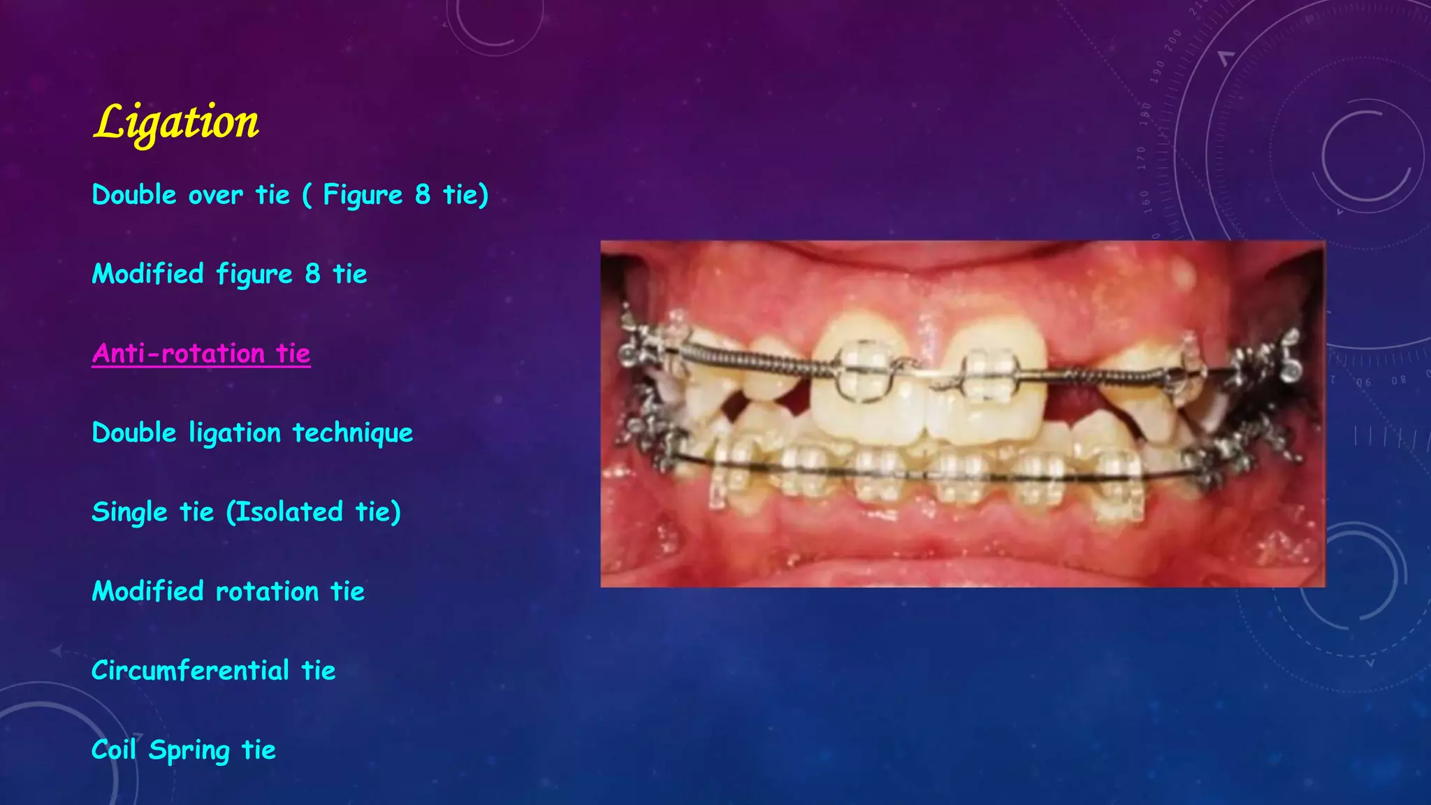
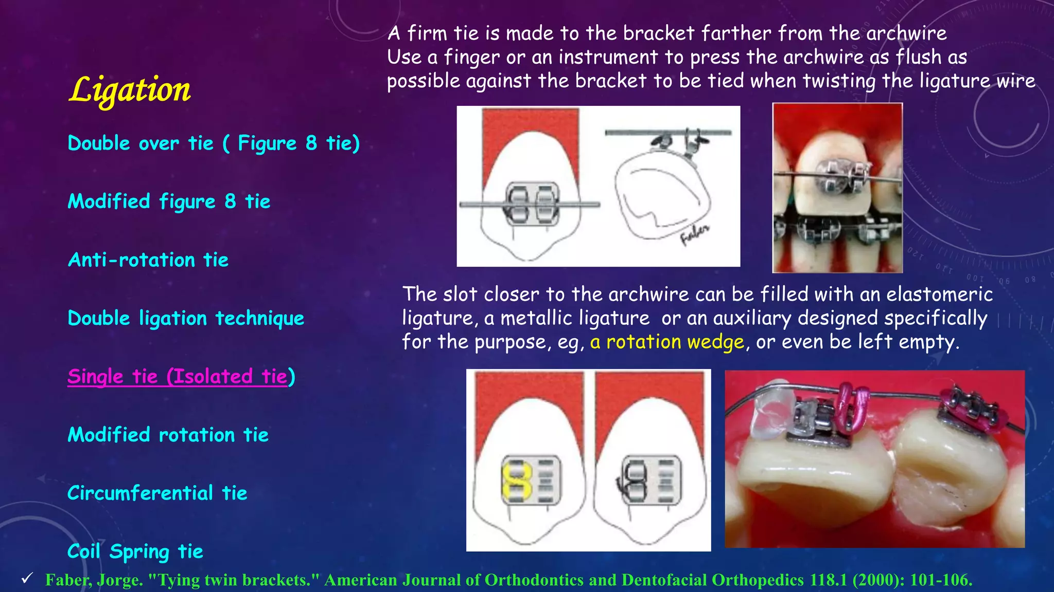
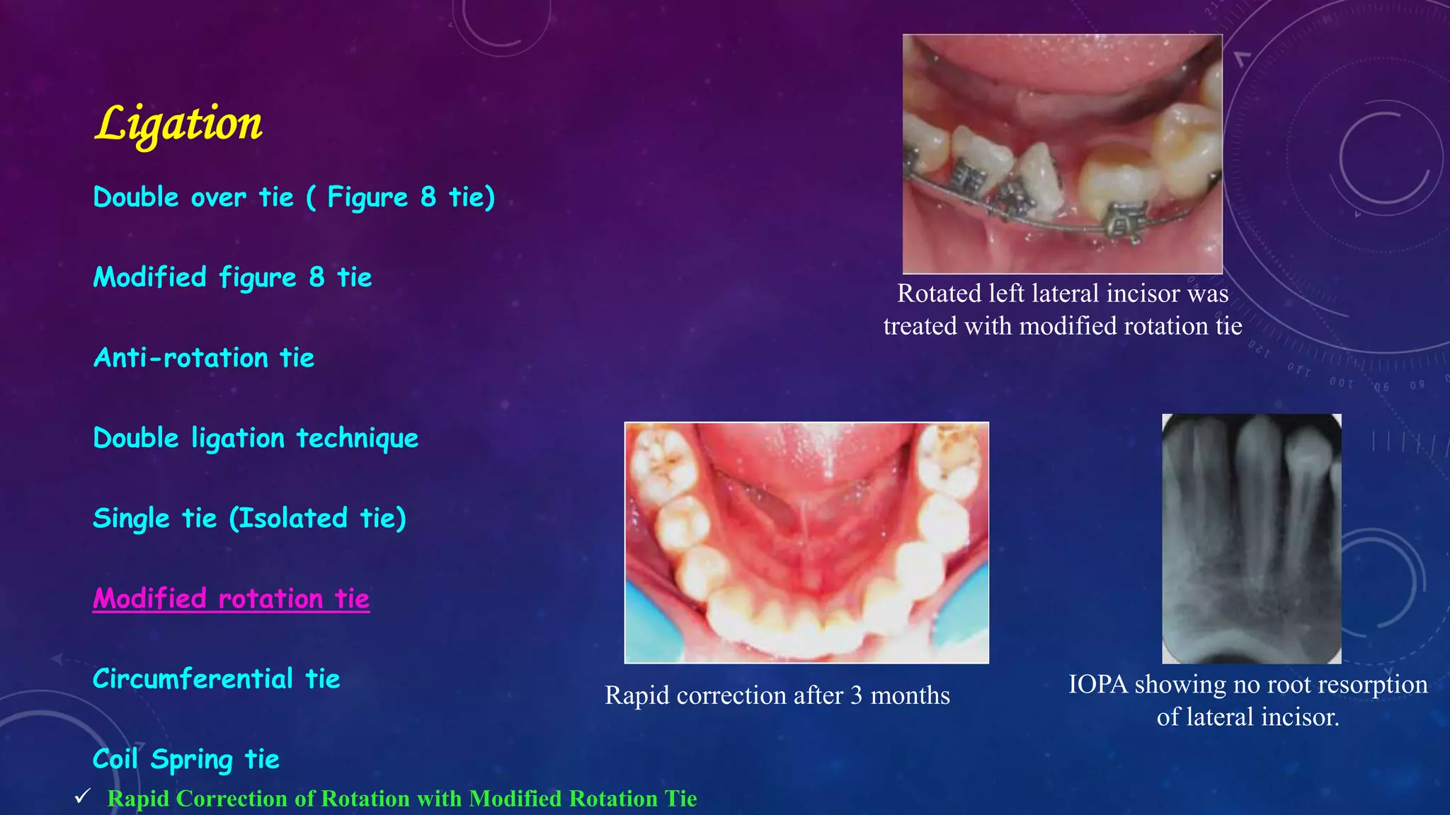
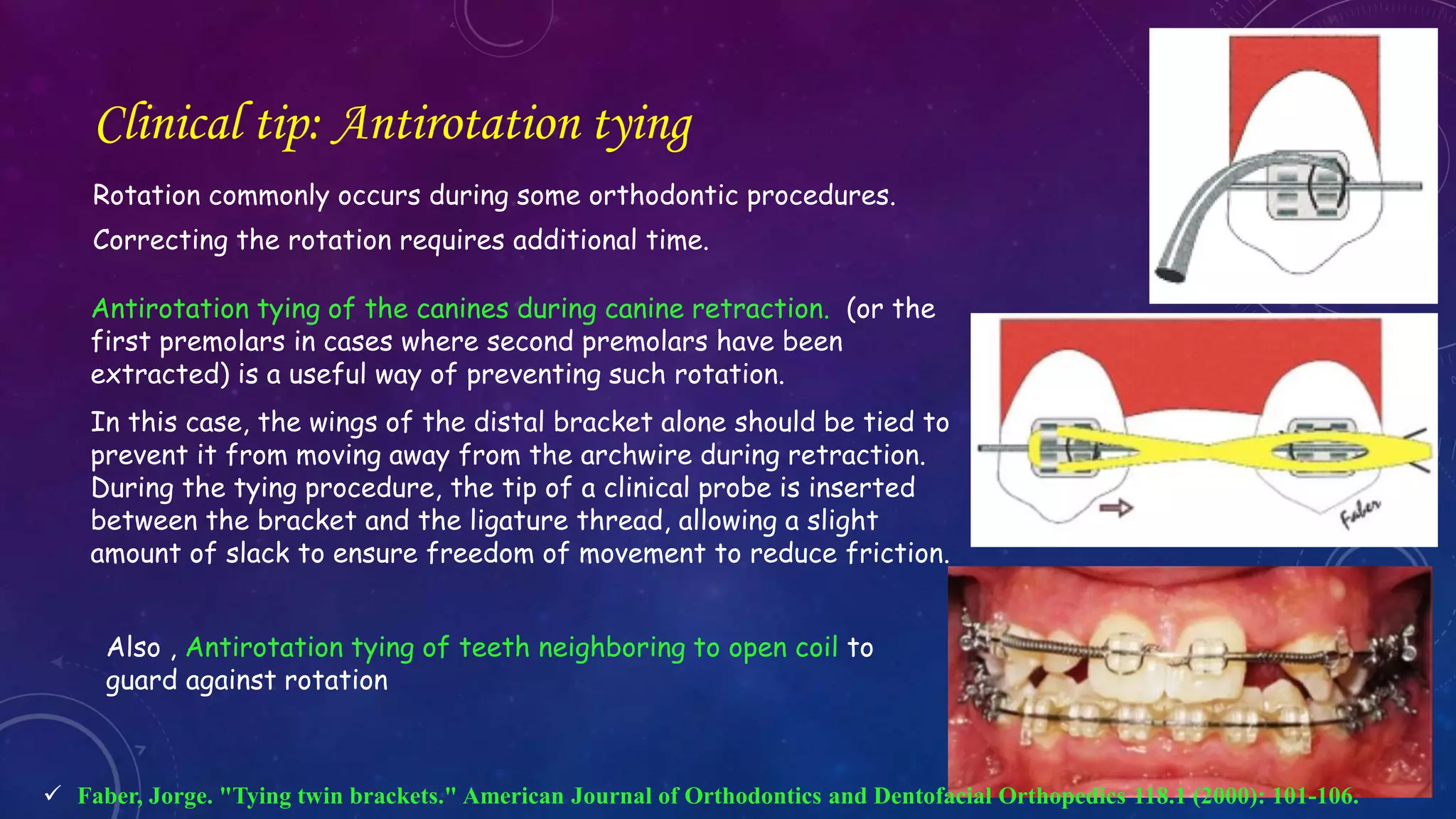
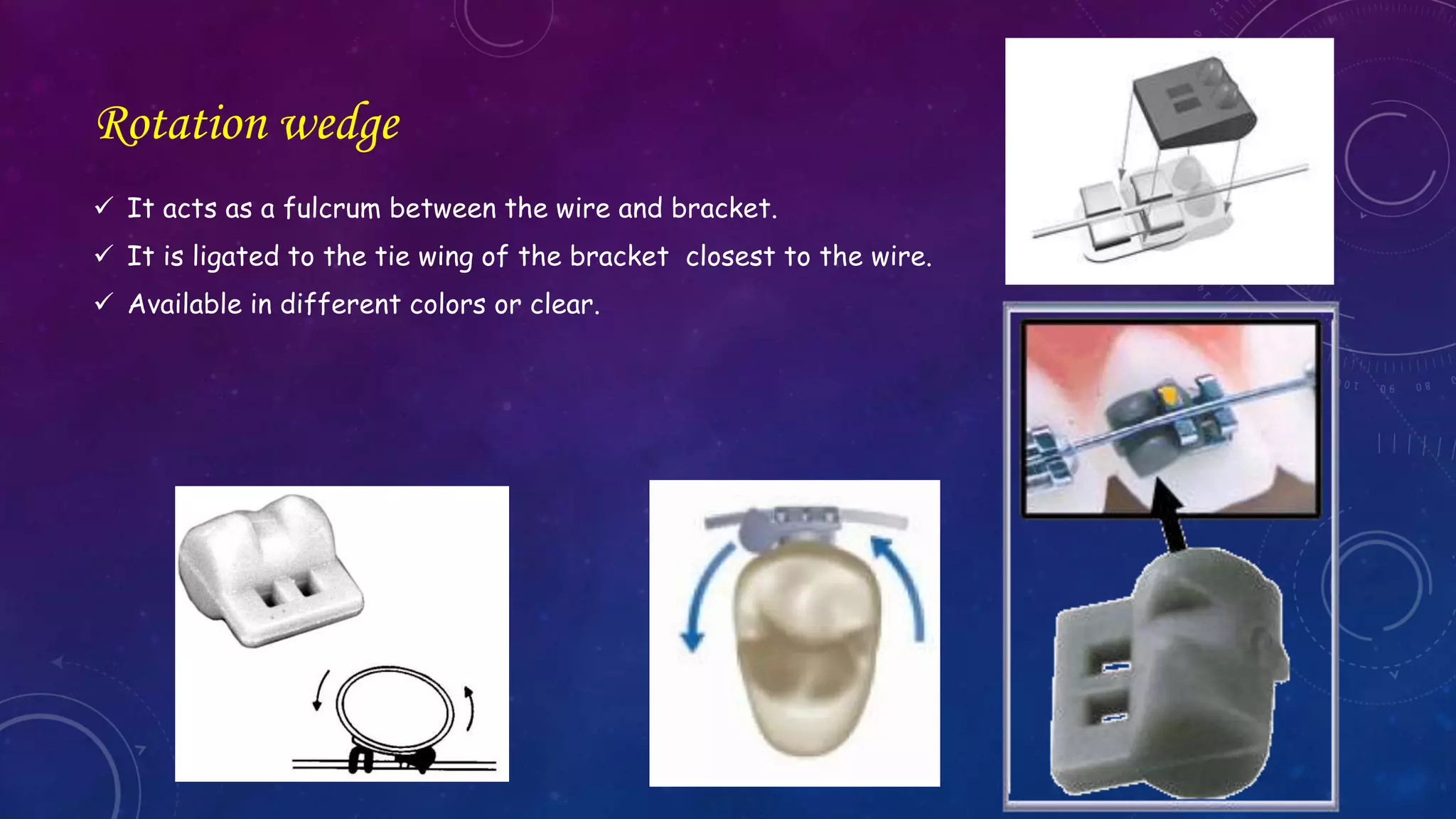
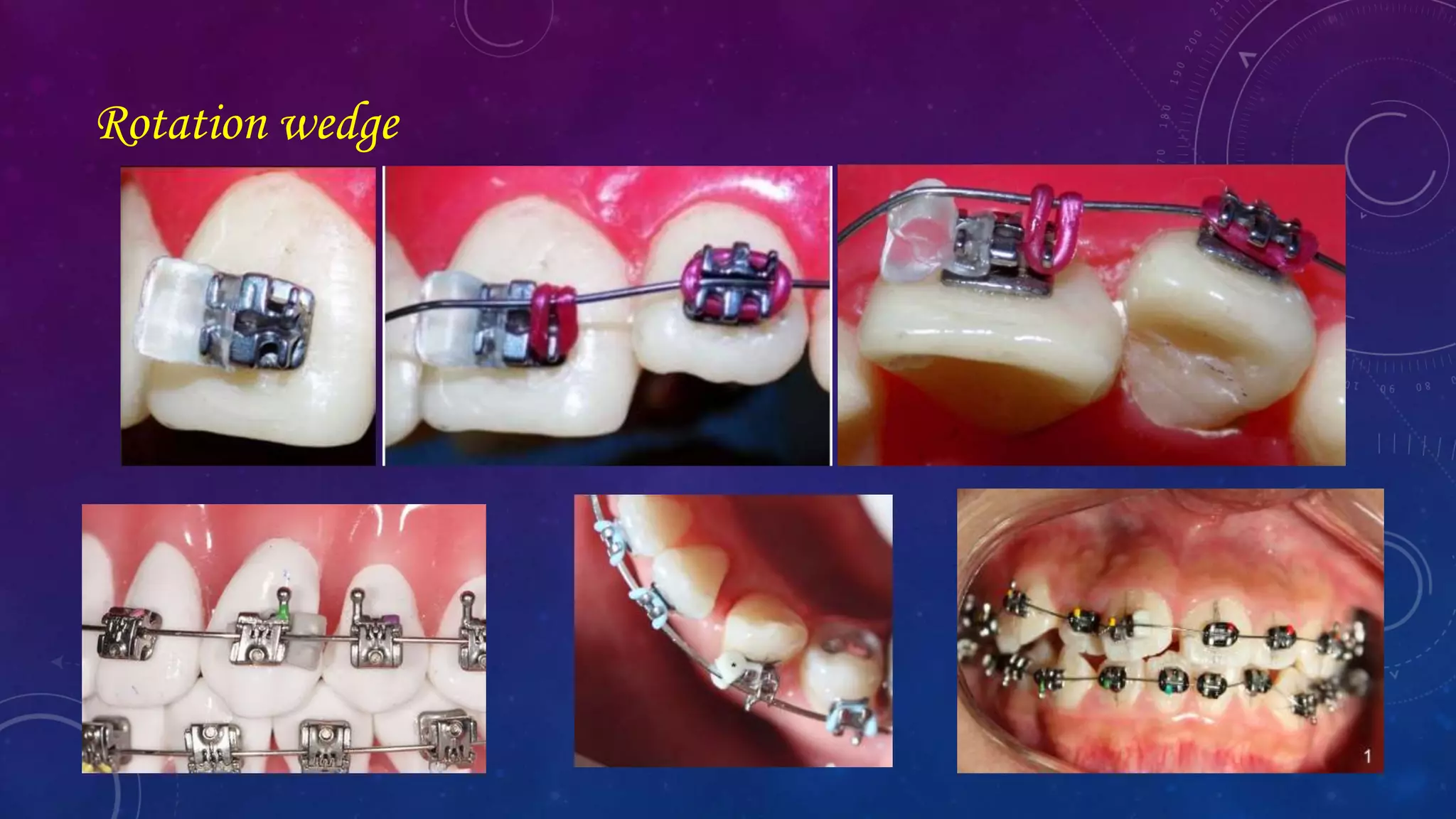
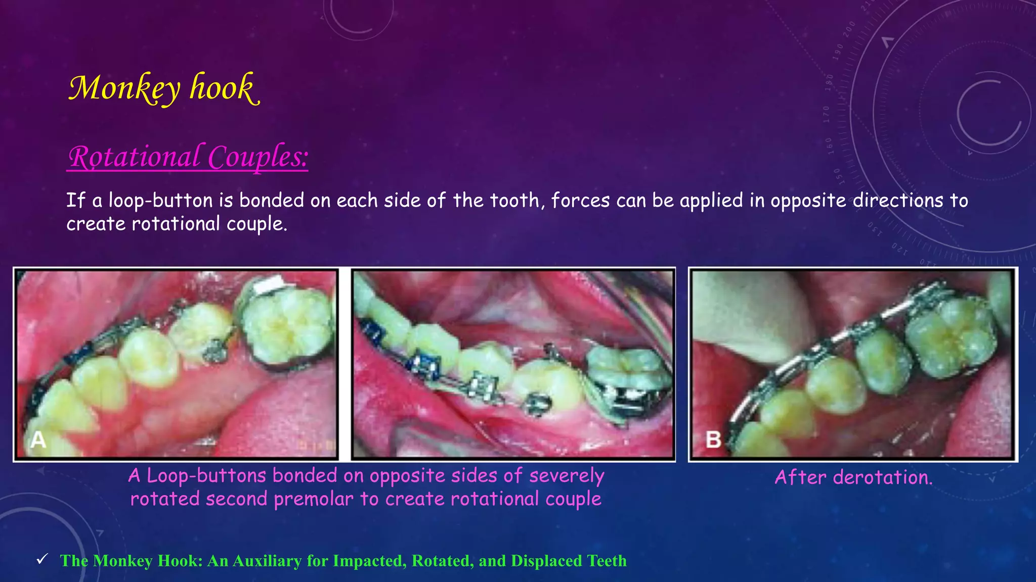
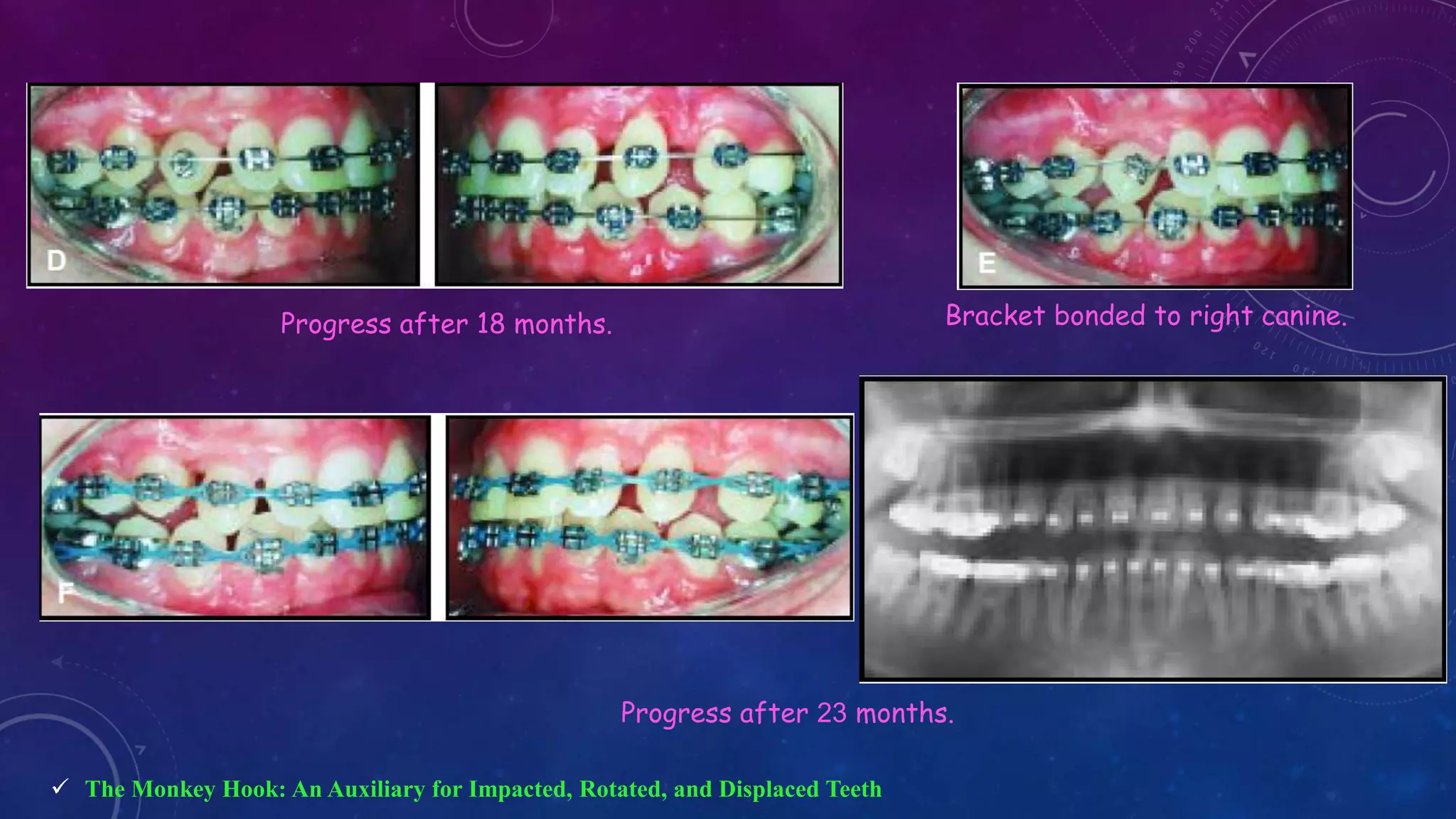
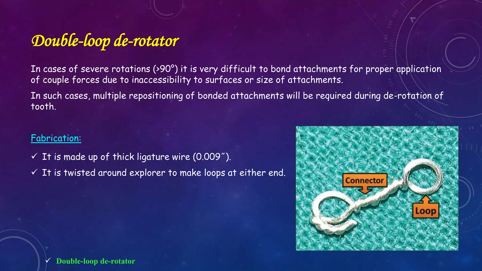
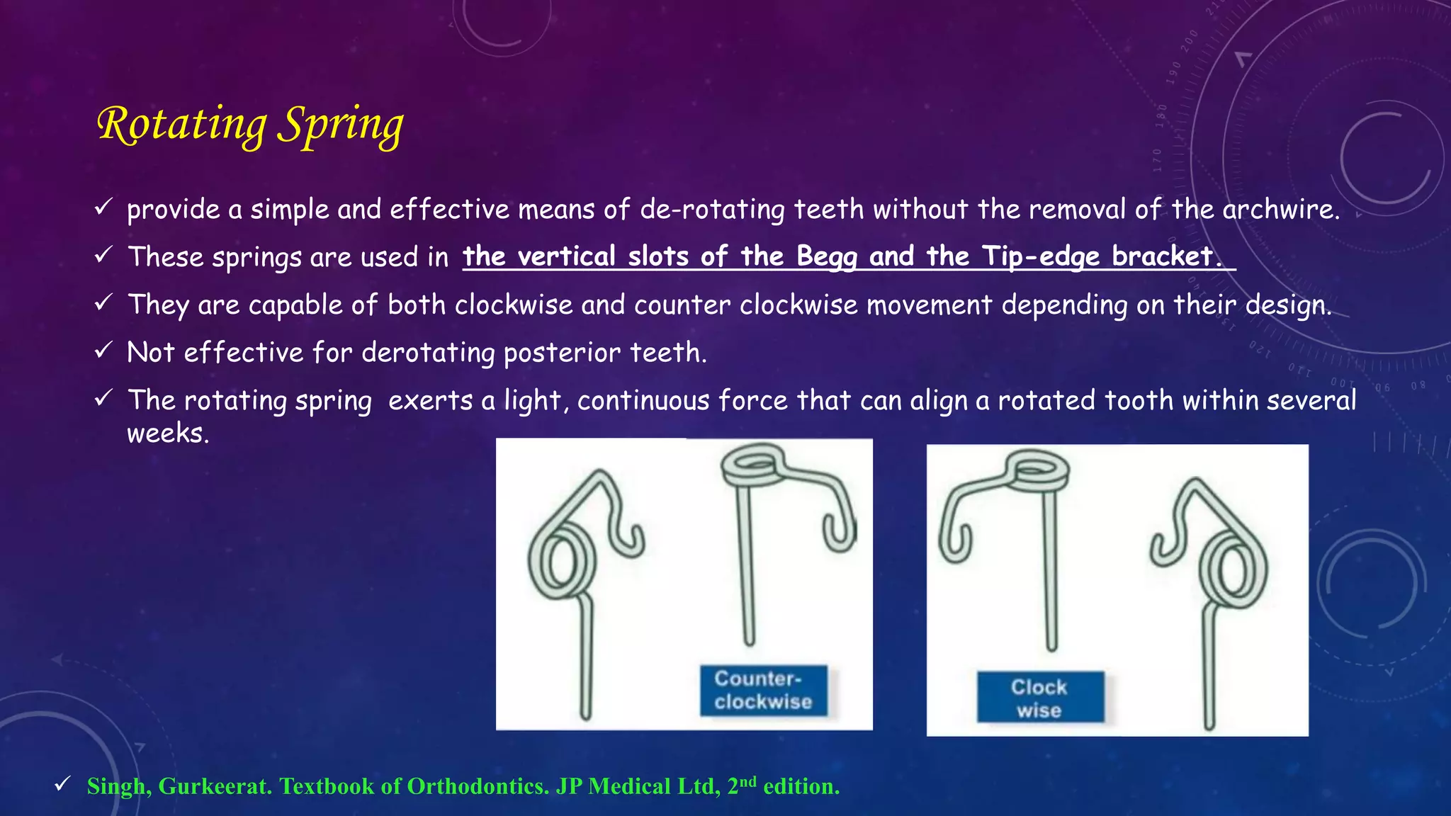
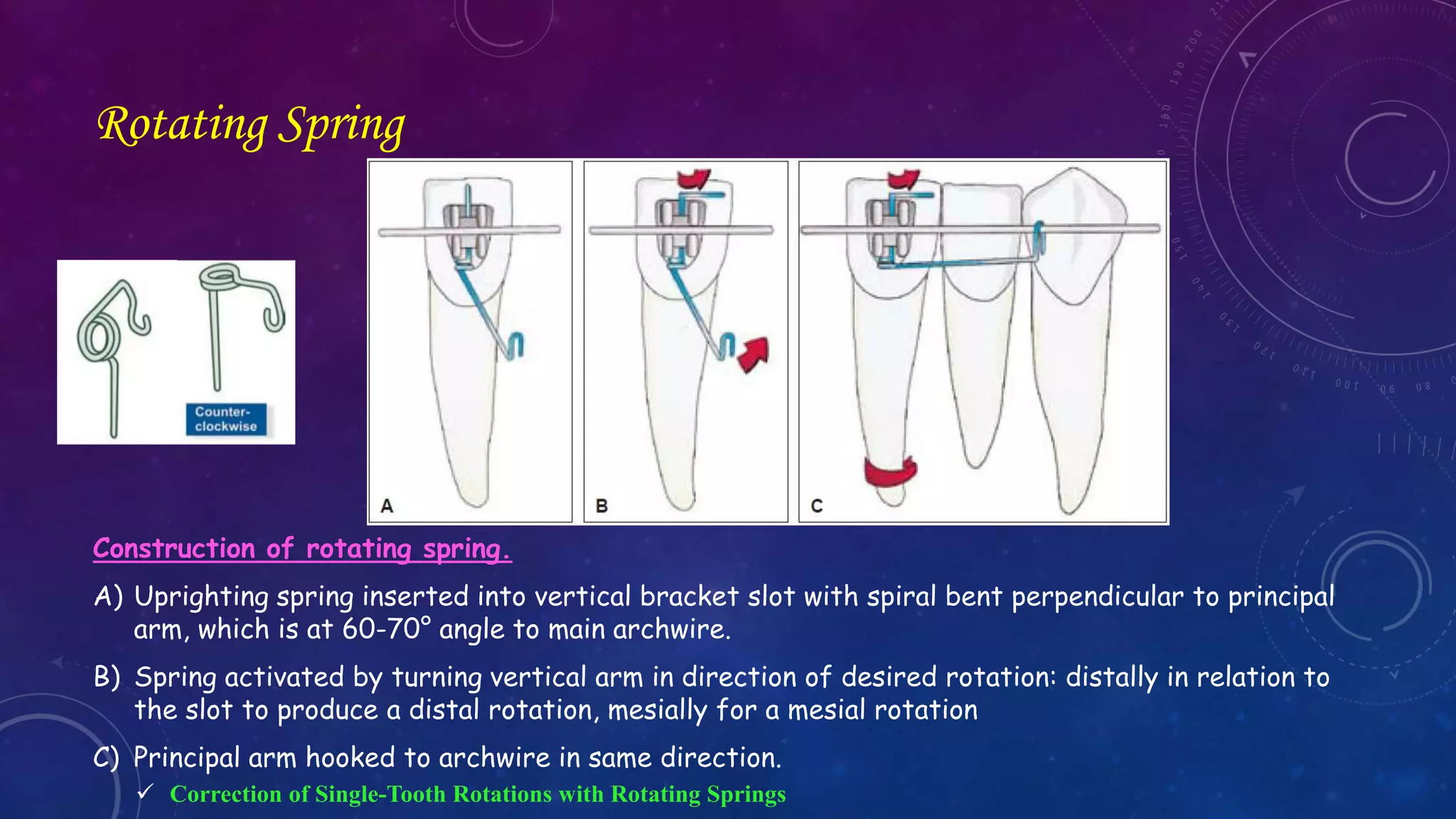
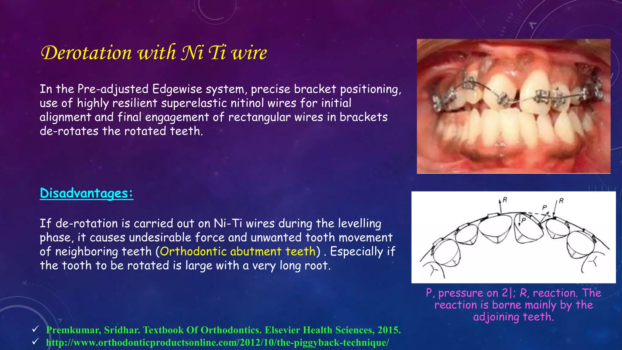
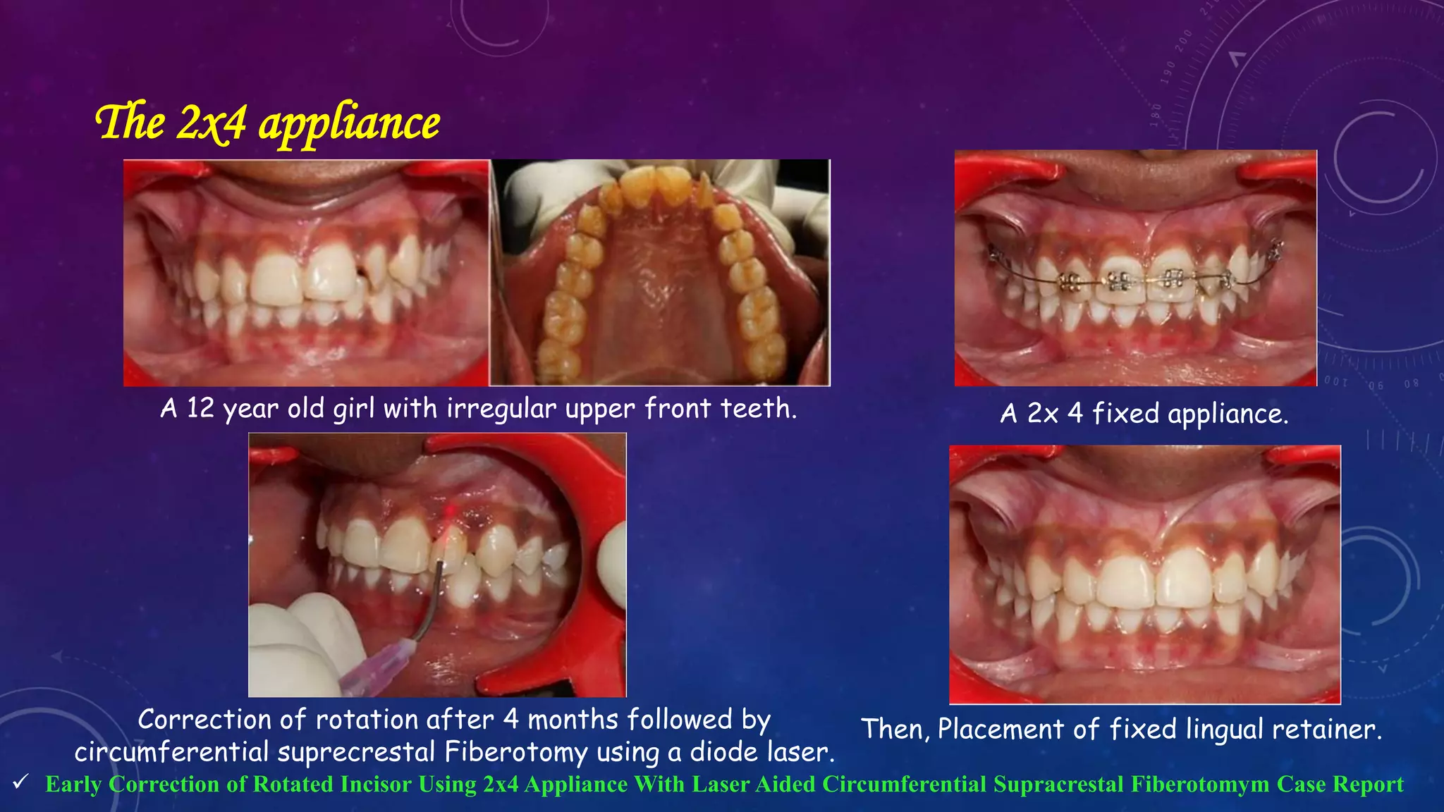
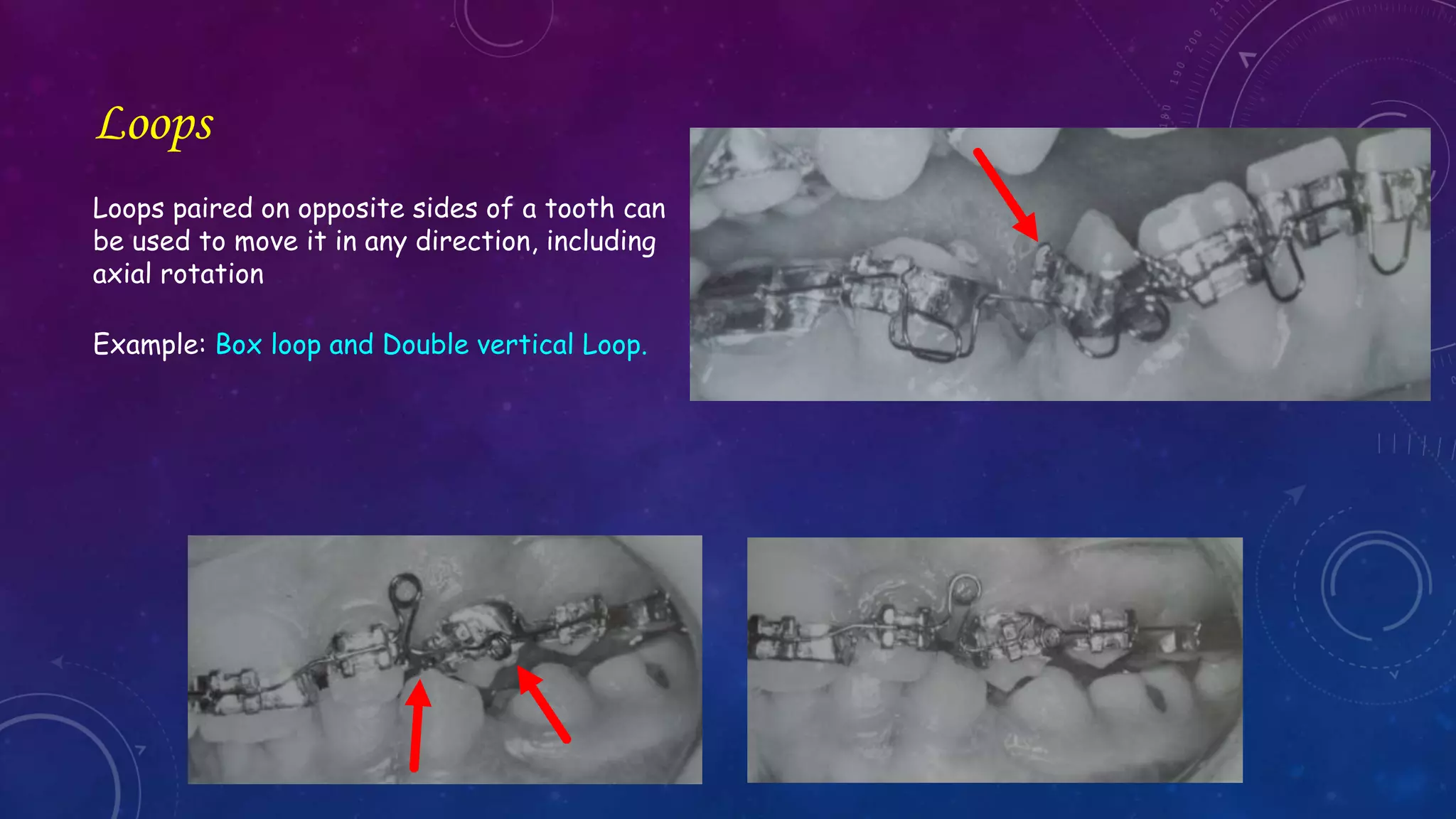
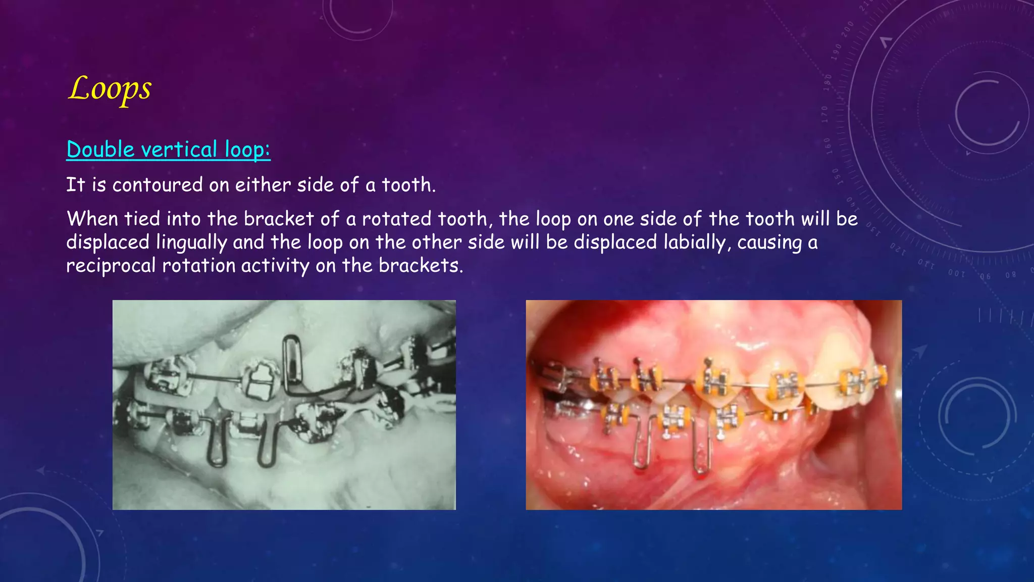
Prepared by: Noha Ali Abd El-Hady
supervision : Prof. Dr/ Maher Fouda
Orthodontic Correction
Of
Rotated Teeth
Contents
Introduction
Treatment Considerations
Treatment options
Fixed appliances
Semifixed appliances
Removable appliances
Retention
Introduction
Rotation
• Tooth rotation can be defined as observable mesio-lingual or disto-lingual intra alveolar
displacement of the tooth around its longitudinal axis (at least 20°) .
 Baccetti, T. "Tooth rotation associated with aplasia of nonadjacent teeth." The Angle Orthodontist 68.5 (1998): 471-474.
 Singh, Gurkeerat. Textbook of Orthodontics. JP Medical Ltd, 2nd edition.
1. Centric rotation only rotation around the long axis.
e.g. (the angulation of the long-axis of the tooth remains unaltered)
2. Eccentric rotation rotation with tipping of the tooth also.
Tooth protrudes from the row of teeth
If the body rotates about its center of resistance, it is called pure rotation.
Rotation
 Premkumar, Sridhar. Textbook Of Orthodontics. Elsevier Health Sciences, 2015.
1. Centric rotation
2. Eccentric rotation
Deviation in the position of individual teeth can be defined as rotation or tipping
depending on the position of the axis.
a) Rotation around an eccentric axis
b) Rotation around a central axis usually the long axis of the tooth.
c) Tipping around a transverse axis , (in a labial or lingual direction)
d) Tipping around a sagittal axis , (in a mesial or distal direction)
Rotation vs. tipping
Rotations are of the following two types:
Mesiolingual or Distolabial
Distolingual or Mesiolabial
 Singh, Gurkeerat. Textbook of Orthodontics. JP Medical Ltd, 2nd edition.
Winging & Counterwinging
Terms by Dahlberg:
If the distal margins of the central incisors are rotated in a labial direction
If the distal margins of the central incisors are rotated in a lingual direction
Winging
Counter winging
 Prasad, Vaishali Nandini, et al. "Winged maxillary central incisors with unusual morphology: a unique presentation and early
treatment." The Angle Orthodontist 75.3 (2005): 478-482
Winging Counter winging
Gupta et al. classified the rotation into three groups:
<45° , 45-90° & >90°.
In his study, rotations were the most common (10.24%) anomaly among the whole study group, that
the majority of tooth rotations were between 45° and 90°, followed by <45° rotations.
In the untreated population, the prevalence of tooth rotation is 2.1-5.1%. Rotations are more
common in patients with hypodontia
The most common rotated teeth were the mandibular second premolars followed by mandibular
first premolars and maxillary central incisors with the same prevalence.
 Baccetti, T. "Tooth rotation associated with aplasia of nonadjacent teeth." The Angle Orthodontist 68.5 (1998): 471-474.
 Parisay, Iman, et al. "Treatment of severe rotations of maxillary central incisors with whip appliance: Report of three
cases." Dental research journal 11.1 (2014): 133.
Etiology
Pre-eruptive disturbances
Injury of the pre-maxillary
region in childhood
The presence of an adjacent
pathology such as cyst, tumor,
odontoma, supernumerary
tooth (mesiodens)
 Parisay, Iman, et al. "Treatment of severe rotations of maxillary central incisors with whip appliance: Report of three
cases." Dental research journal 11.1 (2014): 133.
Can displace and misalign
the developing tooth bud
Can interfere with eruption
of the tooth.
Mesiodens can cause ectopic
eruption, displacement or
rotation of a central incisor
in 28-63% of cases.
Etiology
Post-eruptive disturbances
Habitual factors.
Mechanical factors
Local factors
Environmental factors
 Parisay, Iman, et al. "Treatment of severe rotations of maxillary central incisors with whip appliance: Report of three
cases." Dental research journal 11.1 (2014): 133.
Retained deciduous tooth beyond
normal exfoliation time
space availability for tooth alignment,
path and sequence of tooth eruption
Functional effects or
undesirable forces exerted
by tongue and lips
Mesiodens
Rotated incisor mesiopalataly due to presence of
Mesiodens in the midline.
90° rotation and diastema
Unusual rotation of an erupting maxillary incisor with abnormal diastema >>>>>> Suspect Mesiodens
Unusual rotation of the maxillary lateral incisor >>>>>>> suspect canine impaction.
 Orthodontic Diagnosis and management of malocclusion and Dentofacial deformities
In a dental arch with crowding, rotations are
often present, but in cases of space excess
rotations might also occur.
Rotated and displaced incisors are commonly
seen in the developing crowded malocclusion.
 Staley, Robert N., and Neil T. Reske. Essentials of orthodontics: diagnosis and treatment. John Wiley & Sons, 2010.
 Parisay, Iman, et al. "Treatment of severe rotations of maxillary central incisors with whip appliance: Report of three
cases." Dental research journal 11.1 (2014): 133.
Rotation
In some children, space analysis shows that enough space for all the permanent teeth ultimately
will be available, but relatively large permanent incisors and the clinical reality of the “incisor
liability” cause transient crowding of the permanent incisors.
This crowding is usually expressed as mild faciolingual displacement or rotation of individual
anterior teeth
 Proffit, William R., Henry W. Fields, and David M. Sarver. Contemporary Orthodontics. Elsevier Health Sciences, 2014.
Treatment Considerations
Certain diagnostic questions that require examination prior to beginning biomechanics:
 http://www.orthodonticproductsonline.com/2012/10/the-piggyback-technique/
Enough arch length? To permit correcting the rotation and aligning the tooth.
If not, Consider any method of gaining space as expansion or extraction.
To accommodate the rotation correction (especially buccal or labial bone)
Mavragani et al. suggested that since root shortening due to apical root
resorption is one of the most side effects of orthodontic treatment, it appears
advisable to initiate orthodontic correction of the incisors at a young age
during mixed dentition, in an introductory phase of treatment.
Early correction of rotated teeth before root completion is conducive to
better retention.
Healthy enough to permit the tooth rotation required? Will gingival grafting
and/or bone grafting be required before, during, or after rotation correction?
Enough buccal-lingual
alveolar bone?
Root development?
Gingiva?
Key IV of Andrew’s Six Keys : Rotation
The fourth key to normal occlusion is that the teeth should be free of undesirable rotations.
A rotated molar or bicuspid occupies more space than normal.
A rotated incisor occupies less space than normal.
 Singh, Gurkeerat. Textbook of Orthodontics. JP Medical Ltd, 2nd edition.
Alignment of rotated anterior teeth vs. Alignment of rotated posterior teeth
Broader mesiodistally
Occupy less space when they are rotated.
Alignment of such teeth requires space.
For every millimeter of derotation required, the
same amount of space is required for aligning the
teeth.
 Singh, Gurkeerat. Textbook of Orthodontics. JP Medical Ltd, 2nd edition.
Broader labiolingually.
Occupy more space when they are rotated.
Alignment of such teeth creates space.
The space created depends upon the tooth
(Molar > Premolar)
and the amount of rotation present.
Biomechanics of rotation correction
Rotation can be achieved by two ways:
First by using couple forces.
second by using a single force and a stop.
 Premkumar, Sridhar. Textbook Of Orthodontics. Elsevier Health Sciences, 2015.
Couple
To make rotation movement
What is Couple?
Two parallel forces equal in magnitude but in opposed directions
and separated by a distance (i.e different lines of action) that
act upon a tooth are required.
This is the only force system capable of producing pure rotation
of a body around its center of resistance and the longitudinal axis
of the tooth (seen from the occlusal view).
In this case the tooth maintains its position because both forces
annul each other since both lines of force act at a same distance
perpendicular to the center of resistance, leaving only the pure
Moment (pure rotation).
 Yanez, Esequiel Eduardo Rodriguez. 1,001 tips' for orthodontics and its secrets. 2008.
a couple or coplanar
Fixed vs. Removable appliances
It is frequently held that removable appliances cannot correct rotations.
A force couple can be achieved on a flat large tooth such as a central incisor or large lateral incisor
to correct only a simple rotation of up to about 45°.
Lower incisors are so small in width that the forces even when applied at the extreme ends of the
incisal edge are so close together that an effective mechanical couple cannot be produced.
 Tooth movement with Removable appliances
x
Multiple rotations, more severe individual rotations, and
those in teeth with crowns which are round in cross-
section, which does not offer two points near the outside
of the contour of the tooth to which suitable pressures
may be applied. e.g. premolars and canines, are impossible
to correct with a removable appliance alone.
Fixed vs. Removable appliances
It is important, however, to check that the problem is simply a rotation.
Many rotations have an associated apical malposition which may make the problem impossible without
the control offered by fixed appliances.
If an attempt is made to treat such a problem with a simple whip and band the tooth will tend to upright
over its apex and will probably finish in the wrong position and at the wrong height.
 Tooth movement with Removable appliances
A severe rotation with the
apex in a normal position.
The rotation is less severe but is
combined with apical displacement.
Treatment by simple
means is unsatisfactory.
Fixed vs. Removable appliances
Any fixed appliance system with a two point contact has more efficient rotation control.
Increase control of tooth movements
Movement possible in all three planes of space
Optimum Force
In theory, forces to produce rotation of a tooth
around its long axis could be much larger than
those to produce other tooth movements, since the
force could be distributed over the entire PDL
rather than over a narrow vertical strip.
In fact, however, it is essentially impossible to
apply a rotational force so that the tooth does not
also tip in its socket, and when this happens, an
area of compression is created just as in any other
tipping movement.
For this reason, appropriate forces for rotation
are similar to those for tipping.
 Proffit, William R., Henry W. Fields, and David M. Sarver. Contemporary Orthodontics. Elsevier Health Sciences, 2014.
Clinical Tip
When crowded and rotated maxillary incisors
are corrected orthodontically in adults, the
connector moves incisally and black triangles
may appear, especially if severe crowding was
present.
For that reason, both actual and potential black
triangles should be noted during the orthodontic
examination, and the patient should be prepared
for reshaping of the teeth to minimize this
esthetic problem.
 Proffit, William R., Henry W. Fields, and David M. Sarver. Contemporary Orthodontics. Elsevier Health Sciences, 2014.
Fixed Appliances
Fixed appliances
Ligation
Auxiliaries
Derotation with Ni Ti wire
Sectional fixed wire
The 2x4 appliance
The piggyback technique
Loops
The (Anghileri) technique
Ligation
Ligation
To achieve orthodontic tooth movement, Archwires must be
tied to the bracket slots.
Metal or elastic ligatures are used for this purpose.
Because of their design, twin brackets can be tied in a large
variety of ways.
Traditional elastic O-ring ligation with twin brackets often
fails to correct severe malrotations because of the inability
to fully seat the archwire into the bracket slot.
 Faber, Jorge. "Tying twin brackets." American Journal of Orthodontics and Dentofacial Orthopedics 118.1 (2000): 101-106.
 Mirzakouchaki, Behnam. "Asymmetrical O-Ring Ligation." Journal of clinical orthodontics: JCO 42.2 (2008): 100-100.
Elastic figure-8 technique, in which the O-ring criss-crosses
over the bracket.
Twisting the O-ring in this manner increases its elastic tension,
which helps seat the archwire
Ligation
Double over tie ( Figure 8 tie)
Modified figure 8 tie
Anti-rotation tie
Double ligation technique
Single tie (Isolated tie)
Modified rotation tie
Circumferential tie
Coil Spring tie
With metal / elastic ligature
 Faber, Jorge. "Tying twin brackets." American Journal of Orthodontics and Dentofacial Orthopedics 118.1 (2000): 101-106.
 Rammanohar, Mala. "Bracket Ligation with a Difference." The Journal of Indian Orthodontic Society 48.4 (2014): 286.
Ligation
Double over tie ( Figure 8 tie)
Modified figure 8 tie
Anti-rotation tie
Double ligation technique
Single tie (Isolated tie)
Modified rotation tie
Circumferential tie
Coil Spring tie
 Rammanohar, Mala. "Bracket Ligation with a Difference." The Journal of Indian Orthodontic Society 48.4 (2014): 286.
Patient presented with crowing of lower anterior teeth requiring
the extraction of the first premolars
Modified method of ligation of 31 and 42
Ligation
Double over tie ( Figure 8 tie)
Modified figure 8 tie
Anti-rotation tie
Double ligation technique
Single tie (Isolated tie)
Modified rotation tie
Circumferential tie
Coil Spring tie
 Mirzakouchaki, Behnam. "Asymmetrical O-Ring Ligation." Journal of clinical orthodontics: JCO 42.2 (2008): 100-100.
Double over tie ( Figure 8 tie)
Modified figure 8 tie
Anti-rotation tie
Double ligation technique
Single tie (Isolated tie)
Modified rotation tie
Circumferential tie
Coil Spring tie
Ligation
Assymetrtical O-Ring Ligation:
The force levels of the O-ring are symmetrical, they may
still be inadequate to fully seat the wire.
This modification creates an asymmetrical force to help
fully seat the archwire into the bracket slot.
Tail toward the prominent side
Ligation
Double over tie ( Figure 8 tie)
Modified figure 8 tie
Anti-rotation tie
Double ligation technique
Single tie (Isolated tie)
Modified rotation tie
Circumferential tie
Coil Spring tie
Ligation
Double over tie ( Figure 8 tie)
Modified figure 8 tie
Anti-rotation tie
Double ligation technique
Single tie (Isolated tie)
Modified rotation tie
Circumferential tie
Coil Spring tie
Ligation
 Double-Ligation Technique for Rotated Teeth
Double over tie ( Figure 8 tie)
Modified figure 8 tie
Anti-rotation tie
Double ligation technique
Single tie (Isolated tie)
Modified rotation tie
Circumferential tie
Coil Spring tie
Ligation
 Double-Ligation Technique for Rotated Teeth
Double over tie ( Figure 8 tie)
Modified figure 8 tie
Anti-rotation tie
Double ligation technique
Single tie (Isolated tie)
Modified rotation tie
Circumferential tie
Coil Spring tie
 Faber, Jorge. "Tying twin brackets." American Journal of Orthodontics and Dentofacial Orthopedics 118.1 (2000): 101-106.
Ligation
The slot closer to the archwire can be filled with an elastomeric
ligature, a metallic ligature or an auxiliary designed specifically
for the purpose, eg, a rotation wedge, or even be left empty.
A firm tie is made to the bracket farther from the archwire
Use a finger or an instrument to press the archwire as flush as
possible against the bracket to be tied when twisting the ligature wire
Double over tie ( Figure 8 tie)
Modified figure 8 tie
Anti-rotation tie
Double ligation technique
Single tie (Isolated tie)
Modified rotation tie
Circumferential tie
Coil Spring tie
 Rapid Correction of Rotation with Modified Rotation Tie
Ligation
Double over tie ( Figure 8 tie)
Modified figure 8 tie
Anti-rotation tie
Double ligation technique
Single tie (Isolated tie)
Modified rotation tie
Circumferential tie
Coil Spring tie
Rotation tie was modified by using elastic module or elastic chain
with steel ligature wire .
E chain attached to prominent side of bracket , passes
interproximally then ligature wire is attached to main arch wire on
the other side.
Mesial side
 Rapid Correction of Rotation with Modified Rotation Tie
Ligation
Double over tie ( Figure 8 tie)
Modified figure 8 tie
Anti-rotation tie
Double ligation technique
Single tie (Isolated tie)
Modified rotation tie
Circumferential tie
Coil Spring tie
Rapid correction after 3 months
Rotated left lateral incisor was
treated with modified rotation tie
IOPA showing no root resorption
of lateral incisor.
 Faber, Jorge. "Tying twin brackets." American Journal of Orthodontics and Dentofacial Orthopedics 118.1 (2000): 101-106.
Ligation
Double over tie ( Figure 8 tie)
Modified figure 8 tie
Anti-rotation tie
Double ligation technique
Single tie (Isolated tie)
Modified rotation tie
Circumferential tie
Coil Spring tie
In the case of posterior and canine teeth, a small retention area can be
molded in composite resin or an auxiliary can be placed or soldered onto
the lingual-palatine surface to prevent vertical displacement of the
ligature wires.
 Faber, Jorge. "Tying twin brackets." American Journal of Orthodontics and Dentofacial Orthopedics 118.1 (2000): 101-106.
Ligation
Double over tie ( Figure 8 tie)
Modified figure 8 tie
Anti-rotation tie
Double ligation technique
Single tie (Isolated tie)
Modified rotation tie
Circumferential tie
Coil Spring tie
Circumferential ligations with elastomeric ligature:
Advantages:
it provides prolonged action
of the force applied.
The circumferential ligations should be used with caution because if
the ligature wires pass through the anatomic contact points instead
of the interdental spaces, interproximal pressures may be created
and cause minute irregularities or crowding of the anterior teeth.
Ligation
Double over tie ( Figure 8 tie)
Modified figure 8 tie
Anti-rotation tie
Double ligation technique
Single tie (Isolated tie)
Modified rotation tie
Circumferential tie
Coil Spring tie
• The existing spring section is pressed by the archwire, thus helping
to provide the momentum required for correcting a tooth rotation.
• Prefabricated systems that operate in a similar fashion are available; the
spring is replaced by an elastic wedge.
 Faber, Jorge. "Tying twin brackets." American Journal of Orthodontics and Dentofacial Orthopedics 118.1 (2000): 101-106.
Clinical tip: Antirotation tying
Rotation commonly occurs during some orthodontic procedures.
Correcting the rotation requires additional time.
Antirotation tying of the canines during canine retraction. (or the
first premolars in cases where second premolars have been
extracted) is a useful way of preventing such rotation.
In this case, the wings of the distal bracket alone should be tied to
prevent it from moving away from the archwire during retraction.
During the tying procedure, the tip of a clinical probe is inserted
between the bracket and the ligature thread, allowing a slight
amount of slack to ensure freedom of movement to reduce friction.
Also , Antirotation tying of teeth neighboring to open coil to
guard against rotation
 Faber, Jorge. "Tying twin brackets." American Journal of Orthodontics and Dentofacial Orthopedics 118.1 (2000): 101-106.
Auxiliaries
Auxiliaries
Rotation wedge
Steiner Ligature rotation wedge
Button
Monkey hook
Double-loop de-rotator
Rotation Spring
Rotation wedge
 It acts as a fulcrum between the wire and bracket.
 It is ligated to the tie wing of the bracket closest to the wire.
 Available in different colors or clear.
Rotation wedge
Rotation wedge
Steiner Ligature Rotation Wedges:
In rotation control, higher force levels than elastomeric
materials is required. In rotation cases, the partial
engagement of the arch wire will be difficult with elastic
ligature, so in these cases wire ligature are advised.
The round elastomeric wedge easily attaches to the bracket
under the arch wire. Supplied on preformed ligature wire.
Button
Buttons can be used to produce
By bonding buttons to the buccal and palatal surface of the rotated tooth and use elastic chains
between them and the neighboring teeth or anchorage devices.
a couple force.
Button
TPA:
Nance appliance:
Two hooks incorporated into
the acrylic portion of the
Nance button on either side.
 Dahiya, Amit, and Minakshi Rana. "Modified Nance Appliance for Tooth Derotation."
 Handbook of Orthodontics , 2ed (2016)(2)
Button used with fixed devices to create couple for derotation of teeth.
Quad helix
Button
With Miniscrew:
Depending on the position of the miniscrew and the attachment on the tooth, various combination
of tooth movement may be resulted. Intrusion, mesial, or distal tipping and/or rotation can be
possible.
 Handbook of Orthodontics , 2ed (2016)(2)
 Case Example: Use Microimplant screw for Anchorage, http://healthmantra.com/ic/case4.htm
With the help of screw the premolar rotation was easily corrected in 8 weeks time without loosing
molar anchorage(1-2mm) and gain space for anterior retraction by doing this derotation
Button
A 58 year old male patient with the
chief complaint of the unaesthetic gap
between upper front teeth (5 mm) .
On examination, he had Angle’s Class I
with severe mesio-palatal rotation of 21.
 Gyawali, Rajesh, et al. "Orthodontic correction of severely rotated maxillary central incisor in a diabetic adult."
Radiographs were taken to evaluate the alveolar bone height.
 Gyawali, Rajesh, et al. "Orthodontic correction of severely rotated maxillary central incisor in a diabetic adult."
Journal of College of Medical Sciences-Nepal 11.3 (2015): 30-34.
Periapical Radiograph showing Mesiodens.
Mesiodens was extracted.
After the levelling and alignment of other
maxillary teeth except 21, lingual buttons were
bonded on the labial and palatal surface of 21 and
couple was used to derotate it.
 Gyawali, Rajesh, et al. "Orthodontic correction of severely rotated maxillary central incisor in a diabetic adult."
Journal of College of Medical Sciences-Nepal 11.3 (2015): 30-34.
Fixed lingual retainer after an active
treatment duration of 9 months.
Post treatment photographs intraoral periapical radiograph
 Gyawali, Rajesh, et al. "Orthodontic correction of severely rotated maxillary central incisor in a diabetic adult."
Journal of College of Medical Sciences-Nepal 11.3 (2015): 30-34.
A 7-year-old male with the chief complaint of non-eruption of the permanent maxillary right incisor.
 Fujita, Y., T. Takahashi, and K. Maki. "Orthodontic treatment for an unerupted and severely rotated maxillary central incisor. A
case report." EUROPEAN JOURNAL OF PAEDIATRIC DENTISTRY 9.1 (2008): 43.
A radiographic examination revealed the presence of an inverted
supernumerary tooth overlapping the root of the maxillary left incisor.
Further, the maxillary right incisor was completely impacted and rotated
Surgical exposure with extraction of the supernumerary tooth were done.
Three months later, bilateral central incisors erupted spontaneously.
Orthodontic traction of the tooth with a power chain applied between a button
on the labial surface and an auxiliary spring soldered to the base arch.
After 3 months, the rotation of the upper right incisor was nearly completely
corrected.
 Fujita, Y., T. Takahashi, and K. Maki. "Orthodontic treatment for an unerupted and severely rotated maxillary
central incisor. A case report." EUROPEAN JOURNAL OF PAEDIATRIC DENTISTRY 9.1 (2008): 43.
One week later, a quad helix was applied to the upper arch for
labial movement of the right incisor.
A rapid expansion device was applied after6 months in order
to create adequate space for alignment
 Fujita, Y., T. Takahashi, and K. Maki. "Orthodontic treatment for an unerupted and severely rotated maxillary central
incisor. A case report." EUROPEAN JOURNAL OF PAEDIATRIC DENTISTRY 9.1 (2008): 43.
Bonding of a 2x4 appliance for alignment.
Retention with a Hawley appliance for 6 months
 Fujita, Y., T. Takahashi, and K. Maki. "Orthodontic treatment for an unerupted and severely rotated maxillary central
incisor. A case report." EUROPEAN JOURNAL OF PAEDIATRIC DENTISTRY 9.1 (2008): 43.
Monkey hook
The Monkey Hook is a simple S-shaped auxiliary with an open loop on each end for the attachment of
intraoral elastics or elastomeric chain, or for connecting to a bondable loop-button.
A combination of Monkey Hooks and bondable loop-buttons allows the production of a variety of
different directional forces to assist in the correction of impacted, rotated, or displaced teeth.
 The Monkey Hook: An Auxiliary for Impacted, Rotated, and Displaced Teeth
Monkey hook
Rotational Couples:
If a loop-button is bonded on each side of the tooth, forces can be applied in opposite directions to
create rotational couple.
 The Monkey Hook: An Auxiliary for Impacted, Rotated, and Displaced Teeth
A Loop-buttons bonded on opposite sides of severely
rotated second premolar to create rotational couple
After derotation.
Case
A 15-year-old male patient with palatally impacted maxillary canines.
 The Monkey Hook: An Auxiliary for Impacted, Rotated, and Displaced Teeth
After surgical exposure of canines and direct bonding of loop buttons, Monkey Hooks and
elastomeric chains were attached .Coil springs were used to create and maintain space for canines.
Third Monkey Hook on each side was used for attachment of intermaxillary elastics to produce
vertical eruptive forces, with anchorage from mandibular arch.
After 5 months of eruption, second loop-button was bonded to lingual side of each canine.
Rotational couple was produced using elastic thread to lingual cleat on first molar and thermal
superelastic archwire threaded through buccal loop-button.
 The Monkey Hook: An Auxiliary for Impacted, Rotated, and Displaced Teeth
Progress after 18 months.
 The Monkey Hook: An Auxiliary for Impacted, Rotated, and Displaced Teeth
Bracket bonded to right canine.
Progress after 23 months.
Double-loop de-rotator
In cases of severe rotations (>90°) it is very difficult to bond attachments for proper application
of couple forces due to inaccessibility to surfaces or size of attachments.
In such cases, multiple repositioning of bonded attachments will be required during de-rotation of
tooth.
Fabrication:
 Double-loop de-rotator
 It is made up of thick ligature wire (0.009˝).
 It is twisted around explorer to make loops at either end.
Double-loop de-rotator
Steps in bonding of de-rotator:
 Double-loop de-rotator
 Placement of Separators to get
clearance for bonding.
 Bonding
 contouring on the proximal surface.
 Couple force system application
Derotation completed
Double-loop de-rotator
Advantages:
 Simple and economic technique.
 No need of repositioning of attachments.
Limitations:
 Like any other procedure involving bonding it is
technique sensitive and proper moisture control
is a must.
 Under heavy forces can lead to debonding of the
attachment.
 Double-loop de-rotator
Rotating Spring
 provide a simple and effective means of de-rotating teeth without the removal of the archwire.
 These springs are used in
 They are capable of both clockwise and counter clockwise movement depending on their design.
 Not effective for derotating posterior teeth.
 The rotating spring exerts a light, continuous force that can align a rotated tooth within several
weeks.
 Singh, Gurkeerat. Textbook of Orthodontics. JP Medical Ltd, 2nd edition.
the vertical slots of the Begg and the Tip-edge bracket.
Rotating Spring
Construction of rotating spring.
A) Uprighting spring inserted into vertical bracket slot with spiral bent perpendicular to principal
arm, which is at 60-70° angle to main archwire.
B) Spring activated by turning vertical arm in direction of desired rotation: distally in relation to
the slot to produce a distal rotation, mesially for a mesial rotation
C) Principal arm hooked to archwire in same direction.
 Correction of Single-Tooth Rotations with Rotating Springs
Rotating Spring
A 12-year-old male Class II patient
with mandibular anterior crowding and
rotations before treatment.
Left lateral incisor rotation persists in
finishing phase.
 Correction of Single-Tooth Rotations with Rotating Springs
Rotating Spring
The spring was inserted in the vertical slot of the lateral incisor bracket and
attached to the archwire distal to the canine bracket.
 Correction of Single-Tooth Rotations with Rotating Springs
Rotating Spring
Alignment of rotated incisor in 3 weeks.
Upon completion of active therapy, a mandibular lingual
3-3 retainer was bonded to maintain the result.
 Correction of Single-Tooth Rotations with Rotating Springs
Derotation with Ni Ti wire
In the Pre-adjusted Edgewise system, precise bracket positioning,
use of highly resilient superelastic nitinol wires for initial
alignment and final engagement of rectangular wires in brackets
de-rotates the rotated teeth.
 Premkumar, Sridhar. Textbook Of Orthodontics. Elsevier Health Sciences, 2015.
 http://www.orthodonticproductsonline.com/2012/10/the-piggyback-technique/
Disadvantages:
If de-rotation is carried out on Ni-Ti wires during the levelling
phase, it causes undesirable force and unwanted tooth movement
of neighboring teeth (Orthodontic abutment teeth) . Especially if
the tooth to be rotated is large with a very long root.
P, pressure on 2|; R, reaction. The
reaction is borne mainly by the
adjoining teeth.
Sectional fixed technique
An eight years six-month-old Indian boy with rotated upper central incisors mesiopalatally (winged).
An intraoral periapical radiograph of the region revealed the roots were somewhat convergent.
 Prasad, Vaishali Nandini, et al. "Winged maxillary central incisors with unusual morphology: a unique presentation
and early treatment." The Angle Orthodontist 75.3 (2005): 478-482
Sectional fixed technique
Derotation using only an anterior sectional wire (A 0.018-
inch NiTi ).
Diastema closure and partial derotation were achieved in
four weeks, when the NiTi wire was replaced with a 0.017 x
0.025–inch stainless steel wire.
 Prasad, Vaishali Nandini, et al. "Winged maxillary central incisors with unusual morphology: a unique presentation
and early treatment." The Angle Orthodontist 75.3 (2005): 478-482
Sectional fixed technique
 Prasad, Vaishali Nandini, et al. "Winged maxillary central incisors with unusual morphology: a unique presentation
and early treatment." The Angle Orthodontist 75.3 (2005): 478-482
Complete derotation was achieved after another 10 weeks , and the
appliance was removed after 12 weeks of the retention phase.
There has been no relapse during the 12 months of follow-up period.
A posttreatment intraoral periapical radiograph
showed normal development and divergence of the
roots of 11 and 21.
2 x 4 Appliance
The 2x4 appliance
Design:
The basic 2x4 appliance design is as follows:
 Bands cemented on both maxillary permanent first molars
 Brackets bonded onto the erupted maxillary central and lateral incisors
 Early Correction of Rotated Incisor Using 2x4 Appliance With Laser Aided Circumferential Supracrestal Fiberotomym Case Report
The 2x4 appliance
A 12 year old girl with irregular upper front teeth.
 Early Correction of Rotated Incisor Using 2x4 Appliance With Laser Aided Circumferential Supracrestal Fiberotomym Case Report
A 2x 4 fixed appliance.
Correction of rotation after 4 months followed by
circumferential suprecrestal Fiberotomy using a diode laser.
Then, Placement of fixed lingual retainer.
The 2 x 4 Appliance
Pretreatment extra-oral and
intra-oral photographs
 How to effectively use a 2x4 appliance
The 2 x 4 Appliance
 How to effectively use a 2x4 appliance
Intra-oral photographs after
alignment of incisors
The 2 x 4 Appliance
 How to effectively use a 2x4 appliance
Post-treatment photographs
with Palatal bonded retainer
The Piggyback Technique
The rotational forces have little effect on the abutment teeth.
The traditional approach is to use a smaller-sized main archwire that is flexible enough to engage
the rotated tooth.
The problem is that when rotational forces applied to the rotated tooth create undesired tooth
movements in the abutment teeth, a sequence of wires is usually required to realign all of the
teeth.
The Piggyback Technique helps to avoid this waste of time and resources.
 http://www.orthodonticproductsonline.com/2012/10/the-piggyback-technique/
The Piggyback Technique
Main archwire with open coils to open adequate space to permit the rotation correction.
The piggyback wire was fully engaged into the right and left maxillary lateral incisors and was ligated
on top of the main archwire on the adjacent teeth.
 http://www.orthodonticproductsonline.com/2012/10/the-piggyback-technique/
A patient after Rapid Palatal Expansion.
At this initial stage, the piggyback wire rests on
the lingual surface of the adjacent maxillary
right cuspid (due to the severity of the rotation
on the maxillary right lateral incisor).
After 22 days, the rotated tooth has moved
from a near 90º rotation to near normal.
Note that the piggyback wire is now resting on
the buccal of both cuspids.
 http://www.orthodonticproductsonline.com/2012/10/the-piggyback-technique/
Posttreatment photos
Loops
Loops
Loops paired on opposite sides of a tooth can
be used to move it in any direction, including
axial rotation
Example: Box loop and Double vertical Loop.
Loops
Such rotations almost require significant horizontal movement in addition to the rotation, and some
may require tipping (Uprighting) as well paired loops can be designed to perform all of these actions
simultaneously.
Loops
Double vertical loop:
It is contoured on either side of a tooth.
When tied into the bracket of a rotated tooth, the loop on one side of the tooth will be
displaced lingually and the loop on the other side will be displaced labially, causing a
reciprocal rotation activity on the brackets.
Loops
Box Loop:
The box loop is composed of a series of vertical and horizontal levers contoured in such a manner
to provide a short section of archwire that is freely movable in all planes of space and usually is
contoured to the width of a single tooth.
Canine intrusion using a loop. A rectangular loop welded to a bypass archwire is used to
simultaneously rotate the canine or change mesiodistal axial inclinations during intrusion.
Case
A 14 years old girl with a Class I skeletal pattern and rotation of the mandibular right canine.
 Correction of Canine rotation with box loop
Treatment :
The segmented arch was passive in the slot of the pre-molars and
molar tube, which were joined with metal wire and active in the
canine bracket slot.
 Correction of Canine rotation with box loop
Intermaxillary elastics for intercuspation
Fixed appliance and mandibular lingual arch for anchorage.
Stripping to create space between 4,3,2
a box loop (TMA 0.017x0.025-inch) fixed in the molar auxiliary tube,
premolars and canine brackets.
 Correction of Canine rotation with box loop
AfterBefore
Final result
The “ANG (Anghileri) Technique”
The “ANG (Anghileri) Technique”:
Invented by Dr. Matías Anghileri from Argentina.
 First, place the initial archwire and bond a
button on the buccal surface of the rotated
tooth.
 Second, insert a passive spring between the two
teeth surrounding the rotated tooth.
 The “ANG (Anghileri) Technique”: Making Derotation Easy
 Third, use a ligature from the button to compress the spring
approximately a third of its original length.
 An easy way to do this is by placing the ligature through the
first or second coil. The spring can then exert its force in
the same direction to which the tooth must be rotated.
The “ANG (Anghileri) Technique”:
The spring works continuously to de-rotate the tooth
without adjusting the ligature.
 The “ANG (Anghileri) Technique”: Making Derotation Easy
The “ANG (Anghileri) Technique”:
The spring should be
compressed to provide a
constant mesial light force
towards the rotated bicuspid
 The “ANG (Anghileri) Technique”: Making Derotation Easy
The bicuspid is
rotated distally
Once the tooth is rotated enough to
be engaged in the main archwire, it’s
ready for bonding.
Space is created with derotation
Finally, the total
correction is achieved
after 3 months.
The “ANG (Anghileri) Technique”:
 The “ANG (Anghileri) Technique”: Making Derotation Easy
The ANG Technique is applied to the lower 3s and 5s.
With the ANG technique the directions of the forces of the compressed
springs rotate the bicuspids distally and the cuspids mesially
The tooth where the spring is going to be anchored must have a greater anchorage
than the rotated one to avoid an unwanted rotation.
The “ANG (Anghileri) Technique”:
Two months later, there is enough space to directly engage these teeth.
The ANG Technique doesn’t interfere with the incisors and anterior
guidance is undisturbed.
 The “ANG (Anghileri) Technique”: Making Derotation Easy
After 3.5 months the teeth are in a good alignment
Semi-Fixed appliances
For molar derotation:
For incisor derotation:
For any tooth derotation
Trans-Palatal Arch
The NiTi Molar Rotator
Quad helix Appliance
 Whip appliance
 Hooked appliances
For Molar Derotation
A distal molar relationship could arise due to
the mesiopalatal rotation of the maxillary
molars around the palatal root.
In some patients, an ideal Class I
intercuspation can be achieved with the
opposing molar and a Class II relationship
can be corrected by molar derotation.
Lemons & Holmes reported that a gain of 1–
2 mm of arch length per side may be
achieved following derotation.
 An effective and precise method for rapid molar derotation: Keles TPA
 Proffit, William R., Henry W. Fields, and David M. Sarver. Contemporary Orthodontics. Elsevier Health Sciences, 2014.
Same patient after rotation correction
Class II relation with rotated molar
For Molar Derotation
Ricketts proposed a method of diagnosing mesial rotation of
the upper first molar.
View the upper arch from the occlusal and draw a line from
the distal buccal through the Mesiopalatal cusp of the upper
molar. That line should pass through the opposite canine.
Ricketts’ line passes through the opposite bicuspids.
The molars are mesially rotated
Ricketts’ line passes through the opposite canine.
The molar relationship is Class I.
For Molar Derotation
TPA (Trans-palatal arch)
TPA with U-loop before the molar bands and gives the
appliance more flexibility for adjustment
 Biomechanics in Orthodontics Principles and Practice - Ram Nanda 2010
(a) Equal and opposite moments rule out balancing forces.
(b) If the molars are initially rotated unequally, the balancing
forces tend to move one molar mesially and the other distally.
If this is not desired, the anchorage side should be
reinforced by incorporating adjacent teeth.
(α represents the amount of rotation of the molars relative
to the sagittal plane.)
For Molar Derotation
Keles TPA
Fabrication:
constructed from the Burstone lingual arch
system.The wire consists of 0.032 × 0.032′′
beta-titanium alloy.
 An effective and precise method for rapid molar derotation: Keles TPA
For Molar Derotation
Keles TPA
Activation:
The TPA is placed on a piece of white paper and two
lines are drawn along the terminal ends (rotating
component) of the TPA with a black pen.
Additional lines are drawn with a 20° angle passing
through the distal end of the helix of the wire.
The TPA is activated on both sides with the help of a
bird-beak plier
 An effective and precise method for rapid molar derotation: Keles TPA
Passive stage
Active stage
For Molar Derotation
Keles TPA
The biomechanics of the force moment
system
Two equal and opposite moments are generated on
both molars which would also help to increase the
inter-molar width between the mesial cusp tips of the
first molars.
The activation of the TPA is checked on both sides
and then it is placed in the mouth
 An effective and precise method for rapid molar derotation: Keles TPA
For Molar Derotation
Keles TPA
 An effective and precise method for rapid molar derotation: Keles TPA
Pretreatment With Keles TPA
After molar derotation Posttreatment
For Molar Derotation
The Ni-Ti molar rotator
 Singh, Gurkeerat. Textbook of Orthodontics. JP Medical Ltd, 2nd edition.
For Molar Derotation
Quad helix appliance
Can be used for:
Activation: The amount of activation is checked by inserting one side and observing the relationship
of the retention loop to the sheath on the opposite side.
It is recommended that for an eight week period, activation should not exceed Rotation: 20o
 Orthodontic removable appliance, Sandhya Shyam Lohakare (Talmale)
Unilateral rotation of molar using the
teeth on the opposite side as anchorage
Bilateral rotation of molar
Whip Appliance
Whip Appliance
Introduced by Houston and Isaacson in 1980.
A single rotated tooth in a patient with an otherwise acceptable occlusion may be rotated with a
'whip' where there is adequate space available.
Since whip itself provides no labio-lingual control, labial bow should be adjusted to touch the
labially placed surface of rotated tooth/teeth.
 Tooth movement with Removable appliances
 Fujita, Y., T. Takahashi, and K. Maki. "Orthodontic treatment for an unerupted and severely rotated maxillary
central incisor. A case report." EUROPEAN JOURNAL OF PAEDIATRIC DENTISTRY 9.1 (2008): 43.
 Parisay, Iman, et al. "Treatment of severe rotations of maxillary central incisors with whip appliance: Report
of three cases." Dental research journal 11.1 (2014): 133.
 Whip Spring for Incisor Rotation
Attachment to the
tooth/teeth
Removable appliance
Appliance components:
An oval molar tube ( Mandibular first molar tube ) is bonded directly on
the labial surface of the rotated tooth. Or it can be welded on a band
and adapted to rotated tooth/teeth.
A bonded edgewise bracket can also be used, but it can exert
unnecessary torque during rotation.
Whip Spring
 Whip Spring for Incisor Rotation
Appliance components:
Attachment to the
tooth/teeth
Whip Spring
Cantilever spring.
The recurved mesial end of the whip is inserted into the oval molar tube
or ligated to the bracket on the rotated tooth and is bended towards the
gingiva.
while the distal end formed into a hook to be engaged onto a labial bow or
hooked over the bridge of the upper primary second molar Adams clasp.
Initially, for mesial-in rotations an Adams clasp must be placed on the
molars of the same quadrant and for distal-in rotations it must be placed
on the molars of the opposite quadrant.
Removable appliance
 Whip Spring for Incisor Rotation
 Correction of a severely rotated maxillary central incisor with the Whip device, Case Report
• Simple removable plate with adequate retention using clasps and a
labial bow .
• In designing removable appliance, do not put Adams clasp used for
attachment of the whip spring on first permanent molars because of
its excessive springiness and inadequate strength of the Whip spring
due to increased wire length. Therefore, in order to avoid deformity
of the Whip spring Adams clasps are made on second maxillary primary
molars.
• In case of occlusal interferences, posterior biteplates can be added to
the appliance.
Appliance components:
Attachment to the
tooth/teeth
Removable appliance
Whip Spring
Advantages of Whip Appliance
For a mixed dentition child with a severely rotated central incisor, Whip appliance has several
advantages as follows:
1. This appliance solves the problem in the mixed dentition, relatively in a short duration.
2. Management of anchorage is less critical, a good anchorage is provided from the palate and the
maxillary dentition so, can be used in severe rotations.
3. Force system is relatively simple.
4. It is removable and therefore easier to clean.
5. Patient cooperation is less critical, because when removing the appliance, the distal end of the
whip spring is inserted into the buccal mucosa. The damage of mucosa by wire leads to patient
discomfort.
6. It can be used in emergency situations in the mixed dentition period such as traumatic occlusion
of central incisors.
7. The addition of a band to a single rotated tooth with a whip engaging on to part of the appliance
will allow the correction of more severe rotations and also of canines and premolars.
 Parisay, Iman, et al. "Treatment of severe rotations of maxillary central incisors with whip appliance: Report of
three cases." Dental research journal 11.1 (2014): 133.
 Jahanbin, Arezoo, Bahareh Baghaii, and Iman Parisay. "Correction of a severely rotated maxillary central incisor
with the Whip device." The Saudi dental journal 22.1 (2010): 41-44.
Whip Appliance
Clinical Drawback
• Much attention should be considered not to activate the whip in the vertical plane, otherwise
unwanted mesiodistal crown and root movement may be produced. Extrusion and labial tipping of
the maxillary incisor might occur during treatment.
• Furthermore, the whip spring can wound the mucosa if not adjusted carefully.
• Problems that may be encountered during treatment are debonding of the bracket and
distortion of the spring. However, these problems can be minimized through satisfactory
compliance.
 Parisay, Iman, et al. "Treatment of severe rotations of maxillary central incisors with whip appliance: Report of
three cases." Dental research journal 11.1 (2014): 133.
 Correction of a severely rotated Maxillary Incisor by Elastics in Mixed Dentition Complicated by Mesiodens
Whip Appliance
An 11-year-old boy with severe rotation of
the upper anterior tooth due to a mesiodens
 Correction of a severely rotated maxillary central incisor with the Whip device, Case Report
After 8 months
Whip appliance after
extraction of mesiodens
Whip Appliance
Severe rotation with mesiodens
 Orthodontic Management of a Severely Rotated Maxillary Central Incisor in the Mixed Dentition: A Case Report
Whip Appliance
With whip appliance after
removal of Mesiodens
 Orthodontic Management of a Severely Rotated Maxillary Central Incisor in the Mixed Dentition: A Case Report
During treatment
After removal of appliance
after 8 months
Whip Appliance
70° rotated upper central incisor
 Parisay, Iman, et al. "Treatment of severe rotations of maxillary central incisors with whip appliance: Report
of three cases." Dental research journal 11.1 (2014): 133.
2 month after starting treatment
4 month after starting treatment
Hooked appliances
Hooked appliance
Removable appliances with hooks ( either soldered or incorporated
in the acryl). The rotated tooth is bonded or banded with
attachment to correct the rotation with elastic or power chain
between the attachment and the hook.
Example: Hawley’s retainer with hooks soldered on the labial bow
to rotate an incisor with elastic.
 Orthodontic removable appliance, Sandhya Shyam Lohakare (Talmale)
 O-atlas Dentauram
Hooked appliance
Example: A Hawley’s appliance with a modified labial bow and a palatal hook incorporated in acryl.
 Management of torsiversion of a tooth secondary to a mesiodens
Hooked appliance
An 8 year old with torsiversion of the tooth 21.
Also, the tooth 22 had failed to erupt
An intra-oral periapical radiograph and a
maxillary occlusal radiograph revealed an
unerupted mesiodens which was surgically
extracted.
 Management of torsiversion of a tooth secondary to a mesiodens
Hooked appliance
The affected tooth was banded with attachment for elastics to
create a couple of force to derotate 21.
After the correction of rotation, a retainer was placed.
 Management of torsiversion of a tooth secondary to a mesiodens
Hooked appliance
Another example:
A removable appliance with modified Adams clasp with distal/mesial extension in relation to the
rotated tooth and a loop for engaging elastics.
 Correction of a severely rotated Maxillary Incisor by Elastics in Mixed Dentition Complicated by Mesiodens
Hooked appliance
An 11 years old child with rotated upper right front tooth.
The maxillary right central incisor was rotated with a Mesiodens present, and a maxillary right
lateral incisor was palatally erupted, and in a cross bite.
 Correction of a severely rotated Maxillary Incisor by Elastics in Mixed Dentition Complicated by Mesiodens
Hooked appliance
After extraction of Mesiodens, bondable buttons
were placed on the labial and palatal surfaces of
the incisor.
Fabrication of a removable appliance with modified
Adams clasp with distal extension in relation to the
upper left central incisor and a loop for engaging
elastics.
 Correction of a severely rotated Maxillary Incisor by Elastics in Mixed Dentition Complicated by Mesiodens
Hooked appliance
Elastics were placed between the
palatal bondable button and the distal
extension of the Adam’s clasp. Also
between the labial bondable button
and the loop.
 Correction of a severely rotated Maxillary Incisor by Elastics in Mixed Dentition Complicated by Mesiodens
After 4 months,Circumferential
Supracretal Fiberotomy was
performed.
A Hawley’s appliance with z-spring was fabricated on upper right lateral incisor with a posterior
bite plane for correction of crossbite.
Hooked appliance
 Correction of a severely rotated Maxillary Incisor by Elastics in Mixed Dentition Complicated by Mesiodens
At the end of treatment, a fixed
palatal retainer was placed
Removable appliances
Removable appliances containing any of the following as an active component
 Double Cantilever/Z-Spring
 The labial bow with vertical M-loop
 The labial bow with retractive canine loop
Removable appliances
Advantages:
A simplified and cost effective treatment for successful derotation of anterior teeth in the mixed
dentition stage.
The reactive forces are less. So, there is no particular problem with anchorage.
Better maintenance of oral hygiene.
Limitations:
Ideal case selection is required as it may be indicated only in the case of rotated maxillary central
incisor and probably correct only mild rotations less than 45 degrees.
Rotation has high risk of relapse and because patient compliance is needed , relapse even in the
treatment phase is more likely.
The need for accurate adjustment of the labial bow, palatal spring and acrylic bas plate.
 Correction of a severely rotated Maxillary Incisor by Elastics in Mixed Dentition Complicated by Mesiodens
Double Cantilever/Z-Spring
Construction:
It is made up of 0.5 mm hard round SS wire.
It consists of 2 helices of small internal diameter.
The spring is positioned perpendicular to the palatal surface of the tooth
with a long retentive arm (placed away from tissue) about 12 mm in length.
Activation:
Only one helix may be activated to correct mild rotations.
Use:
the correction of anterior tooth crossbites / rotations where the overlap is
less than the free way space.
The spring is effective only when there is enough space for aligning the teeth.
 Singh, Gurkeerat. Textbook of Orthodontics. JP Medical Ltd, 2nd edition.
Double Cantilever/Z-Spring
Ideal case for correction using ‘Z’ springs,
11 and 21, in negative overbite less than 3
mm and mildly rotated teeth
 Singh, Gurkeerat. Textbook of Orthodontics. JP Medical Ltd, 2nd edition.
Mesio-palatal rotation of 21, leading to
a crossbite treated using an appliance
incorporating a ‘Z’ spring and labial bow
Before After
Spring for molar and premolar derotation
Distal rotation of upper first
premolars about the contact point
with the second premolars.
 The Design, Construction & Use of Removable Orthodontic Appliance
Distal rotation of an upper first molar about the palatal root to
make room for the second premolar
The labial bow with vertical M-loop
Function: Alignment of the canine, if it is labially positioned.
The M- loop moves the tooth primarily in a lingual direction.
Depending on the location of the center part of the loop, the tooth can be rotated.
The loop should only lie on the most prominent part of the crown, avoiding contact with the gingiva.
 O- atlas Dentauram
The labial bow with retractive canine loop
Function: Alignment of labially rotated canine.
To optimize the point of force application, the loop should embrace the
tooth surface as far as possible.
This loop can tip the canine distally as well as lingually.
If the loop is bent the other way around, the canine can be moved
mesially.
 O- atlas Dentauram
Invisalign
Invisalign Teeth with rotations may require a combination of attachments and elastics.
 Clinical Success in Invisalign Orthodontic treatment
Premolars in rotation requiring an additional traction elastic on the aligner
Rotation of the mandibular left second premolar with classic elastic traction.
Invisalign
Correction of rotation
with vertical rectangular
attachments on the
mandibular premolars.
 Clinical Success in Invisalign Orthodontic treatment
Occlusal view before treatment. Occlusal view after placement of attachments.
ClinCheck simulation before treatment.
Simulated results of treatment.
Note the uprighting of the premolars.
Invisalign
 Clinical Success in Invisalign Orthodontic treatment
In case of significant rotation, the location of attachments on the tooth can be
changed during the course of treatment to ensure complete rotation correction
Rotated mandibular right second premolar Placement of attachments.
Stage 13: The original placement of the
attachment is no longer effective for
obtaining the desired rotation.
Stage 14: The attachment is moved.
A template aligner is provided by Invisalign
for both stages at which attachments will
be fabricated (in this case, stages 2 and
14) so that attachments will be placed
accurately and in accordance with the
planned treatment.
Invisalign: Case1
A 27-year-old female patient with a dental
crossbite (24, 34), severe rotations of two lower
incisors (more than 40°) and malalignment of the
upper and lower arches.
 Correction of severe tooth rotations using clear aligners: a case report
Posttreatment photos
A lower fixed retainer was bonded.
Retention in the upper arch with the last
aligner used as a nocturnal removable
retainer.
Invisalign: Case1
The final ClinCheck® provided 17 aligners for the upper arch and 23 aligners for the lower arch
Duration: 12 months. Each aligner was to be worn for two weeks. Rotation correction was about 2° for
each aligner
 Correction of severe tooth rotations using clear aligners: a case report
Initial stage of the ClinCheck®. Final stage of the ClinCheck® with rotational
attachment on teeth in red.
Invisalign: Case1
D. Summary of changes of (E) and (F). The correction of
the rotations on the ClinCheck® and on the photos
are similar.
E. Initial intra-oral photo on the lower arch,
F. Final intra-oral photo on the lower arch
 Correction of severe tooth rotations using clear aligners: a case report
A. Initial ClinCheck®,
B. Final ClinCheck®
C. Superimposition of A and B.
The ClinCheck® simulation shows the degrees of correction
of the rotations.
Invisalign: Case1
 Correction of severe tooth rotations using clear aligners: a case report
A) Initial and (B) final panoramic x-ray (anterior region only).
No obvious root resorption is present after treatment.
A24-year-old patient had a congenitally missing mandibular right lateral incisor and a retained
but failing primary incisor. In the maxillary arch. there was rotation of the right canine.
The plan was stripping of the upper posterior quadrant and extraction of the primary incisor
and closure of the extraction site, using a series of Invisalign aligners and bonded attachments
to produce the necessary rotation and root movement.
 Proffit, William R., Henry W. Fields, and David M. Sarver. Contemporary Orthodontics. Elsevier Health Sciences, 2014.
Note the hard-to-see bonded attachments on
the maxillary right canine and incisors and on
the mandibular right canine and central incisor.
A bonded canine-to-canine mandibular retainer was used, and the final
maxillary aligner was continued at night as the maxillary retainer.
 Proffit, William R., Henry W. Fields, and David M. Sarver. Contemporary Orthodontics. Elsevier Health Sciences, 2014.
Posttreatment
Pretreatment
Retention
Retention
When tooth is rotated about its long axis. The supraalveolar tissue remains
under tension. So, Rotations have a very high risk of relapse due to elastic
recoil of the stretched supra-alveolar and trans-septal gingival fibers, which
readapt very slowly to the new position.
 Orthodontic removable appliance, Sandhya Shyam Lohakare (Talmale)
 Parisay, Iman, et al. "Treatment of severe rotations of maxillary central incisors with whip appliance: Report of three cases." Dental
research journal 11.1 (2014): 133.
Methods suggested to
alleviate the occurrence
of rotational relapse
Early correction of rotated tooth
Long-term retention with bonded lingual retainers
Circumferential supracrestal fiberotomy ( CSF)
Rotations are easy to treat, but very difficult to retain.
Early Correction of Rotated teeth
It is advisable for all rotations to be corrected to ideal or
slightly overcorrected positions in the early stages of
treatment.
The longer the rotated teeth are held in the correct
position, the greater the chances of stability.
The mesiolabial rotations of maxillary lateral incisors in
Class II division 2 malocclusions should be slightly
overcorrected; they relapse very easily.
 Graber, Lee W., et al. Orthodontics: Current Principles and Techniques. Elsevier Health Sciences, 2016.
Circumferential Supra-crestal Fiberotomy ( CSF)
Advocated for the release of soft tissue tension and reattachment of the fibers after orthodontic
correction of tooth rotation.
These procedures are done at the end of the finishing phase of the treatment before the appliance
removal and beginning of the retention phase. i.e: the supra-crestal fibers are sectioned and
allowed to heal and reorient while the teeth are held in the proper position
After the supra-crestal Fiberotomy, the most notable characteristic is an increment in dental
mobility. This mobility is due to the incision of the transeptal fibers that bound teeth with other
teeth; this gradually diminishes in 2 to 4 weeks.
 Proffit, William R., Henry W. Fields, and David M. Sarver. Contemporary Orthodontics. Elsevier Health Sciences, 2014.
 Yanez, Esequiel Eduardo Rodriguez. 1,001 tips' for orthodontics and its secrets. 2008.
 Early Correction of Rotated Incisor Using 2x4 Appliance With Laser Aided
Circumferential Supra-crestal Fiberotomy ( CSF)
Technique:
Edwards’s technique:
 Under local anesthesia, a No. 11 knife is
passed through the gingival sulcus up to the
crest of alveolar bone.
 Cuts are made inter-proximally on each side
of a rotated tooth and along the labial or
lingual gingival margin.
 No periodontal pack is necessary and there
is only minor discomfort after the
Procedure.
 Orthodontic removable appliance, Sandhya Shyam Lohakare (Talmale)
 Textbook of Orthodontics
line of incision to sever the supra alveolar fibers
 Early Correction of Rotated Incisor Using 2x4 Appliance With Laser Aided
 Circumferential Supracrestal Fiberotomy Case Report
Circumferential Supra-crestal Fiberotomy ( CSF)
Experience has demonstrated that sectioning the gingival fibers is an effective method to control
rotational relapse but does not control the tendency for crowded incisors to again become
irregular. The primary indication for gingival surgery therefore is a tooth or teeth that were
severely rotated.
This surgery is not indicated for patients with crowding without rotations.
 Proffit, William R., Henry W. Fields, and David M. Sarver. Contemporary Orthodontics. Elsevier Health Sciences, 2014.
Circumferential Supra-crestal Fiberotomy ( CSF)
The “papilla split” procedure is an alternative to the “around the
tooth” CSF approach.
Technique:
Vertical cuts are made in the gingival papillae without separating the
gingival margin at the papilla tip.
Advantage:
 Reduce the possibility that the height of the gingival attachment will
be reduced after the surgery, and it is particularly indicated for
esthetically sensitive areas (e.g., the maxillary incisor region).
 Easier to perform with an orthodontic appliance and archwire in place.
 From the point of view of improved stability after orthodontic
treatment, the surgical procedures appear to be equivalent.
 Proffit, William R., Henry W. Fields, and David M. Sarver. Contemporary Orthodontics. Elsevier Health Sciences, 2014.
Pretreatment
(Treatment with extraction of
maxillary left 4 and right 5)
 Graber, Lee W., et al. Orthodontics: Current Principles and Techniques. Elsevier Health Sciences, 2016.
Posttreatment
After 18 months of treatment
Repositioning of the maxillary frenum and CSF were carried out with the
orthodontic appliance still in place after alignment.
Three weeks later.
Bonded lingual retainer
A multi-stranded wire individually adjusted and bonded to each tooth in the desired arch segment
for long-term retention.
 Proffit, William R., Henry W. Fields, and David M. Sarver. Contemporary Orthodontics. Elsevier Health Sciences, 2014.
 Graber, Lee W., et al. Orthodontics: Current Principles and Techniques. Elsevier Health Sciences, 2016.
Fixed canine-to-canine retainers.
Bonded lingual retainer
 Graber, Lee W., et al. Orthodontics: Current Principles and Techniques. Elsevier Health Sciences, 2016.
3-3 retainer in stainless steel and gold-coated bar
Bonded lingual retainer
Recommended version of removable plate to be
used with a six-unit bonded lingual retainer for
severely rotated maxillary anterior teeth in
different types of malocclusions.
The labial wire of this plate extends distal to the
bonded retainer to avoid the risk of retainer wire
fracture.
The acrylic of the plate can be ground away from
the teeth involved in the bonded retainer
 Graber, Lee W., et al. Orthodontics: Current Principles and Techniques. Elsevier Health Sciences, 2016.
Bonded lingual retainer
Combination of six-unit bonded lingual retainer
and simplified Crozat appliance.
Used for retention in adult female patient
with an anteriorly constricted maxillary dental
arch and rotated lateral incisors and canines.
 Graber, Lee W., et al. Orthodontics: Current Principles and Techniques. Elsevier Health Sciences, 2016.
Pretreatment
Posttreatment
Retention
The Crozat is optimal for long-
term retention of crossbites
in adults.
Active retention
Spring aligner
Use :
Maintain the anterior teeth aligned and/or to correct small rebounds.
Construction:
like a circumferential or wrap around retainer, but the main difference is that it only includes the
six anterior teeth meanwhile the wrap around retainer includes all the erupted teeth.
N.B: The wires that pass over the incisal edges can interfere with the occlusion and not allow
settlement of the posterior teeth.
 Yanez, Esequiel Eduardo Rodriguez. 1,001 tips' for orthodontics and its secrets. 2008.
Spring aligner
"place- and-take off technique.
For the correction of rotated teeth, we must apply pink wax or block out over the aspect of the
tooth in plaster that we want to rotate, and on the contra-lateral aspect we must wear off the
model. If we want to accelerate this movement, we should perform stripping on the tooth we want
to rotate. However It is not recommended in cases with dental rotations. In these cases place a
fixed retainer.
 Yanez, Esequiel Eduardo Rodriguez. 1,001 tips' for orthodontics and its secrets. 2008.
Right central incisor and left lateral
incisor with rebound problems
Place and take off technique.
The tooth is aligned
Spring aligner
Once the teeth are aligned in the plaster model, the spring aligner is done. The body of the
retainer is made with 0.030“ stainless steel wire.
 Yanez, Esequiel Eduardo Rodriguez. 1,001 tips' for orthodontics and its secrets. 2008.
Essix ® retainer
Esthetic retainer.
This retention system is based on acetate or plastic
plates ( Type “A” or Type "C+” )
In order to correct slight rebounds we can place a
button on the affected tooth , and we can open a
window on the Essix· (on the opposite side) to allow
movement.
In order to accelerate this movement, we must do
some stripping on the tooth we want to move.
 Yanez, Esequiel Eduardo Rodriguez. 1,001 tips' for orthodontics and its secrets. 2008.
Meanwhile we must place a 2 mm to 3 mm in diameter and 1mm
to 3 mm in height resin button on the opposite side of the
tooth on the blue dots in order to "push“ the rotated teeth.
 Yanez, Esequiel Eduardo Rodriguez. 1,001 tips' for orthodontics and its secrets. 2008.
For Rotated teeth correction,
apply Block Out resin on the buccal aspect of
the plaster tooth that we want to rotate.
then Curing of the
block out resin.
Model with the guard.
Orthodontic Correction of Rotated Teeth

Orthodontic Correction of Rotated Teeth

  • 1.
    Prepared by: NohaAli Abd El-Hady supervision : Prof. Dr/ Maher Fouda Orthodontic Correction Of Rotated Teeth
  • 2.
    Contents Introduction Treatment Considerations Treatment options Fixedappliances Semifixed appliances Removable appliances Retention
  • 3.
  • 4.
    Rotation • Tooth rotationcan be defined as observable mesio-lingual or disto-lingual intra alveolar displacement of the tooth around its longitudinal axis (at least 20°) .  Baccetti, T. "Tooth rotation associated with aplasia of nonadjacent teeth." The Angle Orthodontist 68.5 (1998): 471-474.  Singh, Gurkeerat. Textbook of Orthodontics. JP Medical Ltd, 2nd edition.
  • 5.
    1. Centric rotationonly rotation around the long axis. e.g. (the angulation of the long-axis of the tooth remains unaltered) 2. Eccentric rotation rotation with tipping of the tooth also. Tooth protrudes from the row of teeth If the body rotates about its center of resistance, it is called pure rotation. Rotation  Premkumar, Sridhar. Textbook Of Orthodontics. Elsevier Health Sciences, 2015. 1. Centric rotation 2. Eccentric rotation
  • 6.
    Deviation in theposition of individual teeth can be defined as rotation or tipping depending on the position of the axis. a) Rotation around an eccentric axis b) Rotation around a central axis usually the long axis of the tooth. c) Tipping around a transverse axis , (in a labial or lingual direction) d) Tipping around a sagittal axis , (in a mesial or distal direction) Rotation vs. tipping
  • 7.
    Rotations are ofthe following two types: Mesiolingual or Distolabial Distolingual or Mesiolabial  Singh, Gurkeerat. Textbook of Orthodontics. JP Medical Ltd, 2nd edition.
  • 8.
    Winging & Counterwinging Termsby Dahlberg: If the distal margins of the central incisors are rotated in a labial direction If the distal margins of the central incisors are rotated in a lingual direction Winging Counter winging  Prasad, Vaishali Nandini, et al. "Winged maxillary central incisors with unusual morphology: a unique presentation and early treatment." The Angle Orthodontist 75.3 (2005): 478-482 Winging Counter winging
  • 9.
    Gupta et al.classified the rotation into three groups: <45° , 45-90° & >90°. In his study, rotations were the most common (10.24%) anomaly among the whole study group, that the majority of tooth rotations were between 45° and 90°, followed by <45° rotations. In the untreated population, the prevalence of tooth rotation is 2.1-5.1%. Rotations are more common in patients with hypodontia The most common rotated teeth were the mandibular second premolars followed by mandibular first premolars and maxillary central incisors with the same prevalence.  Baccetti, T. "Tooth rotation associated with aplasia of nonadjacent teeth." The Angle Orthodontist 68.5 (1998): 471-474.  Parisay, Iman, et al. "Treatment of severe rotations of maxillary central incisors with whip appliance: Report of three cases." Dental research journal 11.1 (2014): 133.
  • 10.
    Etiology Pre-eruptive disturbances Injury ofthe pre-maxillary region in childhood The presence of an adjacent pathology such as cyst, tumor, odontoma, supernumerary tooth (mesiodens)  Parisay, Iman, et al. "Treatment of severe rotations of maxillary central incisors with whip appliance: Report of three cases." Dental research journal 11.1 (2014): 133. Can displace and misalign the developing tooth bud Can interfere with eruption of the tooth. Mesiodens can cause ectopic eruption, displacement or rotation of a central incisor in 28-63% of cases.
  • 11.
    Etiology Post-eruptive disturbances Habitual factors. Mechanicalfactors Local factors Environmental factors  Parisay, Iman, et al. "Treatment of severe rotations of maxillary central incisors with whip appliance: Report of three cases." Dental research journal 11.1 (2014): 133. Retained deciduous tooth beyond normal exfoliation time space availability for tooth alignment, path and sequence of tooth eruption Functional effects or undesirable forces exerted by tongue and lips
  • 12.
    Mesiodens Rotated incisor mesiopalatalydue to presence of Mesiodens in the midline. 90° rotation and diastema
  • 13.
    Unusual rotation ofan erupting maxillary incisor with abnormal diastema >>>>>> Suspect Mesiodens Unusual rotation of the maxillary lateral incisor >>>>>>> suspect canine impaction.  Orthodontic Diagnosis and management of malocclusion and Dentofacial deformities
  • 14.
    In a dentalarch with crowding, rotations are often present, but in cases of space excess rotations might also occur. Rotated and displaced incisors are commonly seen in the developing crowded malocclusion.  Staley, Robert N., and Neil T. Reske. Essentials of orthodontics: diagnosis and treatment. John Wiley & Sons, 2010.  Parisay, Iman, et al. "Treatment of severe rotations of maxillary central incisors with whip appliance: Report of three cases." Dental research journal 11.1 (2014): 133.
  • 15.
    Rotation In some children,space analysis shows that enough space for all the permanent teeth ultimately will be available, but relatively large permanent incisors and the clinical reality of the “incisor liability” cause transient crowding of the permanent incisors. This crowding is usually expressed as mild faciolingual displacement or rotation of individual anterior teeth  Proffit, William R., Henry W. Fields, and David M. Sarver. Contemporary Orthodontics. Elsevier Health Sciences, 2014.
  • 16.
  • 17.
    Treatment Considerations Certain diagnosticquestions that require examination prior to beginning biomechanics:  http://www.orthodonticproductsonline.com/2012/10/the-piggyback-technique/ Enough arch length? To permit correcting the rotation and aligning the tooth. If not, Consider any method of gaining space as expansion or extraction. To accommodate the rotation correction (especially buccal or labial bone) Mavragani et al. suggested that since root shortening due to apical root resorption is one of the most side effects of orthodontic treatment, it appears advisable to initiate orthodontic correction of the incisors at a young age during mixed dentition, in an introductory phase of treatment. Early correction of rotated teeth before root completion is conducive to better retention. Healthy enough to permit the tooth rotation required? Will gingival grafting and/or bone grafting be required before, during, or after rotation correction? Enough buccal-lingual alveolar bone? Root development? Gingiva?
  • 18.
    Key IV ofAndrew’s Six Keys : Rotation The fourth key to normal occlusion is that the teeth should be free of undesirable rotations. A rotated molar or bicuspid occupies more space than normal. A rotated incisor occupies less space than normal.  Singh, Gurkeerat. Textbook of Orthodontics. JP Medical Ltd, 2nd edition.
  • 19.
    Alignment of rotatedanterior teeth vs. Alignment of rotated posterior teeth Broader mesiodistally Occupy less space when they are rotated. Alignment of such teeth requires space. For every millimeter of derotation required, the same amount of space is required for aligning the teeth.  Singh, Gurkeerat. Textbook of Orthodontics. JP Medical Ltd, 2nd edition. Broader labiolingually. Occupy more space when they are rotated. Alignment of such teeth creates space. The space created depends upon the tooth (Molar > Premolar) and the amount of rotation present.
  • 20.
    Biomechanics of rotationcorrection Rotation can be achieved by two ways: First by using couple forces. second by using a single force and a stop.  Premkumar, Sridhar. Textbook Of Orthodontics. Elsevier Health Sciences, 2015.
  • 21.
    Couple To make rotationmovement What is Couple? Two parallel forces equal in magnitude but in opposed directions and separated by a distance (i.e different lines of action) that act upon a tooth are required. This is the only force system capable of producing pure rotation of a body around its center of resistance and the longitudinal axis of the tooth (seen from the occlusal view). In this case the tooth maintains its position because both forces annul each other since both lines of force act at a same distance perpendicular to the center of resistance, leaving only the pure Moment (pure rotation).  Yanez, Esequiel Eduardo Rodriguez. 1,001 tips' for orthodontics and its secrets. 2008. a couple or coplanar
  • 22.
    Fixed vs. Removableappliances It is frequently held that removable appliances cannot correct rotations. A force couple can be achieved on a flat large tooth such as a central incisor or large lateral incisor to correct only a simple rotation of up to about 45°. Lower incisors are so small in width that the forces even when applied at the extreme ends of the incisal edge are so close together that an effective mechanical couple cannot be produced.  Tooth movement with Removable appliances x Multiple rotations, more severe individual rotations, and those in teeth with crowns which are round in cross- section, which does not offer two points near the outside of the contour of the tooth to which suitable pressures may be applied. e.g. premolars and canines, are impossible to correct with a removable appliance alone.
  • 23.
    Fixed vs. Removableappliances It is important, however, to check that the problem is simply a rotation. Many rotations have an associated apical malposition which may make the problem impossible without the control offered by fixed appliances. If an attempt is made to treat such a problem with a simple whip and band the tooth will tend to upright over its apex and will probably finish in the wrong position and at the wrong height.  Tooth movement with Removable appliances A severe rotation with the apex in a normal position. The rotation is less severe but is combined with apical displacement. Treatment by simple means is unsatisfactory.
  • 24.
    Fixed vs. Removableappliances Any fixed appliance system with a two point contact has more efficient rotation control. Increase control of tooth movements Movement possible in all three planes of space
  • 25.
    Optimum Force In theory,forces to produce rotation of a tooth around its long axis could be much larger than those to produce other tooth movements, since the force could be distributed over the entire PDL rather than over a narrow vertical strip. In fact, however, it is essentially impossible to apply a rotational force so that the tooth does not also tip in its socket, and when this happens, an area of compression is created just as in any other tipping movement. For this reason, appropriate forces for rotation are similar to those for tipping.  Proffit, William R., Henry W. Fields, and David M. Sarver. Contemporary Orthodontics. Elsevier Health Sciences, 2014.
  • 26.
    Clinical Tip When crowdedand rotated maxillary incisors are corrected orthodontically in adults, the connector moves incisally and black triangles may appear, especially if severe crowding was present. For that reason, both actual and potential black triangles should be noted during the orthodontic examination, and the patient should be prepared for reshaping of the teeth to minimize this esthetic problem.  Proffit, William R., Henry W. Fields, and David M. Sarver. Contemporary Orthodontics. Elsevier Health Sciences, 2014.
  • 27.
  • 28.
    Fixed appliances Ligation Auxiliaries Derotation withNi Ti wire Sectional fixed wire The 2x4 appliance The piggyback technique Loops The (Anghileri) technique
  • 29.
  • 30.
    Ligation To achieve orthodontictooth movement, Archwires must be tied to the bracket slots. Metal or elastic ligatures are used for this purpose. Because of their design, twin brackets can be tied in a large variety of ways. Traditional elastic O-ring ligation with twin brackets often fails to correct severe malrotations because of the inability to fully seat the archwire into the bracket slot.  Faber, Jorge. "Tying twin brackets." American Journal of Orthodontics and Dentofacial Orthopedics 118.1 (2000): 101-106.  Mirzakouchaki, Behnam. "Asymmetrical O-Ring Ligation." Journal of clinical orthodontics: JCO 42.2 (2008): 100-100.
  • 31.
    Elastic figure-8 technique,in which the O-ring criss-crosses over the bracket. Twisting the O-ring in this manner increases its elastic tension, which helps seat the archwire Ligation Double over tie ( Figure 8 tie) Modified figure 8 tie Anti-rotation tie Double ligation technique Single tie (Isolated tie) Modified rotation tie Circumferential tie Coil Spring tie With metal / elastic ligature  Faber, Jorge. "Tying twin brackets." American Journal of Orthodontics and Dentofacial Orthopedics 118.1 (2000): 101-106.
  • 32.
     Rammanohar, Mala."Bracket Ligation with a Difference." The Journal of Indian Orthodontic Society 48.4 (2014): 286. Ligation Double over tie ( Figure 8 tie) Modified figure 8 tie Anti-rotation tie Double ligation technique Single tie (Isolated tie) Modified rotation tie Circumferential tie Coil Spring tie
  • 33.
     Rammanohar, Mala."Bracket Ligation with a Difference." The Journal of Indian Orthodontic Society 48.4 (2014): 286. Patient presented with crowing of lower anterior teeth requiring the extraction of the first premolars Modified method of ligation of 31 and 42 Ligation Double over tie ( Figure 8 tie) Modified figure 8 tie Anti-rotation tie Double ligation technique Single tie (Isolated tie) Modified rotation tie Circumferential tie Coil Spring tie
  • 34.
     Mirzakouchaki, Behnam."Asymmetrical O-Ring Ligation." Journal of clinical orthodontics: JCO 42.2 (2008): 100-100. Double over tie ( Figure 8 tie) Modified figure 8 tie Anti-rotation tie Double ligation technique Single tie (Isolated tie) Modified rotation tie Circumferential tie Coil Spring tie Ligation Assymetrtical O-Ring Ligation: The force levels of the O-ring are symmetrical, they may still be inadequate to fully seat the wire. This modification creates an asymmetrical force to help fully seat the archwire into the bracket slot.
  • 35.
    Tail toward theprominent side Ligation Double over tie ( Figure 8 tie) Modified figure 8 tie Anti-rotation tie Double ligation technique Single tie (Isolated tie) Modified rotation tie Circumferential tie Coil Spring tie
  • 36.
    Ligation Double over tie( Figure 8 tie) Modified figure 8 tie Anti-rotation tie Double ligation technique Single tie (Isolated tie) Modified rotation tie Circumferential tie Coil Spring tie
  • 37.
    Ligation  Double-Ligation Techniquefor Rotated Teeth Double over tie ( Figure 8 tie) Modified figure 8 tie Anti-rotation tie Double ligation technique Single tie (Isolated tie) Modified rotation tie Circumferential tie Coil Spring tie
  • 38.
    Ligation  Double-Ligation Techniquefor Rotated Teeth Double over tie ( Figure 8 tie) Modified figure 8 tie Anti-rotation tie Double ligation technique Single tie (Isolated tie) Modified rotation tie Circumferential tie Coil Spring tie
  • 39.
     Faber, Jorge."Tying twin brackets." American Journal of Orthodontics and Dentofacial Orthopedics 118.1 (2000): 101-106. Ligation The slot closer to the archwire can be filled with an elastomeric ligature, a metallic ligature or an auxiliary designed specifically for the purpose, eg, a rotation wedge, or even be left empty. A firm tie is made to the bracket farther from the archwire Use a finger or an instrument to press the archwire as flush as possible against the bracket to be tied when twisting the ligature wire Double over tie ( Figure 8 tie) Modified figure 8 tie Anti-rotation tie Double ligation technique Single tie (Isolated tie) Modified rotation tie Circumferential tie Coil Spring tie
  • 40.
     Rapid Correctionof Rotation with Modified Rotation Tie Ligation Double over tie ( Figure 8 tie) Modified figure 8 tie Anti-rotation tie Double ligation technique Single tie (Isolated tie) Modified rotation tie Circumferential tie Coil Spring tie Rotation tie was modified by using elastic module or elastic chain with steel ligature wire . E chain attached to prominent side of bracket , passes interproximally then ligature wire is attached to main arch wire on the other side. Mesial side
  • 41.
     Rapid Correctionof Rotation with Modified Rotation Tie Ligation Double over tie ( Figure 8 tie) Modified figure 8 tie Anti-rotation tie Double ligation technique Single tie (Isolated tie) Modified rotation tie Circumferential tie Coil Spring tie Rapid correction after 3 months Rotated left lateral incisor was treated with modified rotation tie IOPA showing no root resorption of lateral incisor.
  • 42.
     Faber, Jorge."Tying twin brackets." American Journal of Orthodontics and Dentofacial Orthopedics 118.1 (2000): 101-106. Ligation Double over tie ( Figure 8 tie) Modified figure 8 tie Anti-rotation tie Double ligation technique Single tie (Isolated tie) Modified rotation tie Circumferential tie Coil Spring tie In the case of posterior and canine teeth, a small retention area can be molded in composite resin or an auxiliary can be placed or soldered onto the lingual-palatine surface to prevent vertical displacement of the ligature wires.
  • 43.
     Faber, Jorge."Tying twin brackets." American Journal of Orthodontics and Dentofacial Orthopedics 118.1 (2000): 101-106. Ligation Double over tie ( Figure 8 tie) Modified figure 8 tie Anti-rotation tie Double ligation technique Single tie (Isolated tie) Modified rotation tie Circumferential tie Coil Spring tie Circumferential ligations with elastomeric ligature: Advantages: it provides prolonged action of the force applied. The circumferential ligations should be used with caution because if the ligature wires pass through the anatomic contact points instead of the interdental spaces, interproximal pressures may be created and cause minute irregularities or crowding of the anterior teeth.
  • 44.
    Ligation Double over tie( Figure 8 tie) Modified figure 8 tie Anti-rotation tie Double ligation technique Single tie (Isolated tie) Modified rotation tie Circumferential tie Coil Spring tie • The existing spring section is pressed by the archwire, thus helping to provide the momentum required for correcting a tooth rotation. • Prefabricated systems that operate in a similar fashion are available; the spring is replaced by an elastic wedge.  Faber, Jorge. "Tying twin brackets." American Journal of Orthodontics and Dentofacial Orthopedics 118.1 (2000): 101-106.
  • 45.
    Clinical tip: Antirotationtying Rotation commonly occurs during some orthodontic procedures. Correcting the rotation requires additional time. Antirotation tying of the canines during canine retraction. (or the first premolars in cases where second premolars have been extracted) is a useful way of preventing such rotation. In this case, the wings of the distal bracket alone should be tied to prevent it from moving away from the archwire during retraction. During the tying procedure, the tip of a clinical probe is inserted between the bracket and the ligature thread, allowing a slight amount of slack to ensure freedom of movement to reduce friction. Also , Antirotation tying of teeth neighboring to open coil to guard against rotation  Faber, Jorge. "Tying twin brackets." American Journal of Orthodontics and Dentofacial Orthopedics 118.1 (2000): 101-106.
  • 46.
  • 47.
    Auxiliaries Rotation wedge Steiner Ligaturerotation wedge Button Monkey hook Double-loop de-rotator Rotation Spring
  • 48.
    Rotation wedge  Itacts as a fulcrum between the wire and bracket.  It is ligated to the tie wing of the bracket closest to the wire.  Available in different colors or clear.
  • 49.
  • 50.
    Rotation wedge Steiner LigatureRotation Wedges: In rotation control, higher force levels than elastomeric materials is required. In rotation cases, the partial engagement of the arch wire will be difficult with elastic ligature, so in these cases wire ligature are advised. The round elastomeric wedge easily attaches to the bracket under the arch wire. Supplied on preformed ligature wire.
  • 51.
    Button Buttons can beused to produce By bonding buttons to the buccal and palatal surface of the rotated tooth and use elastic chains between them and the neighboring teeth or anchorage devices. a couple force.
  • 52.
    Button TPA: Nance appliance: Two hooksincorporated into the acrylic portion of the Nance button on either side.  Dahiya, Amit, and Minakshi Rana. "Modified Nance Appliance for Tooth Derotation."  Handbook of Orthodontics , 2ed (2016)(2) Button used with fixed devices to create couple for derotation of teeth. Quad helix
  • 53.
    Button With Miniscrew: Depending onthe position of the miniscrew and the attachment on the tooth, various combination of tooth movement may be resulted. Intrusion, mesial, or distal tipping and/or rotation can be possible.  Handbook of Orthodontics , 2ed (2016)(2)
  • 54.
     Case Example:Use Microimplant screw for Anchorage, http://healthmantra.com/ic/case4.htm With the help of screw the premolar rotation was easily corrected in 8 weeks time without loosing molar anchorage(1-2mm) and gain space for anterior retraction by doing this derotation
  • 55.
    Button A 58 yearold male patient with the chief complaint of the unaesthetic gap between upper front teeth (5 mm) . On examination, he had Angle’s Class I with severe mesio-palatal rotation of 21.  Gyawali, Rajesh, et al. "Orthodontic correction of severely rotated maxillary central incisor in a diabetic adult."
  • 56.
    Radiographs were takento evaluate the alveolar bone height.  Gyawali, Rajesh, et al. "Orthodontic correction of severely rotated maxillary central incisor in a diabetic adult." Journal of College of Medical Sciences-Nepal 11.3 (2015): 30-34. Periapical Radiograph showing Mesiodens. Mesiodens was extracted.
  • 57.
    After the levellingand alignment of other maxillary teeth except 21, lingual buttons were bonded on the labial and palatal surface of 21 and couple was used to derotate it.  Gyawali, Rajesh, et al. "Orthodontic correction of severely rotated maxillary central incisor in a diabetic adult." Journal of College of Medical Sciences-Nepal 11.3 (2015): 30-34.
  • 58.
    Fixed lingual retainerafter an active treatment duration of 9 months. Post treatment photographs intraoral periapical radiograph  Gyawali, Rajesh, et al. "Orthodontic correction of severely rotated maxillary central incisor in a diabetic adult." Journal of College of Medical Sciences-Nepal 11.3 (2015): 30-34.
  • 59.
    A 7-year-old malewith the chief complaint of non-eruption of the permanent maxillary right incisor.  Fujita, Y., T. Takahashi, and K. Maki. "Orthodontic treatment for an unerupted and severely rotated maxillary central incisor. A case report." EUROPEAN JOURNAL OF PAEDIATRIC DENTISTRY 9.1 (2008): 43. A radiographic examination revealed the presence of an inverted supernumerary tooth overlapping the root of the maxillary left incisor. Further, the maxillary right incisor was completely impacted and rotated
  • 60.
    Surgical exposure withextraction of the supernumerary tooth were done. Three months later, bilateral central incisors erupted spontaneously. Orthodontic traction of the tooth with a power chain applied between a button on the labial surface and an auxiliary spring soldered to the base arch. After 3 months, the rotation of the upper right incisor was nearly completely corrected.  Fujita, Y., T. Takahashi, and K. Maki. "Orthodontic treatment for an unerupted and severely rotated maxillary central incisor. A case report." EUROPEAN JOURNAL OF PAEDIATRIC DENTISTRY 9.1 (2008): 43.
  • 61.
    One week later,a quad helix was applied to the upper arch for labial movement of the right incisor. A rapid expansion device was applied after6 months in order to create adequate space for alignment  Fujita, Y., T. Takahashi, and K. Maki. "Orthodontic treatment for an unerupted and severely rotated maxillary central incisor. A case report." EUROPEAN JOURNAL OF PAEDIATRIC DENTISTRY 9.1 (2008): 43.
  • 62.
    Bonding of a2x4 appliance for alignment. Retention with a Hawley appliance for 6 months  Fujita, Y., T. Takahashi, and K. Maki. "Orthodontic treatment for an unerupted and severely rotated maxillary central incisor. A case report." EUROPEAN JOURNAL OF PAEDIATRIC DENTISTRY 9.1 (2008): 43.
  • 63.
    Monkey hook The MonkeyHook is a simple S-shaped auxiliary with an open loop on each end for the attachment of intraoral elastics or elastomeric chain, or for connecting to a bondable loop-button. A combination of Monkey Hooks and bondable loop-buttons allows the production of a variety of different directional forces to assist in the correction of impacted, rotated, or displaced teeth.  The Monkey Hook: An Auxiliary for Impacted, Rotated, and Displaced Teeth
  • 64.
    Monkey hook Rotational Couples: Ifa loop-button is bonded on each side of the tooth, forces can be applied in opposite directions to create rotational couple.  The Monkey Hook: An Auxiliary for Impacted, Rotated, and Displaced Teeth A Loop-buttons bonded on opposite sides of severely rotated second premolar to create rotational couple After derotation.
  • 65.
    Case A 15-year-old malepatient with palatally impacted maxillary canines.  The Monkey Hook: An Auxiliary for Impacted, Rotated, and Displaced Teeth
  • 66.
    After surgical exposureof canines and direct bonding of loop buttons, Monkey Hooks and elastomeric chains were attached .Coil springs were used to create and maintain space for canines. Third Monkey Hook on each side was used for attachment of intermaxillary elastics to produce vertical eruptive forces, with anchorage from mandibular arch. After 5 months of eruption, second loop-button was bonded to lingual side of each canine. Rotational couple was produced using elastic thread to lingual cleat on first molar and thermal superelastic archwire threaded through buccal loop-button.  The Monkey Hook: An Auxiliary for Impacted, Rotated, and Displaced Teeth
  • 67.
    Progress after 18months.  The Monkey Hook: An Auxiliary for Impacted, Rotated, and Displaced Teeth Bracket bonded to right canine. Progress after 23 months.
  • 68.
    Double-loop de-rotator In casesof severe rotations (>90°) it is very difficult to bond attachments for proper application of couple forces due to inaccessibility to surfaces or size of attachments. In such cases, multiple repositioning of bonded attachments will be required during de-rotation of tooth. Fabrication:  Double-loop de-rotator  It is made up of thick ligature wire (0.009˝).  It is twisted around explorer to make loops at either end.
  • 69.
    Double-loop de-rotator Steps inbonding of de-rotator:  Double-loop de-rotator  Placement of Separators to get clearance for bonding.  Bonding  contouring on the proximal surface.  Couple force system application Derotation completed
  • 70.
    Double-loop de-rotator Advantages:  Simpleand economic technique.  No need of repositioning of attachments. Limitations:  Like any other procedure involving bonding it is technique sensitive and proper moisture control is a must.  Under heavy forces can lead to debonding of the attachment.  Double-loop de-rotator
  • 71.
    Rotating Spring  providea simple and effective means of de-rotating teeth without the removal of the archwire.  These springs are used in  They are capable of both clockwise and counter clockwise movement depending on their design.  Not effective for derotating posterior teeth.  The rotating spring exerts a light, continuous force that can align a rotated tooth within several weeks.  Singh, Gurkeerat. Textbook of Orthodontics. JP Medical Ltd, 2nd edition. the vertical slots of the Begg and the Tip-edge bracket.
  • 72.
    Rotating Spring Construction ofrotating spring. A) Uprighting spring inserted into vertical bracket slot with spiral bent perpendicular to principal arm, which is at 60-70° angle to main archwire. B) Spring activated by turning vertical arm in direction of desired rotation: distally in relation to the slot to produce a distal rotation, mesially for a mesial rotation C) Principal arm hooked to archwire in same direction.  Correction of Single-Tooth Rotations with Rotating Springs
  • 73.
    Rotating Spring A 12-year-oldmale Class II patient with mandibular anterior crowding and rotations before treatment. Left lateral incisor rotation persists in finishing phase.  Correction of Single-Tooth Rotations with Rotating Springs
  • 74.
    Rotating Spring The springwas inserted in the vertical slot of the lateral incisor bracket and attached to the archwire distal to the canine bracket.  Correction of Single-Tooth Rotations with Rotating Springs
  • 75.
    Rotating Spring Alignment ofrotated incisor in 3 weeks. Upon completion of active therapy, a mandibular lingual 3-3 retainer was bonded to maintain the result.  Correction of Single-Tooth Rotations with Rotating Springs
  • 76.
  • 77.
    Derotation with NiTi wire In the Pre-adjusted Edgewise system, precise bracket positioning, use of highly resilient superelastic nitinol wires for initial alignment and final engagement of rectangular wires in brackets de-rotates the rotated teeth.  Premkumar, Sridhar. Textbook Of Orthodontics. Elsevier Health Sciences, 2015.  http://www.orthodonticproductsonline.com/2012/10/the-piggyback-technique/ Disadvantages: If de-rotation is carried out on Ni-Ti wires during the levelling phase, it causes undesirable force and unwanted tooth movement of neighboring teeth (Orthodontic abutment teeth) . Especially if the tooth to be rotated is large with a very long root. P, pressure on 2|; R, reaction. The reaction is borne mainly by the adjoining teeth.
  • 78.
  • 79.
    Sectional fixed technique Aneight years six-month-old Indian boy with rotated upper central incisors mesiopalatally (winged). An intraoral periapical radiograph of the region revealed the roots were somewhat convergent.  Prasad, Vaishali Nandini, et al. "Winged maxillary central incisors with unusual morphology: a unique presentation and early treatment." The Angle Orthodontist 75.3 (2005): 478-482
  • 80.
    Sectional fixed technique Derotationusing only an anterior sectional wire (A 0.018- inch NiTi ). Diastema closure and partial derotation were achieved in four weeks, when the NiTi wire was replaced with a 0.017 x 0.025–inch stainless steel wire.  Prasad, Vaishali Nandini, et al. "Winged maxillary central incisors with unusual morphology: a unique presentation and early treatment." The Angle Orthodontist 75.3 (2005): 478-482
  • 81.
    Sectional fixed technique Prasad, Vaishali Nandini, et al. "Winged maxillary central incisors with unusual morphology: a unique presentation and early treatment." The Angle Orthodontist 75.3 (2005): 478-482 Complete derotation was achieved after another 10 weeks , and the appliance was removed after 12 weeks of the retention phase. There has been no relapse during the 12 months of follow-up period. A posttreatment intraoral periapical radiograph showed normal development and divergence of the roots of 11 and 21.
  • 82.
    2 x 4Appliance
  • 83.
    The 2x4 appliance Design: Thebasic 2x4 appliance design is as follows:  Bands cemented on both maxillary permanent first molars  Brackets bonded onto the erupted maxillary central and lateral incisors  Early Correction of Rotated Incisor Using 2x4 Appliance With Laser Aided Circumferential Supracrestal Fiberotomym Case Report
  • 84.
    The 2x4 appliance A12 year old girl with irregular upper front teeth.  Early Correction of Rotated Incisor Using 2x4 Appliance With Laser Aided Circumferential Supracrestal Fiberotomym Case Report A 2x 4 fixed appliance. Correction of rotation after 4 months followed by circumferential suprecrestal Fiberotomy using a diode laser. Then, Placement of fixed lingual retainer.
  • 85.
    The 2 x4 Appliance Pretreatment extra-oral and intra-oral photographs  How to effectively use a 2x4 appliance
  • 86.
    The 2 x4 Appliance  How to effectively use a 2x4 appliance Intra-oral photographs after alignment of incisors
  • 87.
    The 2 x4 Appliance  How to effectively use a 2x4 appliance Post-treatment photographs with Palatal bonded retainer
  • 88.
  • 89.
    The Piggyback Technique Therotational forces have little effect on the abutment teeth. The traditional approach is to use a smaller-sized main archwire that is flexible enough to engage the rotated tooth. The problem is that when rotational forces applied to the rotated tooth create undesired tooth movements in the abutment teeth, a sequence of wires is usually required to realign all of the teeth. The Piggyback Technique helps to avoid this waste of time and resources.  http://www.orthodonticproductsonline.com/2012/10/the-piggyback-technique/
  • 90.
    The Piggyback Technique Mainarchwire with open coils to open adequate space to permit the rotation correction. The piggyback wire was fully engaged into the right and left maxillary lateral incisors and was ligated on top of the main archwire on the adjacent teeth.  http://www.orthodonticproductsonline.com/2012/10/the-piggyback-technique/ A patient after Rapid Palatal Expansion.
  • 91.
    At this initialstage, the piggyback wire rests on the lingual surface of the adjacent maxillary right cuspid (due to the severity of the rotation on the maxillary right lateral incisor). After 22 days, the rotated tooth has moved from a near 90º rotation to near normal. Note that the piggyback wire is now resting on the buccal of both cuspids.  http://www.orthodonticproductsonline.com/2012/10/the-piggyback-technique/ Posttreatment photos
  • 92.
  • 93.
    Loops Loops paired onopposite sides of a tooth can be used to move it in any direction, including axial rotation Example: Box loop and Double vertical Loop.
  • 94.
    Loops Such rotations almostrequire significant horizontal movement in addition to the rotation, and some may require tipping (Uprighting) as well paired loops can be designed to perform all of these actions simultaneously.
  • 95.
    Loops Double vertical loop: Itis contoured on either side of a tooth. When tied into the bracket of a rotated tooth, the loop on one side of the tooth will be displaced lingually and the loop on the other side will be displaced labially, causing a reciprocal rotation activity on the brackets.
  • 96.
    Loops Box Loop: The boxloop is composed of a series of vertical and horizontal levers contoured in such a manner to provide a short section of archwire that is freely movable in all planes of space and usually is contoured to the width of a single tooth. Canine intrusion using a loop. A rectangular loop welded to a bypass archwire is used to simultaneously rotate the canine or change mesiodistal axial inclinations during intrusion.
  • 97.
    Case A 14 yearsold girl with a Class I skeletal pattern and rotation of the mandibular right canine.  Correction of Canine rotation with box loop
  • 98.
    Treatment : The segmentedarch was passive in the slot of the pre-molars and molar tube, which were joined with metal wire and active in the canine bracket slot.  Correction of Canine rotation with box loop Intermaxillary elastics for intercuspation Fixed appliance and mandibular lingual arch for anchorage. Stripping to create space between 4,3,2 a box loop (TMA 0.017x0.025-inch) fixed in the molar auxiliary tube, premolars and canine brackets.
  • 99.
     Correction ofCanine rotation with box loop AfterBefore Final result
  • 100.
  • 101.
    The “ANG (Anghileri)Technique”: Invented by Dr. Matías Anghileri from Argentina.  First, place the initial archwire and bond a button on the buccal surface of the rotated tooth.  Second, insert a passive spring between the two teeth surrounding the rotated tooth.  The “ANG (Anghileri) Technique”: Making Derotation Easy  Third, use a ligature from the button to compress the spring approximately a third of its original length.  An easy way to do this is by placing the ligature through the first or second coil. The spring can then exert its force in the same direction to which the tooth must be rotated.
  • 102.
    The “ANG (Anghileri)Technique”: The spring works continuously to de-rotate the tooth without adjusting the ligature.  The “ANG (Anghileri) Technique”: Making Derotation Easy
  • 103.
    The “ANG (Anghileri)Technique”: The spring should be compressed to provide a constant mesial light force towards the rotated bicuspid  The “ANG (Anghileri) Technique”: Making Derotation Easy The bicuspid is rotated distally Once the tooth is rotated enough to be engaged in the main archwire, it’s ready for bonding. Space is created with derotation Finally, the total correction is achieved after 3 months.
  • 104.
    The “ANG (Anghileri)Technique”:  The “ANG (Anghileri) Technique”: Making Derotation Easy The ANG Technique is applied to the lower 3s and 5s. With the ANG technique the directions of the forces of the compressed springs rotate the bicuspids distally and the cuspids mesially The tooth where the spring is going to be anchored must have a greater anchorage than the rotated one to avoid an unwanted rotation.
  • 105.
    The “ANG (Anghileri)Technique”: Two months later, there is enough space to directly engage these teeth. The ANG Technique doesn’t interfere with the incisors and anterior guidance is undisturbed.  The “ANG (Anghileri) Technique”: Making Derotation Easy After 3.5 months the teeth are in a good alignment
  • 106.
  • 107.
    Semi-Fixed appliances For molarderotation: For incisor derotation: For any tooth derotation Trans-Palatal Arch The NiTi Molar Rotator Quad helix Appliance  Whip appliance  Hooked appliances
  • 108.
    For Molar Derotation Adistal molar relationship could arise due to the mesiopalatal rotation of the maxillary molars around the palatal root. In some patients, an ideal Class I intercuspation can be achieved with the opposing molar and a Class II relationship can be corrected by molar derotation. Lemons & Holmes reported that a gain of 1– 2 mm of arch length per side may be achieved following derotation.  An effective and precise method for rapid molar derotation: Keles TPA  Proffit, William R., Henry W. Fields, and David M. Sarver. Contemporary Orthodontics. Elsevier Health Sciences, 2014. Same patient after rotation correction Class II relation with rotated molar
  • 109.
    For Molar Derotation Rickettsproposed a method of diagnosing mesial rotation of the upper first molar. View the upper arch from the occlusal and draw a line from the distal buccal through the Mesiopalatal cusp of the upper molar. That line should pass through the opposite canine. Ricketts’ line passes through the opposite bicuspids. The molars are mesially rotated Ricketts’ line passes through the opposite canine. The molar relationship is Class I.
  • 110.
    For Molar Derotation TPA(Trans-palatal arch) TPA with U-loop before the molar bands and gives the appliance more flexibility for adjustment  Biomechanics in Orthodontics Principles and Practice - Ram Nanda 2010 (a) Equal and opposite moments rule out balancing forces. (b) If the molars are initially rotated unequally, the balancing forces tend to move one molar mesially and the other distally. If this is not desired, the anchorage side should be reinforced by incorporating adjacent teeth. (α represents the amount of rotation of the molars relative to the sagittal plane.)
  • 111.
    For Molar Derotation KelesTPA Fabrication: constructed from the Burstone lingual arch system.The wire consists of 0.032 × 0.032′′ beta-titanium alloy.  An effective and precise method for rapid molar derotation: Keles TPA
  • 112.
    For Molar Derotation KelesTPA Activation: The TPA is placed on a piece of white paper and two lines are drawn along the terminal ends (rotating component) of the TPA with a black pen. Additional lines are drawn with a 20° angle passing through the distal end of the helix of the wire. The TPA is activated on both sides with the help of a bird-beak plier  An effective and precise method for rapid molar derotation: Keles TPA Passive stage Active stage
  • 113.
    For Molar Derotation KelesTPA The biomechanics of the force moment system Two equal and opposite moments are generated on both molars which would also help to increase the inter-molar width between the mesial cusp tips of the first molars. The activation of the TPA is checked on both sides and then it is placed in the mouth  An effective and precise method for rapid molar derotation: Keles TPA
  • 114.
    For Molar Derotation KelesTPA  An effective and precise method for rapid molar derotation: Keles TPA Pretreatment With Keles TPA After molar derotation Posttreatment
  • 115.
    For Molar Derotation TheNi-Ti molar rotator  Singh, Gurkeerat. Textbook of Orthodontics. JP Medical Ltd, 2nd edition.
  • 116.
    For Molar Derotation Quadhelix appliance Can be used for: Activation: The amount of activation is checked by inserting one side and observing the relationship of the retention loop to the sheath on the opposite side. It is recommended that for an eight week period, activation should not exceed Rotation: 20o  Orthodontic removable appliance, Sandhya Shyam Lohakare (Talmale) Unilateral rotation of molar using the teeth on the opposite side as anchorage Bilateral rotation of molar
  • 117.
  • 118.
    Whip Appliance Introduced byHouston and Isaacson in 1980. A single rotated tooth in a patient with an otherwise acceptable occlusion may be rotated with a 'whip' where there is adequate space available. Since whip itself provides no labio-lingual control, labial bow should be adjusted to touch the labially placed surface of rotated tooth/teeth.  Tooth movement with Removable appliances  Fujita, Y., T. Takahashi, and K. Maki. "Orthodontic treatment for an unerupted and severely rotated maxillary central incisor. A case report." EUROPEAN JOURNAL OF PAEDIATRIC DENTISTRY 9.1 (2008): 43.  Parisay, Iman, et al. "Treatment of severe rotations of maxillary central incisors with whip appliance: Report of three cases." Dental research journal 11.1 (2014): 133.
  • 119.
     Whip Springfor Incisor Rotation Attachment to the tooth/teeth Removable appliance Appliance components: An oval molar tube ( Mandibular first molar tube ) is bonded directly on the labial surface of the rotated tooth. Or it can be welded on a band and adapted to rotated tooth/teeth. A bonded edgewise bracket can also be used, but it can exert unnecessary torque during rotation. Whip Spring
  • 120.
     Whip Springfor Incisor Rotation Appliance components: Attachment to the tooth/teeth Whip Spring Cantilever spring. The recurved mesial end of the whip is inserted into the oval molar tube or ligated to the bracket on the rotated tooth and is bended towards the gingiva. while the distal end formed into a hook to be engaged onto a labial bow or hooked over the bridge of the upper primary second molar Adams clasp. Initially, for mesial-in rotations an Adams clasp must be placed on the molars of the same quadrant and for distal-in rotations it must be placed on the molars of the opposite quadrant. Removable appliance
  • 121.
     Whip Springfor Incisor Rotation  Correction of a severely rotated maxillary central incisor with the Whip device, Case Report • Simple removable plate with adequate retention using clasps and a labial bow . • In designing removable appliance, do not put Adams clasp used for attachment of the whip spring on first permanent molars because of its excessive springiness and inadequate strength of the Whip spring due to increased wire length. Therefore, in order to avoid deformity of the Whip spring Adams clasps are made on second maxillary primary molars. • In case of occlusal interferences, posterior biteplates can be added to the appliance. Appliance components: Attachment to the tooth/teeth Removable appliance Whip Spring
  • 122.
    Advantages of WhipAppliance For a mixed dentition child with a severely rotated central incisor, Whip appliance has several advantages as follows: 1. This appliance solves the problem in the mixed dentition, relatively in a short duration. 2. Management of anchorage is less critical, a good anchorage is provided from the palate and the maxillary dentition so, can be used in severe rotations. 3. Force system is relatively simple. 4. It is removable and therefore easier to clean. 5. Patient cooperation is less critical, because when removing the appliance, the distal end of the whip spring is inserted into the buccal mucosa. The damage of mucosa by wire leads to patient discomfort. 6. It can be used in emergency situations in the mixed dentition period such as traumatic occlusion of central incisors. 7. The addition of a band to a single rotated tooth with a whip engaging on to part of the appliance will allow the correction of more severe rotations and also of canines and premolars.  Parisay, Iman, et al. "Treatment of severe rotations of maxillary central incisors with whip appliance: Report of three cases." Dental research journal 11.1 (2014): 133.  Jahanbin, Arezoo, Bahareh Baghaii, and Iman Parisay. "Correction of a severely rotated maxillary central incisor with the Whip device." The Saudi dental journal 22.1 (2010): 41-44.
  • 123.
    Whip Appliance Clinical Drawback •Much attention should be considered not to activate the whip in the vertical plane, otherwise unwanted mesiodistal crown and root movement may be produced. Extrusion and labial tipping of the maxillary incisor might occur during treatment. • Furthermore, the whip spring can wound the mucosa if not adjusted carefully. • Problems that may be encountered during treatment are debonding of the bracket and distortion of the spring. However, these problems can be minimized through satisfactory compliance.  Parisay, Iman, et al. "Treatment of severe rotations of maxillary central incisors with whip appliance: Report of three cases." Dental research journal 11.1 (2014): 133.  Correction of a severely rotated Maxillary Incisor by Elastics in Mixed Dentition Complicated by Mesiodens
  • 124.
    Whip Appliance An 11-year-oldboy with severe rotation of the upper anterior tooth due to a mesiodens  Correction of a severely rotated maxillary central incisor with the Whip device, Case Report After 8 months Whip appliance after extraction of mesiodens
  • 125.
    Whip Appliance Severe rotationwith mesiodens  Orthodontic Management of a Severely Rotated Maxillary Central Incisor in the Mixed Dentition: A Case Report
  • 126.
    Whip Appliance With whipappliance after removal of Mesiodens  Orthodontic Management of a Severely Rotated Maxillary Central Incisor in the Mixed Dentition: A Case Report During treatment After removal of appliance after 8 months
  • 127.
    Whip Appliance 70° rotatedupper central incisor  Parisay, Iman, et al. "Treatment of severe rotations of maxillary central incisors with whip appliance: Report of three cases." Dental research journal 11.1 (2014): 133. 2 month after starting treatment 4 month after starting treatment
  • 128.
  • 129.
    Hooked appliance Removable applianceswith hooks ( either soldered or incorporated in the acryl). The rotated tooth is bonded or banded with attachment to correct the rotation with elastic or power chain between the attachment and the hook. Example: Hawley’s retainer with hooks soldered on the labial bow to rotate an incisor with elastic.  Orthodontic removable appliance, Sandhya Shyam Lohakare (Talmale)  O-atlas Dentauram
  • 130.
    Hooked appliance Example: AHawley’s appliance with a modified labial bow and a palatal hook incorporated in acryl.  Management of torsiversion of a tooth secondary to a mesiodens
  • 131.
    Hooked appliance An 8year old with torsiversion of the tooth 21. Also, the tooth 22 had failed to erupt An intra-oral periapical radiograph and a maxillary occlusal radiograph revealed an unerupted mesiodens which was surgically extracted.  Management of torsiversion of a tooth secondary to a mesiodens
  • 132.
    Hooked appliance The affectedtooth was banded with attachment for elastics to create a couple of force to derotate 21. After the correction of rotation, a retainer was placed.  Management of torsiversion of a tooth secondary to a mesiodens
  • 133.
    Hooked appliance Another example: Aremovable appliance with modified Adams clasp with distal/mesial extension in relation to the rotated tooth and a loop for engaging elastics.  Correction of a severely rotated Maxillary Incisor by Elastics in Mixed Dentition Complicated by Mesiodens
  • 134.
    Hooked appliance An 11years old child with rotated upper right front tooth. The maxillary right central incisor was rotated with a Mesiodens present, and a maxillary right lateral incisor was palatally erupted, and in a cross bite.  Correction of a severely rotated Maxillary Incisor by Elastics in Mixed Dentition Complicated by Mesiodens
  • 135.
    Hooked appliance After extractionof Mesiodens, bondable buttons were placed on the labial and palatal surfaces of the incisor. Fabrication of a removable appliance with modified Adams clasp with distal extension in relation to the upper left central incisor and a loop for engaging elastics.  Correction of a severely rotated Maxillary Incisor by Elastics in Mixed Dentition Complicated by Mesiodens
  • 136.
    Hooked appliance Elastics wereplaced between the palatal bondable button and the distal extension of the Adam’s clasp. Also between the labial bondable button and the loop.  Correction of a severely rotated Maxillary Incisor by Elastics in Mixed Dentition Complicated by Mesiodens After 4 months,Circumferential Supracretal Fiberotomy was performed.
  • 137.
    A Hawley’s appliancewith z-spring was fabricated on upper right lateral incisor with a posterior bite plane for correction of crossbite. Hooked appliance  Correction of a severely rotated Maxillary Incisor by Elastics in Mixed Dentition Complicated by Mesiodens At the end of treatment, a fixed palatal retainer was placed
  • 138.
  • 139.
    Removable appliances Removable appliancescontaining any of the following as an active component  Double Cantilever/Z-Spring  The labial bow with vertical M-loop  The labial bow with retractive canine loop
  • 140.
    Removable appliances Advantages: A simplifiedand cost effective treatment for successful derotation of anterior teeth in the mixed dentition stage. The reactive forces are less. So, there is no particular problem with anchorage. Better maintenance of oral hygiene. Limitations: Ideal case selection is required as it may be indicated only in the case of rotated maxillary central incisor and probably correct only mild rotations less than 45 degrees. Rotation has high risk of relapse and because patient compliance is needed , relapse even in the treatment phase is more likely. The need for accurate adjustment of the labial bow, palatal spring and acrylic bas plate.  Correction of a severely rotated Maxillary Incisor by Elastics in Mixed Dentition Complicated by Mesiodens
  • 141.
    Double Cantilever/Z-Spring Construction: It ismade up of 0.5 mm hard round SS wire. It consists of 2 helices of small internal diameter. The spring is positioned perpendicular to the palatal surface of the tooth with a long retentive arm (placed away from tissue) about 12 mm in length. Activation: Only one helix may be activated to correct mild rotations. Use: the correction of anterior tooth crossbites / rotations where the overlap is less than the free way space. The spring is effective only when there is enough space for aligning the teeth.  Singh, Gurkeerat. Textbook of Orthodontics. JP Medical Ltd, 2nd edition.
  • 142.
    Double Cantilever/Z-Spring Ideal casefor correction using ‘Z’ springs, 11 and 21, in negative overbite less than 3 mm and mildly rotated teeth  Singh, Gurkeerat. Textbook of Orthodontics. JP Medical Ltd, 2nd edition. Mesio-palatal rotation of 21, leading to a crossbite treated using an appliance incorporating a ‘Z’ spring and labial bow Before After
  • 143.
    Spring for molarand premolar derotation Distal rotation of upper first premolars about the contact point with the second premolars.  The Design, Construction & Use of Removable Orthodontic Appliance Distal rotation of an upper first molar about the palatal root to make room for the second premolar
  • 144.
    The labial bowwith vertical M-loop Function: Alignment of the canine, if it is labially positioned. The M- loop moves the tooth primarily in a lingual direction. Depending on the location of the center part of the loop, the tooth can be rotated. The loop should only lie on the most prominent part of the crown, avoiding contact with the gingiva.  O- atlas Dentauram
  • 145.
    The labial bowwith retractive canine loop Function: Alignment of labially rotated canine. To optimize the point of force application, the loop should embrace the tooth surface as far as possible. This loop can tip the canine distally as well as lingually. If the loop is bent the other way around, the canine can be moved mesially.  O- atlas Dentauram
  • 146.
  • 147.
    Invisalign Teeth withrotations may require a combination of attachments and elastics.  Clinical Success in Invisalign Orthodontic treatment Premolars in rotation requiring an additional traction elastic on the aligner Rotation of the mandibular left second premolar with classic elastic traction.
  • 148.
    Invisalign Correction of rotation withvertical rectangular attachments on the mandibular premolars.  Clinical Success in Invisalign Orthodontic treatment Occlusal view before treatment. Occlusal view after placement of attachments. ClinCheck simulation before treatment. Simulated results of treatment. Note the uprighting of the premolars.
  • 149.
    Invisalign  Clinical Successin Invisalign Orthodontic treatment In case of significant rotation, the location of attachments on the tooth can be changed during the course of treatment to ensure complete rotation correction Rotated mandibular right second premolar Placement of attachments. Stage 13: The original placement of the attachment is no longer effective for obtaining the desired rotation. Stage 14: The attachment is moved. A template aligner is provided by Invisalign for both stages at which attachments will be fabricated (in this case, stages 2 and 14) so that attachments will be placed accurately and in accordance with the planned treatment.
  • 150.
    Invisalign: Case1 A 27-year-oldfemale patient with a dental crossbite (24, 34), severe rotations of two lower incisors (more than 40°) and malalignment of the upper and lower arches.  Correction of severe tooth rotations using clear aligners: a case report Posttreatment photos A lower fixed retainer was bonded. Retention in the upper arch with the last aligner used as a nocturnal removable retainer.
  • 151.
    Invisalign: Case1 The finalClinCheck® provided 17 aligners for the upper arch and 23 aligners for the lower arch Duration: 12 months. Each aligner was to be worn for two weeks. Rotation correction was about 2° for each aligner  Correction of severe tooth rotations using clear aligners: a case report Initial stage of the ClinCheck®. Final stage of the ClinCheck® with rotational attachment on teeth in red.
  • 152.
    Invisalign: Case1 D. Summaryof changes of (E) and (F). The correction of the rotations on the ClinCheck® and on the photos are similar. E. Initial intra-oral photo on the lower arch, F. Final intra-oral photo on the lower arch  Correction of severe tooth rotations using clear aligners: a case report A. Initial ClinCheck®, B. Final ClinCheck® C. Superimposition of A and B. The ClinCheck® simulation shows the degrees of correction of the rotations.
  • 153.
    Invisalign: Case1  Correctionof severe tooth rotations using clear aligners: a case report A) Initial and (B) final panoramic x-ray (anterior region only). No obvious root resorption is present after treatment.
  • 154.
    A24-year-old patient hada congenitally missing mandibular right lateral incisor and a retained but failing primary incisor. In the maxillary arch. there was rotation of the right canine. The plan was stripping of the upper posterior quadrant and extraction of the primary incisor and closure of the extraction site, using a series of Invisalign aligners and bonded attachments to produce the necessary rotation and root movement.  Proffit, William R., Henry W. Fields, and David M. Sarver. Contemporary Orthodontics. Elsevier Health Sciences, 2014. Note the hard-to-see bonded attachments on the maxillary right canine and incisors and on the mandibular right canine and central incisor.
  • 155.
    A bonded canine-to-caninemandibular retainer was used, and the final maxillary aligner was continued at night as the maxillary retainer.  Proffit, William R., Henry W. Fields, and David M. Sarver. Contemporary Orthodontics. Elsevier Health Sciences, 2014. Posttreatment Pretreatment
  • 156.
  • 157.
    Retention When tooth isrotated about its long axis. The supraalveolar tissue remains under tension. So, Rotations have a very high risk of relapse due to elastic recoil of the stretched supra-alveolar and trans-septal gingival fibers, which readapt very slowly to the new position.  Orthodontic removable appliance, Sandhya Shyam Lohakare (Talmale)  Parisay, Iman, et al. "Treatment of severe rotations of maxillary central incisors with whip appliance: Report of three cases." Dental research journal 11.1 (2014): 133. Methods suggested to alleviate the occurrence of rotational relapse Early correction of rotated tooth Long-term retention with bonded lingual retainers Circumferential supracrestal fiberotomy ( CSF) Rotations are easy to treat, but very difficult to retain.
  • 158.
    Early Correction ofRotated teeth It is advisable for all rotations to be corrected to ideal or slightly overcorrected positions in the early stages of treatment. The longer the rotated teeth are held in the correct position, the greater the chances of stability. The mesiolabial rotations of maxillary lateral incisors in Class II division 2 malocclusions should be slightly overcorrected; they relapse very easily.  Graber, Lee W., et al. Orthodontics: Current Principles and Techniques. Elsevier Health Sciences, 2016.
  • 159.
    Circumferential Supra-crestal Fiberotomy( CSF) Advocated for the release of soft tissue tension and reattachment of the fibers after orthodontic correction of tooth rotation. These procedures are done at the end of the finishing phase of the treatment before the appliance removal and beginning of the retention phase. i.e: the supra-crestal fibers are sectioned and allowed to heal and reorient while the teeth are held in the proper position After the supra-crestal Fiberotomy, the most notable characteristic is an increment in dental mobility. This mobility is due to the incision of the transeptal fibers that bound teeth with other teeth; this gradually diminishes in 2 to 4 weeks.  Proffit, William R., Henry W. Fields, and David M. Sarver. Contemporary Orthodontics. Elsevier Health Sciences, 2014.  Yanez, Esequiel Eduardo Rodriguez. 1,001 tips' for orthodontics and its secrets. 2008.  Early Correction of Rotated Incisor Using 2x4 Appliance With Laser Aided
  • 160.
    Circumferential Supra-crestal Fiberotomy( CSF) Technique: Edwards’s technique:  Under local anesthesia, a No. 11 knife is passed through the gingival sulcus up to the crest of alveolar bone.  Cuts are made inter-proximally on each side of a rotated tooth and along the labial or lingual gingival margin.  No periodontal pack is necessary and there is only minor discomfort after the Procedure.  Orthodontic removable appliance, Sandhya Shyam Lohakare (Talmale)  Textbook of Orthodontics line of incision to sever the supra alveolar fibers
  • 161.
     Early Correctionof Rotated Incisor Using 2x4 Appliance With Laser Aided  Circumferential Supracrestal Fiberotomy Case Report
  • 162.
    Circumferential Supra-crestal Fiberotomy( CSF) Experience has demonstrated that sectioning the gingival fibers is an effective method to control rotational relapse but does not control the tendency for crowded incisors to again become irregular. The primary indication for gingival surgery therefore is a tooth or teeth that were severely rotated. This surgery is not indicated for patients with crowding without rotations.  Proffit, William R., Henry W. Fields, and David M. Sarver. Contemporary Orthodontics. Elsevier Health Sciences, 2014.
  • 163.
    Circumferential Supra-crestal Fiberotomy( CSF) The “papilla split” procedure is an alternative to the “around the tooth” CSF approach. Technique: Vertical cuts are made in the gingival papillae without separating the gingival margin at the papilla tip. Advantage:  Reduce the possibility that the height of the gingival attachment will be reduced after the surgery, and it is particularly indicated for esthetically sensitive areas (e.g., the maxillary incisor region).  Easier to perform with an orthodontic appliance and archwire in place.  From the point of view of improved stability after orthodontic treatment, the surgical procedures appear to be equivalent.  Proffit, William R., Henry W. Fields, and David M. Sarver. Contemporary Orthodontics. Elsevier Health Sciences, 2014.
  • 164.
    Pretreatment (Treatment with extractionof maxillary left 4 and right 5)  Graber, Lee W., et al. Orthodontics: Current Principles and Techniques. Elsevier Health Sciences, 2016. Posttreatment After 18 months of treatment Repositioning of the maxillary frenum and CSF were carried out with the orthodontic appliance still in place after alignment. Three weeks later.
  • 165.
    Bonded lingual retainer Amulti-stranded wire individually adjusted and bonded to each tooth in the desired arch segment for long-term retention.  Proffit, William R., Henry W. Fields, and David M. Sarver. Contemporary Orthodontics. Elsevier Health Sciences, 2014.  Graber, Lee W., et al. Orthodontics: Current Principles and Techniques. Elsevier Health Sciences, 2016. Fixed canine-to-canine retainers.
  • 166.
    Bonded lingual retainer Graber, Lee W., et al. Orthodontics: Current Principles and Techniques. Elsevier Health Sciences, 2016. 3-3 retainer in stainless steel and gold-coated bar
  • 167.
    Bonded lingual retainer Recommendedversion of removable plate to be used with a six-unit bonded lingual retainer for severely rotated maxillary anterior teeth in different types of malocclusions. The labial wire of this plate extends distal to the bonded retainer to avoid the risk of retainer wire fracture. The acrylic of the plate can be ground away from the teeth involved in the bonded retainer  Graber, Lee W., et al. Orthodontics: Current Principles and Techniques. Elsevier Health Sciences, 2016.
  • 168.
    Bonded lingual retainer Combinationof six-unit bonded lingual retainer and simplified Crozat appliance. Used for retention in adult female patient with an anteriorly constricted maxillary dental arch and rotated lateral incisors and canines.  Graber, Lee W., et al. Orthodontics: Current Principles and Techniques. Elsevier Health Sciences, 2016. Pretreatment Posttreatment Retention The Crozat is optimal for long- term retention of crossbites in adults.
  • 169.
  • 170.
    Spring aligner Use : Maintainthe anterior teeth aligned and/or to correct small rebounds. Construction: like a circumferential or wrap around retainer, but the main difference is that it only includes the six anterior teeth meanwhile the wrap around retainer includes all the erupted teeth. N.B: The wires that pass over the incisal edges can interfere with the occlusion and not allow settlement of the posterior teeth.  Yanez, Esequiel Eduardo Rodriguez. 1,001 tips' for orthodontics and its secrets. 2008.
  • 171.
    Spring aligner "place- and-takeoff technique. For the correction of rotated teeth, we must apply pink wax or block out over the aspect of the tooth in plaster that we want to rotate, and on the contra-lateral aspect we must wear off the model. If we want to accelerate this movement, we should perform stripping on the tooth we want to rotate. However It is not recommended in cases with dental rotations. In these cases place a fixed retainer.  Yanez, Esequiel Eduardo Rodriguez. 1,001 tips' for orthodontics and its secrets. 2008. Right central incisor and left lateral incisor with rebound problems Place and take off technique. The tooth is aligned
  • 172.
    Spring aligner Once theteeth are aligned in the plaster model, the spring aligner is done. The body of the retainer is made with 0.030“ stainless steel wire.  Yanez, Esequiel Eduardo Rodriguez. 1,001 tips' for orthodontics and its secrets. 2008.
  • 173.
    Essix ® retainer Estheticretainer. This retention system is based on acetate or plastic plates ( Type “A” or Type "C+” ) In order to correct slight rebounds we can place a button on the affected tooth , and we can open a window on the Essix· (on the opposite side) to allow movement. In order to accelerate this movement, we must do some stripping on the tooth we want to move.  Yanez, Esequiel Eduardo Rodriguez. 1,001 tips' for orthodontics and its secrets. 2008.
  • 174.
    Meanwhile we mustplace a 2 mm to 3 mm in diameter and 1mm to 3 mm in height resin button on the opposite side of the tooth on the blue dots in order to "push“ the rotated teeth.  Yanez, Esequiel Eduardo Rodriguez. 1,001 tips' for orthodontics and its secrets. 2008. For Rotated teeth correction, apply Block Out resin on the buccal aspect of the plaster tooth that we want to rotate. then Curing of the block out resin. Model with the guard.