
More Related Content
What's hot
What's hot (20)
Similar to oral and general manifestation of radiated patients - Kelly
Similar to oral and general manifestation of radiated patients - Kelly (20)
More from Kelly Norton
More from Kelly Norton (14)
Recently uploaded
Recently uploaded (20)
oral and general manifestation of radiated patients - Kelly
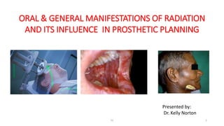
- 1. ORAL & GENERAL MANIFESTATIONS OF RADIATION AND ITS INFLUENCE IN PROSTHETIC PLANNING 52 1 Presented by: Dr. Kelly Norton
- 3. Radiation therapy Electromagnetic - Photons Particulate – Electrons, protons neutrons 52 3
- 4. 52 4 PRIMARY BIOLOGIC EFFECTS OF RADIATION
- 5. Modalities 52 5 1. CONVENTIONAL RADIOTHERAPY 2. INTENSITY MODULATED RADIOTHERAPY
- 7. Role of the dentist • Initial appointment – information source • To make the patient aware of the short term and long term effects of radiation • Manage oral complication after radiation therapy 52 7
- 8. 52 8 DENTAL MANAGEMENT OF RADIATION THERAPY PATIENTS 1. PRE-RADIATION 2. DURING RADIATION 3. POST RADIATION
- 9. 52 9 CRITERIA FOR PRERADIATION EXTRACTION PATIENT RELATED: 1. Residual Dentition 2. Mandible vs maxilla 3. Oral compliance
- 10. Urgency of treatment 52 10 Prognosis for tumor control Mode of radiation therapy RADIATION DELIVERY FACTORS
- 12. 52 12 PROSTHODONTIC ROLE PRIOR TO RADIATION 1. POSITIONING STENT Goel A, Tripathi A, Chand P, Singh SV, Pant MC, Nagar A. Use of positioning stents in lingual carcinoma patients subjected to radiotherapy. International Journal of Prosthodontics. 2010 Sep 1;23(5). Edentulous patientDentulous patient
- 13. 52 13 2. SHIELDING STENT CERROBEND ALLOY Mantri SS, Bhasin AS, Shankaran G, Gupta P. Scope of prosthodontic services for patients with head and neck cancer. Indian journal of cancer. 2012 Jan 1;49(1):39.
- 14. 52 14 3. Recontouring stent 4. Radiation positioning device PRE-LOADED AFTERLOADED
- 15. 52 15 5. TISSUE BOLUS DEVICE Singh BP, Vero N, Singh PK, Verma TR. A simplified technique to fabricate tissue bolus device to manage dose distribution in maxillectomy patient with orbital exenteration. Journal of oral biology and craniofacial research. 2013 Aug 31;3(2):102-4.
- 16. 52 16 DENTAL MANAGEMENT DURING RADIATION THERAPY 1. ORAL MUCOUS MEMBRANE WHO GRADING OF MUCOSITIS Dent Clin North Am. 2008 Jan; 52(1): 61–viii.
- 17. 52 17 PATHOPHYSIOLOGY OF RADIATION MUCOSITIS Current five-phase pathobiologic model of oral mucositis. (Reprinted from Sonis ST. A Biological Approach to Mucositis. J Support Oncol 2004; 2:21–36 Lalla RV, Sonis ST, Peterson DE. Management of oral mucositis in patients who have cancer. Dental Clinics of North America. 2008 Jan 31;52(1):61-77.
- 18. 52 18 BACKSCATTER RADIATION ALTERED ORAL FLORA
- 19. Palifermin for oral mucositis after intensive therapy for hematologic cancers • The aim of this study was to test the ability of palifermin (recombinant human keratinocyte growth factor) to decrease oral mucosal injury induced by cytotoxic therapy. • This double-blind study compared the effect of palifermin with that of a placebo on the development of oral mucositis in 212 patients with hematologic cancers. • The incidence of oral mucositis of World Health Organization (WHO) grade 3 or 4 was 63 percent in the palifermin group and 98 percent in the placebo group (P<0.001). • Palifermin reduced the duration and severity of oral mucositis after intensive chemotherapy and radiotherapy for hematologic cancers 52 19 Spielberger R, Stiff P, Bensinger W, Gentile T, Weisdorf D, Kewalramani T, Shea T, Yanovich S, Hansen K, Noga S, McCarty J. Palifermin for oral mucositis after intensive therapy for hematologic cancers. New England Journal of Medicine. 2004 Dec 16;351(25):2590-8.
- 20. EDEMA 52 20 Tongue-depressing stent in place Tongue-deviating stent in place Treatment planning computed tomographic (CT) image with the CTD stent in place. The planned target volume (PTV) to receive 70 gray of radiation is shown in red Johnson B, Sales L, Winston A, Liao J, Laramore G, Parvathaneni U. Fabrication of customized tongue-displacing stents: considerations for use in patients receiving head and neck radiotherapy. The Journal of the American Dental Association. 2013 Jun 30;144(6):594-600.
- 21. 52 21 TRISMUS • 3-6 months after radiation therapy • Secondary to fibrosis of the muscles of mastication TREATMENT FOR TRISMUS
- 22. PROSTHETIC REHABILITATION FOR A PATIENT WITH MICROSTOMIA: A CLINICAL REPORT. 52 22 Gauri M, Ramandeep D. Prosthodontic management of a completely edentulous patient with microstomia: a case report. The Journal of Indian Prosthodontic Society. 2013 Sep 1;13(3):338-42.
- 23. Irradiated salivary gland SALIVARY GLANDS DYSFUNCTION 52 23 •Radiation caries •Acute and chronic fungal infections
- 24. POST-RADIATION SEVERE XEROSTOMIA RELIEVED BY PILOCARPINE: a prospective study • The aim of the study was: (1) to confirm the action of pilocarpine hydrochloride (Salagen) against xerostomia: (2) to correlate the response to dose/volume radiotherapy parameters. • From June 1995 to February 1996, 156 patients with severe radiation induced xerostomia received pilocarpine hydrochloride orally. IS mg per day with a 5 mg optional increase at S weeks up to a daily dose of 25 mg beyond 9 weeks • No difference was found according to dose/volume radiotherapy parameters suggesting that oral pilocarpine hydrochloride: (1) acts primarily by stimulating minor salivary glands: (2) can be of benefit to patients suffering of severe xerostomia regardless of radiotherapy dose/volume parameters. 52 24 Horiot JC, Lipinski F, Schraub S, Maulard-Durdux C, Bensadoun RJ, Ardiet JM, Bolla M, Coscas Y, Baillet F, Coche-Dequéant B, Urbajtel M. Post- radiation severe xerostomia relieved by pilocarpine: a prospective French cooperative study. Radiotherapy and Oncology. 2000 Jun 1;55(3):233-9.
- 25. Functional salivary reservoir in maxillary complete denture– technique redefined 52 25 Joseph AM, Joseph S, Mathew N, Koshy AT.. Functional salivary reservoir in maxillary complete denture–technique redefined Clinical case reports. 2016 Dec 1;4(12):1082-7. A 60‐year‐old patient reported to the Department of Prosthodontics at Pushpagiri College of Dental Sciences for the replacement of missing teeth. The patient also complained of difficulty in swallowing and mastication and experienced difficulty in opening his mouth. The patient gave a history of radiation therapy 2 months back for focal keratinizing squamous cell carcinoma.
- 26. 52 26 Such bone is essentially non vital and is lacking the capacity for remodeling BONE CHANGES IRREGULAR BONY CONTOURS
- 27. PERIODONTAL CHANGES 52 27 The periodontium is a prime pathway for infection. This patient developed an osteoradionecrosis 4 years post radiation secondary to a periodontal abscess
- 28. DENTAL TISSUE CHANGES 52 28
- 29. 52 29 OSTEORADIONECROSIS • Definition – Exposure of bone within the field of radiation of 3 months duration or longer CLINICAL SYMPTOMS: Pain Swelling Trismus Exposed Bone Pathologic Fracture Orocutaneous fistula formation
- 30. 52 30
- 31. • 1. Conservative treatment: • Irrigation with saline and chlorhexidine • Iodoform gauze packing • Gentle debridement with removal of sharp bony spicules. • Antibiotic coverage if necessary • PEN-TO-CLO- Pentoxyfylline+ tocopherol+ clodronate • Strict oral hygiene measures. 52 31 TREATMENT OF OSTEORADIONECROSIS Robard L, Louis MY, Blanchard D, Babin E, Delanian S. Medical treatment of osteoradionecrosis of the mandible by PENTOCLO: preliminary results. European annals of otorhinolaryngology, head and neck diseases. 2014 Dec 31;131(6):333-8.
- 32. 52 32 • Dr. Marx and his colleagues believe almost all osteoradionecrosis of the mandible require treatment with hyperbaric oxygen Hyperbaric Oxygen Therapy How does HBOT work and why is it effective ? In air, normal 21% oxygen 100% O2 combined with pressure delivers 15 times O2 to all body fluids Enhanced growth of new blood vessels Increased ability of white blood cells to destroy bacteria and remove toxins Increased growth of fibroblasts (cells involved in wound healing)
- 33. 52 33
- 34. Topical Fluoride • Mouth rinse • Toothpaste • Gel applied with custom carriers 52 34 The use of stannous fluoride gel applied with custom carriers and five minute daily applications. DENTAL MAINTAINANCE POST RADIATION CARE
- 35. ROLE OF ENDODONTIC THERAPY 52 35
- 36. Examination findings of unique importance • Condition of oral mucous membranes • Contours of the bony bearing surfaces, presence of bony undercuts • Amount and Viscosity of saliva • Posterior palatal seal area • Trismus • Scarring at the tumor site 52 36 PROSTHODONTIC PROCEDURES • History findings of importance • Fields of radiation • Dose to mandibular bearing surfaces
- 37. Impressions • Border molding • Border mold with a low fusing compound • Apply petrolatum to prevent sticking to the dry mucosa • Develop maximum extensions but avoid overextension at the tumor site • Do not attempt to displace the floor of the mouth to obtain peripheral seal. 52 37Thermoplastic wax Polysulfide • Wash impression materials
- 38. Assessment of Vertical dimension • Determined by phonetics, closest speaking space, swallowing , VDR • The VDO is reduced only in patients with severe trismus so as to facilitate easy entrance of the bolus by increase in interocclusal space. • In case of severe scarring of tongue, lower the occlusal plane 52 38
- 39. Centric relation records are made in the usual manner using wax, ZOE or silicone materials • A facebow transfer record is used to mount the maxillary cast on the articulator. 52 39
- 40. Lingualized / bilateral balance 52 40 Nonanatomic with balancing ramps • Less horizontal forces are generated • Proper horizontal overlap to maintained Occlusal forms
- 41. Delivery and Post-Insertion Care • Pressure indicating paste • Disclosing wax- delineate overextension • Clinical remount • 24 and 48 hour follow up • Leave dentures out at night • Educate the patient 52 41
- 42. Period between Completion of Radiation Therapy and Prosthetic Rehabilitation in Edentulous Patients: A Retrospective Study Purpose: 1. to describe the number and types of complications patients had before and after insertion of a removable prosthesis (i.e., denture) following radiation therapy to the head and neck and 2. to investigate whether the time between radiation therapy and denture insertion might contribute to those complications Materials and Methods: A total of 190 patients met the inclusion criteria with data available for review. Conclusions: • The majority of patients had no complications. • The patients who received their dentures in 180 days or less had the same number of complications when compared with those patients who received their dentures in 181 to 365 days and those who had to wait longer than a year for prosthetic rehabilitation. • Patients with more pre-insertion complications tended to have delayed prosthetic rehabilitation. • The majority of patients who experienced complications before and after denture insertion had greater than 5000 cGy. 52 42 Period between Completion of Radiation Therapy and Prosthetic Rehabilitation in Edentulous Patients: A Retrospective Study Peter J. Gerngross, et al . J Prosthodont 2005;14:110-121
- 43. Implants in irradiated tissues • Radiation effects : • Reduced vasculature • Loss of osteo progenitor cells • Fatty degeneration • Compromised remodeling • Susceptibility to osteoradionecrosis 52 43 Root surfac e Marrow Trabecular bone • Loss of central artery in Haversian systems • Death of osteocytes
- 44. Implants in the irradiated maxilla • Predictability-Maxilla %Success • Roumanas et al, 1998 55 • Nimi et al, 1998 63 52 44 Without HBO Implants in irradiated edentulous maxillectomy patient (UCLA Data) Patients Number of implants Success Treated placed uncovered buried failed % Irradiated 13 50 29 11 10 55.2
- 45. 52 45 In the mandible, one would expect risk to become significant at doses to bone above 6500 cGy. This patient received 6600 cGy for a squamous carcinoma of the lateral tongue. Three years later implants were placed. Eventually, the patient developed an osteoradionecrosis, a pathologic fracture of the mandible and subsequently the mandible was resected. Three years after implant placement the patient developed an infection associated with left posterior implant. Implants in the irradiated mandible
- 46. IMPLANTS IN THE CRANIOFACIAL SITES Success is poor and failures are late because: • Anchorage is mechanical: short implants have higher failure rate • Exposure of flange leads to persistent irritation of perimplant skin or mucosa 52 46 Flange exposure Eventually led to loss of implants
- 47. IRRADIATION OF EXISTING IMPLANTS 52 47 These implants were irradiated 2 years following placement. Note the exposure of the implant flanges. Recommendation: Abutments and superstructures should be removed and skin and/or mucosa closed over the implant fixtures prior to radiation.
- 48. SUMMARY • Implant material : Advanced dental implant surfaces like TPS [titanium plasma spreaded], SLA [sandblasted and acid etched], Ti-Unite and different implant materials like zirconia [zirconium oxide] • Implant position : Implants can be best placed in the mandibular anterior / symphyseal region as it is the area which receives the least amount of radiation. The maximum implant failures are reported in the maxillary jaw [69% to 95%] . • Type of prosthesis : Fixed implant supported prosthesis is advocated in irradiated mandibles. • Effect of radiation dose :. Favorable osseointegration is found in radiation doses lesser than 45-50 Gy. • Effect of smoking: Irradiated patients who continued to smoke must be considered as an absolute contraindication to treatment. 52 48Dholam KP, Gurav SV. Dental implants in irradiated jaws: a literature review. Journal of cancer research and therapeutics. 2012 Jan 1;8(6):85.
- 49. • Soft tissue complications : Gingivitis was more common in these patients than normally observed. Cover-screw mucosal perforations were observed over the areas of 17% of implants during the healing period between stage one and stage two surgery. • Hyperbaric oxygen : Some studies found it useful while others considered it as an additional burden of treatment. Timing of implant placement: One year time interval between tumor therapy and the time of dental implantation seems logical • Timing of abutment placement and loading the implants: Abutment connection, fabrication and loading of the prosthesis should be delayed for six months instead of the traditional three to four months to permit osseointegration. 52 49 Dholam KP, Gurav SV. Dental implants in irradiated jaws: a literature review. Journal of cancer research and therapeutics. 2012 Jan 1;8(6):85.
- 50. Osseointegrated implants in irradiated bone: A case-controlled study using adjunctive hyperbaric oxygen therapy • The current investigation was undertaken to study whether osseointegration of implants in irradiated tissues is subject to a higher failure rate than in non irradiated tissues. It further aimed to study whether hyperbaric oxygen treatment (HBO) can be used to reduce implant failure. • Patients and Methods: • 78 cancer patients who were rehabilitated using osseointegrated implants between 1981 and 1997 were investigated. Three groups of patients were compared: irradiated (A), nonirradiated (B), and irradiated and HBO-treated (C). In addition, 10 irradiated patients who had lost most of their implants received new ones after HBO treatment. These were compared as a case-control group. • Conclusions: Implant insertion in irradiated bone is associated with a higher failure rate. Adjuvant HBO treatment can reduce the failures. 52 50 Granström G, Tjellström A, Brånemark PI. Osseointegrated implants in irradiated bone: a case-controlled study using adjunctive hyperbaric oxygen therapy. Journal of oral and maxillofacial surgery. 1999 May 1;57(5):493-9.
- 51. 52 51 The aim of this systematic review was to evaluate the effect of radiation therapy on osseointegrated dental implant survival in oral cancer patients. MATERIALS AND METHODS: A review of the literature published between 1990 and June 2012 was conducted. Overall implant survival rates were compared with respect to timing of radiation ,site of implant placement, radiation dose, time interval between radiation therapy and implant placement, and the effect of hyperbaric oxygen therapy. CONCLUSION: There, was no significant difference in dental implant survival rates between pre implantation and post implantation radiation therapy. The anatomical site of implant placement in preimplantation radiation therapy was the most pertinent variable affecting implant survival, with a better survival rate in the mandible compared to the maxilla and grafted bone. Dental implant survival in irradiated oral cancer patients: a systematic review of the literature. Nooh N. Dental implant survival in irradiated oral cancer patients: a systematic review of the literature. International Journal of Oral & Maxillofacial Implants. 2013 Sep 1;28(5).
- 52. Implant-prosthetic rehabilitation after radiation treatment in head and neck cancer patients: a case-series report of outcome. • The aim of the study was to review the outcome of implant-prosthetic treatment after radiation therapy. • Patients and methods. Twenty irradiated head and neck cancer patients received a removable implant supported denture • Results. Twenty patients had 100 implants inserted. The estimated implant survival rate was 96% after 1 year and 87% after 5 years. Failures were mostly observed before loading (91.2%). The attachment system and the number of implants did not have a statistically significant influence on the success rate. 52 52 Conclusions. Implant-supported dentures have been shown to be a reliable treatment modality after head and neck cancer surgery and radiation therapy. Possible early failures should be communicated with the patients Cotic J, Jamsek J, Kuhar M, Ihan Hren N, Kansky A, Özcan M, Jevnikar P. Implant-prosthetic rehabilitation after radiation treatment in head and neck cancer patients: a case-series report of outcome. Radiology and oncology. 2017 Mar 1;51(1):94-100.
- 53. Conclusion 52 53 In summary, it is our intention and goal as dentists to minimize and/or prevent potentially devastating side effects of Radiation therapy from occurring and to help the patient maintain the highest possible level of oral health and function both during and after Radiotherapy.
- 54. REFERENCES 52 54 1. Beumer J, Curtis T, and Nishimura R. Prosthetic management - Edentulous patients. In Beumer J, Curtis T, and Marunick M, editors. Maxillofacial Rehabilitation. St. Louis – Tokyo: Ishiyaku EuroAmerica 1996. 2. Thomas T Taylor , Clinical Maxillofacial Prosthetics, First edition,2000, Quintessence publications, Illionis, pp 37 – 52 3. Goel A, Tripathi A, Chand P, Singh SV, Pant MC, Nagar A. Use of positioning stents in lingual carcinoma patients subjected to radiotherapy. International Journal of Prosthodontics. 2010 Sep 1;23(5). 4. Mantri SS, Bhasin AS, Shankaran G, Gupta P. Scope of prosthodontic services for patients with head and neck cancer. Indian journal of cancer. 2012 Jan 1;49(1):39. 5. Singh BP, Vero N, Singh PK, Verma TR. A simplified technique to fabricate tissue bolus device to manage dose distribution in maxillectomy patient with orbital exenteration. Journal of oral biology and craniofacial research. 2013 Aug 31;3(2):102-4. 6. Lalla RV, Sonis ST, Peterson DE. Management of oral mucositis in patients who have cancer. Dental Clinics of North America. 2008 Jan 31;52(1):61-77 7. Spielberger R, Stiff P, Bensinger W, Gentile T, Weisdorf D, Kewalramani T, Shea T, Yanovich S, Hansen K, Noga S, McCarty J. Palifermin for oral mucositis after intensive therapy for hematologic cancers. New England Journal of Medicine. 2004 Dec 16;351(25):2590-8
- 55. 8. Johnson B, Sales L, Winston A, Liao J, Laramore G, Parvathaneni U. Fabrication of customized tongue-displacing stents: considerations for use in patients receiving head and neck radiotherapy. The Journal of the American Dental Association. 2013 Jun 30;144(6):594-600. 9. Gauri M, Ramandeep D. Prosthodontic management of a completely edentulous patient with microstomia: a case report. The Journal of Indian Prosthodontic Society. 2013 Sep 1;13(3):338-42. 10. Horiot JC, Lipinski F, Schraub S, Maulard-Durdux C, Bensadoun RJ, Ardiet JM, Bolla M, Coscas Y, Baillet F, Coche-Dequéant B, Urbajtel M. Post-radiation severe xerostomia relieved by pilocarpine: a prospective French cooperative study. Radiotherapy and Oncology. 2000 Jun 1;55(3):233-9. 11. Joseph AM, Joseph S, Mathew N, Koshy AT.. Functional salivary reservoir in maxillary complete denture–technique redefined Clinical case reports. 2016 Dec 1;4(12):1082-7. 12. Robard L, Louis MY, Blanchard D, Babin E, Delanian S. Medical treatment of osteoradionecrosis of the mandible by PENTOCLO: preliminary results. European annals of otorhinolaryngology, head and neck diseases. 2014 Dec 31;131(6):333-8. 13. Period between Completion of Radiation Therapy and Prosthetic Rehabilitation in Edentulous Patients: A Retrospective Study Peter J. Gerngross, et al . J Prosthodont 2005;14:110-121 14. Dholam KP, Gurav SV. Dental implants in irradiated jaws: a literature review. Journal of cancer research and therapeutics. 2012 Jan 1;8(6):85. 52 55
- 56. 15. Granström G, Tjellström A, Brånemark PI. Osseointegrated implants in irradiated bone: a case-controlled study using adjunctive hyperbaric oxygen therapy. Journal of oral and maxillofacial surgery. 1999 May 1;57(5):493-9. 16. Nooh N. Dental implant survival in irradiated oral cancer patients: a systematic review of the literature. International Journal of Oral & Maxillofacial Implants. 2013 Sep 1;28(5). 17. Cotic J, Jamsek J, Kuhar M, Ihan Hren N, Kansky A, Özcan M, Jevnikar P. Implant- prosthetic rehabilitation after radiation treatment in head and neck cancer patients: a case-series report of outcome. Radiology and oncology. 2017 Mar 1;51(1):94-100. 52 56
Editor's Notes
- Cancer is the 2nd leading cause of death after heart disease with 30 % being oral cancer. Principal methods of treating malignancies of the head neck are: surgical resection and radio therapy. The Prosthodontist is commonly consulted when custom prosthetic devices are used to facilitate the delivery of radiation therapy.
- Direct action results when secondary particles (i.e., recoil electrons and protons) interact with the target molecule, while indirect action results from interaction with water to produce free radicals (hydroxyl and hydrogen), which in turn interact with the target molecule by oxidation reduction reactions.
- Radiation is delivered via an external source IMRT allows delivery of different doses to each volume at the same time
- Radioactive sources are implanted locally within the tissues (encompassed Iridium 192 seeds ) To deliver the dose at a shorter distance They are used primarily in T1 and T2 carcinomas of the oral tongue and floor of the mouth Advantages: Dose to the buccal side of the mandible and the salivary glands is generally limited to the dose delivered by the external therapy. This level (5000-5500cGy) of radiation is not sufficient to totally eliminate the fine vasculature of these tissues.
- Teeth with questionable prognosis . Especially mandible ..higher risk of orn TRISMUS , IMPAIRED OTOR FUNCTIONS AND SURGICAL MORBIDITIES
- This situation occurs very rarely. When it does both the radiation therapist and the dentist must accept the risk of future dental complications
- OCCURS DUE TO CELL DEATH OF THE BASAL LAYER OF THE EPITHELIUM. Oral mucositis initially presents as erythema of the oral mucosa which then often progresses to erosion and ulceration
- Initiation of tissue injury: Radiation and/or chemotherapy induce cellular damage resulting in death of the basal epithelial cells. free radicals activate second messengers that transmit signals from receptors on the cellular surface to the inside of the cell.This leads to upregulation of pro-inflammatory cytokines, tissue injury and cell death. amplify mucosal injury There is a significant inflammatory cell infiltrate associated with the mucosal ulcerations, This phase is characterized by epithelial proliferation as well as cellular and tissue differentiation 15, restoring the integrity of the epithelium.
- loss of central artery in the Haversion systems (arrow) b) loss of osteocytes from their lacunae (arrow)
- Periosteum and overlying soft tissue undergo hyperemia, inflammation and endarteritis. Leading to thrombosis, cellular death progressive hypovascularity, and fibrosis.
- Pentocifylline + tocofeol reduce post radiation fibrosis Clodronate- bisphosphonate – reduces bone resorption allows for bone formation
- 20 dives before treatment 10 dives after treatment at 2.28 atmospheric pressure for 90-120 minutes