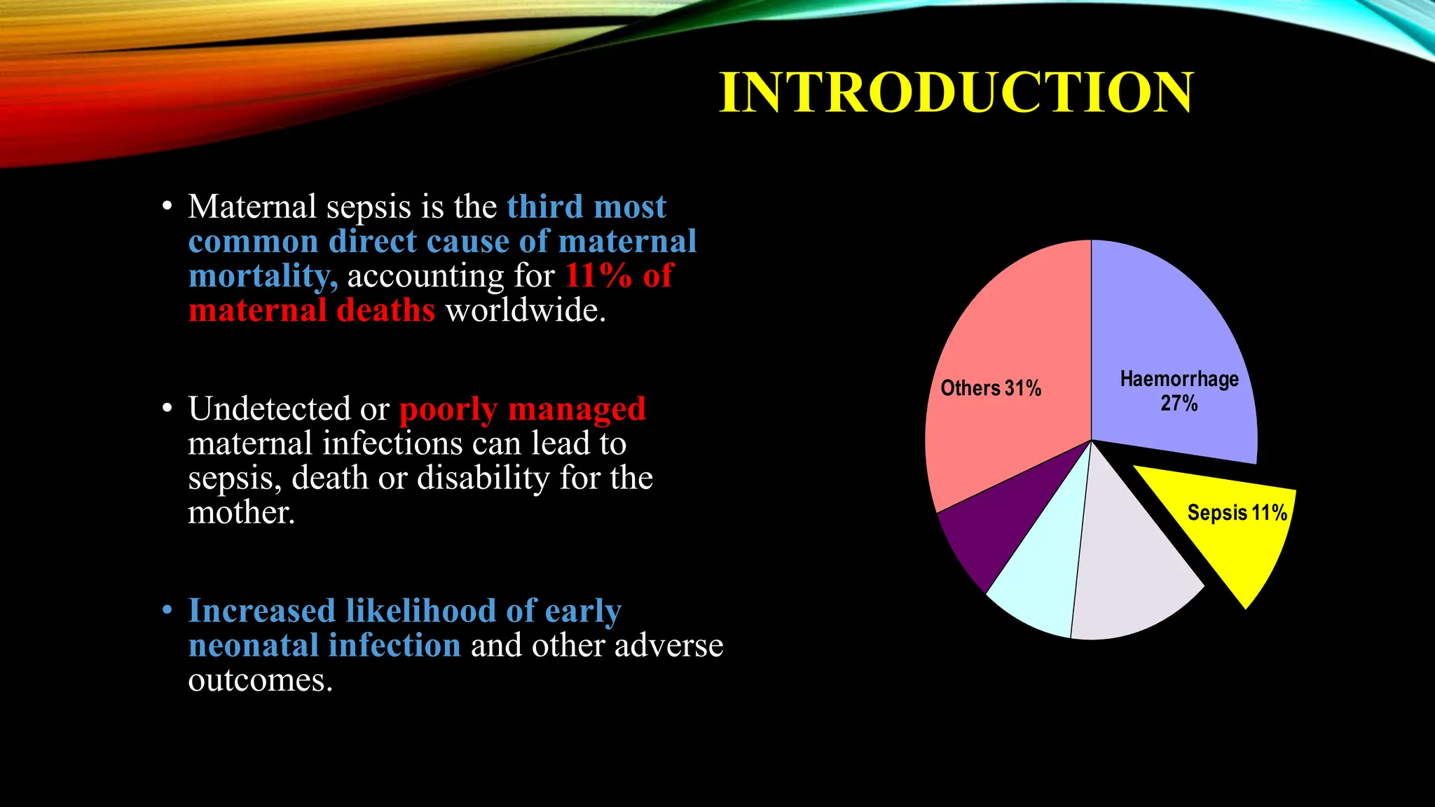
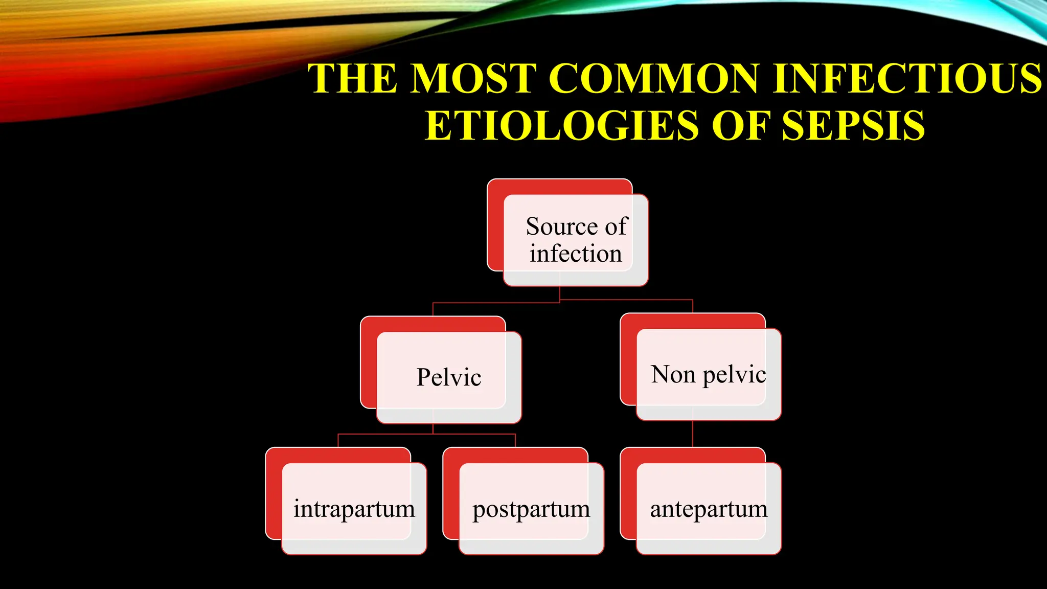
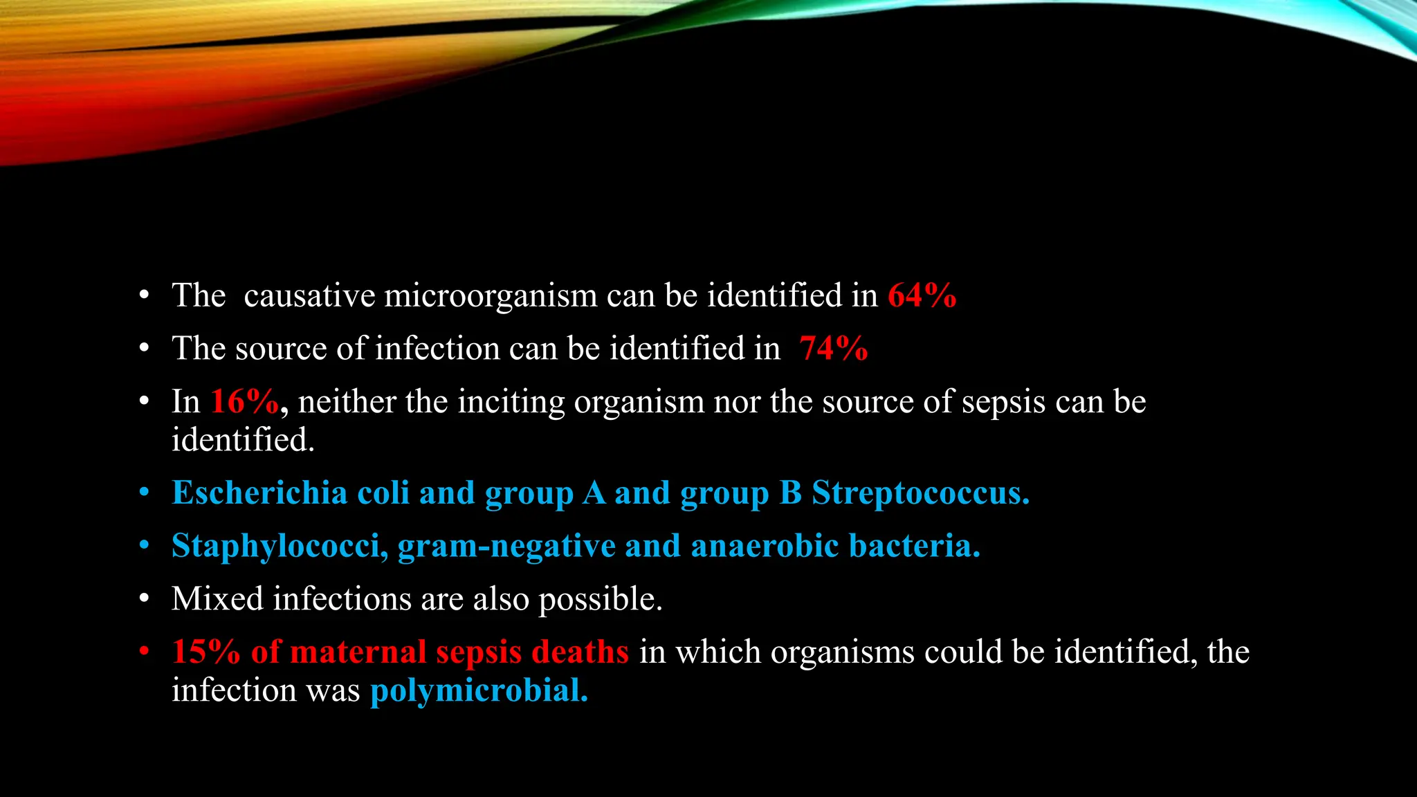
OBSTETRICS SEPSIS –
BUNDLE APPROACH
9TH ANNUAL CONFERENCE OF ANDHRA PRADESH
OBSTETRICS & GYNAECOLOGICAL SOCIETY
Date: 13th July 2024
Venue: Sree Convention Center, Eluru.
Professor and Unit Chief, L.T.M.M.C & L.T.M.G.H, Sion Hospital
President, MOGS (2022-2023)
Joint Treasurer, FOGSI (2021-2025)
Organising Secretary, AICOG Mumbai 2025
Treasurer, AFG (2023-2024)
Member Oncology Committee, SAFOG (2021-2023)
Dean AGOG & Chief Content Director, HIGHGRAD & FEMAS Courses
Editor-in-Chief, FEMAS, JGOG & TOA Journal
87 publications in International and National Journals with 198 Citations
National Coordinator, FOGSI Medical Disorders in Pregnancy Committee (2019-2022)
Chair & Convener, FOGSI Cell Violence Against Doctors (2015-16)
Member, Oncology Committee AOFOG (2013-2015)
Coordinator of 11 batches of MUHS recognized Certificate Course of B.I.M.I.E at
L.T.M.G.H (2010-16)
Member, Managing Committee IAGE (2013-17), (2018-20), (2022-2023)
Editorial Board, European Journal of Gynaec. Oncology (Italy)
Course Coordinator of 3 batches of Advanced Minimal Access Gynaec Surgery
(AMAS) at LTMGH (2018-19)
DR. NIRANJAN CHAVAN
MD, FCPS, DGO, MICOG, DICOG, FICOG, DFP,
DIPLOMA IN ENDOSCOPY (USA)
• Maternal sepsis is the third most
common direct cause of maternal
mortality, accounting for 11% of
maternal deaths worldwide.
• Undetected or poorly managed
maternal infections can lead to
sepsis, death or disability for the
mother.
• Increased likelihood of early
neonatal infection and other adverse
outcomes.
Haemorrhage
27%
Sepsis11%
Hypertensive
disorders14%
Obstructed
labour 9%
Abortion8%
Others 31%
Source- WHO 2014
INTRODUCTION
• In low- and middle-income countries, rates of
fatality after puerperal infection can be as
high as 50%.
• Under new sepsis definitions some
representative reports gave ranges on the
mortality of sepsis in the general
population of 25%–30%.
• The mortality range for septic shock was
40%–70%.
SEPSIS - CONTROVERSIES AND
LIMITATIONS
• There are inherent challenges in defining
sepsis and septic shock.
• First is a broad term applied to a ill
understood process.
• There are yet no simple clinical criteria
or biological, imaging or laboratory
features that can identify a septic patient.
• Maternal sepsis is a life-threatening condition defined as organ dysfunction
resulting from infection during pregnancy, childbirth, post-abortion, or
postpartum period (WHO).
DEFINITION
Dr Komal N. Chavan, Mumbai
CAUSES OF SEPSIS
Obstetric causes
• Retained product of conception (septic
abortion, retained placenta)
• Chorioamnionitis
• Endometritis pelvic abscess and
• Wound infection
Non Obstetric causes
• Pyelonephritis
• Appendicitis
• Bowel infarction
• Pancreatitis and pneumonia (bacterial as
staphylococcus or pneumococcus) or
• Viral (H1 N1,influenza, Herpes)
WHAT ARE THE RISK FACTORS
FOR MATERNAL SEPSIS?
• Obesity
• Advanced age
• Diabetes
• Impaired immunity,
• Anaemia , vaginal infections
• History of pelvic infection,
• History of group B streptococcal
infection
• Caesarean birth
• Amniocentesis
• cervical cerclage
• Prolonged ROM
• Group A streptococcal (GAS)infection
in close contacts
• Retained products of conception
Dr Komal N. Chavan, Mumbai
THE MOST COMMON INFECTIOUS
ETIOLOGIES OF SEPSIS
Source of
infection
Pelvic
intrapartum postpartum
Non pelvic
antepartum
• The causative microorganism can be identified in 64%
• The source of infection can be identified in 74%
• In 16%, neither the inciting organism nor the source of sepsis can be
identified.
• Escherichia coli and group A and group B Streptococcus.
• Staphylococci, gram-negative and anaerobic bacteria.
• Mixed infections are also possible.
• 15% of maternal sepsis deaths in which organisms could be identified, the
infection was polymicrobial.
TYPES OF INFECTION
Infection Features
1. Infected wound (perineal or abdominal) Discharge from wound, pain, erythema and swelling around
wound, wound dehiscence, burst abdomen
2. Endometritis/septic abortion Abdominal pain, vaginal bleeding, offensive cervical discharge,
uterine tenderness, delayed uterine involution, fever
3. Chorioamnionitis Abdominal pain, vaginal bleeding, offensive vaginal discharge
or lochia, uterine tenderness, delayed uterine involution, fetal
tachycardia >160 beats per minute, fever
4. Respiratory tract infection Productive cough, sore throat, shortness of breath/difficulty
breathing, chest pain and fever
5. Breast abscess/mastitis Breast pain and tenderness, erythema, painful induration, nipple
discharge
6. Urinary tract infection Dysuria, increased frequency, urgency, abdominal/flank/back
pain, rigors, fever, nausea and vomiting
7. Malaria Fever, headache, muscle/joint pain, jaundice, anemia
8. Meningitis Headache, rash, photophobia, neck stiffness, confusion, fever,
nausea and vomiting
9. Toxic shock syndrome (streptococcal and staphylococcal) Nausea, vomiting, diarrhea, watery vaginal discharge,
generalized maculopapular rash, conjunctival suffusion
10.Non-specific features of infection Lethargy, reduced appetite, fever, nausea, vomiting
PATHOPHYSIOLOGY OF SEPSIS
Infection
Inflammatory
response
Extravasation of
albumin and
fluid
Organ
dysfunction
Cytokines
Decreased
SVR& increased
cardiac output
Dysregulated
host response
hypovolemia
Septic cardiomyopathy Pulmonary oedema and
hypotension
Dr Komal N. Chavan, Mumbai
SECOND STAGE - ORGAN
DYSFUNCTION
CLINICALAND LABORATORY
DIAGNOSIS OF SEPSIS
Signs and Symptoms
• Fever or Temperature instability(>380 C or
110 beats/min)
• Tachypnoea (>24 breaths/min)
• Diaphoresis
• Clammy or mottled skin
• Nausea or vomiting
• Hypotension or shock
• Oliguria or anuria
• Pain (location based on site of infection)
• Altered mental state (confusion, decreased
alertness)
Laboratory Findings
• Leukocytosis or leukopenia
• Positive culture from infection site and/or blood
• Hypoxemia
• Thrombocytopenia
• Metabolic acidosis: serum lactate, low arterial
pH, base deficit
• Elevated serum creatinine
• Elevated liver enzymes
• Hyperglycemia in the absence of diabetes
• Disseminated intravascular coagulation
INVESTIGATIONS
• Complete blood count
• Electrolytes
• Blood cultures, if sepsis is suspected
• Urinalysis, with cultures and sensitivity tests
• Cervical or uterine cultures
• Wound cultures
• Lactate
• Coagulation studies, if pelvic thrombosis, deep vein thrombosis, pulmonary embolism,
or invasive treatment (eg, surgical procedure) is being considered.
• Pelvic USG- retained products of conception, pelvic abscess, or infected hematoma.
• CT or MRI - septic pelvic thrombosis.
DIFFERENT SCORING SYSTEMS
FOR OBSTETRIC SEPSIS
Dr Komal N. Chavan, Mumbai
Adv Excellent NPV
98.6%
Rapidly rules out need for
ICU
Does not use altered
mental status in criteria
Disadv Complex scoring
system with multiple
variables
Requires laboratory data,
which can delay
diagnosis
OBSTETRICALLY MODIFIED QUICK
SEQUENTIAL ORGAN FAILURE
ASSESSMENT (OM Q-SOFA)
• Presence of 3 clinical criteria:
Systolic<90mm of hg
RR≥25
Altered mental status
(≥ 2 of these associated with ↑ risk & poor outcome)
Advantage: Simple bedside screening tool Uses only clinical data, allowing for rapid
diagnosis.
Disadvantage: Altered mental status in criteria may have nonseptic causes in obstetric
patients may need for secondary testing.
• RCOG –has recommended modified early obstetric warning score (MEOWS) to detect signs
of sepsis
Dr Komal N. Chavan, Mumbai
Advantage:
Simple bedside
screening tool
Disadvantage:
Marked variation of
thresholds and
formats
Validated for
chorioamnionitis
Over detects severe
sepsis
Need for secondary
testing to identify true-
positives
Low specificity.
• The best tool for identifying infection or predicting mortality in pregnant or
postpartum patients remains unknown.
• But simple bedside screening tool such as the MEOWS or OMQSOFA.
• Followed by further evaluation for evidence of end-organ damage are used for early
recognition of sepsis.
ROLE OF BIOMARKERS
• Currently no one biomarker has been identified as the gold standard.
• Commonly used markers such as white cell count and C-reactive protein are neither
specific nor sensitive.
• Procalcitonin - guiding antibiotic therapy -shows promise, NICE guidelines- there is
currently insufficient evidence. Surviving Sepsis Campaign (SSC) Guidelines- low-
quality evidence to support its use. There is absence of an accepted normal range
during pregnancy.
• Serum lactate - is a useful biomarker, elevated lactate levels associated with poor
outcomes in maternal sepsis. Repeat measurement required if initial lactate is greater
than 2 mmol/L.
Dr Komal N. Chavan, Mumbai
1. Screening and Management of Infection: The first step - cultures and blood
samples followed by administering appropriate antibiotics.
2. Screening for Organ Dysfunction and Management of Sepsis : identified by the
organ dysfunction criteria; omqSOFA.
3. Identification and Management of Initial Hypotension: Patients with infection and
hypotension or a lactate level >4mmol/L, are given 30mL/kg crystalloid with
reassessment of volume responsiveness and tissue perfusion.
In the event of persistent hypotension despite fluid resuscitation (septic shock) and/or
lactate >4mmol/l vasopressors are used.
a. Achieve a central venous pressure (CVP) of ≥8mmHg
b. Achieve a central venous oxygen saturation (ScvO2 ) ≥ 70% or mixed venous
oxygen saturation (ScvO2 ) ≥ 65%.
EARLY SCREENING AND
MANAGEMENT
Sepsis Six (Bundle Approach) –
a set of six tasks including
• Oxygen,
• Cultures,
• Antibiotics,
• Fluids,
• Lactate measurement and,
• Urine output monitoring
to be instituted within one hour.
ROLE OF CORTICOSTEROIDS
• Corticosteroids in septic patients is subject of controversy.
• Hydrocortisone – reserved for septic patients with refractory shock (those who
remain hypotensive following initial fluid resuscitation and vasopressors).
• IV infusion of hydrocortisone at a dose of 200mg per day.
• Monitored for hyperglycemia and hypernatremia.
• Must be given within the first 7 days of treatment and should be interrupted as soon
as the patient shows signs of clinical improvement.
• Another benefit is the need for corticoids to accelerate fetal lung maturation.
SOURCE CONTROL
• Once a source of sepsis is identified,
• source control is a priority and may involve
• abscess drainage or
• the delivery of the fetus
• if the uterus is found to be the source of the
infection.
DEEP VENOUS THROMBOSIS
PROPHYLAXIS
• Prevention of DVT is essential in septic pregnant
patients as both pregnancy and sepsis are associated
with hypercoagulability.
• Methods of prophylaxis are the use of compression
stockings, intermittent lower limb compression, and
LMWH or unfractionated heparin.
• The role of intravenous immunoglobulin (IVIG):
IVIG is recommended for severe invasive streptococcal
or staphylococcal infection if other therapies have failed
because of its immunomodulatory effect.
DELIVERY DECISION
• Influenced by
• patient’s condition,
• gestational age,
• fetal status,
• presence of chorioamnionitis, and
• labour.
• In a critically ill woman, delivery is considered if it
would be beneficial to the mother or the baby or to
both.
• Attempting delivery of unstable mother increases the
maternal and fetal mortality rates unless the source of
infection is intrauterine.
• Indicators of delivery are
• Intrauterine infection,
• DIC,
• hepatic/ renal problems,
• heart failure,
• compartmental syndrome,
• multifetal gestation and,
• ARDS
• While foetal factors include foetal demise and a gestationally viable fetus.
• During the intrapartum period, continuous electronic fetal monitoring is recommended.
• Epidural/spinal anaesthesia should be avoided in women with sepsis and a general
anaesthetia will usually be required for caesarean section.
EXTRACORPOREAL MEMBRANE
OXYGENATION (ECMO)
• Respiratory failure in patients in the intensive
care unit, ECMO has been used increasingly
during pregnancy and the puerperium,
• ECMO may be a choice for the treatment of
refractory sepsis.
SUMMARY LIST OF
WHO RECOMMENDATIONS
These recommendations are given by WHO 2023 for prevention of puerperal sepsis:
• Routine perineal/pubic shaving prior to giving vaginal birth is not recommended.
• Digital vaginal examination at intervals of four hours is recommended for routine
assessment of active first stage of labour in low-risk women.
• Routine vaginal cleansing with chlorhexidine during labour for the purpose of preventing
infectious morbidities is not recommended.
• Routine vaginal cleansing with chlorhexidine during labour in women with group B
Streptococcus (GBS) colonization is not recommended for prevention of early neonatal
GBS infection.
• Vaginal cleansing with povidone-iodine immediately before caesarean section is
recommended.
MATERNALAND PERINATAL
COMPLICATIONS OF SEVERE
SEPSIS AND SEPTIC SHOCK
• Pulmonary oedema and adult respiratory distress
syndrome.
• Myocardial ischemia and left ventricular dysfunction.
• Renal failure
• Disseminated intravascular coagulation.
• Multiple organ failure is associated with a high rate of
maternal mortality.
• Preterm labour result of release of endotoxins.
• Neonatal hypoxia, sepsis or death.
CASE 1
• A 25-year-old patient, P2L2,
• Day 7 of LSCS done i/v/o meconium-stained amniotic fluid
• Referred from a peripheral hospital i/v/o multiple fever spikes with deranged creatinine
likely s/o puerperal sepsis
• O/E
General condition fair
Febrile
P- 130/min
BP- 100/60 mmHg
CVS- S1S2 +
RS- Bilateral basal crept +
• P/A
Soft
uterus well retracted
Skin necrosis present
Suture line unhealthy
• Per speculum
No abnormal bleeding
No foul-smelling lochia
Investigations:
CBC- 8.7/ 24000/ 120000
T. bili- 2.3
SGOT/SGPT- 200/230
BUN/Creat- 28/ 3.8
PT/INR- 16/1.2
The patient was admitted to MICU for evaluation and management.
Decision of suture removal taken
Post-debridement in MICU under local
anesthesia
Post-suture removal (Day 8 of LSCS)
• Wound culture: MRSA (Methicillin-resistant
Staphylococcus Aureus)
• Patient started on higher antibiotics in renal corrected
doses. (Inj Meropenem and Inj Vancomycin)
• 2 hemodialysis cycles received over a period of 1
week.
• Patient had breathlessness and SPO2 drop on day 4
of admission and hence was intubated and started on
VCAC mode of ventilation.
• On wound examination rectus sheath was open, rectus muscle intact.
• Daily BD wound dressing was done.
• Surgery reference taken:
Surgical debridement in OT under anesthesia.
Patient shifted to OT for surgical debridement under anaesthesia.
Before debridement After debridement
• Patient condition improved.
• Patient extubated on day 10 of admission.
• Lab parameters improved.
• Patient shifted to the ward on day 15 and daily wound dressing with debridace ointment
continued.
Improvement in wound seen due to regular dressing
Present wound (day 16 of surgical debridement)
• POA: Vacuum-assisted closure (VAC dressing).
• Vacuum-assisted closure (VAC) is a method of
decreasing air pressure around a wound to assist the
healing.
• It's also referred to as negative pressure wound therapy.
• During a VAC procedure, a healthcare professional
applies a foam bandage over an open wound, and a
vacuum pump creates negative pressure around the
wound.
CASE 2
• A 30-year-old patient, P3L3 with day 6 of normal vaginal delivery presented with
• C/o intermittent high-grade fever with breathlessness.
• C/o foul-smelling discharge per vagina post-delivery.
• O/e:
Conscious, oriented, pallor +
Febrile SPO2- 94% on room air
P- 140/min
BP- 100/70 mmHg
• P/A
Soft
Uterus well retracted
No guarding, rigidity, or tenderness.
• Per speculum:
Minimal bleeding present
Lochia foul smelling
• Breast:
No engorgement, no tenderness. Yellow foul-smelling discharge coming
from the vagina
• Investigation:
CBC- 7.8/30000/93000
BUN/Creat- 9/0.9
T. Bili- 1
SGOT/SGPT- 26/30
PT/INR- 12/0.8
A high vaginal swab was sent for culture and sensitivity.
Blood and urine cultures were sent.
• USG-
Endometrial thickness 24mm
No e/o retained placental tissue, blood clots, or hemoperitoneum.
• The patient started on Inj Piptaz and Inj. Metronidazole
• Oxygen by nasal prongs started.
• Endometrial curettage was done.
• Vaginal douching with betadine solution done daily.
• Fever spikes continued.
• On day 4 of admission she developed gross abdominal distension with absent bowel
sounds.
• Contrast-enhanced computerized
tomogram (CECT) abdomen and pelvis
I. Features of paralytic ileus,
II. Grossly distended stomach,
III. Moderate ascites,
IV. No endometrial collection.
• Surgery reference taken and conservative
management done. CECT abdomen and pelvis sagittal section with,
grossly distended stomach, dilated bowel loops
with no endometrial collection.
• IV antibiotics continued.
• Inj clindamycin added as per culture sensitivity report.
• Abdominal distension settled gradually.
• Fever settled.
• 1 unit-packed red blood cell was given on day 12 of admission because of moderate
anemia (Hb- 7.8 g/dl).
• Antibiotics were stopped on day 18 as she
improved clinically.
• Her CBC was serially monitored.
• WBC Count decreased from 30,000 at admission
to 11,000 at discharge on day 21.
Recognition is key
• Pearl 1. Always maintain a high index of suspicion for sepsis.
• Pearl 2. Implement a rapid bedside tool for detection of maternal deterioration.
Move fast during the golden hour
• Pearl 3. Implement sepsis bundles to facilitate rapid escalation of care.
• Pearl 4. Laboratory and radiologic studies are keys to search for etiology and
source control.
• Pearl 5. Know microbes and likely origin, and that group A streptococcus can kill
quickly.
Top 10 Pearls for Managing
Maternal Sepsis
Take Home Message
• Pearl 6. Choose antimicrobials tailored to the most likely diagnosis.
• Pearl 7. Fluid resuscitation should be initiated rapidly for patients with a blood
lactate greater than 4 mmol/L or mean arterial pressure less than 65 mm Hg.
Beyond the golden hour
• Pearl 8. Escalation of care is critical to survival.
• Pearl 9. Once the patient is stabilized, get to the source of the problem.
• Pearl 10. Anticipate and prevent adverse pregnancy outcomes.
Dr Komal N. Chavan, Mumbai
OBSTETRICS SEPSIS - BUNDLE APPROACH.pptx

OBSTETRICS SEPSIS - BUNDLE APPROACH.pptx

  • 1.
    OBSTETRICS SEPSIS – BUNDLEAPPROACH 9TH ANNUAL CONFERENCE OF ANDHRA PRADESH OBSTETRICS & GYNAECOLOGICAL SOCIETY Date: 13th July 2024 Venue: Sree Convention Center, Eluru.
  • 2.
    Professor and UnitChief, L.T.M.M.C & L.T.M.G.H, Sion Hospital President, MOGS (2022-2023) Joint Treasurer, FOGSI (2021-2025) Organising Secretary, AICOG Mumbai 2025 Treasurer, AFG (2023-2024) Member Oncology Committee, SAFOG (2021-2023) Dean AGOG & Chief Content Director, HIGHGRAD & FEMAS Courses Editor-in-Chief, FEMAS, JGOG & TOA Journal 87 publications in International and National Journals with 198 Citations National Coordinator, FOGSI Medical Disorders in Pregnancy Committee (2019-2022) Chair & Convener, FOGSI Cell Violence Against Doctors (2015-16) Member, Oncology Committee AOFOG (2013-2015) Coordinator of 11 batches of MUHS recognized Certificate Course of B.I.M.I.E at L.T.M.G.H (2010-16) Member, Managing Committee IAGE (2013-17), (2018-20), (2022-2023) Editorial Board, European Journal of Gynaec. Oncology (Italy) Course Coordinator of 3 batches of Advanced Minimal Access Gynaec Surgery (AMAS) at LTMGH (2018-19) DR. NIRANJAN CHAVAN MD, FCPS, DGO, MICOG, DICOG, FICOG, DFP, DIPLOMA IN ENDOSCOPY (USA)
  • 3.
    • Maternal sepsisis the third most common direct cause of maternal mortality, accounting for 11% of maternal deaths worldwide. • Undetected or poorly managed maternal infections can lead to sepsis, death or disability for the mother. • Increased likelihood of early neonatal infection and other adverse outcomes. Haemorrhage 27% Sepsis11% Hypertensive disorders14% Obstructed labour 9% Abortion8% Others 31% Source- WHO 2014 INTRODUCTION
  • 4.
    • In low-and middle-income countries, rates of fatality after puerperal infection can be as high as 50%. • Under new sepsis definitions some representative reports gave ranges on the mortality of sepsis in the general population of 25%–30%. • The mortality range for septic shock was 40%–70%.
  • 5.
    SEPSIS - CONTROVERSIESAND LIMITATIONS • There are inherent challenges in defining sepsis and septic shock. • First is a broad term applied to a ill understood process. • There are yet no simple clinical criteria or biological, imaging or laboratory features that can identify a septic patient.
  • 6.
    • Maternal sepsisis a life-threatening condition defined as organ dysfunction resulting from infection during pregnancy, childbirth, post-abortion, or postpartum period (WHO). DEFINITION
  • 8.
    Dr Komal N.Chavan, Mumbai
  • 9.
    CAUSES OF SEPSIS Obstetriccauses • Retained product of conception (septic abortion, retained placenta) • Chorioamnionitis • Endometritis pelvic abscess and • Wound infection Non Obstetric causes • Pyelonephritis • Appendicitis • Bowel infarction • Pancreatitis and pneumonia (bacterial as staphylococcus or pneumococcus) or • Viral (H1 N1,influenza, Herpes)
  • 10.
    WHAT ARE THERISK FACTORS FOR MATERNAL SEPSIS? • Obesity • Advanced age • Diabetes • Impaired immunity, • Anaemia , vaginal infections • History of pelvic infection, • History of group B streptococcal infection • Caesarean birth • Amniocentesis • cervical cerclage • Prolonged ROM • Group A streptococcal (GAS)infection in close contacts • Retained products of conception Dr Komal N. Chavan, Mumbai
  • 11.
    THE MOST COMMONINFECTIOUS ETIOLOGIES OF SEPSIS Source of infection Pelvic intrapartum postpartum Non pelvic antepartum
  • 12.
    • The causativemicroorganism can be identified in 64% • The source of infection can be identified in 74% • In 16%, neither the inciting organism nor the source of sepsis can be identified. • Escherichia coli and group A and group B Streptococcus. • Staphylococci, gram-negative and anaerobic bacteria. • Mixed infections are also possible. • 15% of maternal sepsis deaths in which organisms could be identified, the infection was polymicrobial.
  • 13.
    TYPES OF INFECTION InfectionFeatures 1. Infected wound (perineal or abdominal) Discharge from wound, pain, erythema and swelling around wound, wound dehiscence, burst abdomen 2. Endometritis/septic abortion Abdominal pain, vaginal bleeding, offensive cervical discharge, uterine tenderness, delayed uterine involution, fever 3. Chorioamnionitis Abdominal pain, vaginal bleeding, offensive vaginal discharge or lochia, uterine tenderness, delayed uterine involution, fetal tachycardia >160 beats per minute, fever 4. Respiratory tract infection Productive cough, sore throat, shortness of breath/difficulty breathing, chest pain and fever 5. Breast abscess/mastitis Breast pain and tenderness, erythema, painful induration, nipple discharge
  • 14.
    6. Urinary tractinfection Dysuria, increased frequency, urgency, abdominal/flank/back pain, rigors, fever, nausea and vomiting 7. Malaria Fever, headache, muscle/joint pain, jaundice, anemia 8. Meningitis Headache, rash, photophobia, neck stiffness, confusion, fever, nausea and vomiting 9. Toxic shock syndrome (streptococcal and staphylococcal) Nausea, vomiting, diarrhea, watery vaginal discharge, generalized maculopapular rash, conjunctival suffusion 10.Non-specific features of infection Lethargy, reduced appetite, fever, nausea, vomiting
  • 15.
    PATHOPHYSIOLOGY OF SEPSIS Infection Inflammatory response Extravasationof albumin and fluid Organ dysfunction Cytokines Decreased SVR& increased cardiac output Dysregulated host response hypovolemia Septic cardiomyopathy Pulmonary oedema and hypotension
  • 16.
    Dr Komal N.Chavan, Mumbai
  • 17.
    SECOND STAGE -ORGAN DYSFUNCTION
  • 18.
    CLINICALAND LABORATORY DIAGNOSIS OFSEPSIS Signs and Symptoms • Fever or Temperature instability(>380 C or 110 beats/min) • Tachypnoea (>24 breaths/min) • Diaphoresis • Clammy or mottled skin • Nausea or vomiting • Hypotension or shock • Oliguria or anuria • Pain (location based on site of infection) • Altered mental state (confusion, decreased alertness) Laboratory Findings • Leukocytosis or leukopenia • Positive culture from infection site and/or blood • Hypoxemia • Thrombocytopenia • Metabolic acidosis: serum lactate, low arterial pH, base deficit • Elevated serum creatinine • Elevated liver enzymes • Hyperglycemia in the absence of diabetes • Disseminated intravascular coagulation
  • 19.
    INVESTIGATIONS • Complete bloodcount • Electrolytes • Blood cultures, if sepsis is suspected • Urinalysis, with cultures and sensitivity tests • Cervical or uterine cultures • Wound cultures • Lactate • Coagulation studies, if pelvic thrombosis, deep vein thrombosis, pulmonary embolism, or invasive treatment (eg, surgical procedure) is being considered. • Pelvic USG- retained products of conception, pelvic abscess, or infected hematoma. • CT or MRI - septic pelvic thrombosis.
  • 20.
  • 21.
    Dr Komal N.Chavan, Mumbai Adv Excellent NPV 98.6% Rapidly rules out need for ICU Does not use altered mental status in criteria Disadv Complex scoring system with multiple variables Requires laboratory data, which can delay diagnosis
  • 22.
    OBSTETRICALLY MODIFIED QUICK SEQUENTIALORGAN FAILURE ASSESSMENT (OM Q-SOFA) • Presence of 3 clinical criteria: Systolic<90mm of hg RR≥25 Altered mental status (≥ 2 of these associated with ↑ risk & poor outcome) Advantage: Simple bedside screening tool Uses only clinical data, allowing for rapid diagnosis. Disadvantage: Altered mental status in criteria may have nonseptic causes in obstetric patients may need for secondary testing.
  • 23.
    • RCOG –hasrecommended modified early obstetric warning score (MEOWS) to detect signs of sepsis Dr Komal N. Chavan, Mumbai Advantage: Simple bedside screening tool Disadvantage: Marked variation of thresholds and formats Validated for chorioamnionitis Over detects severe sepsis Need for secondary testing to identify true- positives Low specificity.
  • 27.
    • The besttool for identifying infection or predicting mortality in pregnant or postpartum patients remains unknown. • But simple bedside screening tool such as the MEOWS or OMQSOFA. • Followed by further evaluation for evidence of end-organ damage are used for early recognition of sepsis.
  • 28.
    ROLE OF BIOMARKERS •Currently no one biomarker has been identified as the gold standard. • Commonly used markers such as white cell count and C-reactive protein are neither specific nor sensitive. • Procalcitonin - guiding antibiotic therapy -shows promise, NICE guidelines- there is currently insufficient evidence. Surviving Sepsis Campaign (SSC) Guidelines- low- quality evidence to support its use. There is absence of an accepted normal range during pregnancy. • Serum lactate - is a useful biomarker, elevated lactate levels associated with poor outcomes in maternal sepsis. Repeat measurement required if initial lactate is greater than 2 mmol/L.
  • 29.
    Dr Komal N.Chavan, Mumbai
  • 30.
    1. Screening andManagement of Infection: The first step - cultures and blood samples followed by administering appropriate antibiotics. 2. Screening for Organ Dysfunction and Management of Sepsis : identified by the organ dysfunction criteria; omqSOFA. 3. Identification and Management of Initial Hypotension: Patients with infection and hypotension or a lactate level >4mmol/L, are given 30mL/kg crystalloid with reassessment of volume responsiveness and tissue perfusion. In the event of persistent hypotension despite fluid resuscitation (septic shock) and/or lactate >4mmol/l vasopressors are used. a. Achieve a central venous pressure (CVP) of ≥8mmHg b. Achieve a central venous oxygen saturation (ScvO2 ) ≥ 70% or mixed venous oxygen saturation (ScvO2 ) ≥ 65%. EARLY SCREENING AND MANAGEMENT
  • 31.
    Sepsis Six (BundleApproach) – a set of six tasks including • Oxygen, • Cultures, • Antibiotics, • Fluids, • Lactate measurement and, • Urine output monitoring to be instituted within one hour.
  • 34.
    ROLE OF CORTICOSTEROIDS •Corticosteroids in septic patients is subject of controversy. • Hydrocortisone – reserved for septic patients with refractory shock (those who remain hypotensive following initial fluid resuscitation and vasopressors). • IV infusion of hydrocortisone at a dose of 200mg per day. • Monitored for hyperglycemia and hypernatremia. • Must be given within the first 7 days of treatment and should be interrupted as soon as the patient shows signs of clinical improvement. • Another benefit is the need for corticoids to accelerate fetal lung maturation.
  • 35.
    SOURCE CONTROL • Oncea source of sepsis is identified, • source control is a priority and may involve • abscess drainage or • the delivery of the fetus • if the uterus is found to be the source of the infection.
  • 36.
    DEEP VENOUS THROMBOSIS PROPHYLAXIS •Prevention of DVT is essential in septic pregnant patients as both pregnancy and sepsis are associated with hypercoagulability. • Methods of prophylaxis are the use of compression stockings, intermittent lower limb compression, and LMWH or unfractionated heparin. • The role of intravenous immunoglobulin (IVIG): IVIG is recommended for severe invasive streptococcal or staphylococcal infection if other therapies have failed because of its immunomodulatory effect.
  • 37.
    DELIVERY DECISION • Influencedby • patient’s condition, • gestational age, • fetal status, • presence of chorioamnionitis, and • labour. • In a critically ill woman, delivery is considered if it would be beneficial to the mother or the baby or to both. • Attempting delivery of unstable mother increases the maternal and fetal mortality rates unless the source of infection is intrauterine.
  • 38.
    • Indicators ofdelivery are • Intrauterine infection, • DIC, • hepatic/ renal problems, • heart failure, • compartmental syndrome, • multifetal gestation and, • ARDS • While foetal factors include foetal demise and a gestationally viable fetus. • During the intrapartum period, continuous electronic fetal monitoring is recommended. • Epidural/spinal anaesthesia should be avoided in women with sepsis and a general anaesthetia will usually be required for caesarean section.
  • 39.
    EXTRACORPOREAL MEMBRANE OXYGENATION (ECMO) •Respiratory failure in patients in the intensive care unit, ECMO has been used increasingly during pregnancy and the puerperium, • ECMO may be a choice for the treatment of refractory sepsis.
  • 40.
    SUMMARY LIST OF WHORECOMMENDATIONS These recommendations are given by WHO 2023 for prevention of puerperal sepsis: • Routine perineal/pubic shaving prior to giving vaginal birth is not recommended. • Digital vaginal examination at intervals of four hours is recommended for routine assessment of active first stage of labour in low-risk women. • Routine vaginal cleansing with chlorhexidine during labour for the purpose of preventing infectious morbidities is not recommended. • Routine vaginal cleansing with chlorhexidine during labour in women with group B Streptococcus (GBS) colonization is not recommended for prevention of early neonatal GBS infection. • Vaginal cleansing with povidone-iodine immediately before caesarean section is recommended.
  • 41.
    MATERNALAND PERINATAL COMPLICATIONS OFSEVERE SEPSIS AND SEPTIC SHOCK • Pulmonary oedema and adult respiratory distress syndrome. • Myocardial ischemia and left ventricular dysfunction. • Renal failure • Disseminated intravascular coagulation. • Multiple organ failure is associated with a high rate of maternal mortality. • Preterm labour result of release of endotoxins. • Neonatal hypoxia, sepsis or death.
  • 42.
    CASE 1 • A25-year-old patient, P2L2, • Day 7 of LSCS done i/v/o meconium-stained amniotic fluid • Referred from a peripheral hospital i/v/o multiple fever spikes with deranged creatinine likely s/o puerperal sepsis • O/E General condition fair Febrile P- 130/min BP- 100/60 mmHg CVS- S1S2 + RS- Bilateral basal crept +
  • 43.
    • P/A Soft uterus wellretracted Skin necrosis present Suture line unhealthy • Per speculum No abnormal bleeding No foul-smelling lochia
  • 44.
    Investigations: CBC- 8.7/ 24000/120000 T. bili- 2.3 SGOT/SGPT- 200/230 BUN/Creat- 28/ 3.8 PT/INR- 16/1.2 The patient was admitted to MICU for evaluation and management.
  • 45.
    Decision of sutureremoval taken Post-debridement in MICU under local anesthesia Post-suture removal (Day 8 of LSCS)
  • 46.
    • Wound culture:MRSA (Methicillin-resistant Staphylococcus Aureus) • Patient started on higher antibiotics in renal corrected doses. (Inj Meropenem and Inj Vancomycin) • 2 hemodialysis cycles received over a period of 1 week. • Patient had breathlessness and SPO2 drop on day 4 of admission and hence was intubated and started on VCAC mode of ventilation.
  • 47.
    • On woundexamination rectus sheath was open, rectus muscle intact. • Daily BD wound dressing was done. • Surgery reference taken: Surgical debridement in OT under anesthesia.
  • 48.
    Patient shifted toOT for surgical debridement under anaesthesia. Before debridement After debridement
  • 49.
    • Patient conditionimproved. • Patient extubated on day 10 of admission. • Lab parameters improved. • Patient shifted to the ward on day 15 and daily wound dressing with debridace ointment continued.
  • 50.
    Improvement in woundseen due to regular dressing
  • 51.
    Present wound (day16 of surgical debridement)
  • 52.
    • POA: Vacuum-assistedclosure (VAC dressing). • Vacuum-assisted closure (VAC) is a method of decreasing air pressure around a wound to assist the healing. • It's also referred to as negative pressure wound therapy. • During a VAC procedure, a healthcare professional applies a foam bandage over an open wound, and a vacuum pump creates negative pressure around the wound.
  • 53.
    CASE 2 • A30-year-old patient, P3L3 with day 6 of normal vaginal delivery presented with • C/o intermittent high-grade fever with breathlessness. • C/o foul-smelling discharge per vagina post-delivery. • O/e: Conscious, oriented, pallor + Febrile SPO2- 94% on room air P- 140/min BP- 100/70 mmHg
  • 54.
    • P/A Soft Uterus wellretracted No guarding, rigidity, or tenderness. • Per speculum: Minimal bleeding present Lochia foul smelling • Breast: No engorgement, no tenderness. Yellow foul-smelling discharge coming from the vagina
  • 55.
    • Investigation: CBC- 7.8/30000/93000 BUN/Creat-9/0.9 T. Bili- 1 SGOT/SGPT- 26/30 PT/INR- 12/0.8 A high vaginal swab was sent for culture and sensitivity. Blood and urine cultures were sent. • USG- Endometrial thickness 24mm No e/o retained placental tissue, blood clots, or hemoperitoneum.
  • 56.
    • The patientstarted on Inj Piptaz and Inj. Metronidazole • Oxygen by nasal prongs started. • Endometrial curettage was done. • Vaginal douching with betadine solution done daily. • Fever spikes continued. • On day 4 of admission she developed gross abdominal distension with absent bowel sounds.
  • 57.
    • Contrast-enhanced computerized tomogram(CECT) abdomen and pelvis I. Features of paralytic ileus, II. Grossly distended stomach, III. Moderate ascites, IV. No endometrial collection. • Surgery reference taken and conservative management done. CECT abdomen and pelvis sagittal section with, grossly distended stomach, dilated bowel loops with no endometrial collection.
  • 58.
    • IV antibioticscontinued. • Inj clindamycin added as per culture sensitivity report. • Abdominal distension settled gradually. • Fever settled. • 1 unit-packed red blood cell was given on day 12 of admission because of moderate anemia (Hb- 7.8 g/dl).
  • 59.
    • Antibiotics werestopped on day 18 as she improved clinically. • Her CBC was serially monitored. • WBC Count decreased from 30,000 at admission to 11,000 at discharge on day 21.
  • 61.
    Recognition is key •Pearl 1. Always maintain a high index of suspicion for sepsis. • Pearl 2. Implement a rapid bedside tool for detection of maternal deterioration. Move fast during the golden hour • Pearl 3. Implement sepsis bundles to facilitate rapid escalation of care. • Pearl 4. Laboratory and radiologic studies are keys to search for etiology and source control. • Pearl 5. Know microbes and likely origin, and that group A streptococcus can kill quickly. Top 10 Pearls for Managing Maternal Sepsis Take Home Message
  • 62.
    • Pearl 6.Choose antimicrobials tailored to the most likely diagnosis. • Pearl 7. Fluid resuscitation should be initiated rapidly for patients with a blood lactate greater than 4 mmol/L or mean arterial pressure less than 65 mm Hg. Beyond the golden hour • Pearl 8. Escalation of care is critical to survival. • Pearl 9. Once the patient is stabilized, get to the source of the problem. • Pearl 10. Anticipate and prevent adverse pregnancy outcomes.
  • 63.
    Dr Komal N.Chavan, Mumbai

Editor's Notes

  • #16 SVR Systemic Vascular Resistance COP = Cardiac Output
  • #22 NPV= Negative Predictive Value