SlideShare a Scribd company logo
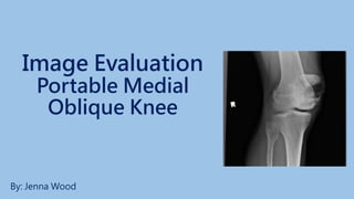
Image Evaluation
Portable Medial
Oblique Knee
By: Jenna Wood
HIPAA Compliance
• This image is HIPAA compliant
• There is no information provided
on this image that could identify a
patient or an imaging facility
• It does not violate patient
confidentiality
Marker and Patient ID
• An anatomical marker is visible on
this image
Marker and Patient ID
• The correct right anatomical marker
should be placed on the lateral side of
the patient’s body, indicating it is a right
knee, so it is displayed on the viewer’s
left side
• Marker should include the technologist’s
identification
• It should not superimpose any pertinent
anatomy
• A portable marker should have been
used along with the time in some
hospital protocols
• The image is now displayed correctly
based on my marker placement
JW
Portable
time
Radiation Hygiene
• An image must have a minimum of three
sides of beam restriction and (1) of these
sides should be closest to the patient’s
gonads
• This is especially true while doing
portables (Technologist’s should also
wear a minimum of 0.25 mm/Pb eq)
• Gonadal shielding is required if the gonads
are within 5 cm of the primary beam
• This image does not display appropriate
beam restriction and is only clearly visible on
two sides of the border
• There is no evidence of primary or additional
shielding
JW
Portable
time
Completeness of
Position/Projection
• Routine imaging of the knee includes:
• AP projection, posterior position
• 45° AP (medial/internal) oblique
• 45° AP (lateral/external) oblique
• Mediolateral projection
• Additional images depending on hospital/facility
protocol:
• PA/AP axial intercondylar fossa projection
• Tangential patella projection
• This image is a routine projection and all
anatomical parts are visualized on the
image, but not correctly positioned
time
Artifact
Identification
• There appears to be no preventable physical
artifacts present on the image
• There appears to be no body parts
superimposed over the image
• There appears to be no hospital
paraphernalia present
• There appears to be no patient clothing
visible
• There appears to be no indwelling
artifacts/foreign bodies visible on this image
time
Artifact Identification
• There appears to be no
excessive fog affecting
the quality of the image
• There appears to be no
CR/DR artifact on the
image
time
Image Sharpness
• There appears to be no
gross voluntary motion
visible on the image
• There appears to be no
excessive quantum mottle
present in the image
time
Image Sharpness
• There appears to be no
evidence of a double or
previous exposure
• Grid lines, grid artifact, &/or
grid cut-off are not visible on
the image because a grid
might not have been used
&/or a high frequency could
have been used since it was
done portably
time
Image Sharpness
• Size distortion does not appear to be greater than
expected for this image because OID is minimal in this
position
• The CR is not properly centered to femorotibial joint
space, but less than 1 cm
• There is evidence of slight shape distortion due to the
CR/part misalignment
• Due to the insufficient 5 degree cephalic angle that
should have been used because the patient’s thigh
thickness appears to be >25 cm the tibial plateau
does not appear to be perpendicular to IR
• The patellar apex sits closer to the upper joint space
also indicating the CR was not proper angled
time
Accurate Part Position
• The part does appear to be
aligned parallel to the image
media
• The part is slightly off-centered
to the image media
time
Accurate Part Position
• The CR is off centered slightly laterally to the
femorotibial joint space, but within 1 cm of the
anatomical part
• The CR is adequately aligned to the image
media
• The CR’s alignment does not appear to
conform to an acceptable IR exposure
recognition field due to the lack of only two
sides of collimation
time
Accurate Part Position
According to Kathy McQuillen Martensen’s Radiographic Image Analysis and Merrill’s Atlas:
• 40” SID and 10 x 12” IR
• Position the patient supine, with the knee joint centered to the IR
• Internally rotate the leg until the femoral epicondyles are at a 45 degree angle with the IR
• Angle the CR to align it parallel with the tibial plateau
• To do so measure the patient from the ASIS to the table after the patient has been
accurately positioned to determine the correct CR angulation (use a caliper and do not
include abdominal tissue in measurement)
• 5 degree caudal if ASIS to tabletop measurement is 18 cm below
• Perpendicular if ASIS to tabletop is between 19-24 cm
• 5 degrees cephalic angle if ASIS to tabletop is 25 cm or greater
** It is not uncommon to require a cephalic angle for the AP medial oblique when a
perpendicular or caudal angle was used for an AP projection because the patients hips
are elevated
• Center the CR to the midline of the knee 1 inch distal to the medial epicondyle (Martensen)
• Center the CR to the knee joint at ½ inch distal to the patellar apex (Merrill’s)
• Longitudinally collimate to include ¼ of the distal femus and proximal lower leg
• Transversely collimate to 0.5 inch of the knee skin line
• Table bucky and a grid should be used if the knee is greater than 10 cm part thickness,
however when done portable a stationary grid would be used
Accurate Part Position
According to Kathy McQuillen Martensen’s Radiographic Image
Analysis and Merrill’s Atlas:
Evaluation Criteria:
• Fibular head is seen free of tibial superimposition (Martensen)
• Lateral femoral condyle is in profile without superimposing the medial
condyle
• Knee joint space is open: when correct obliquity is used (Bontrager)
• The anterior and posterior condylar margins of the tibia are aligned
• Fibular head is approximately 0.5 in distal to the tibial plateau
• Knee joint is at the center of the exposure field
• Tibia and fibula should be separated at their proximal articulations
• Posterior tibia should be visible
• ¼ of the distal femur and proximal lower leg included
• Margin of the patella projecting slightly beyond the medial side of the
femoral condylar leg and the surrounding knee soft tissue are
included with the exposure field
Accurate Part Position
Evaluation• A minimum of three sides of beam restriction should be
demonstrated
• The CR is centered slightly laterally to the femorotibial joint
• The CR appears to have been directed perpendicular
causing the tibial plateau to not appear perpendicular to the
IR, therefore a 5 degree cephalic angle should have been
used due to the patient’s thigh thickness >25cm
• The anterior and posterior condyles of the tibia are not
superimposed due to incorrect CR angulation
• The patellar apex sits closer to the upper joint space also
indicating the CR was not proper angled
• The knee does not appear to be sufficiently rotated to 45
degrees because the tibia is superimposed over the fibula
head and also demonstrating a partially closed tibiofibular
articulation
time
Accurate Part Position
Evaluation
• All pertinent anatomy is included on
the image
• The anatomical part is not positioned
correctly based on my assessment of
the evaluation criteria
time
Accurate Part Position
Evaluation
• The most radiolucent structure is
the surrounding soft tissue and
knee joint (which should be open)
and it is visible on the image
• The most radiopaque structure is
the bony cortex and it is visible
on the image time
Judicious Exposure Technique
• Assessment of Image Contrast
(Window Width)
• Contrast is determined by the
number of grays produced on an
image
• Extremities should be displayed
with short scale contrast,
showing many shades of gray
• The image appears to have
adequate contrast
time
Judicious Exposure Technique
• Assessment of Image’s
Brightness (Window Level)
• The image’s brightness is the
balance between light and dark
areas on the image and it
appears to be acceptable
because you are able to visualize
the soft tissue, bony trabeculi and
cortex
• I would expect the overall EI
value to be within normal range
time
Accept/Reject?
This image should be repeated because it does not meet
minimal established standards
Required corrections:
• Use a correctly placed right marker with technologist
identification and a portable marker along with the time
the exposure was taken
• Include a minimum of three sides of collimation
• Center the CR to the femoroibial joint space
• Rotate the knee internally to 45 degrees so the tibia
does not superimpose over the fibula head articulation
• Angle to CR approximately 5 degrees cephalically to
ensure the tibial plateau is perpendicular to the IR
• Aligning the anterior and posterior condyles of the tibia to
superimpose one another
• Angulation will also rotate the patellar apex superiorly and
medially away from the joint space
time
Sources
Frank, E, Long, B, & Smith, B. Merrill’s atlas of radiographic
positioning and procedures. 12th ed. St. Louis, MO:
Mosby, 2012.
McQuillen-Martensen, K. (2015). Radiographic image analysis.
Vol 4. St. Louis, MO: Elsevier
Saint Mary’s Medical Center

More Related Content

Viewers also liked

Redes sociales en la educación
Redes sociales en la educaciónRedes sociales en la educación
Redes sociales en la educación
ZoeBeatriz7
 
Работы Студии МартДизайн - Печатные материалы
Работы Студии МартДизайн - Печатные материалыРаботы Студии МартДизайн - Печатные материалы
Работы Студии МартДизайн - Печатные материалы
Nick Zaporozsky
 
brochure
brochurebrochure
brochure
Laleh Dehghani
 
DOODLEBOARD LAUNCH PLAN
DOODLEBOARD LAUNCH PLANDOODLEBOARD LAUNCH PLAN
DOODLEBOARD LAUNCH PLAN
Tiffany Tapia
 
r am jun 16
r am jun 16r am jun 16
r am jun 16
Adam Mueller
 
Proposed red kitchen
Proposed red kitchenProposed red kitchen
Proposed red kitchen
Lilian Kuya
 

Viewers also liked (7)

Redes sociales en la educación
Redes sociales en la educaciónRedes sociales en la educación
Redes sociales en la educación
 
Работы Студии МартДизайн - Печатные материалы
Работы Студии МартДизайн - Печатные материалыРаботы Студии МартДизайн - Печатные материалы
Работы Студии МартДизайн - Печатные материалы
 
brochure
brochurebrochure
brochure
 
EEP Brochure
EEP BrochureEEP Brochure
EEP Brochure
 
DOODLEBOARD LAUNCH PLAN
DOODLEBOARD LAUNCH PLANDOODLEBOARD LAUNCH PLAN
DOODLEBOARD LAUNCH PLAN
 
r am jun 16
r am jun 16r am jun 16
r am jun 16
 
Proposed red kitchen
Proposed red kitchenProposed red kitchen
Proposed red kitchen
 

Similar to Oblique knee final evaluation

Image Evaluation: Stretcher Chest
Image Evaluation: Stretcher ChestImage Evaluation: Stretcher Chest
Image Evaluation: Stretcher Chest
Rochelle Mazzella
 
Final Image Evaluation: Left Lateral Lumbar Spine
Final Image Evaluation: Left Lateral Lumbar SpineFinal Image Evaluation: Left Lateral Lumbar Spine
Final Image Evaluation: Left Lateral Lumbar Spine
NBlankz
 
D torres lower_leg_oralppt3
D torres lower_leg_oralppt3D torres lower_leg_oralppt3
D torres lower_leg_oralppt3
dtorres2017
 
Image Evaluation: AP Axial C-Spine
Image Evaluation: AP Axial C-SpineImage Evaluation: AP Axial C-Spine
Image Evaluation: AP Axial C-Spine
alexagerg
 
Kristen Steever PA Chest
Kristen Steever PA ChestKristen Steever PA Chest
Kristen Steever PA Chest
Kristen Steever RT(R)
 
Final Image Evaluation
Final Image EvaluationFinal Image Evaluation
Final Image Evaluation
Nicolette Brennan
 
Kauffman ap knee
Kauffman   ap kneeKauffman   ap knee
Kauffman ap knee
pkauffman94
 
Lateral t spine kaleigh
Lateral t spine kaleighLateral t spine kaleigh
Lateral t spine kaleigh
Kaleigh Taylor BSRS, R.T., (R)
 
Sutliff - Final - RADS216
Sutliff - Final - RADS216Sutliff - Final - RADS216
Sutliff - Final - RADS216
bobsutliff
 
Image evaluation lateral ankle mattingly
Image evaluation  lateral ankle mattinglyImage evaluation  lateral ankle mattingly
Image evaluation lateral ankle mattingly
Heather Mattingly
 
Hip and sacroiliac joints_compressed.pdf
Hip and sacroiliac joints_compressed.pdfHip and sacroiliac joints_compressed.pdf
Hip and sacroiliac joints_compressed.pdf
christapherjohn1433
 
Various X-ray views of Knee Joint
Various X-ray views of Knee JointVarious X-ray views of Knee Joint
Various X-ray views of Knee Joint
Vinayak Sharad Abhishek Punuganti
 
Keratometry
KeratometryKeratometry
Keratometry
Karan Bhatia
 
Pilla Final
Pilla FinalPilla Final
Pilla Final
danapilla
 
Mammography positioning technique for MLO View
Mammography positioning technique for MLO View Mammography positioning technique for MLO View
Mammography positioning technique for MLO View
Selin Prasad
 
Andresen image evalfinal_16
Andresen image evalfinal_16Andresen image evalfinal_16
Andresen image evalfinal_16
Kristen Andresen
 
DOC-20230223-WA0019. copy.pptx
DOC-20230223-WA0019. copy.pptxDOC-20230223-WA0019. copy.pptx
DOC-20230223-WA0019. copy.pptx
shyam sunder
 
Spine_Radiography.pptx
Spine_Radiography.pptxSpine_Radiography.pptx
Spine_Radiography.pptx
shyam sunder
 
Spine radiography
Spine radiographySpine radiography
Spine radiography
Dr. Muhammad Bin Zulfiqar
 
Seminar new
Seminar newSeminar new
Seminar new
Ganesh Gadag
 

Similar to Oblique knee final evaluation (20)

Image Evaluation: Stretcher Chest
Image Evaluation: Stretcher ChestImage Evaluation: Stretcher Chest
Image Evaluation: Stretcher Chest
 
Final Image Evaluation: Left Lateral Lumbar Spine
Final Image Evaluation: Left Lateral Lumbar SpineFinal Image Evaluation: Left Lateral Lumbar Spine
Final Image Evaluation: Left Lateral Lumbar Spine
 
D torres lower_leg_oralppt3
D torres lower_leg_oralppt3D torres lower_leg_oralppt3
D torres lower_leg_oralppt3
 
Image Evaluation: AP Axial C-Spine
Image Evaluation: AP Axial C-SpineImage Evaluation: AP Axial C-Spine
Image Evaluation: AP Axial C-Spine
 
Kristen Steever PA Chest
Kristen Steever PA ChestKristen Steever PA Chest
Kristen Steever PA Chest
 
Final Image Evaluation
Final Image EvaluationFinal Image Evaluation
Final Image Evaluation
 
Kauffman ap knee
Kauffman   ap kneeKauffman   ap knee
Kauffman ap knee
 
Lateral t spine kaleigh
Lateral t spine kaleighLateral t spine kaleigh
Lateral t spine kaleigh
 
Sutliff - Final - RADS216
Sutliff - Final - RADS216Sutliff - Final - RADS216
Sutliff - Final - RADS216
 
Image evaluation lateral ankle mattingly
Image evaluation  lateral ankle mattinglyImage evaluation  lateral ankle mattingly
Image evaluation lateral ankle mattingly
 
Hip and sacroiliac joints_compressed.pdf
Hip and sacroiliac joints_compressed.pdfHip and sacroiliac joints_compressed.pdf
Hip and sacroiliac joints_compressed.pdf
 
Various X-ray views of Knee Joint
Various X-ray views of Knee JointVarious X-ray views of Knee Joint
Various X-ray views of Knee Joint
 
Keratometry
KeratometryKeratometry
Keratometry
 
Pilla Final
Pilla FinalPilla Final
Pilla Final
 
Mammography positioning technique for MLO View
Mammography positioning technique for MLO View Mammography positioning technique for MLO View
Mammography positioning technique for MLO View
 
Andresen image evalfinal_16
Andresen image evalfinal_16Andresen image evalfinal_16
Andresen image evalfinal_16
 
DOC-20230223-WA0019. copy.pptx
DOC-20230223-WA0019. copy.pptxDOC-20230223-WA0019. copy.pptx
DOC-20230223-WA0019. copy.pptx
 
Spine_Radiography.pptx
Spine_Radiography.pptxSpine_Radiography.pptx
Spine_Radiography.pptx
 
Spine radiography
Spine radiographySpine radiography
Spine radiography
 
Seminar new
Seminar newSeminar new
Seminar new
 

Recently uploaded

clinical examination of hip joint (1).pdf
clinical examination of hip joint (1).pdfclinical examination of hip joint (1).pdf
clinical examination of hip joint (1).pdf
Priyankaranawat4
 
BBR 2024 Summer Sessions Interview Training
BBR  2024 Summer Sessions Interview TrainingBBR  2024 Summer Sessions Interview Training
BBR 2024 Summer Sessions Interview Training
Katrina Pritchard
 
C1 Rubenstein AP HuG xxxxxxxxxxxxxx.pptx
C1 Rubenstein AP HuG xxxxxxxxxxxxxx.pptxC1 Rubenstein AP HuG xxxxxxxxxxxxxx.pptx
C1 Rubenstein AP HuG xxxxxxxxxxxxxx.pptx
mulvey2
 
Exploiting Artificial Intelligence for Empowering Researchers and Faculty, In...
Exploiting Artificial Intelligence for Empowering Researchers and Faculty, In...Exploiting Artificial Intelligence for Empowering Researchers and Faculty, In...
Exploiting Artificial Intelligence for Empowering Researchers and Faculty, In...
Dr. Vinod Kumar Kanvaria
 
Azure Interview Questions and Answers PDF By ScholarHat
Azure Interview Questions and Answers PDF By ScholarHatAzure Interview Questions and Answers PDF By ScholarHat
Azure Interview Questions and Answers PDF By ScholarHat
Scholarhat
 
Cognitive Development Adolescence Psychology
Cognitive Development Adolescence PsychologyCognitive Development Adolescence Psychology
Cognitive Development Adolescence Psychology
paigestewart1632
 
How to Build a Module in Odoo 17 Using the Scaffold Method
How to Build a Module in Odoo 17 Using the Scaffold MethodHow to Build a Module in Odoo 17 Using the Scaffold Method
How to Build a Module in Odoo 17 Using the Scaffold Method
Celine George
 
Pengantar Penggunaan Flutter - Dart programming language1.pptx
Pengantar Penggunaan Flutter - Dart programming language1.pptxPengantar Penggunaan Flutter - Dart programming language1.pptx
Pengantar Penggunaan Flutter - Dart programming language1.pptx
Fajar Baskoro
 
বাংলাদেশ অর্থনৈতিক সমীক্ষা (Economic Review) ২০২৪ UJS App.pdf
বাংলাদেশ অর্থনৈতিক সমীক্ষা (Economic Review) ২০২৪ UJS App.pdfবাংলাদেশ অর্থনৈতিক সমীক্ষা (Economic Review) ২০২৪ UJS App.pdf
বাংলাদেশ অর্থনৈতিক সমীক্ষা (Economic Review) ২০২৪ UJS App.pdf
eBook.com.bd (প্রয়োজনীয় বাংলা বই)
 
The Diamonds of 2023-2024 in the IGRA collection
The Diamonds of 2023-2024 in the IGRA collectionThe Diamonds of 2023-2024 in the IGRA collection
The Diamonds of 2023-2024 in the IGRA collection
Israel Genealogy Research Association
 
The History of Stoke Newington Street Names
The History of Stoke Newington Street NamesThe History of Stoke Newington Street Names
The History of Stoke Newington Street Names
History of Stoke Newington
 
DRUGS AND ITS classification slide share
DRUGS AND ITS classification slide shareDRUGS AND ITS classification slide share
DRUGS AND ITS classification slide share
taiba qazi
 
Advanced Java[Extra Concepts, Not Difficult].docx
Advanced Java[Extra Concepts, Not Difficult].docxAdvanced Java[Extra Concepts, Not Difficult].docx
Advanced Java[Extra Concepts, Not Difficult].docx
adhitya5119
 
Main Java[All of the Base Concepts}.docx
Main Java[All of the Base Concepts}.docxMain Java[All of the Base Concepts}.docx
Main Java[All of the Base Concepts}.docx
adhitya5119
 
What is Digital Literacy? A guest blog from Andy McLaughlin, University of Ab...
What is Digital Literacy? A guest blog from Andy McLaughlin, University of Ab...What is Digital Literacy? A guest blog from Andy McLaughlin, University of Ab...
What is Digital Literacy? A guest blog from Andy McLaughlin, University of Ab...
GeorgeMilliken2
 
Walmart Business+ and Spark Good for Nonprofits.pdf
Walmart Business+ and Spark Good for Nonprofits.pdfWalmart Business+ and Spark Good for Nonprofits.pdf
Walmart Business+ and Spark Good for Nonprofits.pdf
TechSoup
 
South African Journal of Science: Writing with integrity workshop (2024)
South African Journal of Science: Writing with integrity workshop (2024)South African Journal of Science: Writing with integrity workshop (2024)
South African Journal of Science: Writing with integrity workshop (2024)
Academy of Science of South Africa
 
Digital Artefact 1 - Tiny Home Environmental Design
Digital Artefact 1 - Tiny Home Environmental DesignDigital Artefact 1 - Tiny Home Environmental Design
Digital Artefact 1 - Tiny Home Environmental Design
amberjdewit93
 
A Independência da América Espanhola LAPBOOK.pdf
A Independência da América Espanhola LAPBOOK.pdfA Independência da América Espanhola LAPBOOK.pdf
A Independência da América Espanhola LAPBOOK.pdf
Jean Carlos Nunes Paixão
 
ANATOMY AND BIOMECHANICS OF HIP JOINT.pdf
ANATOMY AND BIOMECHANICS OF HIP JOINT.pdfANATOMY AND BIOMECHANICS OF HIP JOINT.pdf
ANATOMY AND BIOMECHANICS OF HIP JOINT.pdf
Priyankaranawat4
 

Recently uploaded (20)

clinical examination of hip joint (1).pdf
clinical examination of hip joint (1).pdfclinical examination of hip joint (1).pdf
clinical examination of hip joint (1).pdf
 
BBR 2024 Summer Sessions Interview Training
BBR  2024 Summer Sessions Interview TrainingBBR  2024 Summer Sessions Interview Training
BBR 2024 Summer Sessions Interview Training
 
C1 Rubenstein AP HuG xxxxxxxxxxxxxx.pptx
C1 Rubenstein AP HuG xxxxxxxxxxxxxx.pptxC1 Rubenstein AP HuG xxxxxxxxxxxxxx.pptx
C1 Rubenstein AP HuG xxxxxxxxxxxxxx.pptx
 
Exploiting Artificial Intelligence for Empowering Researchers and Faculty, In...
Exploiting Artificial Intelligence for Empowering Researchers and Faculty, In...Exploiting Artificial Intelligence for Empowering Researchers and Faculty, In...
Exploiting Artificial Intelligence for Empowering Researchers and Faculty, In...
 
Azure Interview Questions and Answers PDF By ScholarHat
Azure Interview Questions and Answers PDF By ScholarHatAzure Interview Questions and Answers PDF By ScholarHat
Azure Interview Questions and Answers PDF By ScholarHat
 
Cognitive Development Adolescence Psychology
Cognitive Development Adolescence PsychologyCognitive Development Adolescence Psychology
Cognitive Development Adolescence Psychology
 
How to Build a Module in Odoo 17 Using the Scaffold Method
How to Build a Module in Odoo 17 Using the Scaffold MethodHow to Build a Module in Odoo 17 Using the Scaffold Method
How to Build a Module in Odoo 17 Using the Scaffold Method
 
Pengantar Penggunaan Flutter - Dart programming language1.pptx
Pengantar Penggunaan Flutter - Dart programming language1.pptxPengantar Penggunaan Flutter - Dart programming language1.pptx
Pengantar Penggunaan Flutter - Dart programming language1.pptx
 
বাংলাদেশ অর্থনৈতিক সমীক্ষা (Economic Review) ২০২৪ UJS App.pdf
বাংলাদেশ অর্থনৈতিক সমীক্ষা (Economic Review) ২০২৪ UJS App.pdfবাংলাদেশ অর্থনৈতিক সমীক্ষা (Economic Review) ২০২৪ UJS App.pdf
বাংলাদেশ অর্থনৈতিক সমীক্ষা (Economic Review) ২০২৪ UJS App.pdf
 
The Diamonds of 2023-2024 in the IGRA collection
The Diamonds of 2023-2024 in the IGRA collectionThe Diamonds of 2023-2024 in the IGRA collection
The Diamonds of 2023-2024 in the IGRA collection
 
The History of Stoke Newington Street Names
The History of Stoke Newington Street NamesThe History of Stoke Newington Street Names
The History of Stoke Newington Street Names
 
DRUGS AND ITS classification slide share
DRUGS AND ITS classification slide shareDRUGS AND ITS classification slide share
DRUGS AND ITS classification slide share
 
Advanced Java[Extra Concepts, Not Difficult].docx
Advanced Java[Extra Concepts, Not Difficult].docxAdvanced Java[Extra Concepts, Not Difficult].docx
Advanced Java[Extra Concepts, Not Difficult].docx
 
Main Java[All of the Base Concepts}.docx
Main Java[All of the Base Concepts}.docxMain Java[All of the Base Concepts}.docx
Main Java[All of the Base Concepts}.docx
 
What is Digital Literacy? A guest blog from Andy McLaughlin, University of Ab...
What is Digital Literacy? A guest blog from Andy McLaughlin, University of Ab...What is Digital Literacy? A guest blog from Andy McLaughlin, University of Ab...
What is Digital Literacy? A guest blog from Andy McLaughlin, University of Ab...
 
Walmart Business+ and Spark Good for Nonprofits.pdf
Walmart Business+ and Spark Good for Nonprofits.pdfWalmart Business+ and Spark Good for Nonprofits.pdf
Walmart Business+ and Spark Good for Nonprofits.pdf
 
South African Journal of Science: Writing with integrity workshop (2024)
South African Journal of Science: Writing with integrity workshop (2024)South African Journal of Science: Writing with integrity workshop (2024)
South African Journal of Science: Writing with integrity workshop (2024)
 
Digital Artefact 1 - Tiny Home Environmental Design
Digital Artefact 1 - Tiny Home Environmental DesignDigital Artefact 1 - Tiny Home Environmental Design
Digital Artefact 1 - Tiny Home Environmental Design
 
A Independência da América Espanhola LAPBOOK.pdf
A Independência da América Espanhola LAPBOOK.pdfA Independência da América Espanhola LAPBOOK.pdf
A Independência da América Espanhola LAPBOOK.pdf
 
ANATOMY AND BIOMECHANICS OF HIP JOINT.pdf
ANATOMY AND BIOMECHANICS OF HIP JOINT.pdfANATOMY AND BIOMECHANICS OF HIP JOINT.pdf
ANATOMY AND BIOMECHANICS OF HIP JOINT.pdf
 

Oblique knee final evaluation

  • 2. HIPAA Compliance • This image is HIPAA compliant • There is no information provided on this image that could identify a patient or an imaging facility • It does not violate patient confidentiality
  • 3. Marker and Patient ID • An anatomical marker is visible on this image
  • 4. Marker and Patient ID • The correct right anatomical marker should be placed on the lateral side of the patient’s body, indicating it is a right knee, so it is displayed on the viewer’s left side • Marker should include the technologist’s identification • It should not superimpose any pertinent anatomy • A portable marker should have been used along with the time in some hospital protocols • The image is now displayed correctly based on my marker placement JW Portable time
  • 5. Radiation Hygiene • An image must have a minimum of three sides of beam restriction and (1) of these sides should be closest to the patient’s gonads • This is especially true while doing portables (Technologist’s should also wear a minimum of 0.25 mm/Pb eq) • Gonadal shielding is required if the gonads are within 5 cm of the primary beam • This image does not display appropriate beam restriction and is only clearly visible on two sides of the border • There is no evidence of primary or additional shielding JW Portable time
  • 6. Completeness of Position/Projection • Routine imaging of the knee includes: • AP projection, posterior position • 45° AP (medial/internal) oblique • 45° AP (lateral/external) oblique • Mediolateral projection • Additional images depending on hospital/facility protocol: • PA/AP axial intercondylar fossa projection • Tangential patella projection • This image is a routine projection and all anatomical parts are visualized on the image, but not correctly positioned time
  • 7. Artifact Identification • There appears to be no preventable physical artifacts present on the image • There appears to be no body parts superimposed over the image • There appears to be no hospital paraphernalia present • There appears to be no patient clothing visible • There appears to be no indwelling artifacts/foreign bodies visible on this image time
  • 8. Artifact Identification • There appears to be no excessive fog affecting the quality of the image • There appears to be no CR/DR artifact on the image time
  • 9. Image Sharpness • There appears to be no gross voluntary motion visible on the image • There appears to be no excessive quantum mottle present in the image time
  • 10. Image Sharpness • There appears to be no evidence of a double or previous exposure • Grid lines, grid artifact, &/or grid cut-off are not visible on the image because a grid might not have been used &/or a high frequency could have been used since it was done portably time
  • 11. Image Sharpness • Size distortion does not appear to be greater than expected for this image because OID is minimal in this position • The CR is not properly centered to femorotibial joint space, but less than 1 cm • There is evidence of slight shape distortion due to the CR/part misalignment • Due to the insufficient 5 degree cephalic angle that should have been used because the patient’s thigh thickness appears to be >25 cm the tibial plateau does not appear to be perpendicular to IR • The patellar apex sits closer to the upper joint space also indicating the CR was not proper angled time
  • 12. Accurate Part Position • The part does appear to be aligned parallel to the image media • The part is slightly off-centered to the image media time
  • 13. Accurate Part Position • The CR is off centered slightly laterally to the femorotibial joint space, but within 1 cm of the anatomical part • The CR is adequately aligned to the image media • The CR’s alignment does not appear to conform to an acceptable IR exposure recognition field due to the lack of only two sides of collimation time
  • 14. Accurate Part Position According to Kathy McQuillen Martensen’s Radiographic Image Analysis and Merrill’s Atlas: • 40” SID and 10 x 12” IR • Position the patient supine, with the knee joint centered to the IR • Internally rotate the leg until the femoral epicondyles are at a 45 degree angle with the IR • Angle the CR to align it parallel with the tibial plateau • To do so measure the patient from the ASIS to the table after the patient has been accurately positioned to determine the correct CR angulation (use a caliper and do not include abdominal tissue in measurement) • 5 degree caudal if ASIS to tabletop measurement is 18 cm below • Perpendicular if ASIS to tabletop is between 19-24 cm • 5 degrees cephalic angle if ASIS to tabletop is 25 cm or greater ** It is not uncommon to require a cephalic angle for the AP medial oblique when a perpendicular or caudal angle was used for an AP projection because the patients hips are elevated • Center the CR to the midline of the knee 1 inch distal to the medial epicondyle (Martensen) • Center the CR to the knee joint at ½ inch distal to the patellar apex (Merrill’s) • Longitudinally collimate to include ¼ of the distal femus and proximal lower leg • Transversely collimate to 0.5 inch of the knee skin line • Table bucky and a grid should be used if the knee is greater than 10 cm part thickness, however when done portable a stationary grid would be used
  • 15. Accurate Part Position According to Kathy McQuillen Martensen’s Radiographic Image Analysis and Merrill’s Atlas: Evaluation Criteria: • Fibular head is seen free of tibial superimposition (Martensen) • Lateral femoral condyle is in profile without superimposing the medial condyle • Knee joint space is open: when correct obliquity is used (Bontrager) • The anterior and posterior condylar margins of the tibia are aligned • Fibular head is approximately 0.5 in distal to the tibial plateau • Knee joint is at the center of the exposure field • Tibia and fibula should be separated at their proximal articulations • Posterior tibia should be visible • ¼ of the distal femur and proximal lower leg included • Margin of the patella projecting slightly beyond the medial side of the femoral condylar leg and the surrounding knee soft tissue are included with the exposure field
  • 16. Accurate Part Position Evaluation• A minimum of three sides of beam restriction should be demonstrated • The CR is centered slightly laterally to the femorotibial joint • The CR appears to have been directed perpendicular causing the tibial plateau to not appear perpendicular to the IR, therefore a 5 degree cephalic angle should have been used due to the patient’s thigh thickness >25cm • The anterior and posterior condyles of the tibia are not superimposed due to incorrect CR angulation • The patellar apex sits closer to the upper joint space also indicating the CR was not proper angled • The knee does not appear to be sufficiently rotated to 45 degrees because the tibia is superimposed over the fibula head and also demonstrating a partially closed tibiofibular articulation time
  • 17. Accurate Part Position Evaluation • All pertinent anatomy is included on the image • The anatomical part is not positioned correctly based on my assessment of the evaluation criteria time
  • 18. Accurate Part Position Evaluation • The most radiolucent structure is the surrounding soft tissue and knee joint (which should be open) and it is visible on the image • The most radiopaque structure is the bony cortex and it is visible on the image time
  • 19. Judicious Exposure Technique • Assessment of Image Contrast (Window Width) • Contrast is determined by the number of grays produced on an image • Extremities should be displayed with short scale contrast, showing many shades of gray • The image appears to have adequate contrast time
  • 20. Judicious Exposure Technique • Assessment of Image’s Brightness (Window Level) • The image’s brightness is the balance between light and dark areas on the image and it appears to be acceptable because you are able to visualize the soft tissue, bony trabeculi and cortex • I would expect the overall EI value to be within normal range time
  • 21. Accept/Reject? This image should be repeated because it does not meet minimal established standards Required corrections: • Use a correctly placed right marker with technologist identification and a portable marker along with the time the exposure was taken • Include a minimum of three sides of collimation • Center the CR to the femoroibial joint space • Rotate the knee internally to 45 degrees so the tibia does not superimpose over the fibula head articulation • Angle to CR approximately 5 degrees cephalically to ensure the tibial plateau is perpendicular to the IR • Aligning the anterior and posterior condyles of the tibia to superimpose one another • Angulation will also rotate the patellar apex superiorly and medially away from the joint space time
  • 22. Sources Frank, E, Long, B, & Smith, B. Merrill’s atlas of radiographic positioning and procedures. 12th ed. St. Louis, MO: Mosby, 2012. McQuillen-Martensen, K. (2015). Radiographic image analysis. Vol 4. St. Louis, MO: Elsevier Saint Mary’s Medical Center