SlideShare a Scribd company logo
Organic &
Biomolecular Chemistry
PAPER
Cite this: DOI: 10.1039/c5ob00513b
Received 14th March 2015,
Accepted 18th May 2015
DOI: 10.1039/c5ob00513b
www.rsc.org/obc
RNA nucleosides as chiral sensing agents in NMR
spectroscopy†
N. Lokesh,a,b
S. L. Sachin,a
L. V. Narendra,a
K. Aruna
and N. Suryaprakash*a,b
The study reports chiral sensing properties of RNA nucleosides. Adenosine, guanosine, uridine and cyti-
dine are used as chiral derivatizing agents to differentiate chiral 1°-amines. A three component protocol
has been adopted for complexation of nucleosides and amines. The chiral differentiating ability of nucleo-
sides is examined for different amines based on the 1
H NMR chemical shift differences of diastereomers
(ΔδR,S
). Enantiomeric differentiation has been observed at multiple chemically distinct proton sites.
Adenosine and guanosine exhibit large chiral differentiation (ΔδR,S
) due to the presence of a purine ring.
The diastereomeric excess (de) measured by using adenosine is in good agreement with the gravimetric
values.
Introduction
Most of the drug interactions with biological systems are
enantiospecific due to the inherent chiral nature of biological
molecules, such as RNA, DNA and proteins.1,2
Therefore,
chiral sensing or enantiomeric differentiation has gained
enormous importance in the asymmetric synthesis and in
pharmaceutical research.3–6
Most commonly used techniques
to analyze enantiomers are NMR,7–10
X-ray,11,12
ORD, CD13
and
chromatographic methods.14,15
The easy accessibility and
available experimental techniques make NMR an important
analytical tool for chiral analysis. NMR differentiates enantio-
mers through chiral auxiliaries7–10,16–18
and through chiral
alignment media,19–21
which induce diastereomeric inter-
actions and a differential ordering effect (DOE) respectively.
Number of chiral auxiliaries have been reported for efficient
differentiation of enantiomers, assignment of the absolute
configuration and also for the measurement of the enantio-
meric excess (ee).7–10,16–18
Most of these auxiliaries are syn-
thetically designed that requires more time, synthetic skills
and is also uneconomical. On the other hand chiral bio-
molecules are cheaply available, more abundant and can be
utilized for many applications.22–32
Recently, several studies
have been reported on bio-molecular chiral sensing, which
used DNA, xanthan, folic acid and guanosine based weak
chiral aligning media to differentiate enantiomers using
NMR.19,20,33,34
There are also reports of DNA and polysacchar-
ide based chiral stationary phases in chromatography to
differentiate enantiomeric peptides and other chiral mole-
cules,27,35,36
epigallocatechin gallate (EGCG) as a chiral solvat-
ing agent to differentiate chiral amino acids24
and nucleotides
for recognition of L-cysteine and D-cysteine by the colorimetric
technique.32
In general chiral auxiliaries contain chiral units (viz. chiral
center, chiral axis) to induce diastereomeric interactions and
delocalize electrons (e.g. aromatic ring) to induce shielding
and deshielding effects, causing enantiomeric differentiation.
Nucleosides, on the other hand, possess both chiral units
(ribose sugar) and delocalized electrons (nuclear bases), and
promise to be efficient chiral sensing agents.
The present study is aimed at exploring the chiral sensing
ability of RNA nucleosides, such as adenosine, guanosine,
uridine and cytidine, to differentiate chiral 1°-amines using
1
H NMR. A reported three component protocol was employed
to complex nucleosides and chiral amines in DMSO-d6 using
2-formylphenylboronic acid.37–39
The general scheme for the
preparation of a 3-component complex is given in Scheme 1.
Each nucleoside was tested for its ability to differentiate chiral
amines, and the 1
H chemical shift separation (ΔδR,S
) of dia-
stereomers formed from R- and S- amines was used as an indi-
cator. The unambiguous measurement of the diastereomeric
excess (de) was demonstrated on a nucleoside and amine
pair. The formed diastereomeric complexes of R and S amines
exhibit discrimination at multiple proton sites; the peak
assignment and the structure of complexes are reported
using 2D NMR (COSY, NOESY) techniques, spiking experi-
ments and utilizing the knowledge of the reported analogous
structures.37–39
†Electronic supplementary information (ESI) available. See DOI: 10.1039/
c5ob00513b
a
NMR Research Centre, Indian Institute of Science, Bangalore 560012, India.
E-mail: nsp@nrc.iisc.ernet.in; Fax: +91 80 23601550;
Tel: +91 80 22933300, +91 98 45124802
b
Solid State and Structural Chemistry Unit, Indian Institute of Science, Bangalore
560012, India
This journal is © The Royal Society of Chemistry 2015 Org. Biomol. Chem.
Publishedon18May2015.DownloadedbyNationalChiaoTungUniversityon10/06/201508:37:44.
View Article Online
View Journal
Experimental section
All the chemicals were purchased and used without any
further purification. The nucleosides used are adenosine,
guanosine, cytidine and uridine. The chiral amines used for
testing enantiodiscrimination are α-methylbenzylamine,
4-methoxy-α-methylbenzylamine, 1-(2-naphthyl)ethanamine,
1-cyclohexylethylamine and sec-butylamine. Other chemicals
used are 2-formylphenylboronic acid (purchased from com-
mercial sources and used as received) and DMSO-d6. NMR
spectra were recorded at 298 K on 500 MHz and 800 MHz
spectrometers. DMSO-d5 was used as a 1
H reference in all the
spectra. For diastereomeric excess (de) measurements a longer
inter-scan delay (10 s) was used.
For derivatization, 1.3 equivalent of each nucleoside,
1 equivalence of a racemic mixture of each amine and 1.3
equivalence of 2-formylphenylboronic acid were taken in
600 μl DMSO-d6 and stirred at 110 °C for about 10 minutes.
The excess quantities of nucleosides and 2-formylphenylboro-
nic acid were used to ensure maximum conversion of enantio-
meric amines to diastereomeric complexes. The high boiling
point of DMSO helped to carry out the reaction above 100 °C,
which removed any existing or formed water in the reaction.
This completed the reaction. The prepared solutions were used
for NMR experiments without any further purification.
Results and discussion
Initially guanosine was used as a chiral derivatizing agent
to differentiate α-methylbenzylamine. 10 mg of guanosine,
5.5 mg of 2-formylphenylboronic acid and 3.3 mg of a racemic
mixture of α-methylbenzylamine (α-MBA) were taken in 600 μl
of DMSO-d6 and were stirred for 10 minutes at about 110 °C.
The possible complex formation is reported in Scheme 1,
which was further substantiated by mass and NMR spectro-
scopic analysis (the spectrum is given in the ESI†). The
recorded 1
H NMR spectrum is given in Fig. 1, the peaks were
assigned based on 2D NMR (COSY, NOESY, spectra are
given in the ESI†) experiments and the spiking technique. The
spectrum shows two doublets for methyl protons (peak ‘c’
in Fig. 1), suggesting the formation of two diastereomeric
complexes from two enantiomeric (R & S) forms of α-methyl-
benzylamine, which unambiguously confirms the enantio-
meric differentiation. The chiral differentiation is observed at
many proton sites (shown as expanded regions in Fig. 1). The
peak identified as ‘a’ pertains to the imine proton and that
identified with number ‘8’ corresponds to the nuclear base
proton. The peaks marked 1′, 2′, 3′, 4′ 5′ and 6′ are from the
protons of the sugar unit. Better discrimination (ΔδR,S
in ppm)
was observed for sugar ring protons and nuclear base protons.
For quick observation of chiral discrimination and also for dia-
stereomeric excess (de) measurement, one can use the methyl
proton peaks (marked as ‘c’ around 1.5 ppm) or the imine
proton peaks (marked as ‘a’ around 9 ppm).
In the subsequent step the study was focused on verifying
the chiral discrimination ability of other three nucleosides,
adenosine, uridine and cytidine. Individual samples of three-
component diastereomeric complexes of α-methylbenzylamine
were prepared with each of the nucleoside (using the identical
procedure discussed earlier). The recorded 1
H NMR spectra for
all the samples are given in Fig. 2. The spectrum given in the
Scheme 1 A three-component protocol for derivatization of 1°-chiral amines and RNA nucleosides using 2-formylphenylboronic acid * indicates
the chiral center; R1 and R2 are different alkyl groups.
Paper Organic & Biomolecular Chemistry
Org. Biomol. Chem. This journal is © The Royal Society of Chemistry 2015
Publishedon18May2015.DownloadedbyNationalChiaoTungUniversityon10/06/201508:37:44.
View Article Online
Fig. 1 800 MHz 1
H NMR spectrum and chemical structure of the diastereomeric complex formed from guanosine, α-methylbenzylamine and
2-formylphenylboronic acid. The protons and peaks showing enantiomeric differentiation are labeled with alphabets and numbers.
Fig. 2 From bottom to top: 800 MHz 1
H NMR spectra of α-methylbenzylamine (MBA), and diastereomeric complex spectra of guanosine: α-methyl-
benzylamine : 2-formylphenylboronic acid (G), adenosine: α-methylbenzylamine : 2-formylphenylboronic acid (A), uridine: α-methylbenzylamine :
2-formylphenylboronic acid (U) and cytidine: α-methylbenzylamine : 2-formylphenylboronic acid (C).
Organic & Biomolecular Chemistry Paper
This journal is © The Royal Society of Chemistry 2015 Org. Biomol. Chem.
Publishedon18May2015.DownloadedbyNationalChiaoTungUniversityon10/06/201508:37:44.
View Article Online
bottom trace pertains to pure α-methylbenzylamine in DMSO-
d6. The other labeled spectra G, A, U and C were recorded from
the solutions of diastereomeric complexes of α-methyl-
benzylamine and the respective nucleosides (G-guanosine,
A-adenosine, U-uridine and C-cytidine). The methyl and imine
proton peaks were monitored for quick and easy identification
of discrimination, which are reported in the expanded scale.
The appearance of a pair of doublets for methyl protons and a
doublet for imine protons (expanded region) in each spectrum
confirms the enantiomeric discrimination ability of all the
four nucleosides. The extent of chemical shift separation
(ΔδR,S
in ppm) between two doublets suggests the chiral dis-
crimination ability. As expected, more separation was observed
in the case of adenosine, then guanosine followed by uracil
and cytosine. This is attributed to the presence of the purine
ring (more delocalized electrons) in adenosine and guanosine.
However an opposite pattern was observed for imine proton
peaks.
For establishing the wide generality of the enantiomeric dis-
crimination properties of nucleosides, many diastereomeric
Fig. 3 The plot of chemical shift separation of the selected protons (identified in red colour in the structures above) of diastereomeric complexes
formed from R and S amines versus different diastereomeric complexes (left). Nucleosides are labeled as A, G, U and C for adenosine, guanosine,
uridine and cytidine respectively. The chemical structures of amines, 4-methoxy-α-methylbenzylamine (1), 1-(2-naphthyl)ethanamine (2), α-methyl-
benzylamine (3), sec-butylamine (4) and 1-cyclohexylethylamine (5) are also given.
Fig. 4 800 MHz 1
H NMR spectrum pertaining to methyl peaks of adenosine: methoxy-α-methylbenzylamine : 2-formylphenylboronic acid diaster-
eomeric complex with varied R and S concentrations of 4-methoxy α-methylbenzylamine, the gravimetric amine ratios S : R used are shown (left).
A plot of the diastereomeric excess of R-4-methoxy-α-methylbenzylamine measured by the gravimetric method versus NMR experimental measure-
ment (right).
Paper Organic & Biomolecular Chemistry
Org. Biomol. Chem. This journal is © The Royal Society of Chemistry 2015
Publishedon18May2015.DownloadedbyNationalChiaoTungUniversityon10/06/201508:37:44.
View Article Online
complexes were prepared from different types of 1°-amines
(aliphatic-, cyclic- and aromatic-amines) with each of the
selected nucleosides using the identical procedure discussed
earlier. All the recorded 1
H NMR spectra are given in the ESI.†
The enantiomeric discrimination and the extent of discrimi-
nation of all the nucleosides with different amines are sum-
marized in Fig. 3. The plot in Fig. 3 represents 1
H chemical
shift separation (ΔδR,S
) of R and S diastereomeric complexes of
the chosen peaks of amines with the corresponding nucleo-
sides. The derivatized nucleosides are indicated as A, G, U and
C for adenosine, guanosine, uridine and cytidine respectively.
The amines used are 4-methoxy-α-methylbenzylamine, 1-(2-
naphthyl)ethanamine, α-methylbenzylamine, sec-butylamine
and 1-cyclohexylethylamine, which are numbered as 1, 2, 3, 4
and 5 respectively (chemical structures are given in Fig. 3). The
chemical shift separation was measured for CH3 protons
marked in red colour. Adenosine exhibited more enantiomeric
discrimination for all the five amines investigated, which was
followed by guanosine, uridine and cytidine. In cases of gua-
nosine and adenosine, the methoxy-protons of 4-methoxy-
α-methylbenzylamine showed better discrimination. In all
nucleosides the methyl protons of 1-(2-naphthyl)ethanamine
are better discriminated, which is due to the aromaticity of the
naphthyl ring, which induces more shielding and deshielding
effects. The observed R and S chemical shift separation (ΔδR,S
)
in other three amines is almost the same.
Finally the nucleoside was tested for the precise measure-
ment of the diastereomeric excess (de). A series of adenosine
and 4-methoxy-α-methylbenzylamine derivative complexes
were prepared with varying ratios of the R and S mixture of
amines (details are given in the ESI†). For each sample the 1D
1
H NMR spectra were recorded and the methyl peaks were
used to calculate the diastereomeric excess. The intensity vari-
ation of R and S methyl peaks with varying R and S amine con-
centration is reported in Fig. 4. The figure also contains the
plot of the diastereomeric excess calculated by gravimetric
measurements and those derived from the 1
H NMR spectrum.
The R2
= 0.9993 obtained suggests excellent agreement
between the gravimetric measurement and those obtained by
NMR experiment. This clearly establishes the applicability of
nucleosides for ee measurements.
Conclusions
In summary, RNA nucleosides have been demonstrated as
chiral derivatizing agents for efficient NMR spectroscopic dis-
crimination of chiral 1°-amines. A detailed analysis of the
differentiation ability of nucleosides has been carried out
using 1
H NMR. The enantiomeric differentiation ability was
observed for all RNA nucleosides. The better differentiation
(Δδ in ppm for R & S) was observed for adenosine and guano-
sine than that of uridine and cytidine in the methyl proton
region due to the presence of the purine ring but an opposite
pattern was observed for imine protons. Adenosine has been
used to demonstrate the precise measurement of the diastereo-
meric excess, which is in good agreement with the gravimetri-
cally determined ratios. The present study demonstrated
another important application of biomolecules helping in
exploring the possibility of using different chiral biomolecules
for chiral sensing application.
Acknowledgements
NL thanks IISc for a fellowship and Karthik for helpful discus-
sions. NS gratefully acknowledges the generous financial
support by the Board of Research in Nuclear Sciences,
Mumbai (Grant no. 2013/37C/4/BRNS) and the Science and
Engineering Research Board, Department of Science and Tech-
nology, New Delhi (grant no. SR/S1/PC-42/2011).
References
1 A. J. Hutt and S. C. Tan, Drugs, 1996, 52, 1–12.
2 L. A. Nguyen, H. He and C. Pham-Huy, Int. J. Biomed. Sci.,
2006, 2, 85–100.
3 P. M. Dewick, Medicinal Natural Products, John Wiley &
Sons, Ltd, 2009.
4 J. Gal, Chirality Drug Research, Wiley-VCH Verlag GmbH &
Co. KGaA, 2006.
5 R. E. Gawley and J. Aubé, in Principles of Asymmetric Syn-
thesis, ed. R. E. G. Aubé, Elsevier, Oxford, 2nd edn, 2012.
6 C. M. Reeves and B. M. Stoltz, in Asymmetric Synthesis II,
Wiley-VCH Verlag GmbH & Co. KGaA, 2012.
7 J. M. Seco, E. Quiñoá and R. Riguera, Chem. Rev., 2004,
104, 17–118.
8 J. M. Seco, E. Quiñoá and R. Riguera, Chem. Rev., 2012,
112, 4603–4641.
9 T. J. Wenzel and C. D. Chisholm, Prog. Nucl. Magn. Reson.
Spectrosc., 2011, 59, 1–63.
10 T. J. Wenzel and C. D. Chisholm, Chirality, 2011, 23, 190–
214.
11 N. Harada, Chirality, 2008, 20, 691–723.
12 J. Trotter, Acta Crystallogr., Sect. B: Struct. Sci., 1981, 37,
493–494.
13 N. Harada, K. Nakanishi and N. Berova, Comprehensive
Chiroptical Spectroscopy, John Wiley & Sons, Inc., 2012.
14 T. J. Ward and D.-M. Hamburg, Anal. Chem., 2004, 76,
4635–4644.
15 T. J. Ward and K. D. Ward, Anal. Chem., 2010, 82, 4712–
4722.
16 J. Chin, D. C. Kim, H.-J. Kim, F. B. Panosyan and
K. M. Kim, Org. Lett., 2004, 6, 2591–2593.
17 T. Ema, D. Tanida and T. Sakai, J. Am. Chem. Soc., 2007,
129, 10591–10596.
18 Q. Ma, M. Ma, H. Tian, X. Ye, H. Xiao, L.-h. Chen and
X. Lei, Org. Lett., 2012, 14, 5813–5815.
19 P. Lesot, U. Venkateswara Reddy and N. Suryaprakash,
Chem. Commun., 2011, 47, 11736–11738.
Organic & Biomolecular Chemistry Paper
This journal is © The Royal Society of Chemistry 2015 Org. Biomol. Chem.
Publishedon18May2015.DownloadedbyNationalChiaoTungUniversityon10/06/201508:37:44.
View Article Online
20 Lokesh and N. Suryaprakash, Chem. Commun., 2013, 49,
2049–2051.
21 M. Sarfati, P. Lesot, D. Merlet and J. Courtieu, Chem.
Commun., 2000, 2069–2081.
22 Z. Guo, I. De Cat, B. Van Averbeke, J. Lin, G. Wang, H. Xu,
R. Lazzaroni, D. Beljonne, E. W. Meijer,
A. P. H. J. Schenning and S. De Feyter, J. Am. Chem. Soc.,
2011, 133, 17764–17771.
23 V. B. Kandimalla, V. S. Tripathi and H. Ju, Crit. Rev. Anal.
Chem., 2006, 36, 73–106.
24 D. Kumari, P. Bandyopadhyay and N. Suryaprakash, J. Org.
Chem., 2013, 78, 2373–2378.
25 L. Liu and X. Zhong, Chem. Commun., 2012, 48, 5718–5720.
26 N. Metzler-Nolte, Angew. Chem., Int. Ed., 2001, 40, 1040–
1043.
27 M. Michaud, E. Jourdan, C. Ravelet, A. Villet, A. Ravel,
C. Grosset and E. Peyrin, Anal. Chem., 2004, 76, 1015–1020.
28 S. V. Patwardhan, G. Patwardhan and C. C. Perry, J. Mater.
Chem., 2007, 17, 2875–2884.
29 J. L. Rouge, B. E. Eaton and D. L. Feldheim, Energy Environ.
Sci., 2011, 4, 398–402.
30 G. Shemer, O. Krichevski, G. Markovich, T. Molotsky,
I. Lubitz and A. B. Kotlyar, J. Am. Chem. Soc., 2006, 128,
11006–11007.
31 J. Zhang, M. T. Albelda, Y. Liu and J. W. Canary, Chirality,
2005, 17, 404–420.
32 M. Zhang and B.-C. Ye, Anal. Chem., 2011, 83, 1504–
1509.
33 Lokesh and N. Suryaprakash, Chem. – Eur. J., 2012, 18,
11560–11563.
34 U. V. Reddy and N. Suryaprakash, Chem. Commun., 2011,
47, 8364–8366.
35 B. Chankvetadze, in Chiral Separations, ed. G. K. E. Scriba,
Humana Press, 2013.
36 S. Pedotti and A. Patti, J. Sep. Sci., 2014, 37, 3451–3460.
37 A. M. Kelly, Y. Pérez-Fuertes, S. Arimori, S. D. Bull and
T. D. James, Org. Lett., 2006, 8, 1971–1974.
38 A. M. Kelly, Y. Perez-Fuertes, J. S. Fossey, S. L. Yeste,
S. D. Bull and T. D. James, Nat. Protocols, 2008, 3, 215–
219.
39 Y. Pérez-Fuertes, A. M. Kelly, A. L. Johnson, S. Arimori,
S. D. Bull and T. D. James, Org. Lett., 2006, 8, 609–612.
Paper Organic & Biomolecular Chemistry
Org. Biomol. Chem. This journal is © The Royal Society of Chemistry 2015
Publishedon18May2015.DownloadedbyNationalChiaoTungUniversityon10/06/201508:37:44.
View Article Online

More Related Content

What's hot

prakash_JMS_2015
prakash_JMS_2015prakash_JMS_2015
prakash_JMS_2015
omshamli
 
SYNTHESIS, CHARACTERIZATION AND ANTIMICROBIAL ACTIVITIES OF{FE(II),CO(II),NI(...
SYNTHESIS, CHARACTERIZATION AND ANTIMICROBIAL ACTIVITIES OF{FE(II),CO(II),NI(...SYNTHESIS, CHARACTERIZATION AND ANTIMICROBIAL ACTIVITIES OF{FE(II),CO(II),NI(...
SYNTHESIS, CHARACTERIZATION AND ANTIMICROBIAL ACTIVITIES OF{FE(II),CO(II),NI(...
International Journal of Technical Research & Application
 
UV resonance raman study of streptavidin binding of biotin and 2 iminobiotin ...
UV resonance raman study of streptavidin binding of biotin and 2 iminobiotin ...UV resonance raman study of streptavidin binding of biotin and 2 iminobiotin ...
UV resonance raman study of streptavidin binding of biotin and 2 iminobiotin ...
John Clarkson
 
schiff base ligand
schiff  base ligandschiff  base ligand
polyaniline nanofiber based pH sensors
polyaniline nanofiber  based pH sensorspolyaniline nanofiber  based pH sensors
polyaniline nanofiber based pH sensors
SanwarHossain10
 
Structure elucidation of steroids
Structure elucidation of steroidsStructure elucidation of steroids
Structure elucidation of steroids
Sidratal Muntaha
 
Multinuclear liquid and solid-state NMR of Fructoborate complex
Multinuclear liquid and solid-state NMR of Fructoborate complexMultinuclear liquid and solid-state NMR of Fructoborate complex
Multinuclear liquid and solid-state NMR of Fructoborate complex
John Edwards
 
Synthesis and characterization of water-soluble free-base, zinc and copper
Synthesis and characterization of water-soluble free-base, zinc and copperSynthesis and characterization of water-soluble free-base, zinc and copper
Synthesis and characterization of water-soluble free-base, zinc and copper
Angela Mammana
 
Growth and characterization of EDTA doped BupropionHydrochloride
Growth and characterization of EDTA doped BupropionHydrochlorideGrowth and characterization of EDTA doped BupropionHydrochloride
Growth and characterization of EDTA doped BupropionHydrochloride
inventy
 
Org Let PASS
Org Let PASSOrg Let PASS
Org Let PASS
Peter ten Holte
 
ja2041139_paper1
ja2041139_paper1ja2041139_paper1
ja2041139_paper1
Ashley Zuniga
 
PRISM Spring 2012 drug DNA binding poster
PRISM Spring 2012 drug DNA binding poster PRISM Spring 2012 drug DNA binding poster
PRISM Spring 2012 drug DNA binding poster
Bob Rawal
 
The Synthesis and Characterization of Highly Fluorescent Polycyclic Azaborine...
The Synthesis and Characterization of Highly Fluorescent Polycyclic Azaborine...The Synthesis and Characterization of Highly Fluorescent Polycyclic Azaborine...
The Synthesis and Characterization of Highly Fluorescent Polycyclic Azaborine...
Nicolle Jackson
 
Photosynthetic Pigments under Nitrogen Stress in Spirulina Platensis -Udaya B...
Photosynthetic Pigments under Nitrogen Stress in Spirulina Platensis -Udaya B...Photosynthetic Pigments under Nitrogen Stress in Spirulina Platensis -Udaya B...
Photosynthetic Pigments under Nitrogen Stress in Spirulina Platensis -Udaya B...
IRJET Journal
 
Protein Purification Lecture
Protein Purification LectureProtein Purification Lecture
Protein Purification Lecture
ouopened
 
SIMONA CAVALU_Raman and Surface Enhanced Raman Spectroscopy of 2,2,5,5-Tetram...
SIMONA CAVALU_Raman and Surface Enhanced Raman Spectroscopy of 2,2,5,5-Tetram...SIMONA CAVALU_Raman and Surface Enhanced Raman Spectroscopy of 2,2,5,5-Tetram...
SIMONA CAVALU_Raman and Surface Enhanced Raman Spectroscopy of 2,2,5,5-Tetram...
Simona Cavalu
 
Capillary Electrophoresis by Sachin Kuhire
Capillary Electrophoresis by Sachin KuhireCapillary Electrophoresis by Sachin Kuhire
Capillary Electrophoresis by Sachin Kuhire
National Chemical Laboratory, Pune
 
Protein sequencing methods
Protein sequencing methodsProtein sequencing methods
Protein sequencing methods
sepidehsaroghi
 
Proteomics course 2
Proteomics course 2Proteomics course 2
Proteomics course 2
utpaltatu
 
PROTEIN ANALYSIS
PROTEIN ANALYSISPROTEIN ANALYSIS
PROTEIN ANALYSIS
VIPIN E V
 

What's hot (20)

prakash_JMS_2015
prakash_JMS_2015prakash_JMS_2015
prakash_JMS_2015
 
SYNTHESIS, CHARACTERIZATION AND ANTIMICROBIAL ACTIVITIES OF{FE(II),CO(II),NI(...
SYNTHESIS, CHARACTERIZATION AND ANTIMICROBIAL ACTIVITIES OF{FE(II),CO(II),NI(...SYNTHESIS, CHARACTERIZATION AND ANTIMICROBIAL ACTIVITIES OF{FE(II),CO(II),NI(...
SYNTHESIS, CHARACTERIZATION AND ANTIMICROBIAL ACTIVITIES OF{FE(II),CO(II),NI(...
 
UV resonance raman study of streptavidin binding of biotin and 2 iminobiotin ...
UV resonance raman study of streptavidin binding of biotin and 2 iminobiotin ...UV resonance raman study of streptavidin binding of biotin and 2 iminobiotin ...
UV resonance raman study of streptavidin binding of biotin and 2 iminobiotin ...
 
schiff base ligand
schiff  base ligandschiff  base ligand
schiff base ligand
 
polyaniline nanofiber based pH sensors
polyaniline nanofiber  based pH sensorspolyaniline nanofiber  based pH sensors
polyaniline nanofiber based pH sensors
 
Structure elucidation of steroids
Structure elucidation of steroidsStructure elucidation of steroids
Structure elucidation of steroids
 
Multinuclear liquid and solid-state NMR of Fructoborate complex
Multinuclear liquid and solid-state NMR of Fructoborate complexMultinuclear liquid and solid-state NMR of Fructoborate complex
Multinuclear liquid and solid-state NMR of Fructoborate complex
 
Synthesis and characterization of water-soluble free-base, zinc and copper
Synthesis and characterization of water-soluble free-base, zinc and copperSynthesis and characterization of water-soluble free-base, zinc and copper
Synthesis and characterization of water-soluble free-base, zinc and copper
 
Growth and characterization of EDTA doped BupropionHydrochloride
Growth and characterization of EDTA doped BupropionHydrochlorideGrowth and characterization of EDTA doped BupropionHydrochloride
Growth and characterization of EDTA doped BupropionHydrochloride
 
Org Let PASS
Org Let PASSOrg Let PASS
Org Let PASS
 
ja2041139_paper1
ja2041139_paper1ja2041139_paper1
ja2041139_paper1
 
PRISM Spring 2012 drug DNA binding poster
PRISM Spring 2012 drug DNA binding poster PRISM Spring 2012 drug DNA binding poster
PRISM Spring 2012 drug DNA binding poster
 
The Synthesis and Characterization of Highly Fluorescent Polycyclic Azaborine...
The Synthesis and Characterization of Highly Fluorescent Polycyclic Azaborine...The Synthesis and Characterization of Highly Fluorescent Polycyclic Azaborine...
The Synthesis and Characterization of Highly Fluorescent Polycyclic Azaborine...
 
Photosynthetic Pigments under Nitrogen Stress in Spirulina Platensis -Udaya B...
Photosynthetic Pigments under Nitrogen Stress in Spirulina Platensis -Udaya B...Photosynthetic Pigments under Nitrogen Stress in Spirulina Platensis -Udaya B...
Photosynthetic Pigments under Nitrogen Stress in Spirulina Platensis -Udaya B...
 
Protein Purification Lecture
Protein Purification LectureProtein Purification Lecture
Protein Purification Lecture
 
SIMONA CAVALU_Raman and Surface Enhanced Raman Spectroscopy of 2,2,5,5-Tetram...
SIMONA CAVALU_Raman and Surface Enhanced Raman Spectroscopy of 2,2,5,5-Tetram...SIMONA CAVALU_Raman and Surface Enhanced Raman Spectroscopy of 2,2,5,5-Tetram...
SIMONA CAVALU_Raman and Surface Enhanced Raman Spectroscopy of 2,2,5,5-Tetram...
 
Capillary Electrophoresis by Sachin Kuhire
Capillary Electrophoresis by Sachin KuhireCapillary Electrophoresis by Sachin Kuhire
Capillary Electrophoresis by Sachin Kuhire
 
Protein sequencing methods
Protein sequencing methodsProtein sequencing methods
Protein sequencing methods
 
Proteomics course 2
Proteomics course 2Proteomics course 2
Proteomics course 2
 
PROTEIN ANALYSIS
PROTEIN ANALYSISPROTEIN ANALYSIS
PROTEIN ANALYSIS
 

Similar to OBC

CENT-1206APP11.15-A_Characterizing RNA Nanoparticles by Analytical Ultracentr...
CENT-1206APP11.15-A_Characterizing RNA Nanoparticles by Analytical Ultracentr...CENT-1206APP11.15-A_Characterizing RNA Nanoparticles by Analytical Ultracentr...
CENT-1206APP11.15-A_Characterizing RNA Nanoparticles by Analytical Ultracentr...
Chad Schwartz
 
Bukovec_Melanie_ResearchPaper
Bukovec_Melanie_ResearchPaperBukovec_Melanie_ResearchPaper
Bukovec_Melanie_ResearchPaper
Melanie Bukovec
 
Si-Imidazole-HSO4 Functionalized Magnetic Fe3O4 Nanoparticles as an Efficient...
Si-Imidazole-HSO4 Functionalized Magnetic Fe3O4 Nanoparticles as an Efficient...Si-Imidazole-HSO4 Functionalized Magnetic Fe3O4 Nanoparticles as an Efficient...
Si-Imidazole-HSO4 Functionalized Magnetic Fe3O4 Nanoparticles as an Efficient...
Iranian Chemical Society
 
1-s2.0-S0022286015000939-main
1-s2.0-S0022286015000939-main1-s2.0-S0022286015000939-main
1-s2.0-S0022286015000939-main
govindarasu kannan
 
LCMS PHYTOCHEMICAL ANALYSIS
LCMS PHYTOCHEMICAL ANALYSISLCMS PHYTOCHEMICAL ANALYSIS
LCMS PHYTOCHEMICAL ANALYSIS
BushraYasin4
 
Applications of gold nanoparticles in medicine and therapy
Applications of gold nanoparticles in medicine and therapyApplications of gold nanoparticles in medicine and therapy
Applications of gold nanoparticles in medicine and therapy
Al Baha University
 
Ppij 06-00172
Ppij 06-00172Ppij 06-00172
Ppij 06-00172
Al Baha University
 
Jacs 3 pot_synthesis_article
Jacs 3 pot_synthesis_articleJacs 3 pot_synthesis_article
Jacs 3 pot_synthesis_article
AVIVE, INC.
 
Green synthesis of well dispersed nanoparticles using leaf extract of medicin...
Green synthesis of well dispersed nanoparticles using leaf extract of medicin...Green synthesis of well dispersed nanoparticles using leaf extract of medicin...
Green synthesis of well dispersed nanoparticles using leaf extract of medicin...
tshankar20134
 
Ijaret 06 10_018
Ijaret 06 10_018Ijaret 06 10_018
Ijaret 06 10_018
IAEME Publication
 
Synthesis, Spectroscopic study & Biological Activity Of Some Organotin(Iv) De...
Synthesis, Spectroscopic study & Biological Activity Of Some Organotin(Iv) De...Synthesis, Spectroscopic study & Biological Activity Of Some Organotin(Iv) De...
Synthesis, Spectroscopic study & Biological Activity Of Some Organotin(Iv) De...
IOSR Journals
 
Targeted Intermediates of Eudesmic Acid: Synthesis and X-ray Investigations
Targeted Intermediates of Eudesmic Acid: Synthesis and X-ray InvestigationsTargeted Intermediates of Eudesmic Acid: Synthesis and X-ray Investigations
Targeted Intermediates of Eudesmic Acid: Synthesis and X-ray Investigations
ijtsrd
 
Polymer
PolymerPolymer
SYNTHESIS OF NICKEL NANOPARTICLES AND APPLICATION IN MALACHITE GREEN DYE COLO...
SYNTHESIS OF NICKEL NANOPARTICLES AND APPLICATION IN MALACHITE GREEN DYE COLO...SYNTHESIS OF NICKEL NANOPARTICLES AND APPLICATION IN MALACHITE GREEN DYE COLO...
SYNTHESIS OF NICKEL NANOPARTICLES AND APPLICATION IN MALACHITE GREEN DYE COLO...
vinubhanu
 
sugar - 1
sugar - 1sugar - 1
sugar - 1
Thierry Fouquet
 
A STUDY TO EVALUATE THE IN VITRO ANTIMICROBIAL ACTIVITY AND ANTIANDROGENIC E...
A STUDY TO EVALUATE THE IN VITRO ANTIMICROBIAL ACTIVITY AND  ANTIANDROGENIC E...A STUDY TO EVALUATE THE IN VITRO ANTIMICROBIAL ACTIVITY AND  ANTIANDROGENIC E...
A STUDY TO EVALUATE THE IN VITRO ANTIMICROBIAL ACTIVITY AND ANTIANDROGENIC E...
Dr. Pradeep mitharwal
 
D04010021033
D04010021033D04010021033
D04010021033
iosrphr_editor
 
Antibacterial Activity of Schiff Bases Derived from OrthoDiaminocyclohexane, ...
Antibacterial Activity of Schiff Bases Derived from OrthoDiaminocyclohexane, ...Antibacterial Activity of Schiff Bases Derived from OrthoDiaminocyclohexane, ...
Antibacterial Activity of Schiff Bases Derived from OrthoDiaminocyclohexane, ...
inventionjournals
 
IRJET- Green Synthesis and Characterization of Nanocomposites
IRJET- Green Synthesis and Characterization of NanocompositesIRJET- Green Synthesis and Characterization of Nanocomposites
IRJET- Green Synthesis and Characterization of Nanocomposites
IRJET Journal
 
Detailed characterization of saponins isolated from Zygophyllum Propinqueem D...
Detailed characterization of saponins isolated from Zygophyllum Propinqueem D...Detailed characterization of saponins isolated from Zygophyllum Propinqueem D...
Detailed characterization of saponins isolated from Zygophyllum Propinqueem D...
Open Access Research Paper
 

Similar to OBC (20)

CENT-1206APP11.15-A_Characterizing RNA Nanoparticles by Analytical Ultracentr...
CENT-1206APP11.15-A_Characterizing RNA Nanoparticles by Analytical Ultracentr...CENT-1206APP11.15-A_Characterizing RNA Nanoparticles by Analytical Ultracentr...
CENT-1206APP11.15-A_Characterizing RNA Nanoparticles by Analytical Ultracentr...
 
Bukovec_Melanie_ResearchPaper
Bukovec_Melanie_ResearchPaperBukovec_Melanie_ResearchPaper
Bukovec_Melanie_ResearchPaper
 
Si-Imidazole-HSO4 Functionalized Magnetic Fe3O4 Nanoparticles as an Efficient...
Si-Imidazole-HSO4 Functionalized Magnetic Fe3O4 Nanoparticles as an Efficient...Si-Imidazole-HSO4 Functionalized Magnetic Fe3O4 Nanoparticles as an Efficient...
Si-Imidazole-HSO4 Functionalized Magnetic Fe3O4 Nanoparticles as an Efficient...
 
1-s2.0-S0022286015000939-main
1-s2.0-S0022286015000939-main1-s2.0-S0022286015000939-main
1-s2.0-S0022286015000939-main
 
LCMS PHYTOCHEMICAL ANALYSIS
LCMS PHYTOCHEMICAL ANALYSISLCMS PHYTOCHEMICAL ANALYSIS
LCMS PHYTOCHEMICAL ANALYSIS
 
Applications of gold nanoparticles in medicine and therapy
Applications of gold nanoparticles in medicine and therapyApplications of gold nanoparticles in medicine and therapy
Applications of gold nanoparticles in medicine and therapy
 
Ppij 06-00172
Ppij 06-00172Ppij 06-00172
Ppij 06-00172
 
Jacs 3 pot_synthesis_article
Jacs 3 pot_synthesis_articleJacs 3 pot_synthesis_article
Jacs 3 pot_synthesis_article
 
Green synthesis of well dispersed nanoparticles using leaf extract of medicin...
Green synthesis of well dispersed nanoparticles using leaf extract of medicin...Green synthesis of well dispersed nanoparticles using leaf extract of medicin...
Green synthesis of well dispersed nanoparticles using leaf extract of medicin...
 
Ijaret 06 10_018
Ijaret 06 10_018Ijaret 06 10_018
Ijaret 06 10_018
 
Synthesis, Spectroscopic study & Biological Activity Of Some Organotin(Iv) De...
Synthesis, Spectroscopic study & Biological Activity Of Some Organotin(Iv) De...Synthesis, Spectroscopic study & Biological Activity Of Some Organotin(Iv) De...
Synthesis, Spectroscopic study & Biological Activity Of Some Organotin(Iv) De...
 
Targeted Intermediates of Eudesmic Acid: Synthesis and X-ray Investigations
Targeted Intermediates of Eudesmic Acid: Synthesis and X-ray InvestigationsTargeted Intermediates of Eudesmic Acid: Synthesis and X-ray Investigations
Targeted Intermediates of Eudesmic Acid: Synthesis and X-ray Investigations
 
Polymer
PolymerPolymer
Polymer
 
SYNTHESIS OF NICKEL NANOPARTICLES AND APPLICATION IN MALACHITE GREEN DYE COLO...
SYNTHESIS OF NICKEL NANOPARTICLES AND APPLICATION IN MALACHITE GREEN DYE COLO...SYNTHESIS OF NICKEL NANOPARTICLES AND APPLICATION IN MALACHITE GREEN DYE COLO...
SYNTHESIS OF NICKEL NANOPARTICLES AND APPLICATION IN MALACHITE GREEN DYE COLO...
 
sugar - 1
sugar - 1sugar - 1
sugar - 1
 
A STUDY TO EVALUATE THE IN VITRO ANTIMICROBIAL ACTIVITY AND ANTIANDROGENIC E...
A STUDY TO EVALUATE THE IN VITRO ANTIMICROBIAL ACTIVITY AND  ANTIANDROGENIC E...A STUDY TO EVALUATE THE IN VITRO ANTIMICROBIAL ACTIVITY AND  ANTIANDROGENIC E...
A STUDY TO EVALUATE THE IN VITRO ANTIMICROBIAL ACTIVITY AND ANTIANDROGENIC E...
 
D04010021033
D04010021033D04010021033
D04010021033
 
Antibacterial Activity of Schiff Bases Derived from OrthoDiaminocyclohexane, ...
Antibacterial Activity of Schiff Bases Derived from OrthoDiaminocyclohexane, ...Antibacterial Activity of Schiff Bases Derived from OrthoDiaminocyclohexane, ...
Antibacterial Activity of Schiff Bases Derived from OrthoDiaminocyclohexane, ...
 
IRJET- Green Synthesis and Characterization of Nanocomposites
IRJET- Green Synthesis and Characterization of NanocompositesIRJET- Green Synthesis and Characterization of Nanocomposites
IRJET- Green Synthesis and Characterization of Nanocomposites
 
Detailed characterization of saponins isolated from Zygophyllum Propinqueem D...
Detailed characterization of saponins isolated from Zygophyllum Propinqueem D...Detailed characterization of saponins isolated from Zygophyllum Propinqueem D...
Detailed characterization of saponins isolated from Zygophyllum Propinqueem D...
 

OBC

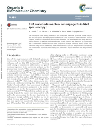
  • 1. Organic & Biomolecular Chemistry PAPER Cite this: DOI: 10.1039/c5ob00513b Received 14th March 2015, Accepted 18th May 2015 DOI: 10.1039/c5ob00513b www.rsc.org/obc RNA nucleosides as chiral sensing agents in NMR spectroscopy† N. Lokesh,a,b S. L. Sachin,a L. V. Narendra,a K. Aruna and N. Suryaprakash*a,b The study reports chiral sensing properties of RNA nucleosides. Adenosine, guanosine, uridine and cyti- dine are used as chiral derivatizing agents to differentiate chiral 1°-amines. A three component protocol has been adopted for complexation of nucleosides and amines. The chiral differentiating ability of nucleo- sides is examined for different amines based on the 1 H NMR chemical shift differences of diastereomers (ΔδR,S ). Enantiomeric differentiation has been observed at multiple chemically distinct proton sites. Adenosine and guanosine exhibit large chiral differentiation (ΔδR,S ) due to the presence of a purine ring. The diastereomeric excess (de) measured by using adenosine is in good agreement with the gravimetric values. Introduction Most of the drug interactions with biological systems are enantiospecific due to the inherent chiral nature of biological molecules, such as RNA, DNA and proteins.1,2 Therefore, chiral sensing or enantiomeric differentiation has gained enormous importance in the asymmetric synthesis and in pharmaceutical research.3–6 Most commonly used techniques to analyze enantiomers are NMR,7–10 X-ray,11,12 ORD, CD13 and chromatographic methods.14,15 The easy accessibility and available experimental techniques make NMR an important analytical tool for chiral analysis. NMR differentiates enantio- mers through chiral auxiliaries7–10,16–18 and through chiral alignment media,19–21 which induce diastereomeric inter- actions and a differential ordering effect (DOE) respectively. Number of chiral auxiliaries have been reported for efficient differentiation of enantiomers, assignment of the absolute configuration and also for the measurement of the enantio- meric excess (ee).7–10,16–18 Most of these auxiliaries are syn- thetically designed that requires more time, synthetic skills and is also uneconomical. On the other hand chiral bio- molecules are cheaply available, more abundant and can be utilized for many applications.22–32 Recently, several studies have been reported on bio-molecular chiral sensing, which used DNA, xanthan, folic acid and guanosine based weak chiral aligning media to differentiate enantiomers using NMR.19,20,33,34 There are also reports of DNA and polysacchar- ide based chiral stationary phases in chromatography to differentiate enantiomeric peptides and other chiral mole- cules,27,35,36 epigallocatechin gallate (EGCG) as a chiral solvat- ing agent to differentiate chiral amino acids24 and nucleotides for recognition of L-cysteine and D-cysteine by the colorimetric technique.32 In general chiral auxiliaries contain chiral units (viz. chiral center, chiral axis) to induce diastereomeric interactions and delocalize electrons (e.g. aromatic ring) to induce shielding and deshielding effects, causing enantiomeric differentiation. Nucleosides, on the other hand, possess both chiral units (ribose sugar) and delocalized electrons (nuclear bases), and promise to be efficient chiral sensing agents. The present study is aimed at exploring the chiral sensing ability of RNA nucleosides, such as adenosine, guanosine, uridine and cytidine, to differentiate chiral 1°-amines using 1 H NMR. A reported three component protocol was employed to complex nucleosides and chiral amines in DMSO-d6 using 2-formylphenylboronic acid.37–39 The general scheme for the preparation of a 3-component complex is given in Scheme 1. Each nucleoside was tested for its ability to differentiate chiral amines, and the 1 H chemical shift separation (ΔδR,S ) of dia- stereomers formed from R- and S- amines was used as an indi- cator. The unambiguous measurement of the diastereomeric excess (de) was demonstrated on a nucleoside and amine pair. The formed diastereomeric complexes of R and S amines exhibit discrimination at multiple proton sites; the peak assignment and the structure of complexes are reported using 2D NMR (COSY, NOESY) techniques, spiking experi- ments and utilizing the knowledge of the reported analogous structures.37–39 †Electronic supplementary information (ESI) available. See DOI: 10.1039/ c5ob00513b a NMR Research Centre, Indian Institute of Science, Bangalore 560012, India. E-mail: nsp@nrc.iisc.ernet.in; Fax: +91 80 23601550; Tel: +91 80 22933300, +91 98 45124802 b Solid State and Structural Chemistry Unit, Indian Institute of Science, Bangalore 560012, India This journal is © The Royal Society of Chemistry 2015 Org. Biomol. Chem. Publishedon18May2015.DownloadedbyNationalChiaoTungUniversityon10/06/201508:37:44. View Article Online View Journal
  • 2. Experimental section All the chemicals were purchased and used without any further purification. The nucleosides used are adenosine, guanosine, cytidine and uridine. The chiral amines used for testing enantiodiscrimination are α-methylbenzylamine, 4-methoxy-α-methylbenzylamine, 1-(2-naphthyl)ethanamine, 1-cyclohexylethylamine and sec-butylamine. Other chemicals used are 2-formylphenylboronic acid (purchased from com- mercial sources and used as received) and DMSO-d6. NMR spectra were recorded at 298 K on 500 MHz and 800 MHz spectrometers. DMSO-d5 was used as a 1 H reference in all the spectra. For diastereomeric excess (de) measurements a longer inter-scan delay (10 s) was used. For derivatization, 1.3 equivalent of each nucleoside, 1 equivalence of a racemic mixture of each amine and 1.3 equivalence of 2-formylphenylboronic acid were taken in 600 μl DMSO-d6 and stirred at 110 °C for about 10 minutes. The excess quantities of nucleosides and 2-formylphenylboro- nic acid were used to ensure maximum conversion of enantio- meric amines to diastereomeric complexes. The high boiling point of DMSO helped to carry out the reaction above 100 °C, which removed any existing or formed water in the reaction. This completed the reaction. The prepared solutions were used for NMR experiments without any further purification. Results and discussion Initially guanosine was used as a chiral derivatizing agent to differentiate α-methylbenzylamine. 10 mg of guanosine, 5.5 mg of 2-formylphenylboronic acid and 3.3 mg of a racemic mixture of α-methylbenzylamine (α-MBA) were taken in 600 μl of DMSO-d6 and were stirred for 10 minutes at about 110 °C. The possible complex formation is reported in Scheme 1, which was further substantiated by mass and NMR spectro- scopic analysis (the spectrum is given in the ESI†). The recorded 1 H NMR spectrum is given in Fig. 1, the peaks were assigned based on 2D NMR (COSY, NOESY, spectra are given in the ESI†) experiments and the spiking technique. The spectrum shows two doublets for methyl protons (peak ‘c’ in Fig. 1), suggesting the formation of two diastereomeric complexes from two enantiomeric (R & S) forms of α-methyl- benzylamine, which unambiguously confirms the enantio- meric differentiation. The chiral differentiation is observed at many proton sites (shown as expanded regions in Fig. 1). The peak identified as ‘a’ pertains to the imine proton and that identified with number ‘8’ corresponds to the nuclear base proton. The peaks marked 1′, 2′, 3′, 4′ 5′ and 6′ are from the protons of the sugar unit. Better discrimination (ΔδR,S in ppm) was observed for sugar ring protons and nuclear base protons. For quick observation of chiral discrimination and also for dia- stereomeric excess (de) measurement, one can use the methyl proton peaks (marked as ‘c’ around 1.5 ppm) or the imine proton peaks (marked as ‘a’ around 9 ppm). In the subsequent step the study was focused on verifying the chiral discrimination ability of other three nucleosides, adenosine, uridine and cytidine. Individual samples of three- component diastereomeric complexes of α-methylbenzylamine were prepared with each of the nucleoside (using the identical procedure discussed earlier). The recorded 1 H NMR spectra for all the samples are given in Fig. 2. The spectrum given in the Scheme 1 A three-component protocol for derivatization of 1°-chiral amines and RNA nucleosides using 2-formylphenylboronic acid * indicates the chiral center; R1 and R2 are different alkyl groups. Paper Organic & Biomolecular Chemistry Org. Biomol. Chem. This journal is © The Royal Society of Chemistry 2015 Publishedon18May2015.DownloadedbyNationalChiaoTungUniversityon10/06/201508:37:44. View Article Online
  • 3. Fig. 1 800 MHz 1 H NMR spectrum and chemical structure of the diastereomeric complex formed from guanosine, α-methylbenzylamine and 2-formylphenylboronic acid. The protons and peaks showing enantiomeric differentiation are labeled with alphabets and numbers. Fig. 2 From bottom to top: 800 MHz 1 H NMR spectra of α-methylbenzylamine (MBA), and diastereomeric complex spectra of guanosine: α-methyl- benzylamine : 2-formylphenylboronic acid (G), adenosine: α-methylbenzylamine : 2-formylphenylboronic acid (A), uridine: α-methylbenzylamine : 2-formylphenylboronic acid (U) and cytidine: α-methylbenzylamine : 2-formylphenylboronic acid (C). Organic & Biomolecular Chemistry Paper This journal is © The Royal Society of Chemistry 2015 Org. Biomol. Chem. Publishedon18May2015.DownloadedbyNationalChiaoTungUniversityon10/06/201508:37:44. View Article Online
  • 4. bottom trace pertains to pure α-methylbenzylamine in DMSO- d6. The other labeled spectra G, A, U and C were recorded from the solutions of diastereomeric complexes of α-methyl- benzylamine and the respective nucleosides (G-guanosine, A-adenosine, U-uridine and C-cytidine). The methyl and imine proton peaks were monitored for quick and easy identification of discrimination, which are reported in the expanded scale. The appearance of a pair of doublets for methyl protons and a doublet for imine protons (expanded region) in each spectrum confirms the enantiomeric discrimination ability of all the four nucleosides. The extent of chemical shift separation (ΔδR,S in ppm) between two doublets suggests the chiral dis- crimination ability. As expected, more separation was observed in the case of adenosine, then guanosine followed by uracil and cytosine. This is attributed to the presence of the purine ring (more delocalized electrons) in adenosine and guanosine. However an opposite pattern was observed for imine proton peaks. For establishing the wide generality of the enantiomeric dis- crimination properties of nucleosides, many diastereomeric Fig. 3 The plot of chemical shift separation of the selected protons (identified in red colour in the structures above) of diastereomeric complexes formed from R and S amines versus different diastereomeric complexes (left). Nucleosides are labeled as A, G, U and C for adenosine, guanosine, uridine and cytidine respectively. The chemical structures of amines, 4-methoxy-α-methylbenzylamine (1), 1-(2-naphthyl)ethanamine (2), α-methyl- benzylamine (3), sec-butylamine (4) and 1-cyclohexylethylamine (5) are also given. Fig. 4 800 MHz 1 H NMR spectrum pertaining to methyl peaks of adenosine: methoxy-α-methylbenzylamine : 2-formylphenylboronic acid diaster- eomeric complex with varied R and S concentrations of 4-methoxy α-methylbenzylamine, the gravimetric amine ratios S : R used are shown (left). A plot of the diastereomeric excess of R-4-methoxy-α-methylbenzylamine measured by the gravimetric method versus NMR experimental measure- ment (right). Paper Organic & Biomolecular Chemistry Org. Biomol. Chem. This journal is © The Royal Society of Chemistry 2015 Publishedon18May2015.DownloadedbyNationalChiaoTungUniversityon10/06/201508:37:44. View Article Online
  • 5. complexes were prepared from different types of 1°-amines (aliphatic-, cyclic- and aromatic-amines) with each of the selected nucleosides using the identical procedure discussed earlier. All the recorded 1 H NMR spectra are given in the ESI.† The enantiomeric discrimination and the extent of discrimi- nation of all the nucleosides with different amines are sum- marized in Fig. 3. The plot in Fig. 3 represents 1 H chemical shift separation (ΔδR,S ) of R and S diastereomeric complexes of the chosen peaks of amines with the corresponding nucleo- sides. The derivatized nucleosides are indicated as A, G, U and C for adenosine, guanosine, uridine and cytidine respectively. The amines used are 4-methoxy-α-methylbenzylamine, 1-(2- naphthyl)ethanamine, α-methylbenzylamine, sec-butylamine and 1-cyclohexylethylamine, which are numbered as 1, 2, 3, 4 and 5 respectively (chemical structures are given in Fig. 3). The chemical shift separation was measured for CH3 protons marked in red colour. Adenosine exhibited more enantiomeric discrimination for all the five amines investigated, which was followed by guanosine, uridine and cytidine. In cases of gua- nosine and adenosine, the methoxy-protons of 4-methoxy- α-methylbenzylamine showed better discrimination. In all nucleosides the methyl protons of 1-(2-naphthyl)ethanamine are better discriminated, which is due to the aromaticity of the naphthyl ring, which induces more shielding and deshielding effects. The observed R and S chemical shift separation (ΔδR,S ) in other three amines is almost the same. Finally the nucleoside was tested for the precise measure- ment of the diastereomeric excess (de). A series of adenosine and 4-methoxy-α-methylbenzylamine derivative complexes were prepared with varying ratios of the R and S mixture of amines (details are given in the ESI†). For each sample the 1D 1 H NMR spectra were recorded and the methyl peaks were used to calculate the diastereomeric excess. The intensity vari- ation of R and S methyl peaks with varying R and S amine con- centration is reported in Fig. 4. The figure also contains the plot of the diastereomeric excess calculated by gravimetric measurements and those derived from the 1 H NMR spectrum. The R2 = 0.9993 obtained suggests excellent agreement between the gravimetric measurement and those obtained by NMR experiment. This clearly establishes the applicability of nucleosides for ee measurements. Conclusions In summary, RNA nucleosides have been demonstrated as chiral derivatizing agents for efficient NMR spectroscopic dis- crimination of chiral 1°-amines. A detailed analysis of the differentiation ability of nucleosides has been carried out using 1 H NMR. The enantiomeric differentiation ability was observed for all RNA nucleosides. The better differentiation (Δδ in ppm for R & S) was observed for adenosine and guano- sine than that of uridine and cytidine in the methyl proton region due to the presence of the purine ring but an opposite pattern was observed for imine protons. Adenosine has been used to demonstrate the precise measurement of the diastereo- meric excess, which is in good agreement with the gravimetri- cally determined ratios. The present study demonstrated another important application of biomolecules helping in exploring the possibility of using different chiral biomolecules for chiral sensing application. Acknowledgements NL thanks IISc for a fellowship and Karthik for helpful discus- sions. NS gratefully acknowledges the generous financial support by the Board of Research in Nuclear Sciences, Mumbai (Grant no. 2013/37C/4/BRNS) and the Science and Engineering Research Board, Department of Science and Tech- nology, New Delhi (grant no. SR/S1/PC-42/2011). References 1 A. J. Hutt and S. C. Tan, Drugs, 1996, 52, 1–12. 2 L. A. Nguyen, H. He and C. Pham-Huy, Int. J. Biomed. Sci., 2006, 2, 85–100. 3 P. M. Dewick, Medicinal Natural Products, John Wiley & Sons, Ltd, 2009. 4 J. Gal, Chirality Drug Research, Wiley-VCH Verlag GmbH & Co. KGaA, 2006. 5 R. E. Gawley and J. Aubé, in Principles of Asymmetric Syn- thesis, ed. R. E. G. Aubé, Elsevier, Oxford, 2nd edn, 2012. 6 C. M. Reeves and B. M. Stoltz, in Asymmetric Synthesis II, Wiley-VCH Verlag GmbH & Co. KGaA, 2012. 7 J. M. Seco, E. Quiñoá and R. Riguera, Chem. Rev., 2004, 104, 17–118. 8 J. M. Seco, E. Quiñoá and R. Riguera, Chem. Rev., 2012, 112, 4603–4641. 9 T. J. Wenzel and C. D. Chisholm, Prog. Nucl. Magn. Reson. Spectrosc., 2011, 59, 1–63. 10 T. J. Wenzel and C. D. Chisholm, Chirality, 2011, 23, 190– 214. 11 N. Harada, Chirality, 2008, 20, 691–723. 12 J. Trotter, Acta Crystallogr., Sect. B: Struct. Sci., 1981, 37, 493–494. 13 N. Harada, K. Nakanishi and N. Berova, Comprehensive Chiroptical Spectroscopy, John Wiley & Sons, Inc., 2012. 14 T. J. Ward and D.-M. Hamburg, Anal. Chem., 2004, 76, 4635–4644. 15 T. J. Ward and K. D. Ward, Anal. Chem., 2010, 82, 4712– 4722. 16 J. Chin, D. C. Kim, H.-J. Kim, F. B. Panosyan and K. M. Kim, Org. Lett., 2004, 6, 2591–2593. 17 T. Ema, D. Tanida and T. Sakai, J. Am. Chem. Soc., 2007, 129, 10591–10596. 18 Q. Ma, M. Ma, H. Tian, X. Ye, H. Xiao, L.-h. Chen and X. Lei, Org. Lett., 2012, 14, 5813–5815. 19 P. Lesot, U. Venkateswara Reddy and N. Suryaprakash, Chem. Commun., 2011, 47, 11736–11738. Organic & Biomolecular Chemistry Paper This journal is © The Royal Society of Chemistry 2015 Org. Biomol. Chem. Publishedon18May2015.DownloadedbyNationalChiaoTungUniversityon10/06/201508:37:44. View Article Online
  • 6. 20 Lokesh and N. Suryaprakash, Chem. Commun., 2013, 49, 2049–2051. 21 M. Sarfati, P. Lesot, D. Merlet and J. Courtieu, Chem. Commun., 2000, 2069–2081. 22 Z. Guo, I. De Cat, B. Van Averbeke, J. Lin, G. Wang, H. Xu, R. Lazzaroni, D. Beljonne, E. W. Meijer, A. P. H. J. Schenning and S. De Feyter, J. Am. Chem. Soc., 2011, 133, 17764–17771. 23 V. B. Kandimalla, V. S. Tripathi and H. Ju, Crit. Rev. Anal. Chem., 2006, 36, 73–106. 24 D. Kumari, P. Bandyopadhyay and N. Suryaprakash, J. Org. Chem., 2013, 78, 2373–2378. 25 L. Liu and X. Zhong, Chem. Commun., 2012, 48, 5718–5720. 26 N. Metzler-Nolte, Angew. Chem., Int. Ed., 2001, 40, 1040– 1043. 27 M. Michaud, E. Jourdan, C. Ravelet, A. Villet, A. Ravel, C. Grosset and E. Peyrin, Anal. Chem., 2004, 76, 1015–1020. 28 S. V. Patwardhan, G. Patwardhan and C. C. Perry, J. Mater. Chem., 2007, 17, 2875–2884. 29 J. L. Rouge, B. E. Eaton and D. L. Feldheim, Energy Environ. Sci., 2011, 4, 398–402. 30 G. Shemer, O. Krichevski, G. Markovich, T. Molotsky, I. Lubitz and A. B. Kotlyar, J. Am. Chem. Soc., 2006, 128, 11006–11007. 31 J. Zhang, M. T. Albelda, Y. Liu and J. W. Canary, Chirality, 2005, 17, 404–420. 32 M. Zhang and B.-C. Ye, Anal. Chem., 2011, 83, 1504– 1509. 33 Lokesh and N. Suryaprakash, Chem. – Eur. J., 2012, 18, 11560–11563. 34 U. V. Reddy and N. Suryaprakash, Chem. Commun., 2011, 47, 8364–8366. 35 B. Chankvetadze, in Chiral Separations, ed. G. K. E. Scriba, Humana Press, 2013. 36 S. Pedotti and A. Patti, J. Sep. Sci., 2014, 37, 3451–3460. 37 A. M. Kelly, Y. Pérez-Fuertes, S. Arimori, S. D. Bull and T. D. James, Org. Lett., 2006, 8, 1971–1974. 38 A. M. Kelly, Y. Perez-Fuertes, J. S. Fossey, S. L. Yeste, S. D. Bull and T. D. James, Nat. Protocols, 2008, 3, 215– 219. 39 Y. Pérez-Fuertes, A. M. Kelly, A. L. Johnson, S. Arimori, S. D. Bull and T. D. James, Org. Lett., 2006, 8, 609–612. Paper Organic & Biomolecular Chemistry Org. Biomol. Chem. This journal is © The Royal Society of Chemistry 2015 Publishedon18May2015.DownloadedbyNationalChiaoTungUniversityon10/06/201508:37:44. View Article Online