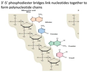
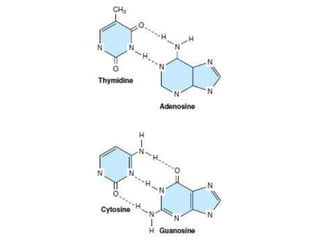
Nucleotides & Nucleic Acids
Nucleotides—the monomer units or building blocks
of nucleic acids—serve multiple additional functions.
They form a part of many coenzymes and serve as
donors of phosphoryl groups (eg, ATP or GTP),
of sugars (eg,UDP- or GDP-sugars),
or of lipid (eg, CDP-acylglycerol).
Regulatory nucleotides include
the second messengers cAMP and cGMP,
allosteric regulation of enzyme activity by ATP, AMP,
and CTP.
• Synthetic purine and pyrimidine analogs that
contain halogens, thiols, or additional nitrogen
are employed for chemotherapy of cancer and
AIDS and
• as suppressors of the immune response during
organ transplantation.
• There are 2 types of Nucleic acids :
• Deoxyribonucleic Acid ( DNA ) &
• Ribonucleic acid ( RNA)
• Components of Nucleic acids :
• Nucleic acids are polymers of nucleotides held
by 3’ & 5’ phosphate bridges.
• Nucleotides are composed of
• nitrogenous base + pentose sugar + phosphate
• Nucleoside refers to base + sugar
• So, Nucleotide = Nucleoside + phosphate
• Nitrogenous bases found in nucleotides are
aromatic heterocyclic compounds.
• Bases are of 2 types :
• Purines & Pyrimidines
• Thymine & Uracil differ in structure by the presence
(in T) or absence of (in U) a methyl (--CH3 ) group.
• Sugars of Nucleic acids :
• RNA contains D- ribose &
• DNA contains D- deoxyribose
• Nucleic Acids Are Polynucleotides
• Nucleic acids are linear polymers of nucleotides linked 3’ to
5’ by phosphodiester bridges.
They are formed as 5’-nucleoside monophosphates
are successively added to the 3’-OH group of the preceding
nucleotide, a process that gives the polymer a directional
sense.
By definition, the 5’ end lacks a nucleotide at the 5’ position
& the 3’ end lacks a nucleotide at the 3’ position.
Other groups (most often one or more phosphates) may be
present on one or both ends.
• Polymers of ribonucleotides are named ribonucleic acid,
or RNA.
• Deoxyribonucleotide polymers are called
• deoxyribonucleic acid, or DNA.
3’-5’ phosphodiester bridges link nucleotides together to
form polynucleotide chains
• Chargaff’s rule of DNA composition :
• He observed (late 1940s) that in all the species,
• DNA had equal numbers of adenine & thymine
residues(A=T) &
• equal numbers of guanine & cytosine(G=C)
residues.
• This is known as Chargaff’s rule of molar
equivalence between the purines & pyrimidines in
DNA structure.
DNA DOUBLE HELIX
• Double helical structure of DNA was proposed by
James Watson & Francis Crick in 1953( Nobel Prize,
1962)
• The main features of Watson – Crick model of DNA
(Now known as B- DNA ) are :
• 1. DNA is right handed double helix .It consists of 2
polydeoxynucleotide chains(strands) twisted
around each other on a common axis.
• 2. Two strands are antiparallel
• i.e. 5’---------------3’ & 3’------------------5’
• 3.Width or diameter of a double helix is 20 A0 (2nm)
• 4. Each turn (pitch) of the helix is 34 A0 (3.4nm)
with 10 pairs of nucleotides, each pair placed at a
distance of about 3.4A0
• 5. Each strand of DNA has a hydrophilic deoxyribose
phosphate backbone(3’—5’ phosphodiester bonds)
on the outside(periphery) of the molecule while the
hydrophobic bases are stacked inside(core).
• 6. Two polynucleotide chains are not identical but
complementary to each other due to base pairing.
• 7. Two strands are held together by H- bonds
formed by complementary base pairs.
• A-T pair has 2 Hydrogen bonds & G-C pair has 3
hydrogen bonds.
• G = C is stronger by about 50% than A = T.
• 8. H Bonds are formed between a purine & a
pyrimidine only. A-T, T-A, G-C & C-G.
• 9. Complementary base pairing in DNA helix proves
Chargaff’s rule.
• 10. Genetic information resides on one of the 2
strands, known as template strand or sense strand.
• Opposite strand is antisense strand.
Conformations of DNA Double helix
Double helical structure of DNA exists in at least
6 different forms A- E & Z.
Among these, B, A & Z forms are important.
B- form of DNA double helix described by
Watson & Crick , is the most predominant form
under physiological conditions.
Other Types of DNA structure
It is now recognized that besides double helical
structure , DNA also exists in certain unusual
structures.
• Bent DNA
• Triple stranded DNA
• Four stranded DNA
Polynucleotides with very high contents of
guanine can form G- quartets(parallel)
G- tetraplexes(Antiparallel)
Structure of RNA
• RNA is a polymer of ribonucleotides held together
by 3’,5’-phosphodiester bridges.
• Although RNA has certain similarities with DNA
structure, they have specific differences.
• 1. Pentoses : Suger in RNA is ribose in contrast to
deoxyribose in DNA.
• 2. Pyrimidine : RNA contains the pyrimidine uracil
in place of thymine (in DNA).
• 3. Single strand : RNA is usually a single stranded
polynucleotide. However this strand may fold at
certain places to give a double stranded structure ,
if complementary base pairs are in close proximity.
4. Chargaff’s rule ---- not obeyed : Due to the single
stranded nature , there is no specific relation between
purine & pyrimidine contents.
Thus Guanine content is not equal to cytosine(as in
the case in DNA)
5. Susceptibility to alkali hydrolysis : Alkali can
hydrolyse RNA to 2’,3’-cyclic diesters. This is possible
due to the presence of a hydroxyl group at 2’ position.
DNA can not be subjected to alkali hydrolysis due to
lack of this group.
6. Orcinol color reaction : RNAs can be histologically
identified by orcinol color reaction due to the
presence of ribose.
• Types of RNA :
• 3 major types of RNAs with their respective cellular
composition ;--
• 1. Messenger RNA( m RNA) : 5 – 10 %
• 2. Transfer RNA ( t RNA ) : 10 – 20 %
• 3. Ribosomal RNA (r RNA) : 50 – 80 %
• Besides the above 3 , other RNAs are also present in
the cells.
• RNAs are synthesized from DNA, and are primarily
involved in the process of protein biosynthesis.
Cellular RNAs & their functions
• Messenger RNA ( mRNA) :
• Transfers genetic information from genes to
ribosomes to synthesize proteins.
• Heterogeneous nuclear RNA ( hnRNA) :
• Serves as precursor for mRNA & other RNAs.
• Transfer RNA( tRNA) :
• Transfers amino aid to mRNA for protein
biosynthesis.
• Ribosomal RNA(rRNA) :
• Provides structural framework for ribosomes.
• Small nuclear RNA( snRNA) :
• Involved in mRNA processing.
• Small nucleolar RNA (snoRNA) :
• Playes a key role in the processing of rRNA
molecules.
• Small cytoplasmic RNA(scRNA) :
• Involved in the selection of proteins for export.
• Transfer- messenger RNA(tmRNA) :
• Mostly present in bacteria. Adds short peptide tags
to proteins to facilitate the degradation of
incorrectly synthesized proteins.
Messenger RNA(mRNA)
• m RNA is synthesized in the nucleus (in eukaryotes)
as heterogeneous nuclear RNA( hnRNA).
• hn RNA, on processing ,liberates the functional
m RNA which enters the cytoplasm to participate in
protein synthesis .
• m RNA has high mol wt. with a short half –life.
• Eukaryotic mRNA is capped at the 5’ terminal end
by 7-methylguanosine triphosphate.
• This cap helps to prevent the hydrolysis of mRNA by
5’-exonucleases.
• Cap may also be involved in the recognition of
mRNA for protein biosynthesis.
• 3’-terminal end of m-RNA contains a polymer of
adenylate residues( 20 – 250 nucleotides) which is
known as poly(A) tail.
• This tail may provide stability to mRNA , besides
preventing it from the attack of 3’-exonucleases.
• mRNA molecules often contain certain modified
bases such as 6-methyladenylates in the internal
structure.
Above picture, The cap structure attached to
the 5’ terminal of most eukaryotic messenger
RNA molecules.
A 7-methylguanosine triphosphate (black) is
attached at the 5’ terminal of the mRNA (shown
in blue), which usually contains a
2’-O-methylpurine nucleotide.
These modifications (the cap and methyl group)
are added after the mRNA is transcribed from
DNA.
Transfer RNA (tRNA)
• tRNA molecules vary in length from 74 to 95
nucleotides.
• They also are generated by nuclear processing
of a precursor molecule .
• The tRNA molecules serve as adapters for the
translation of the information in the sequence of
nucleotides of the mRNA into specific amino
acids.
• There are at least 20 species of tRNA
molecules in every cell, at least one (and often
several) corresponding to each of the 20 amino
acids required for protein synthesis.
• The structure of tRNA, resembles that of a
clover leaf.
• tRNA contains 4 arms,each with a base paired
stem.
Structure of tRNA
• 1. The acceptor arm : This arm is capped with a
sequence CCA(5’ to 3’). The amino acid is attached
to the acceptor arm.
• 2. The anticodon arm : This arm with the 3 specific
nucleotide bases (anticodon), is responsible for the
recognition of triplet codon of mRNA.
• Codon and anticodon are complementary to each
other.
• 3. The D arm: It is so named due to the presence of
dihydrouridine.
• 4. The TψC arm : This arm contains a sequence of T,
pseudouridine(represented by ψ) and C.
• 5. The variable arm : This arm is the most variable
in tRNA.
• Based on this variability,
tRNAs are classified into 2 categories:
a) Class I tRNAs :
The most predominant(about 75%) form with 3-5
base pairs length.
b) Class II tRNAs :
They contain 13- 20 base pair long arm.
• Base pairs in tRNA :
• Structure of tRNA is maintained due to the
complementary base pairing in the arms.
• The 4 arms with their repective base pairs are
given below :
• The acceptor arm - 7 bp
• The TψC arm - 5 bp
• The anticodon arm - 5 bp
• The D arm - 4 bp
Ribosomal RNA ( rRNA)
• Ribosomes are the factories of protein synthesis.
• Eukaryotic ribosomes are composed of 2 major
nucleoprotein complexes----
60S subunit & 40s subunit.
• 60S subunit contains 28S rRNA, 5S rRNA &
5.8S rRNA.
• 40S subunit contains 18S rRNA.
• Most probably rRNAs play a significant role in the
binding of mRNA to ribosomes & protein synthesis.

Nucleotides & nucleic acids

  • 1.
    Nucleotides & NucleicAcids Nucleotides—the monomer units or building blocks of nucleic acids—serve multiple additional functions. They form a part of many coenzymes and serve as donors of phosphoryl groups (eg, ATP or GTP), of sugars (eg,UDP- or GDP-sugars), or of lipid (eg, CDP-acylglycerol). Regulatory nucleotides include the second messengers cAMP and cGMP, allosteric regulation of enzyme activity by ATP, AMP, and CTP.
  • 2.
    • Synthetic purineand pyrimidine analogs that contain halogens, thiols, or additional nitrogen are employed for chemotherapy of cancer and AIDS and • as suppressors of the immune response during organ transplantation. • There are 2 types of Nucleic acids : • Deoxyribonucleic Acid ( DNA ) & • Ribonucleic acid ( RNA) • Components of Nucleic acids : • Nucleic acids are polymers of nucleotides held by 3’ & 5’ phosphate bridges.
  • 3.
    • Nucleotides arecomposed of • nitrogenous base + pentose sugar + phosphate • Nucleoside refers to base + sugar • So, Nucleotide = Nucleoside + phosphate • Nitrogenous bases found in nucleotides are aromatic heterocyclic compounds. • Bases are of 2 types : • Purines & Pyrimidines
  • 5.
    • Thymine &Uracil differ in structure by the presence (in T) or absence of (in U) a methyl (--CH3 ) group. • Sugars of Nucleic acids : • RNA contains D- ribose & • DNA contains D- deoxyribose
  • 6.
    • Nucleic AcidsAre Polynucleotides • Nucleic acids are linear polymers of nucleotides linked 3’ to 5’ by phosphodiester bridges. They are formed as 5’-nucleoside monophosphates are successively added to the 3’-OH group of the preceding nucleotide, a process that gives the polymer a directional sense. By definition, the 5’ end lacks a nucleotide at the 5’ position & the 3’ end lacks a nucleotide at the 3’ position. Other groups (most often one or more phosphates) may be present on one or both ends. • Polymers of ribonucleotides are named ribonucleic acid, or RNA. • Deoxyribonucleotide polymers are called • deoxyribonucleic acid, or DNA.
  • 8.
    3’-5’ phosphodiester bridgeslink nucleotides together to form polynucleotide chains
  • 11.
    • Chargaff’s ruleof DNA composition : • He observed (late 1940s) that in all the species, • DNA had equal numbers of adenine & thymine residues(A=T) & • equal numbers of guanine & cytosine(G=C) residues. • This is known as Chargaff’s rule of molar equivalence between the purines & pyrimidines in DNA structure.
  • 12.
    DNA DOUBLE HELIX •Double helical structure of DNA was proposed by James Watson & Francis Crick in 1953( Nobel Prize, 1962) • The main features of Watson – Crick model of DNA (Now known as B- DNA ) are : • 1. DNA is right handed double helix .It consists of 2 polydeoxynucleotide chains(strands) twisted around each other on a common axis. • 2. Two strands are antiparallel • i.e. 5’---------------3’ & 3’------------------5’
  • 16.
    • 3.Width ordiameter of a double helix is 20 A0 (2nm) • 4. Each turn (pitch) of the helix is 34 A0 (3.4nm) with 10 pairs of nucleotides, each pair placed at a distance of about 3.4A0 • 5. Each strand of DNA has a hydrophilic deoxyribose phosphate backbone(3’—5’ phosphodiester bonds) on the outside(periphery) of the molecule while the hydrophobic bases are stacked inside(core). • 6. Two polynucleotide chains are not identical but complementary to each other due to base pairing.
  • 17.
    • 7. Twostrands are held together by H- bonds formed by complementary base pairs. • A-T pair has 2 Hydrogen bonds & G-C pair has 3 hydrogen bonds. • G = C is stronger by about 50% than A = T. • 8. H Bonds are formed between a purine & a pyrimidine only. A-T, T-A, G-C & C-G. • 9. Complementary base pairing in DNA helix proves Chargaff’s rule. • 10. Genetic information resides on one of the 2 strands, known as template strand or sense strand. • Opposite strand is antisense strand.
  • 18.
    Conformations of DNADouble helix Double helical structure of DNA exists in at least 6 different forms A- E & Z. Among these, B, A & Z forms are important. B- form of DNA double helix described by Watson & Crick , is the most predominant form under physiological conditions.
  • 19.
    Other Types ofDNA structure It is now recognized that besides double helical structure , DNA also exists in certain unusual structures. • Bent DNA • Triple stranded DNA • Four stranded DNA Polynucleotides with very high contents of guanine can form G- quartets(parallel) G- tetraplexes(Antiparallel)
  • 23.
    Structure of RNA •RNA is a polymer of ribonucleotides held together by 3’,5’-phosphodiester bridges. • Although RNA has certain similarities with DNA structure, they have specific differences. • 1. Pentoses : Suger in RNA is ribose in contrast to deoxyribose in DNA. • 2. Pyrimidine : RNA contains the pyrimidine uracil in place of thymine (in DNA). • 3. Single strand : RNA is usually a single stranded polynucleotide. However this strand may fold at certain places to give a double stranded structure , if complementary base pairs are in close proximity.
  • 24.
    4. Chargaff’s rule---- not obeyed : Due to the single stranded nature , there is no specific relation between purine & pyrimidine contents. Thus Guanine content is not equal to cytosine(as in the case in DNA) 5. Susceptibility to alkali hydrolysis : Alkali can hydrolyse RNA to 2’,3’-cyclic diesters. This is possible due to the presence of a hydroxyl group at 2’ position. DNA can not be subjected to alkali hydrolysis due to lack of this group. 6. Orcinol color reaction : RNAs can be histologically identified by orcinol color reaction due to the presence of ribose.
  • 25.
    • Types ofRNA : • 3 major types of RNAs with their respective cellular composition ;-- • 1. Messenger RNA( m RNA) : 5 – 10 % • 2. Transfer RNA ( t RNA ) : 10 – 20 % • 3. Ribosomal RNA (r RNA) : 50 – 80 % • Besides the above 3 , other RNAs are also present in the cells. • RNAs are synthesized from DNA, and are primarily involved in the process of protein biosynthesis.
  • 26.
    Cellular RNAs &their functions • Messenger RNA ( mRNA) : • Transfers genetic information from genes to ribosomes to synthesize proteins. • Heterogeneous nuclear RNA ( hnRNA) : • Serves as precursor for mRNA & other RNAs. • Transfer RNA( tRNA) : • Transfers amino aid to mRNA for protein biosynthesis. • Ribosomal RNA(rRNA) : • Provides structural framework for ribosomes.
  • 27.
    • Small nuclearRNA( snRNA) : • Involved in mRNA processing. • Small nucleolar RNA (snoRNA) : • Playes a key role in the processing of rRNA molecules. • Small cytoplasmic RNA(scRNA) : • Involved in the selection of proteins for export. • Transfer- messenger RNA(tmRNA) : • Mostly present in bacteria. Adds short peptide tags to proteins to facilitate the degradation of incorrectly synthesized proteins.
  • 28.
  • 30.
    • m RNAis synthesized in the nucleus (in eukaryotes) as heterogeneous nuclear RNA( hnRNA). • hn RNA, on processing ,liberates the functional m RNA which enters the cytoplasm to participate in protein synthesis . • m RNA has high mol wt. with a short half –life. • Eukaryotic mRNA is capped at the 5’ terminal end by 7-methylguanosine triphosphate. • This cap helps to prevent the hydrolysis of mRNA by 5’-exonucleases. • Cap may also be involved in the recognition of mRNA for protein biosynthesis.
  • 31.
    • 3’-terminal endof m-RNA contains a polymer of adenylate residues( 20 – 250 nucleotides) which is known as poly(A) tail. • This tail may provide stability to mRNA , besides preventing it from the attack of 3’-exonucleases. • mRNA molecules often contain certain modified bases such as 6-methyladenylates in the internal structure.
  • 33.
    Above picture, Thecap structure attached to the 5’ terminal of most eukaryotic messenger RNA molecules. A 7-methylguanosine triphosphate (black) is attached at the 5’ terminal of the mRNA (shown in blue), which usually contains a 2’-O-methylpurine nucleotide. These modifications (the cap and methyl group) are added after the mRNA is transcribed from DNA.
  • 34.
    Transfer RNA (tRNA) •tRNA molecules vary in length from 74 to 95 nucleotides. • They also are generated by nuclear processing of a precursor molecule . • The tRNA molecules serve as adapters for the translation of the information in the sequence of nucleotides of the mRNA into specific amino acids.
  • 35.
    • There areat least 20 species of tRNA molecules in every cell, at least one (and often several) corresponding to each of the 20 amino acids required for protein synthesis. • The structure of tRNA, resembles that of a clover leaf. • tRNA contains 4 arms,each with a base paired stem.
  • 36.
  • 39.
    • 1. Theacceptor arm : This arm is capped with a sequence CCA(5’ to 3’). The amino acid is attached to the acceptor arm. • 2. The anticodon arm : This arm with the 3 specific nucleotide bases (anticodon), is responsible for the recognition of triplet codon of mRNA. • Codon and anticodon are complementary to each other. • 3. The D arm: It is so named due to the presence of dihydrouridine. • 4. The TψC arm : This arm contains a sequence of T, pseudouridine(represented by ψ) and C.
  • 40.
    • 5. Thevariable arm : This arm is the most variable in tRNA. • Based on this variability, tRNAs are classified into 2 categories: a) Class I tRNAs : The most predominant(about 75%) form with 3-5 base pairs length. b) Class II tRNAs : They contain 13- 20 base pair long arm.
  • 41.
    • Base pairsin tRNA : • Structure of tRNA is maintained due to the complementary base pairing in the arms. • The 4 arms with their repective base pairs are given below : • The acceptor arm - 7 bp • The TψC arm - 5 bp • The anticodon arm - 5 bp • The D arm - 4 bp
  • 42.
    Ribosomal RNA (rRNA) • Ribosomes are the factories of protein synthesis. • Eukaryotic ribosomes are composed of 2 major nucleoprotein complexes---- 60S subunit & 40s subunit. • 60S subunit contains 28S rRNA, 5S rRNA & 5.8S rRNA. • 40S subunit contains 18S rRNA. • Most probably rRNAs play a significant role in the binding of mRNA to ribosomes & protein synthesis.