This document provides information about different views of the skull:
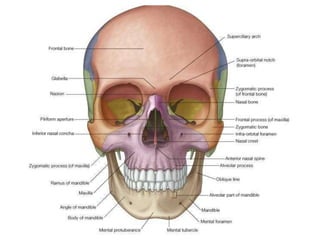
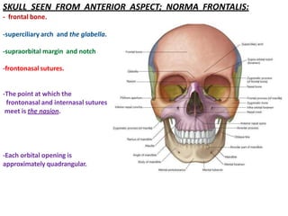
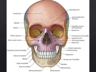
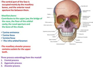
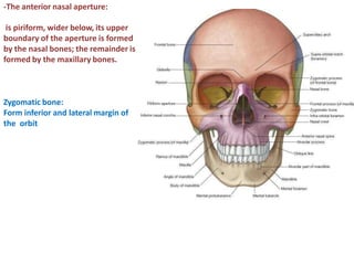
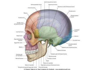
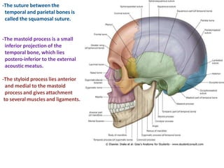
- The norma frontalis view shows features of the frontal bone and maxillary bones including the frontal process, zygomatic process, and alveolar process.
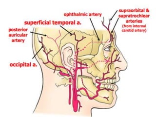
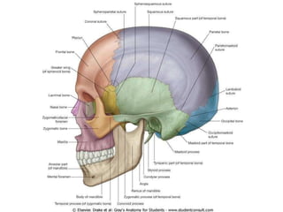
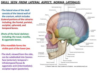
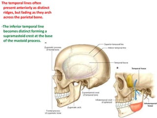
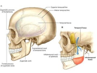
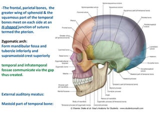
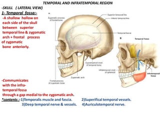
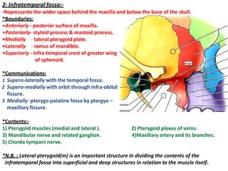
- The norma lateralis view outlines the temporal fossa and infratemporal fossa, separated by the zygomatic arch, and their contents like muscles and nerves.
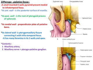
- The pterygo-palatine fossa is also described as a small space medial to the infratemporal fossa containing blood vessels and nerves.