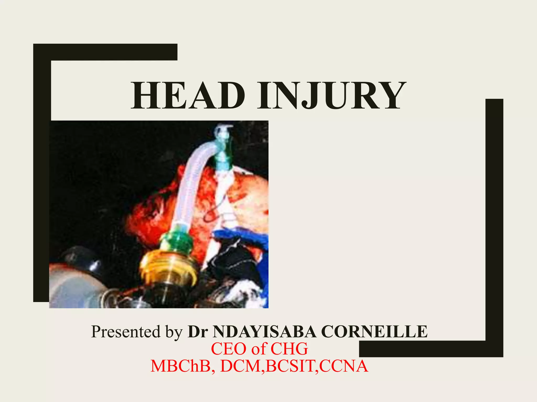
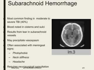
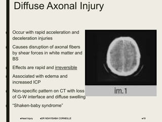
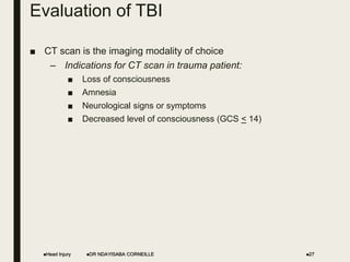
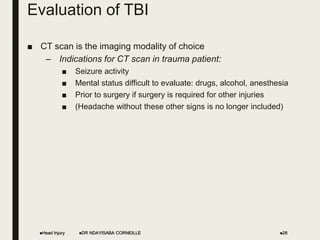
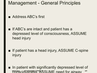
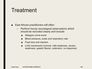
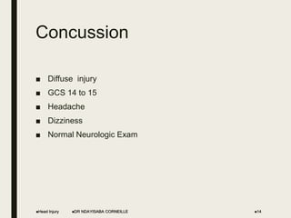
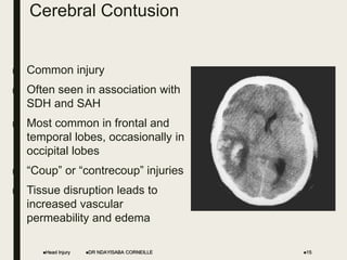
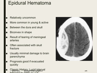
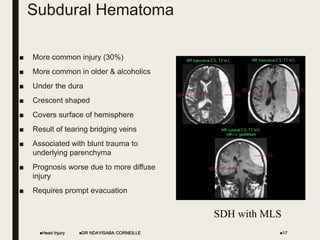
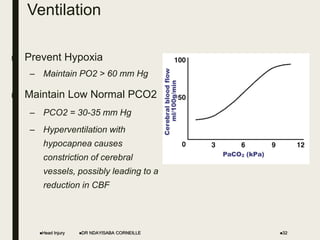
HEAD INJURY
Presented by Dr NDAYISABA CORNEILLE
CEO of CHG
MBChB, DCM,BCSIT,CCNA
Introduction
■ Definition: Head injury is Alteration in mental or physical
functioning related to trauma
■ Central nervous system injuries remain the leading cause of
morbidity and mortality for young people throughout the
world.
■ The risk of incurring a traumatic brain injury (TBI) is
especially high among adolescents, young adults, and elderly
people.
– For example, the management of depressed skull fractures in
children at Muhimbili Medical Centre, Dar es Salaam, Tanzania is
described by Mlay and Sayi.
– The fractures were located in the frontal or parietal bone in 27
(76.3%) patients.
– Casualty officers should note the indications for surgery were
cosmetics in 15 patients, compound fracture in 3 patients, focal
neurological deficit in 2 patients, and torn dura presenting with
Pseudo-meningocele in 2 patients.
Head Injury DR NDAYISABA CORNEILLE 2
Anatomy:
Axial T1-weighted MRI
Head Injury DR NDAYISABA CORNEILLE 3
Parietal
Occipital
Temporal
Frontal
Anatomy: Brain
■ Brain: Cerebrum
■ Frontal Lobe
– Emotions
– Motor Function
– Expression of
speech
■ Temporal Lobe
– Comprehension of
speech
– Memory
■ Parietal Lobe
– Sensory function
– Spacial orientation
■ Occipital Lobe
– Vision
Head Injury 4
DR NDAYISABA CORNEILLE
Anatomy: Brain
■ Brain:
– Cerebellum:
■ Coordination and balance
– Brainstem (Midbrain,
Pons, Medulla)
■ Alertness (RAS)
■ Cardiorespiratory centers
Head Injury DR NDAYISABA CORNEILLE 5
Anatomy: Meninges
■ The meninges of the brain and potential sites for
hemorrhage
Head Injury DR NDAYISABA CORNEILLE 6
Brain
Calvarium
Dura Mater
Arachnoid
Pia Mater
Potential Space
CSF
Potential Space
Epidural Hematoma (meningeal arteries)
Subdural Hematoma (bridging veins)
Subarachnoid Hemorrhage into CSF
Physiology
■ Cerebral Blood Flow (CBF):
– Normal CBF = 50 ml/100 gm of tissue
– Loss of cell function occurs at < 20-25
– Cell death of irreversible damage occurs at < 5
■ Autoregulation:
– the brain can maintain a constant CBF with a CCP
(Cerebral Perfusion Pressure) of 50-150 mm Hg
through vasoconstriction and vasodilatation
Head Injury DR NDAYISABA CORNEILLE 7
Physiology
■ Cerebral Perfusion Pressure (CPP): CPP = MAP – ICP
■ Intracranial Pressure (ICP):
■ Normal 5-15 mm Hg
■ Abnormal > 20
■ Severe > 40
■ CCP must be maintained > 70 mm Hg
■ The autoregulatory function is often lost in TBI patients
■ MAP and ICP must be carefully monitored in TBI patients
■ Low CPP must be treated aggressively to maintain CBF
– Increase MAP
– Decrease ICP
Head Injury DR NDAYISABA CORNEILLE 8
Classification of TBI - Mechanism
■ Mechanism:
– Blunt
■ High velocity (MVC)
■ Low velocity (fall, assault)
– Penetrating
■ Gunshot wounds
■ Other penetrating injuries
Head Injury DR NDAYISABA CORNEILLE 9
Classification of TBI - Morphology
■ Scalp Lacerations
– Can result in major blood loss
– Treatment
■ Direct pressure
■ Ligation of the bleeding vessels
■ Injection of lidocaine with epinephrine into the bleeding
areas
■ Multi-layer closure of the wound (always close torn
galea)
Head Injury DR NDAYISABA CORNEILLE 10
Skull Fractures
■ Cranial vault fractures
■ Linear or stellate
■ Open or closed
■ Depressed or non-depressed
– Fragments depressed greater than skull
thickness require elevation
■ Basilar skull fractures (BSF)
■ Periorbital ecchymosis (raccoon eyes)
■ Retroauricular ecchymosis (Battle’s sign)
■ CSF leaks (rhinorrhea, otorrhea)
■ Hemotympanum (blood behind the eardrum)
Head Injury DR NDAYISABA CORNEILLE 11
Basilar Skull Fractures (BSF)
■ DO NOT place NGT’s in
patients with suspected or
known BSF!!!
■ This patient clearly has
physical signs of a BSF, yet
inappropriately had an NGT
placed!!!!!
Head Injury 12
DR NDAYISABA CORNEILLE
Basilar Skull Fractures (BSF)
Head Injury DR NDAYISABA CORNEILLE 13
What is the PROBLEM in this picture?
Concussion
■ Diffuse injury
■ GCS 14 to 15
■ Headache
■ Dizziness
■ Normal Neurologic Exam
Head Injury DR NDAYISABA CORNEILLE 14
Cerebral Contusion
■ Common injury
■ Often seen in association with
SDH and SAH
■ Most common in frontal and
temporal lobes, occasionally in
occipital lobes
■ “Coup” or “contrecoup” injuries
■ Tissue disruption leads to
increased vascular
permeability and edema
Head Injury 15
DR NDAYISABA CORNEILLE
Epidural Hematoma
■ Relatively uncommon
■ More common in young & active
■ Between the dura and skull
■ Biconvex in shape
■ Result of tearing of meningeal
arteries
■ Often associated with skull
fracture
■ Usually minimal damage to brain
parenchyma
■ Prognosis good if evacuated
quickly
■ Classic History: Lucid interval
Head Injury 16
DR NDAYISABA CORNEILLE
Subdural Hematoma
■ More common injury (30%)
■ More common in older & alcoholics
■ Under the dura
■ Crescent shaped
■ Covers surface of hemisphere
■ Result of tearing bridging veins
■ Associated with blunt trauma to
underlying parenchyma
■ Prognosis worse due to more diffuse
injury
■ Requires prompt evacuation
Head Injury 17
DR NDAYISABA CORNEILLE
SDH with MLS
Subarachnoid Hemorrhage
■ Most common finding in moderate to
severe TBI (40%)
■ Blood noted in cisterns and sulci
■ Results from tear in subarachnoid
vessels
■ May precipitate vasospasm
■ Often associated with meningeal
signs:
– Photophobia
– Neck stiffness
– Headache
■ Requires neurosurgical consultation
Head Injury 18
DR NDAYISABA CORNEILLE
Diffuse Axonal Injury
■ Occur with rapid acceleration and
deceleration injuries
■ Causes disruption of axonal fibers
by shear forces in white matter and
BS
■ Effects are rapid and irreversible
■ Associated with edema and
increased ICP
■ Non-specific pattern on CT with loss
of G-W interface and diffuse swelling
■ “Shaken-baby syndrome”
Head Injury 19
DR NDAYISABA CORNEILLE
Glasgow Coma Scale (GCS)
■ Main Components
– Eyes
– Verbal
– Motor
■ Scoring
– Mild Injury 14-15
– Moderate Injury 9-13
– Severe Injury 3-8
Head Injury DR NDAYISABA CORNEILLE 20
Glasgow Coma Scale (GCS)
■ Eyes
– 1 – Closed
– 2 - Opens to pain
– 3 - Opens to voice
– 4 - Open
Head Injury DR NDAYISABA CORNEILLE 21
Glasgow Coma Scale (GCS)
■ Verbal
– 1 – Silent
– 2 – Moans
– 3 - Inappropriate words
– 4 - Disoriented or confused
– 5 - Oriented and appropriate
Head Injury DR NDAYISABA CORNEILLE 22
Glasgow Coma Scale (GCS)
■ Motor
– 1 - No response
– 2 - Extension to pain
– 3 - Flexion to pain
– 4 - Withdraws from pain
– 5 - Localizing pain
– 6 - Follows commands
Head Injury DR NDAYISABA CORNEILLE 23
Evaluation
■ Critical Components of History:
■ Age
■ Sex
■ Mechanism of injury
■ Time of injury
■ Loss of consciousness
■ Level of alertness
■ Amnesia
■ Headache
■ Seizures
Head Injury DR NDAYISABA CORNEILLE 24
Evaluation
■ Critical components of physical exam:
■ GCS (Eye, Verbal, Motor)
■ Pupillary light reaction (brainstem)
Head Injury DR NDAYISABA CORNEILLE 25
Pupil Evaluation
Pupil Size Light Response Interpretation
Unilateral dilation Sluggish or fixed 3rd nerve compression due
to tentorial herniation
Bilateral dilation Sluggish of fixed Inadequate brain perfusion
Bilateral 3rd nerve palsy
Unilateral dilation or
equal
Swinging flashlight +
(Marcus-Gunn)
Optic nerve injury
Bilateral constriction None or minimal Drugs (opiates)
Metabolic encephalopathy
Pontine hemorrhage
Unilateral
constriction
Intact Injured sympathetic path
(ie: carotid sheath injury)
Head Injury 26
DR NDAYISABA CORNEILLE
Evaluation of TBI
■ CT scan is the imaging modality of choice
– Indications for CT scan in trauma patient:
■ Loss of consciousness
■ Amnesia
■ Neurological signs or symptoms
■ Decreased level of consciousness (GCS < 14)
Head Injury DR NDAYISABA CORNEILLE 27
Evaluation of TBI
■ CT scan is the imaging modality of choice
– Indications for CT scan in trauma patient:
■ Seizure activity
■ Mental status difficult to evaluate: drugs, alcohol, anesthesia
■ Prior to surgery if surgery is required for other injuries
■ (Headache without these other signs is no longer included)
Head Injury DR NDAYISABA CORNEILLE 28
Management - General Principles
■ Address ABC’s first
■ If ABC’s are intact and patient has a
depressed level of consciousness, ASSUME
head injury
■ If patient has a head injury, ASSUME C-spine
injury
■ In patient with significantly depressed level of
Head Injury DR NDAYISABA CORNEILLE 29
Treatment
■ East African practitioners will often:
– Perform hourly neurological observations which
should be recorded clearly and include:
■ Glasgow coma score
■ Blood pressure, pulse and respiratory rate
■ Pupil size and reaction
■ Limb movements (normal, mild weakness, severe
weakness, spastic flexion, extension, no response)
Head Injury DR NDAYISABA CORNEILLE 30
Treatment to Control ICP
■ Intravenous fluids
■ Mannitol
■ Anesthesia and sedation
■ Anticonvulsants
■ ICP monitoring
■ Raise the head of the bed 30 degrees for increased ICP
Head Injury DR NDAYISABA CORNEILLE 31
Ventilation
■ Prevent Hypoxia
– Maintain PO2 > 60 mm Hg
■ Maintain Low Normal PCO2
– PCO2 = 30-35 mm Hg
– Hyperventilation with
hypocapnea causes
constriction of cerebral
vessels, possibly leading to a
reduction in CBF
Head Injury 32
DR NDAYISABA CORNEILLE
Intravenous Fluids
■ Volume Status:
– Maintain the patient NORMOVOLEMIC
■ Use venous or Swan-Gantz catheter to monitor volume
status
■ Fluid Types:
– Isotonic only
■ NS or LR
– No glucose containing solutions
■ Glucose is broken down into lactic acid in the damaged
tissue and further increases brain injury
Head Injury DR NDAYISABA CORNEILLE 33
Mannitol
■ Mechanisms
■ Immediate:
– Expands circulating volume and decreases viscosity
– Increases CBF and O2 delivery
■ Delayed (15-30 min):
– Osmotic diuretic
– Assists in drawing free water from CNS across BBB
■ Dose:
■ 0.5-1 gm/kg
■ Given as one time bolus
■ Do NOT use for long term
Head Injury DR NDAYISABA CORNEILLE 34
Sedation and Anesthesia
■ Keep patient sedated and anesthetized
■ Reduces muscles tone and contractions to minimize ICP
elevation
■ Barbituate Coma
■ Indications:
– Increase ICP with no surgical lesion
– Other methods have failed to control ICP
– Low GCS
■ Benefits:
– Decreases cerebral metabolic rate
– Decreases cerebral blood volume and ICP
– Promotes hypothermia
Head Injury DR NDAYISABA CORNEILLE 35
Anticonvulsants
■ Seizure can increase tissue damage by:
■ Increasing hypoxia
■ Increasing ischemia
■ Increasing ICP
■ Risk factors:
■ Intracranial hemorrhage
■ Depressed skull fracture
■ Recommendations for severe TBI:
■ Anticonvulsant prophylaxis for 1 week post event
Head Injury DR NDAYISABA CORNEILLE 36
ICP Monitoring
■ Intraventricular Catheter (IVC):
■ Invasive measure of ICP
■ Most accurate and reliable method to
monitor ICP
■ Can be used for continuous monitoring
■ Catheter placed through burr hole in
calvarium and through parenchyma into
lateral ventricle
Head Injury 37
DR NDAYISABA CORNEILLE
Burr Hole
■ Criteria for
explorative burr hole
– No CT scan
– No neurosurgical
services
– Rapid deterioration
– Herniation
Head Injury 38
DR NDAYISABA CORNEILLE
Burr holes
Head Injury DR NDAYISABA CORNEILLE 39
Head Injury DR NDAYISABA CORNEILLE 40
Summary
■ Address the ABC’s First
■ Primary Goals:
■ Identify life-threatening injuries and treat immediately
■ Prevent secondary brain injury by avoiding hypoxia
and hypotension
■ Identify decompressible mass lesions early by CT
■ Intubate early
■ Treat hypotension aggressively
■ Frequently reassess the patient’s neuro status and
vitals
Head Injury DR NDAYISABA CORNEILLE 41
THANKS FOR BEING
ATTENTION
Head Injury DR NDAYISABA CORNEILLE 42

Head injury

  • 1.
    HEAD INJURY Presented byDr NDAYISABA CORNEILLE CEO of CHG MBChB, DCM,BCSIT,CCNA
  • 2.
    Introduction ■ Definition: Headinjury is Alteration in mental or physical functioning related to trauma ■ Central nervous system injuries remain the leading cause of morbidity and mortality for young people throughout the world. ■ The risk of incurring a traumatic brain injury (TBI) is especially high among adolescents, young adults, and elderly people. – For example, the management of depressed skull fractures in children at Muhimbili Medical Centre, Dar es Salaam, Tanzania is described by Mlay and Sayi. – The fractures were located in the frontal or parietal bone in 27 (76.3%) patients. – Casualty officers should note the indications for surgery were cosmetics in 15 patients, compound fracture in 3 patients, focal neurological deficit in 2 patients, and torn dura presenting with Pseudo-meningocele in 2 patients. Head Injury DR NDAYISABA CORNEILLE 2
  • 3.
    Anatomy: Axial T1-weighted MRI HeadInjury DR NDAYISABA CORNEILLE 3 Parietal Occipital Temporal Frontal
  • 4.
    Anatomy: Brain ■ Brain:Cerebrum ■ Frontal Lobe – Emotions – Motor Function – Expression of speech ■ Temporal Lobe – Comprehension of speech – Memory ■ Parietal Lobe – Sensory function – Spacial orientation ■ Occipital Lobe – Vision Head Injury 4 DR NDAYISABA CORNEILLE
  • 5.
    Anatomy: Brain ■ Brain: –Cerebellum: ■ Coordination and balance – Brainstem (Midbrain, Pons, Medulla) ■ Alertness (RAS) ■ Cardiorespiratory centers Head Injury DR NDAYISABA CORNEILLE 5
  • 6.
    Anatomy: Meninges ■ Themeninges of the brain and potential sites for hemorrhage Head Injury DR NDAYISABA CORNEILLE 6 Brain Calvarium Dura Mater Arachnoid Pia Mater Potential Space CSF Potential Space Epidural Hematoma (meningeal arteries) Subdural Hematoma (bridging veins) Subarachnoid Hemorrhage into CSF
  • 7.
    Physiology ■ Cerebral BloodFlow (CBF): – Normal CBF = 50 ml/100 gm of tissue – Loss of cell function occurs at < 20-25 – Cell death of irreversible damage occurs at < 5 ■ Autoregulation: – the brain can maintain a constant CBF with a CCP (Cerebral Perfusion Pressure) of 50-150 mm Hg through vasoconstriction and vasodilatation Head Injury DR NDAYISABA CORNEILLE 7
  • 8.
    Physiology ■ Cerebral PerfusionPressure (CPP): CPP = MAP – ICP ■ Intracranial Pressure (ICP): ■ Normal 5-15 mm Hg ■ Abnormal > 20 ■ Severe > 40 ■ CCP must be maintained > 70 mm Hg ■ The autoregulatory function is often lost in TBI patients ■ MAP and ICP must be carefully monitored in TBI patients ■ Low CPP must be treated aggressively to maintain CBF – Increase MAP – Decrease ICP Head Injury DR NDAYISABA CORNEILLE 8
  • 9.
    Classification of TBI- Mechanism ■ Mechanism: – Blunt ■ High velocity (MVC) ■ Low velocity (fall, assault) – Penetrating ■ Gunshot wounds ■ Other penetrating injuries Head Injury DR NDAYISABA CORNEILLE 9
  • 10.
    Classification of TBI- Morphology ■ Scalp Lacerations – Can result in major blood loss – Treatment ■ Direct pressure ■ Ligation of the bleeding vessels ■ Injection of lidocaine with epinephrine into the bleeding areas ■ Multi-layer closure of the wound (always close torn galea) Head Injury DR NDAYISABA CORNEILLE 10
  • 11.
    Skull Fractures ■ Cranialvault fractures ■ Linear or stellate ■ Open or closed ■ Depressed or non-depressed – Fragments depressed greater than skull thickness require elevation ■ Basilar skull fractures (BSF) ■ Periorbital ecchymosis (raccoon eyes) ■ Retroauricular ecchymosis (Battle’s sign) ■ CSF leaks (rhinorrhea, otorrhea) ■ Hemotympanum (blood behind the eardrum) Head Injury DR NDAYISABA CORNEILLE 11
  • 12.
    Basilar Skull Fractures(BSF) ■ DO NOT place NGT’s in patients with suspected or known BSF!!! ■ This patient clearly has physical signs of a BSF, yet inappropriately had an NGT placed!!!!! Head Injury 12 DR NDAYISABA CORNEILLE
  • 13.
    Basilar Skull Fractures(BSF) Head Injury DR NDAYISABA CORNEILLE 13 What is the PROBLEM in this picture?
  • 14.
    Concussion ■ Diffuse injury ■GCS 14 to 15 ■ Headache ■ Dizziness ■ Normal Neurologic Exam Head Injury DR NDAYISABA CORNEILLE 14
  • 15.
    Cerebral Contusion ■ Commoninjury ■ Often seen in association with SDH and SAH ■ Most common in frontal and temporal lobes, occasionally in occipital lobes ■ “Coup” or “contrecoup” injuries ■ Tissue disruption leads to increased vascular permeability and edema Head Injury 15 DR NDAYISABA CORNEILLE
  • 16.
    Epidural Hematoma ■ Relativelyuncommon ■ More common in young & active ■ Between the dura and skull ■ Biconvex in shape ■ Result of tearing of meningeal arteries ■ Often associated with skull fracture ■ Usually minimal damage to brain parenchyma ■ Prognosis good if evacuated quickly ■ Classic History: Lucid interval Head Injury 16 DR NDAYISABA CORNEILLE
  • 17.
    Subdural Hematoma ■ Morecommon injury (30%) ■ More common in older & alcoholics ■ Under the dura ■ Crescent shaped ■ Covers surface of hemisphere ■ Result of tearing bridging veins ■ Associated with blunt trauma to underlying parenchyma ■ Prognosis worse due to more diffuse injury ■ Requires prompt evacuation Head Injury 17 DR NDAYISABA CORNEILLE SDH with MLS
  • 18.
    Subarachnoid Hemorrhage ■ Mostcommon finding in moderate to severe TBI (40%) ■ Blood noted in cisterns and sulci ■ Results from tear in subarachnoid vessels ■ May precipitate vasospasm ■ Often associated with meningeal signs: – Photophobia – Neck stiffness – Headache ■ Requires neurosurgical consultation Head Injury 18 DR NDAYISABA CORNEILLE
  • 19.
    Diffuse Axonal Injury ■Occur with rapid acceleration and deceleration injuries ■ Causes disruption of axonal fibers by shear forces in white matter and BS ■ Effects are rapid and irreversible ■ Associated with edema and increased ICP ■ Non-specific pattern on CT with loss of G-W interface and diffuse swelling ■ “Shaken-baby syndrome” Head Injury 19 DR NDAYISABA CORNEILLE
  • 20.
    Glasgow Coma Scale(GCS) ■ Main Components – Eyes – Verbal – Motor ■ Scoring – Mild Injury 14-15 – Moderate Injury 9-13 – Severe Injury 3-8 Head Injury DR NDAYISABA CORNEILLE 20
  • 21.
    Glasgow Coma Scale(GCS) ■ Eyes – 1 – Closed – 2 - Opens to pain – 3 - Opens to voice – 4 - Open Head Injury DR NDAYISABA CORNEILLE 21
  • 22.
    Glasgow Coma Scale(GCS) ■ Verbal – 1 – Silent – 2 – Moans – 3 - Inappropriate words – 4 - Disoriented or confused – 5 - Oriented and appropriate Head Injury DR NDAYISABA CORNEILLE 22
  • 23.
    Glasgow Coma Scale(GCS) ■ Motor – 1 - No response – 2 - Extension to pain – 3 - Flexion to pain – 4 - Withdraws from pain – 5 - Localizing pain – 6 - Follows commands Head Injury DR NDAYISABA CORNEILLE 23
  • 24.
    Evaluation ■ Critical Componentsof History: ■ Age ■ Sex ■ Mechanism of injury ■ Time of injury ■ Loss of consciousness ■ Level of alertness ■ Amnesia ■ Headache ■ Seizures Head Injury DR NDAYISABA CORNEILLE 24
  • 25.
    Evaluation ■ Critical componentsof physical exam: ■ GCS (Eye, Verbal, Motor) ■ Pupillary light reaction (brainstem) Head Injury DR NDAYISABA CORNEILLE 25
  • 26.
    Pupil Evaluation Pupil SizeLight Response Interpretation Unilateral dilation Sluggish or fixed 3rd nerve compression due to tentorial herniation Bilateral dilation Sluggish of fixed Inadequate brain perfusion Bilateral 3rd nerve palsy Unilateral dilation or equal Swinging flashlight + (Marcus-Gunn) Optic nerve injury Bilateral constriction None or minimal Drugs (opiates) Metabolic encephalopathy Pontine hemorrhage Unilateral constriction Intact Injured sympathetic path (ie: carotid sheath injury) Head Injury 26 DR NDAYISABA CORNEILLE
  • 27.
    Evaluation of TBI ■CT scan is the imaging modality of choice – Indications for CT scan in trauma patient: ■ Loss of consciousness ■ Amnesia ■ Neurological signs or symptoms ■ Decreased level of consciousness (GCS < 14) Head Injury DR NDAYISABA CORNEILLE 27
  • 28.
    Evaluation of TBI ■CT scan is the imaging modality of choice – Indications for CT scan in trauma patient: ■ Seizure activity ■ Mental status difficult to evaluate: drugs, alcohol, anesthesia ■ Prior to surgery if surgery is required for other injuries ■ (Headache without these other signs is no longer included) Head Injury DR NDAYISABA CORNEILLE 28
  • 29.
    Management - GeneralPrinciples ■ Address ABC’s first ■ If ABC’s are intact and patient has a depressed level of consciousness, ASSUME head injury ■ If patient has a head injury, ASSUME C-spine injury ■ In patient with significantly depressed level of Head Injury DR NDAYISABA CORNEILLE 29
  • 30.
    Treatment ■ East Africanpractitioners will often: – Perform hourly neurological observations which should be recorded clearly and include: ■ Glasgow coma score ■ Blood pressure, pulse and respiratory rate ■ Pupil size and reaction ■ Limb movements (normal, mild weakness, severe weakness, spastic flexion, extension, no response) Head Injury DR NDAYISABA CORNEILLE 30
  • 31.
    Treatment to ControlICP ■ Intravenous fluids ■ Mannitol ■ Anesthesia and sedation ■ Anticonvulsants ■ ICP monitoring ■ Raise the head of the bed 30 degrees for increased ICP Head Injury DR NDAYISABA CORNEILLE 31
  • 32.
    Ventilation ■ Prevent Hypoxia –Maintain PO2 > 60 mm Hg ■ Maintain Low Normal PCO2 – PCO2 = 30-35 mm Hg – Hyperventilation with hypocapnea causes constriction of cerebral vessels, possibly leading to a reduction in CBF Head Injury 32 DR NDAYISABA CORNEILLE
  • 33.
    Intravenous Fluids ■ VolumeStatus: – Maintain the patient NORMOVOLEMIC ■ Use venous or Swan-Gantz catheter to monitor volume status ■ Fluid Types: – Isotonic only ■ NS or LR – No glucose containing solutions ■ Glucose is broken down into lactic acid in the damaged tissue and further increases brain injury Head Injury DR NDAYISABA CORNEILLE 33
  • 34.
    Mannitol ■ Mechanisms ■ Immediate: –Expands circulating volume and decreases viscosity – Increases CBF and O2 delivery ■ Delayed (15-30 min): – Osmotic diuretic – Assists in drawing free water from CNS across BBB ■ Dose: ■ 0.5-1 gm/kg ■ Given as one time bolus ■ Do NOT use for long term Head Injury DR NDAYISABA CORNEILLE 34
  • 35.
    Sedation and Anesthesia ■Keep patient sedated and anesthetized ■ Reduces muscles tone and contractions to minimize ICP elevation ■ Barbituate Coma ■ Indications: – Increase ICP with no surgical lesion – Other methods have failed to control ICP – Low GCS ■ Benefits: – Decreases cerebral metabolic rate – Decreases cerebral blood volume and ICP – Promotes hypothermia Head Injury DR NDAYISABA CORNEILLE 35
  • 36.
    Anticonvulsants ■ Seizure canincrease tissue damage by: ■ Increasing hypoxia ■ Increasing ischemia ■ Increasing ICP ■ Risk factors: ■ Intracranial hemorrhage ■ Depressed skull fracture ■ Recommendations for severe TBI: ■ Anticonvulsant prophylaxis for 1 week post event Head Injury DR NDAYISABA CORNEILLE 36
  • 37.
    ICP Monitoring ■ IntraventricularCatheter (IVC): ■ Invasive measure of ICP ■ Most accurate and reliable method to monitor ICP ■ Can be used for continuous monitoring ■ Catheter placed through burr hole in calvarium and through parenchyma into lateral ventricle Head Injury 37 DR NDAYISABA CORNEILLE
  • 38.
    Burr Hole ■ Criteriafor explorative burr hole – No CT scan – No neurosurgical services – Rapid deterioration – Herniation Head Injury 38 DR NDAYISABA CORNEILLE
  • 39.
    Burr holes Head InjuryDR NDAYISABA CORNEILLE 39
  • 40.
    Head Injury DRNDAYISABA CORNEILLE 40
  • 41.
    Summary ■ Address theABC’s First ■ Primary Goals: ■ Identify life-threatening injuries and treat immediately ■ Prevent secondary brain injury by avoiding hypoxia and hypotension ■ Identify decompressible mass lesions early by CT ■ Intubate early ■ Treat hypotension aggressively ■ Frequently reassess the patient’s neuro status and vitals Head Injury DR NDAYISABA CORNEILLE 41
  • 42.
    THANKS FOR BEING ATTENTION HeadInjury DR NDAYISABA CORNEILLE 42