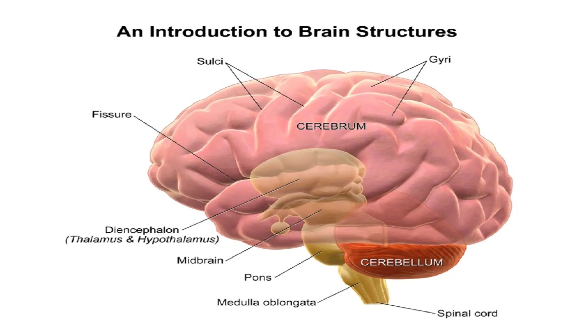
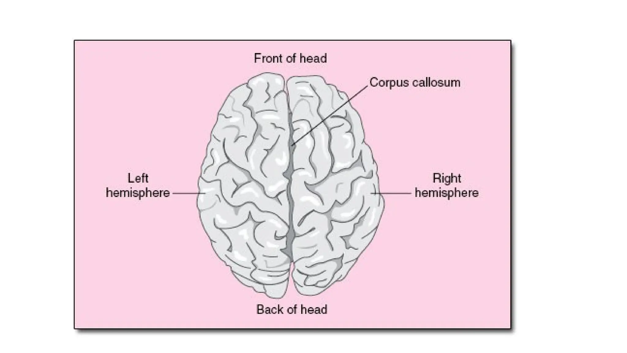
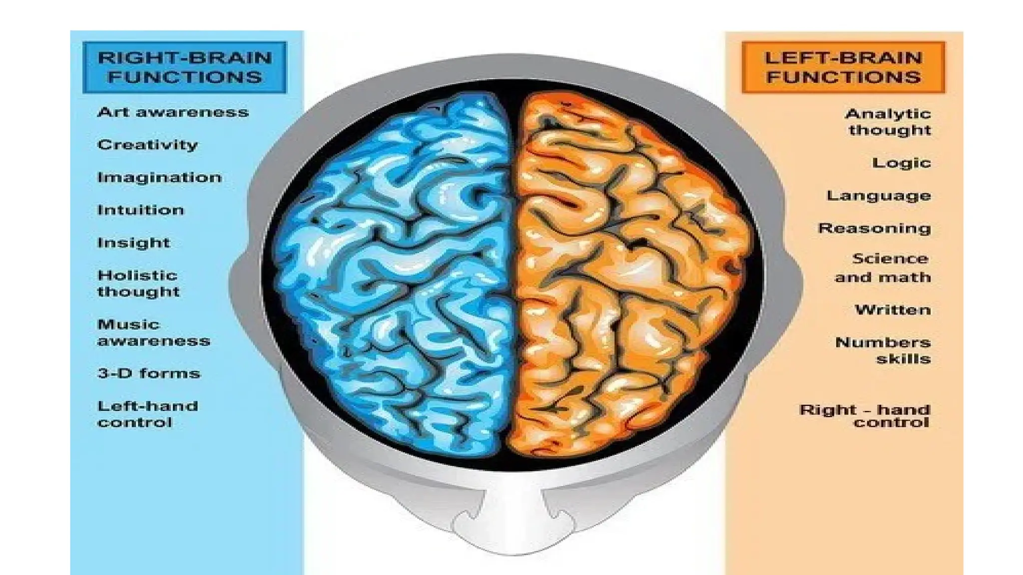
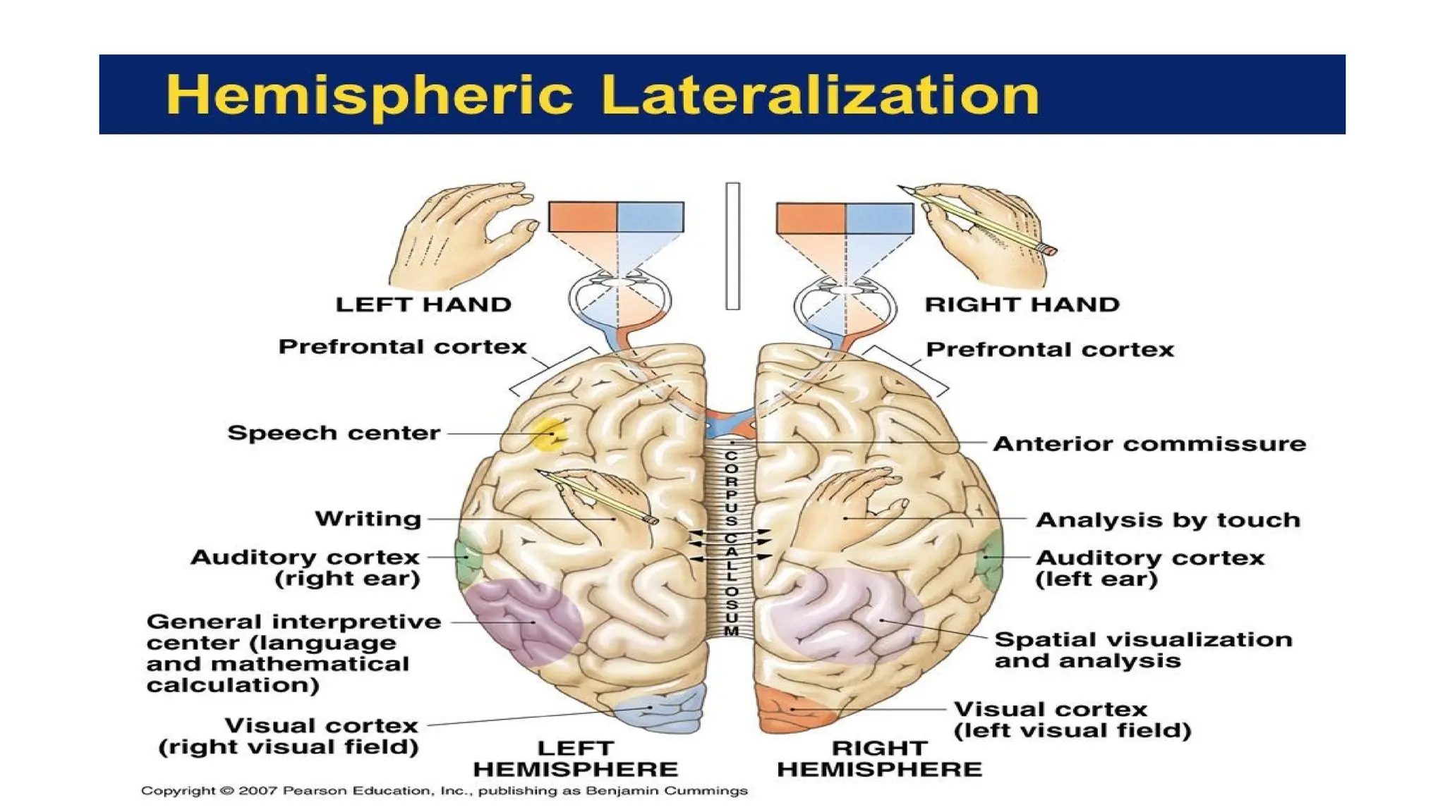
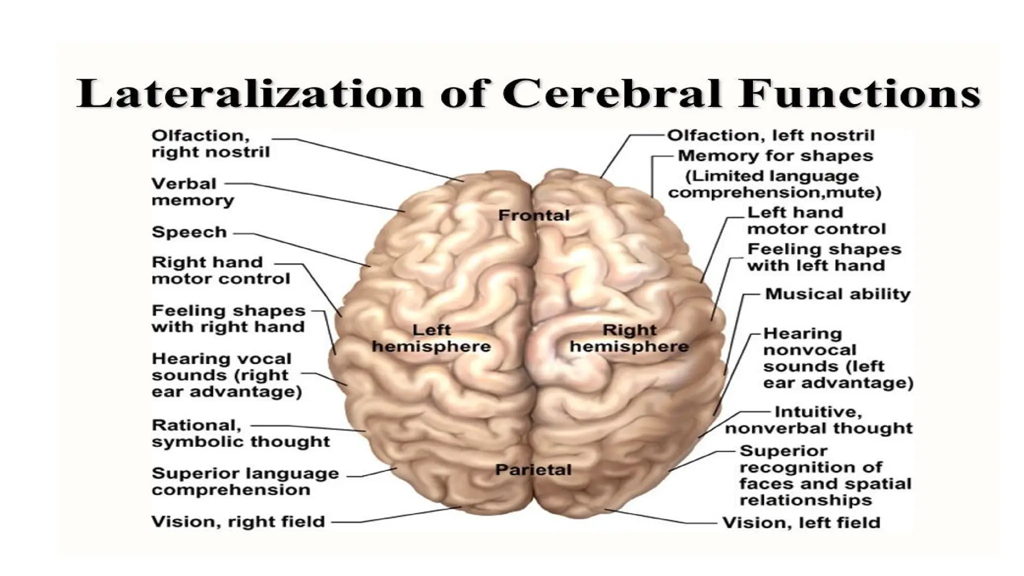
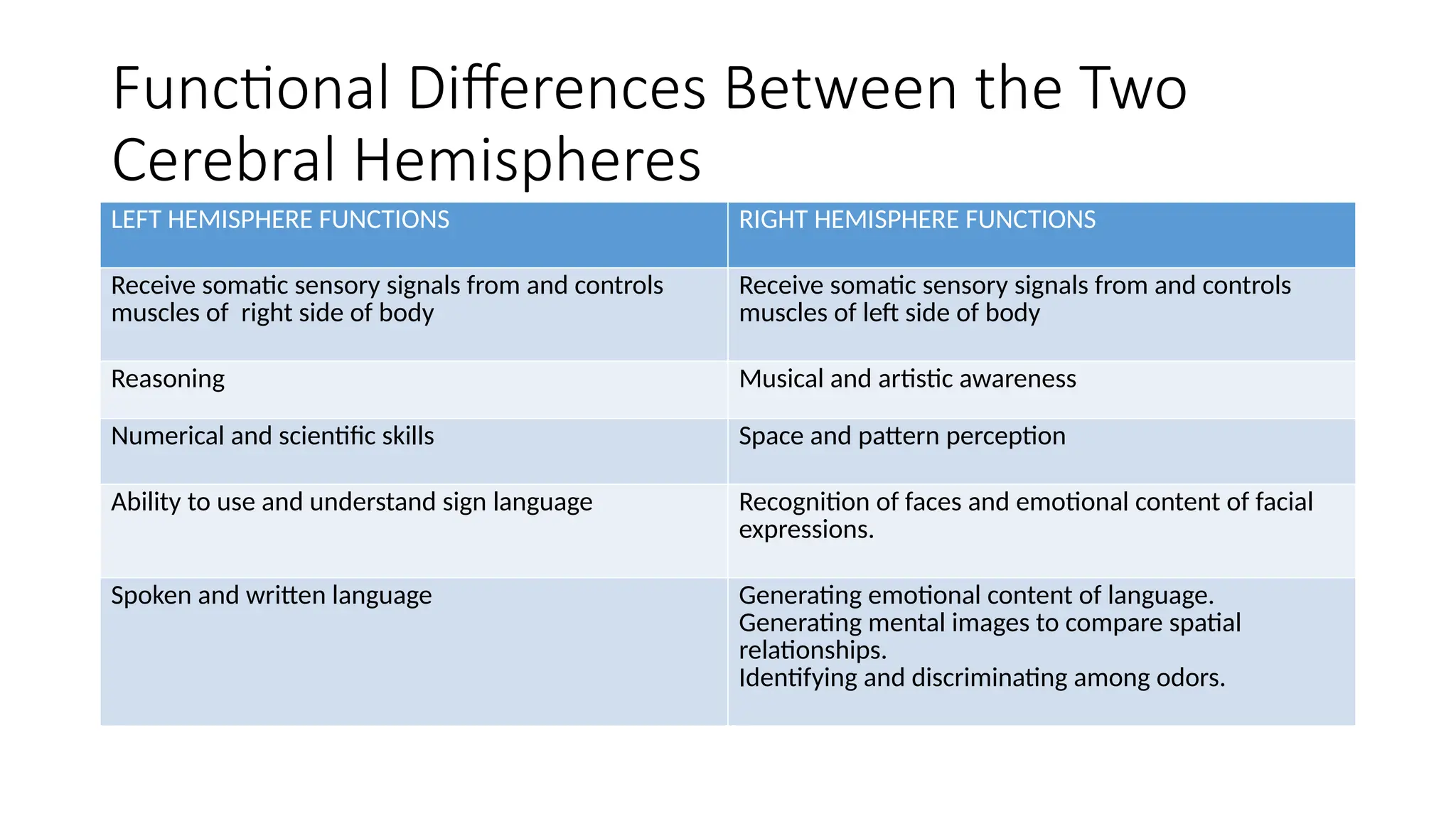
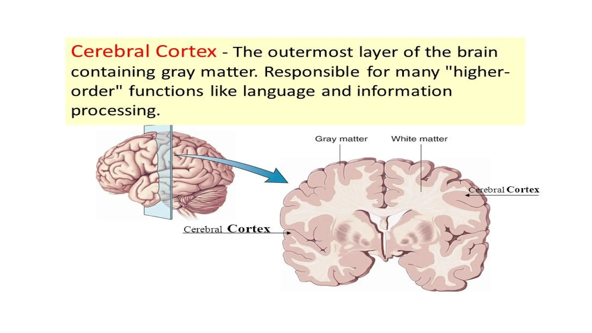
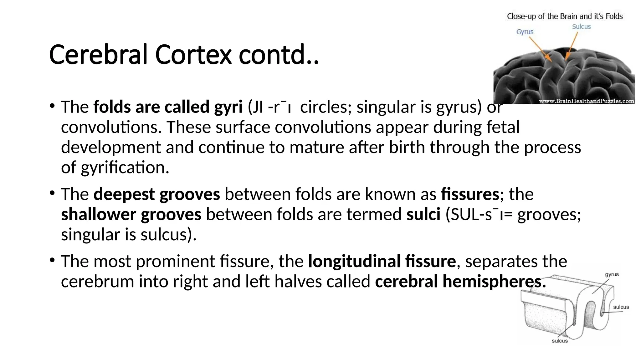
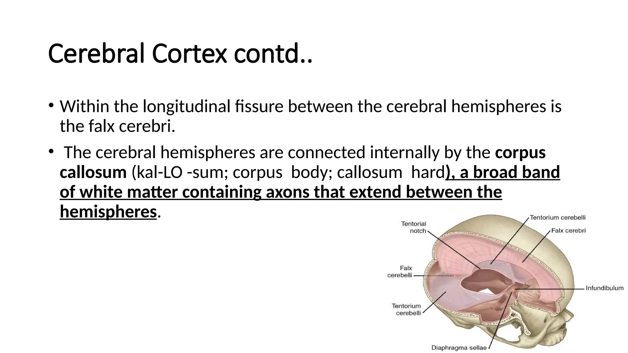
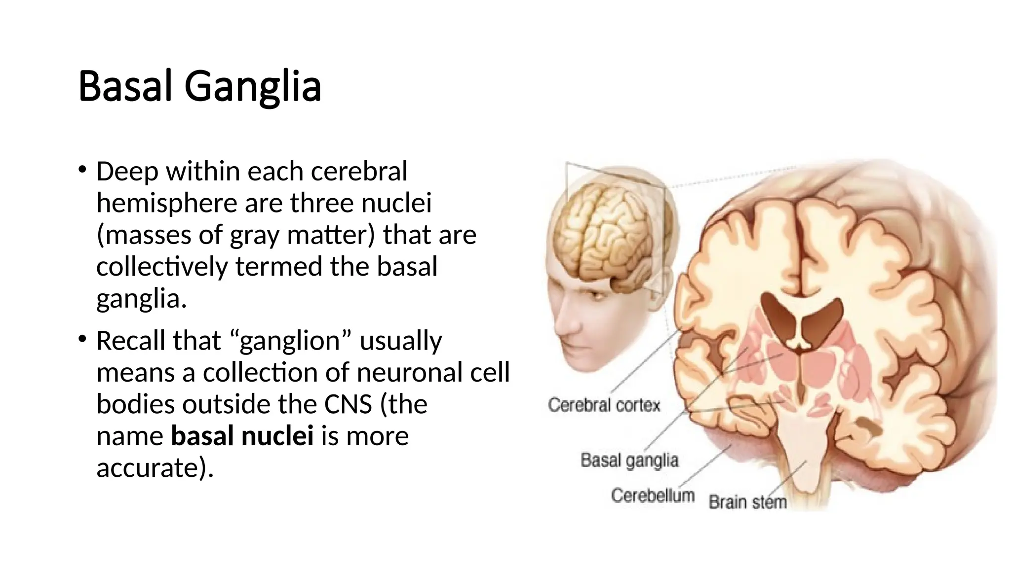
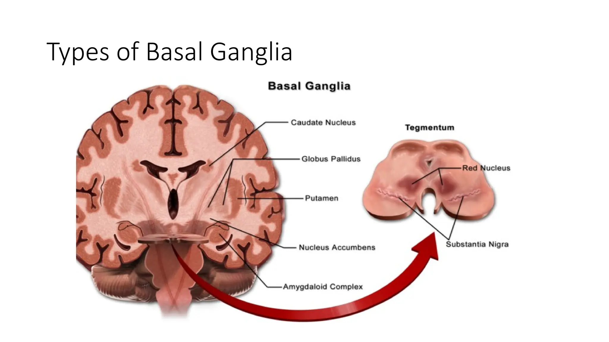
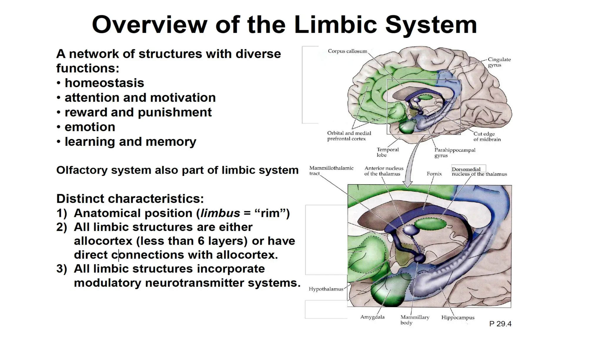
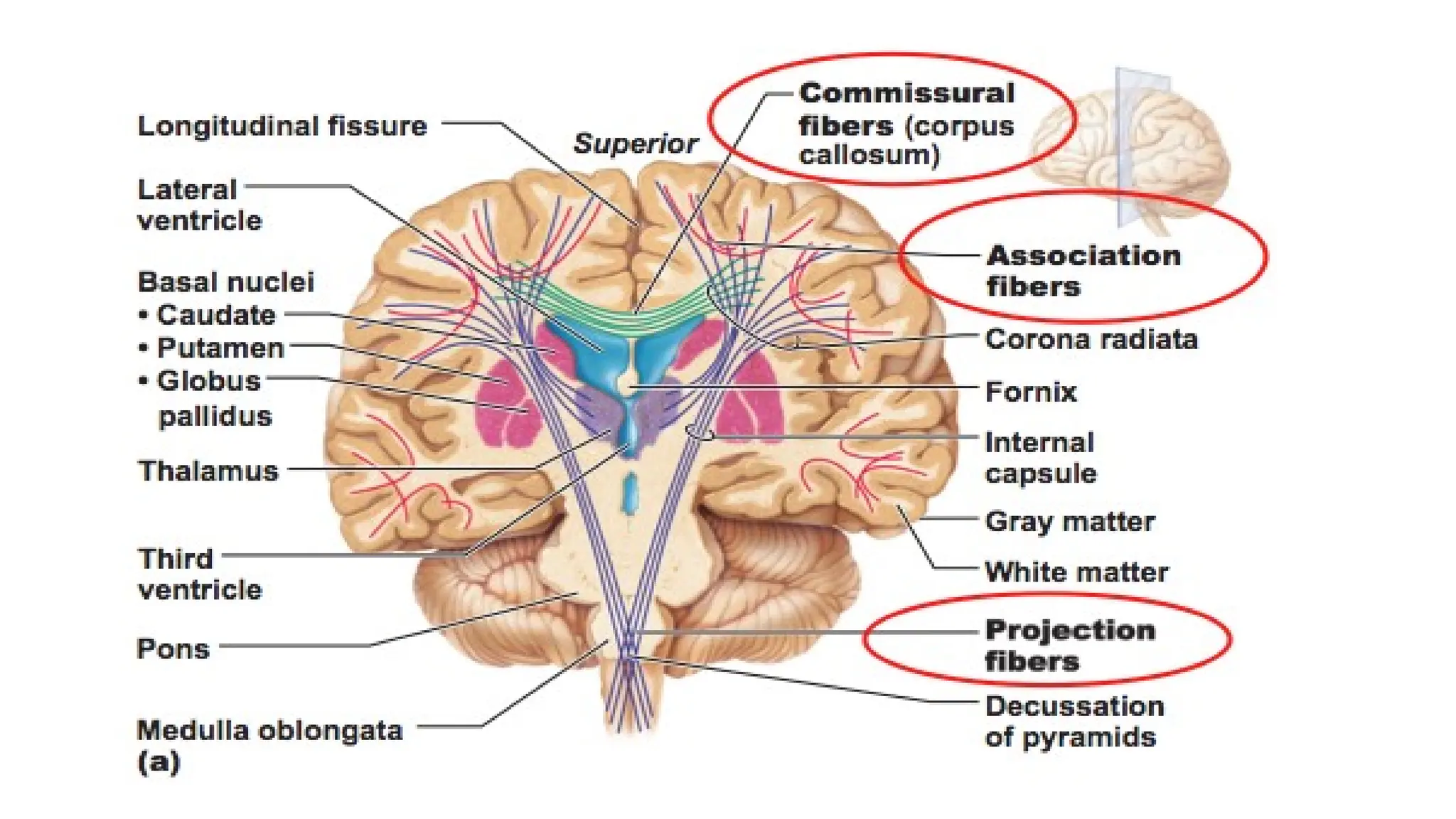
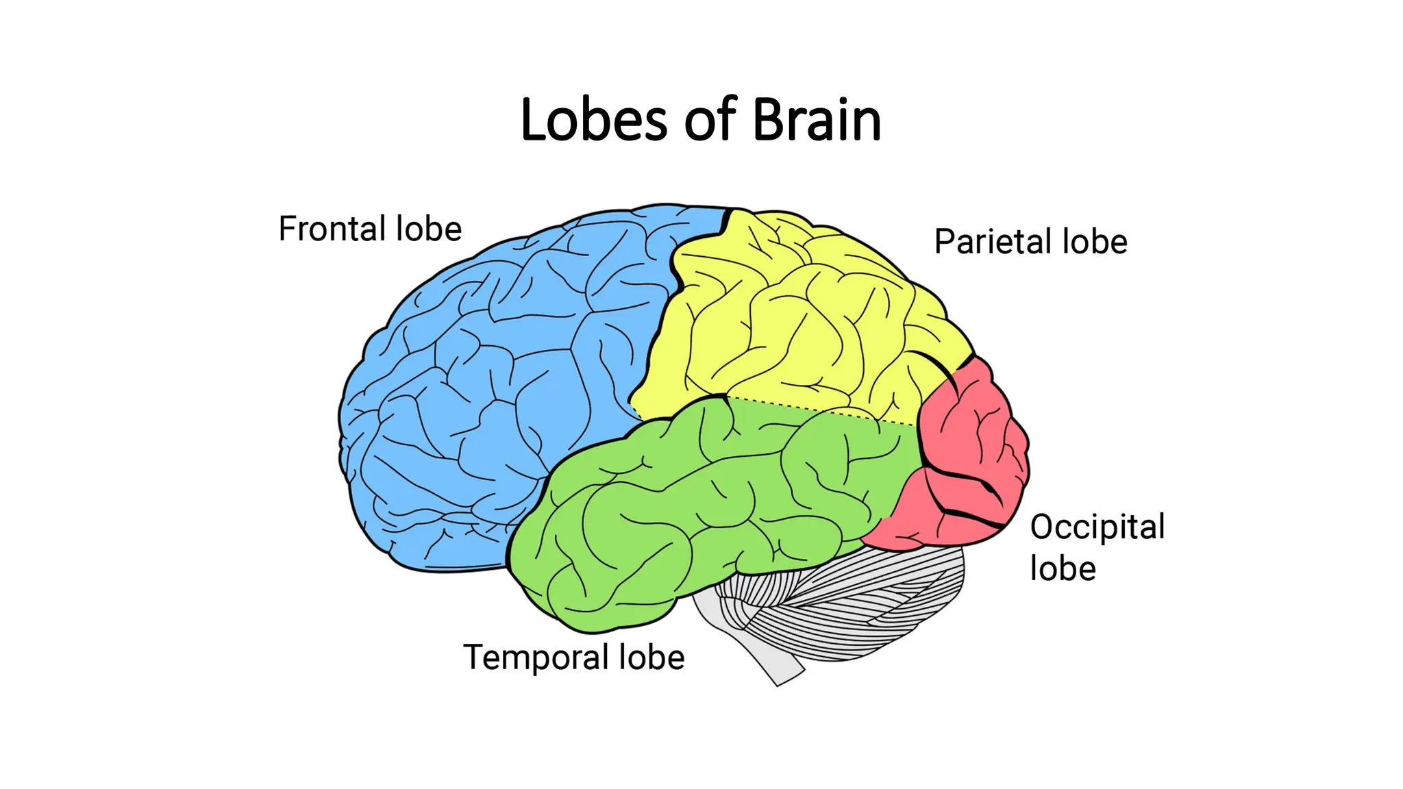
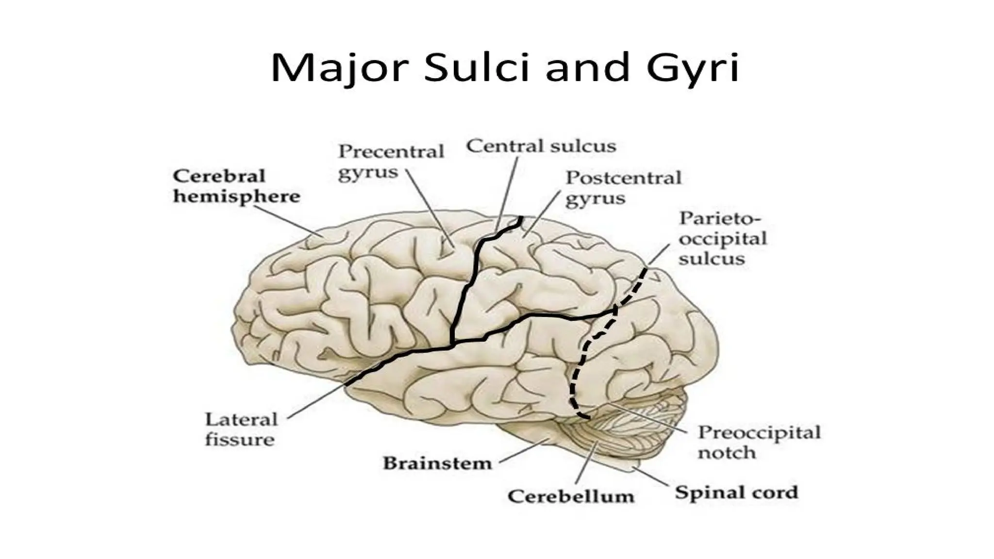
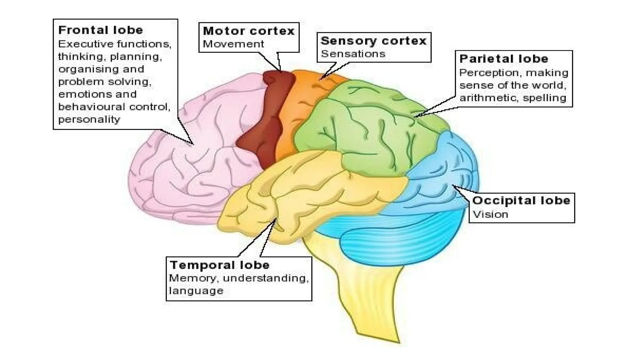
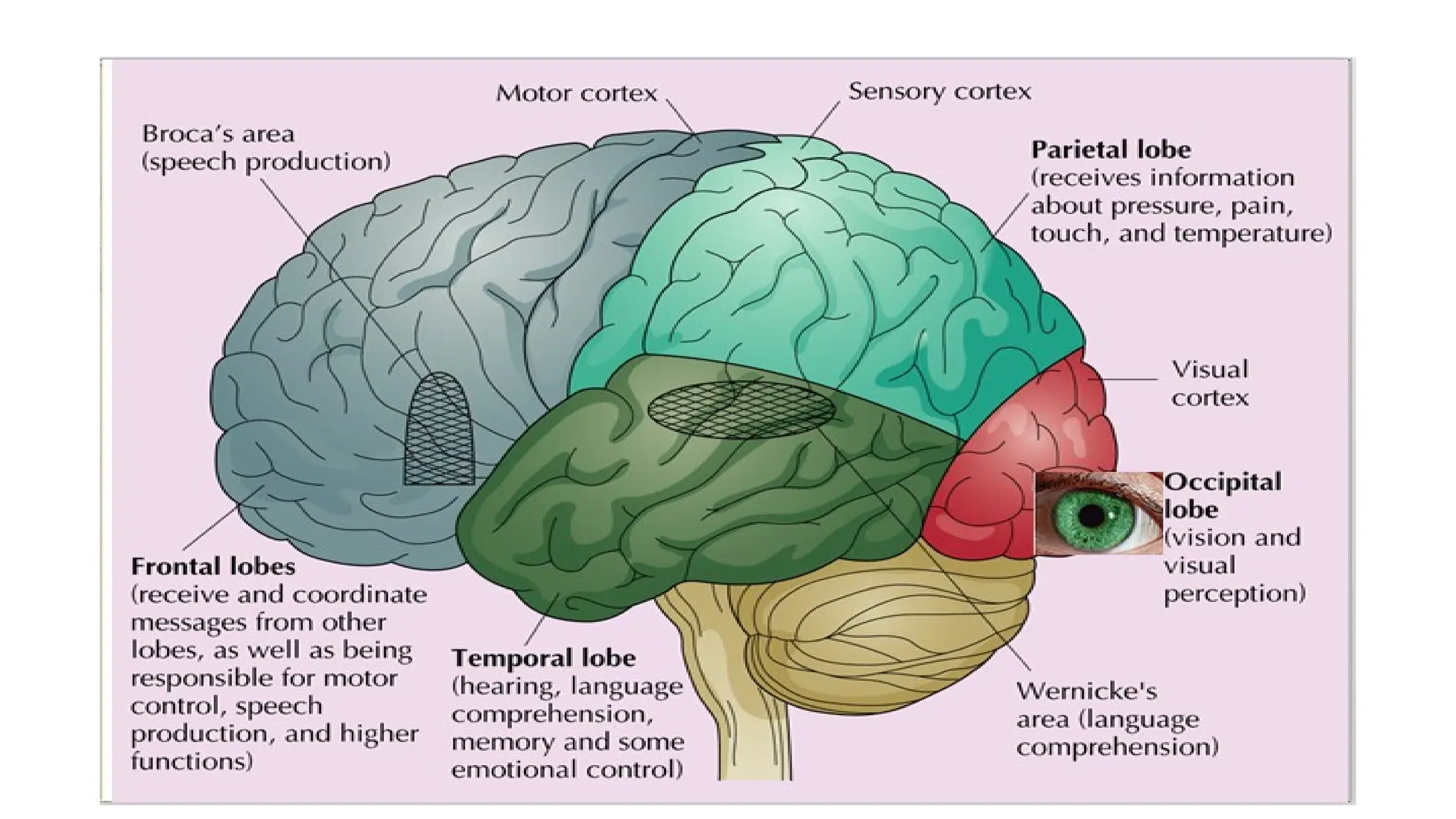
The Cerebrum
Prepared and presented by:
Ms Kanwal Qaiser
The Cerebrum
• The cerebrum or telencephlon is the “seat of intelligence.”
• It provides us with the ability to read, write, and speak; to make
calculations and compose music; and to remember the past, plan for
the future, and imagine things that have never existed before.
• The cerebrum consists of an outer cerebral cortex, an internal region
of cerebral white matter, and gray matter nuclei deep within the
white matter.
Cerebrum
• This is the largest part of the brain and it occupies the anterior and middle
cranial fossae.
• It is divided by a deep cleft, the longitudinal cerebral fissure, into right and
left cerebral hemispheres (outer layers of grey matter and the underlying
regions of white matter), each containing one of the lateral ventricles..
• Deep within the brain, the hemispheres are connected by a mass of
white matter (nerve fibres) called the corpus callosum.
• The cerebrum is made up of the two cerebral hemispheres and
their cortices, ( Its subcortical structures include the hippocampus, basal
ganglia and olfactory bulb).
Cerebrum
• The falx cerebri is formed by the dura mater.
• It separates the two cerebral hemispheres and penetrates to the
depth of the corpus callosum.
• The superficial part of the cerebrum is composed of nerve cell bodies
(grey matter), forming the cerebral cortex, and the deeper layers
consist of nerve fibres (axons, white matter).
• The cerebral cortex shows many infoldings or furrows of varying
depth.
Cerebrum
• The exposed areas of the folds are the gyri (convolutions) and these
are separated by sulci (fissures).
• These convolutions greatly increase the surface area of the cerebrum.
• For descriptive purposes each hemisphere of the cerebrum is divided
into lobes which take the names of the bones of the cranium under
which they lie: • frontal • parietal • temporal • occipital.
• The boundaries of the lobes are marked by deep sulci.
Cerebral Hemispheres
• The cerebrum is divided by the medial longitudinal fissure into
two cerebral hemispheres, the right and the left.
• The cerebrum is contralaterally organized, i.e., right hemisphere
controls and processes signals from the left side of the body, while the
left hemisphere controls and processes signals from the right side of
the body.
• There is a strong but not complete bilateral symmetry between the
hemispheres.
• The lateralization of brain function looks at the known and possible
differences between the two.
Hemispheric Lateralization or Dominance or
Specialization
• Although the brain is almost symmetrical on its right and left sides,
subtle anatomical differences between the two hemispheres exist.
• For example, in about two-thirds of the population, the planum
temporale, a region of the temporal lobe that includes Wernicke’s
area, is 50% larger on the left side than on the right side.
• This asymmetry appears in the human fetus at about 30 weeks of
gestation.
Hemispheric Lateralization contd..
• Physiological differences also exist; although the two hemispheres
share performance of many functions, each hemisphere also
specializes in performing certain unique functions.
• This functional asymmetry is termed hemispheric lateralization.
Functional Differences Between the Two
Cerebral Hemispheres
LEFT HEMISPHERE FUNCTIONS RIGHT HEMISPHERE FUNCTIONS
Receive somatic sensory signals from and controls
muscles of right side of body
Receive somatic sensory signals from and controls
muscles of left side of body
Reasoning Musical and artistic awareness
Numerical and scientific skills Space and pattern perception
Ability to use and understand sign language Recognition of faces and emotional content of facial
expressions.
Spoken and written language Generating emotional content of language.
Generating mental images to compare spatial
relationships.
Identifying and discriminating among odors.
The Cerebral Cortex
• The cerebral cortex (cortex rind or bark) or cerebral mantle is a region of
gray matter that forms the outer rim of the cerebrum.
• Although only 2–4 mm (0.08–0.16 in.) thick, the cerebral cortex contains
billions of neurons. During embryonic development, when brain size
increases rapidly, the gray matter of the cortex enlarges much faster than
the deeper white matter.
• As a result, the cortical region rolls and folds upon itself.
• The cerebral cortex is the largest site of neural integration in the central
nervous system.
• Functions are
attention, perception, awareness, thought, memory, language,
and consciousness.
Cerebral Cortex contd..
• The folds are called gyri (JI -r¯ı circles; singular is gyrus) or
convolutions. These surface convolutions appear during fetal
development and continue to mature after birth through the process
of gyrification.
• The deepest grooves between folds are known as fissures; the
shallower grooves between folds are termed sulci (SUL-s¯ı= grooves;
singular is sulcus).
• The most prominent fissure, the longitudinal fissure, separates the
cerebrum into right and left halves called cerebral hemispheres.
Cerebral Cortex contd..
• Within the longitudinal fissure between the cerebral hemispheres is
the falx cerebri.
• The cerebral hemispheres are connected internally by the corpus
callosum (kal-LO -sum; corpus body; callosum hard), a broad band
of white matter containing axons that extend between the
hemispheres.
Cerebral Cortex contd..
• There are between 14 and 16 billion neurons in the cerebral cortex.
• These are organised into cortical columns and minicolumns of
neurons that make up the layers of the cortex.
• Most of the cerebral cortex consists of the six-layered neocortex.
• Cortical areas have specific functions.
Types of Cerebral Cortex
• Neocortex: The six cortical layers of the neocortex each contain a
characteristic distribution of different neurons and their connections
with other cortical and subcortical regions. There are direct
connections between different cortical areas and indirect connections
via the thalamus.
• It is involved in higher-order brain functions such as sensory
perception, cognition, generation of motor commands, spatial
reasoning and language.
• Allocortex: Contains limbic system
Cortical neuron development
• Neurogenesis and Neuroepithelial cell
• Cortical neurons are generated within the ventricular zone, next to
the ventricles.
• At first, this zone contains neural stem cells, that transition to radial
glial cells–progenitor cells, which divide to produce glial cells and
neurons.
Functional Organization of Cerebral Cortex
• Specific types of sensory, motor, and integrative signals are processed
in certain regions of the cerebral cortex.
• Generally, sensory areas receive sensory information and are
involved in perception, the conscious awareness of a sensation;
motor areas control the execution of voluntary movements; and
association areas deal with more complex integrative functions such
as memory, emotions, reasoning, will, judgment, personality traits,
and intelligence.
Basal Ganglia
• Deep within each cerebral
hemisphere are three nuclei
(masses of gray matter) that are
collectively termed the basal
ganglia.
• Recall that “ganglion” usually
means a collection of neuronal cell
bodies outside the CNS (the
name basal nuclei is more
accurate).
Types of Basal Ganglia
Functions of Basal Ganglia
• The basal ganglia receive input from the cerebral cortex and provide
output back to motor areas of the cerebral cortex via neural
connections with nuclei of the thalamus.
• In addition, the nuclei of the basal ganglia have extensive
connections with one another.
• Axons from the substantia nigra terminate in the caudate nucleus and
putamen.
Functions of Basal Ganglia contd..
• A major function of the basal ganglia is to help initiate and
terminate movements of the body.
• The basal ganglia also suppress unwanted movements and regulate
muscle tone.
• In addition, the basal ganglia influence many aspects of cortical
functions, including: sensory, limbic, cognitive, and linguistic
functions.
Functions of Basal Ganglia contd..
• The basal ganglia have a limbic sector as well.
• This limbic sector is thought to play a central role in reward learning,
particularly a pathway from the Ventral Tegmental Area (VTA) to the
nucleus accumbens that uses the neurotransmitter dopamine.
• A number of highly addictive drugs, including cocaine, amphetamine,
and nicotine, are thought to work by increasing the efficacy of this
dopamine signal.
The Limbic System
• Encircling the upper part of the brain stem and the corpus callosum is
a ring of structures on the inner border of the cerebrum and floor of
the diencephalon that constitutes the limbic system (limbic border).
• The main components of the limbic system are as follows:
• The so-called limbic lobe is a rim of cerebral cortex on the medial
surface of each hemisphere.
The primary structures within the limbic
system
• The limbic system, located just beneath the cerebrum on both sides
of the thalamus, is not only responsible for our emotional lives but
also many higher mental functions, such as learning and formation of
memories.
• The primary structures within the limbic system include the amygdala,
hippocampus, thalamus, hypothalamus, basal ganglia, and cingulate
gyrus.
The primary structures within the limbic
system
• The thalamus and hypothalamus are associated with changes in
emotional reactivity.
• The cingulate gyrus coordinates smells and sights with pleasant
memories, induces an emotional reaction to pain, and helps regulate
aggressive behavior.
• The basal ganglia is a group of nuclei lying deep in the subcortical
white matter of the frontal lobes; its functions include organizing
motor behavior and coordinating rule-based, habit learning.
The Limbic System contd..
• The hippocampus ( seahorse) is a portion of the parahippocampal
gyrus that extends into the floor of the lateral ventricle.
• The amygdala (amygda- almond-shaped) is composed of several
groups of neurons located close to the tail of the caudate nucleus.
The Limbic System contd..
• The limbic system is sometimes called the “emotional brain” because
it plays a primary role in a range of emotions, including pleasure, pain,
docility, affection, fear, and anger.
• It also is involved in olfaction (smell) and memory.
• A person whose amygdala is damaged fails to recognize fearful
expressions in others or to express fear in appropriate situations.
The Limbic System contd..
• The hippocampus, together with other parts of the cerebrum,
functions in memory.
• People with damage to certain limbic system structures forget recent
events and cannot commit anything to memory.
Grey Matter
• Grey matter (or gray matter) is a major component of the central
nervous system, consisting of neuronal cell
bodies, neuropil (dendrites and myelinated as well as
unmyelinated axons), glial
cells (astrocytes and oligodendrocytes), synapses, and capillaries.
• Grey matter is distinguished from white matter in that it contains
numerous cell bodies and relatively few myelinated axons, while
white matter contains relatively few cell bodies and is composed
chiefly of long-range myelinated axons.
• The colour difference arises mainly from the whiteness of myelin.
White Matter
• White matter refers to areas of the central nervous system (CNS) that
are mainly made up of myelinated axons, also called tracts.
• Long thought to be passive tissue, white matter affects learning and
brain functions, modulating the distribution of action potentials,
acting as a relay and coordinating communication between different
brain regions.
• White matter is named for its relatively light appearance resulting
from the lipid content of myelin.
White Matter
• White matter is the tissue through which messages pass between
different areas of gray matter within the central nervous system. This
myelin is found in almost all long nerve fibers, and acts as an electrical
insulation.
• This is important because it allows the messages to pass quickly from
place to place.
• Unlike gray matter, which peaks in development in a person's
twenties, the white matter continues to develop, and peaks in middle
age
Cerebral White Matter
• The cerebral white matter consists primarily of myelinated axons in
three types of tracts:
1. Association tracts contain axons that conduct nerve impulses
between gyri in the same hemisphere.
2. Commissural tracts contain axons that conduct nerve impulses from
gyri in one cerebral hemisphere to corresponding gyri in the other
cerebral hemisphere.
• Three important groups of commissural tracts are the corpus
callosum (the largest fiber bundle in the brain, containing about 300
million fibers), anterior commissure, and posterior commissure.
Cerebral White Matter
3. Projection tracts contain axons that conduct nerve impulses from
the cerebrum to lower parts of the CNS (thalamus, brain stem, or spinal
cord) or from lower parts of the CNS to the cerebrum.
• An example is the internal capsule, a thick band of white matter that
contains both ascending and descending axons.
Lobes of Brain
Lobes of Cerebrum
• Each cerebral hemisphere can be further subdivided into four lobes.
The lobes are named after the bones that cover them: frontal,
parietal, temporal, and occipital lobes .
• The central sulcus (SUL-kus) separates the frontal lobe from the
parietal lobe.
• A major gyrus, the precentral gyrus—located immediately anterior to
the central sulcus—contains the primary motor area of the cerebral
cortex.
Lobes of Cerebrum contd..
• Another major gyrus, the postcentral gyrus, which is located
immediately posterior to the central sulcus, contains the primary
somatosensory area of the cerebral cortex.
• The lateral cerebral sulcus (fissure) separates the frontal lobe from
the temporal lobe.
• The parieto-occipital sulcus separates the parietal lobe from the
occipital lobe.
• A fifth part of the cerebrum, the insula, cannot be seen at the surface
of the brain because it lies within the lateral cerebral sulcus, deep to
the parietal, frontal, and temporal lobes.
Lobes of Brain and their Functions
• The lobes of the brain were originally
a purely anatomical classification, but
have been shown also to be related
to different brain functions.
• The telencephalon (cerebrum), the
largest portion of the human brain, is
divided into lobes, but so is
the cerebellum.
• If not specified, the expression
"lobes of the brain" refers to the
telencephalon.
The Frontal Lobe
• The frontal lobe is located at the front of each cerebral
hemisphere and positioned in front of the parietal lobe and above
and in front of the temporal lobe. It carries out executive functions.
• The precentral gyrus, forming the posterior border of the frontal lobe,
contains the primary motor cortex, which controls voluntary
movements of specific body parts.
The Frontal Lobe contd..
• The frontal lobe contains most of the dopamine-delicate neurons in
the cerebral cortex.
• The dopamine system is associated with reward, attention, short-
term memory tasks, planning, and motivation.
The Parietal Lobe
• The parietal lobe is positioned above the occipital lobe and behind
the frontal lobe and central sulcus.
• The parietal lobe integrates sensory information among
various modalities, including spatial sense and navigation
(proprioception), the main sensory receptive area for the sense of
touch (mechanoreception) in the somatosensory cortex which is just
posterior to the central sulcus in the postcentral gyrus, and the dorsal
stream of the visual system.
The Parietal Lobe contd..
• The major sensory inputs from the skin (touch, temperature,
and pain receptors), relay through the thalamus to the parietal lobe.
• Several areas of the parietal lobe are important in language
processing.
The Occipital Lobe
• The occipital lobe is the visual processing center of
the mammalian brain containing most of the anatomical region of
the visual cortex.
The Temporal Lobe
• The temporal lobe is located beneath the lateral fissure on
both cerebral hemispheres of the mammalian brain.
• The temporal lobe is involved in processing sensory input into derived
meanings for the appropriate retention of visual memories, language
comprehension, and emotion association.
Functional Organization of Cerebral Cortex
• Specific types of sensory, motor, and integrative signals are processed
in certain regions of the cerebral cortex.
• Generally, sensory areas receive sensory information and are
involved in perception, the conscious awareness of a sensation;
motor areas control the execution of voluntary movements; and
association areas deal with more complex integrative functions such
as memory, emotions, reasoning, will, judgment, personality traits,
and intelligence.
Sensory Areas
• Sensory information arrives mainly in the posterior half of both
cerebral hemispheres, in regions behind the central sulci.
• In the cerebral cortex, primary sensory areas receive sensory
information that has been relayed from peripheral sensory receptors
through lower regions of the brain.
The following are some important sensory
areas
• The primary somatosensory area (areas 1, 2, and 3) is located directly
posterior to the central sulcus of each cerebral hemisphere in the
postcentral gyrus of each parietal lobe.
• The primary somatosensory area receives nerve impulses for touch,
pressure, vibration, itch, tickle, temperature (coldness and warmth),
pain, and proprioception (joint and muscle position) and is involved in
the perception of these somatic sensations.
• A “map” of the entire body is present in the primary somatosensory
area.
The following are some important sensory
areas
• The primary somatosensory area allows you to pinpoint where
somatic sensations originate, so that you know exactly where on your
body to swat that mosquito.
• The primary visual area (area 17), located at the posterior tip of the
occipital lobe mainly on the medial surface (next to the longitudinal
fissure), receives visual information and is involved in visual
perception.
The following are some important sensory
areas
• The primary auditory area (areas 41 and 42), located in the superior
part of the temporal lobe near the lateral cerebral sulcus, receives
information for sound and is involved in auditory perception.
• The primary gustatory area (area 43), located at the base of the
postcentral gyrus superior to the lateral cerebral sulcus in the parietal
cortex, receives impulses for taste and is involved in gustatory
perception and taste discrimination
• The primary olfactory area (area 28), located in the temporal lobe on
the medial aspect, receives impulses for smell and is involved in
olfactory perception.
The Motor Areas
• The primary motor area (area 4) is located in the precentral gyrus of
the frontal lobe.
• Each region in the primary motor area controls voluntary
contractions of specific muscles or groups of muscles .
• Electrical stimulation of any point in the primary motor area causes
contraction of specific skeletal muscle fibers on the opposite side of
the body.
The Motor Areas contd..
• More cortical area is devoted to those muscles involved in skilled,
complex, or delicate movement.
• For instance, the cortical region devoted to muscles that move the
fingers is much larger than the region for muscles that move the toes.
The Motor Areas contd..
• Broca’s (BRO -kaz) speech area (areas 44 and 45), located in the
frontal lobe close to the lateral cerebral sulcus, is involved in the
articulation of speech.
• In most people, Broca’s speech area is localized in the left cerebral
hemisphere.
• Neural circuits established between Broca’s speech area, the
premotor area, and primary motor area activate muscles of the
larynx, pharynx, and mouth and breathing muscles.
The Motor Areas contd..
• The coordinated contractions of your speech and breathing muscles
enable you to speak your thoughts.
• People who suffer a cerebrovascular accident (CVA) or stroke in this
area can still have clear thoughts, but are unable to form words.
The Association Areas
• The somatosensory association area
• Visual association area
• Facial recognition area
• Auditory association area
• Prefrontal cortex (frontal association area)
The Association Areas contd..
• Wernicke’s (posterior language) area (VER-ni-ke -z), a broad region in
the left temporal and parietal lobes, interprets the meaning of speech
by recognizing spoken words.
• It is active as you translate words into thoughts.
The Association Areas contd..
• The regions in the right hemisphere that correspond to Broca’s and
Wernicke’s areas in the left hemisphere also contribute to verbal
communication by adding emotional content, such as anger or joy, to
spoken words.
• Unlike those who have CVAs in Broca’s area, people who suffer
strokes in Wernicke’s area can still speak, but cannot arrange words in
a coherent fashion
The Cerebrum.pptx by sir waqar ahmed of brain

The Cerebrum.pptx by sir waqar ahmed of brain

  • 1.
    The Cerebrum Prepared andpresented by: Ms Kanwal Qaiser
  • 3.
    The Cerebrum • Thecerebrum or telencephlon is the “seat of intelligence.” • It provides us with the ability to read, write, and speak; to make calculations and compose music; and to remember the past, plan for the future, and imagine things that have never existed before. • The cerebrum consists of an outer cerebral cortex, an internal region of cerebral white matter, and gray matter nuclei deep within the white matter.
  • 4.
    Cerebrum • This isthe largest part of the brain and it occupies the anterior and middle cranial fossae. • It is divided by a deep cleft, the longitudinal cerebral fissure, into right and left cerebral hemispheres (outer layers of grey matter and the underlying regions of white matter), each containing one of the lateral ventricles.. • Deep within the brain, the hemispheres are connected by a mass of white matter (nerve fibres) called the corpus callosum. • The cerebrum is made up of the two cerebral hemispheres and their cortices, ( Its subcortical structures include the hippocampus, basal ganglia and olfactory bulb).
  • 6.
    Cerebrum • The falxcerebri is formed by the dura mater. • It separates the two cerebral hemispheres and penetrates to the depth of the corpus callosum. • The superficial part of the cerebrum is composed of nerve cell bodies (grey matter), forming the cerebral cortex, and the deeper layers consist of nerve fibres (axons, white matter). • The cerebral cortex shows many infoldings or furrows of varying depth.
  • 8.
    Cerebrum • The exposedareas of the folds are the gyri (convolutions) and these are separated by sulci (fissures). • These convolutions greatly increase the surface area of the cerebrum. • For descriptive purposes each hemisphere of the cerebrum is divided into lobes which take the names of the bones of the cranium under which they lie: • frontal • parietal • temporal • occipital. • The boundaries of the lobes are marked by deep sulci.
  • 9.
    Cerebral Hemispheres • Thecerebrum is divided by the medial longitudinal fissure into two cerebral hemispheres, the right and the left. • The cerebrum is contralaterally organized, i.e., right hemisphere controls and processes signals from the left side of the body, while the left hemisphere controls and processes signals from the right side of the body. • There is a strong but not complete bilateral symmetry between the hemispheres. • The lateralization of brain function looks at the known and possible differences between the two.
  • 11.
    Hemispheric Lateralization orDominance or Specialization • Although the brain is almost symmetrical on its right and left sides, subtle anatomical differences between the two hemispheres exist. • For example, in about two-thirds of the population, the planum temporale, a region of the temporal lobe that includes Wernicke’s area, is 50% larger on the left side than on the right side. • This asymmetry appears in the human fetus at about 30 weeks of gestation.
  • 12.
    Hemispheric Lateralization contd.. •Physiological differences also exist; although the two hemispheres share performance of many functions, each hemisphere also specializes in performing certain unique functions. • This functional asymmetry is termed hemispheric lateralization.
  • 15.
    Functional Differences Betweenthe Two Cerebral Hemispheres LEFT HEMISPHERE FUNCTIONS RIGHT HEMISPHERE FUNCTIONS Receive somatic sensory signals from and controls muscles of right side of body Receive somatic sensory signals from and controls muscles of left side of body Reasoning Musical and artistic awareness Numerical and scientific skills Space and pattern perception Ability to use and understand sign language Recognition of faces and emotional content of facial expressions. Spoken and written language Generating emotional content of language. Generating mental images to compare spatial relationships. Identifying and discriminating among odors.
  • 16.
    The Cerebral Cortex •The cerebral cortex (cortex rind or bark) or cerebral mantle is a region of gray matter that forms the outer rim of the cerebrum. • Although only 2–4 mm (0.08–0.16 in.) thick, the cerebral cortex contains billions of neurons. During embryonic development, when brain size increases rapidly, the gray matter of the cortex enlarges much faster than the deeper white matter. • As a result, the cortical region rolls and folds upon itself. • The cerebral cortex is the largest site of neural integration in the central nervous system. • Functions are attention, perception, awareness, thought, memory, language, and consciousness.
  • 18.
    Cerebral Cortex contd.. •The folds are called gyri (JI -r¯ı circles; singular is gyrus) or convolutions. These surface convolutions appear during fetal development and continue to mature after birth through the process of gyrification. • The deepest grooves between folds are known as fissures; the shallower grooves between folds are termed sulci (SUL-s¯ı= grooves; singular is sulcus). • The most prominent fissure, the longitudinal fissure, separates the cerebrum into right and left halves called cerebral hemispheres.
  • 19.
    Cerebral Cortex contd.. •Within the longitudinal fissure between the cerebral hemispheres is the falx cerebri. • The cerebral hemispheres are connected internally by the corpus callosum (kal-LO -sum; corpus body; callosum hard), a broad band of white matter containing axons that extend between the hemispheres.
  • 20.
    Cerebral Cortex contd.. •There are between 14 and 16 billion neurons in the cerebral cortex. • These are organised into cortical columns and minicolumns of neurons that make up the layers of the cortex. • Most of the cerebral cortex consists of the six-layered neocortex. • Cortical areas have specific functions.
  • 21.
    Types of CerebralCortex • Neocortex: The six cortical layers of the neocortex each contain a characteristic distribution of different neurons and their connections with other cortical and subcortical regions. There are direct connections between different cortical areas and indirect connections via the thalamus. • It is involved in higher-order brain functions such as sensory perception, cognition, generation of motor commands, spatial reasoning and language. • Allocortex: Contains limbic system
  • 22.
    Cortical neuron development •Neurogenesis and Neuroepithelial cell • Cortical neurons are generated within the ventricular zone, next to the ventricles. • At first, this zone contains neural stem cells, that transition to radial glial cells–progenitor cells, which divide to produce glial cells and neurons.
  • 24.
    Functional Organization ofCerebral Cortex • Specific types of sensory, motor, and integrative signals are processed in certain regions of the cerebral cortex. • Generally, sensory areas receive sensory information and are involved in perception, the conscious awareness of a sensation; motor areas control the execution of voluntary movements; and association areas deal with more complex integrative functions such as memory, emotions, reasoning, will, judgment, personality traits, and intelligence.
  • 26.
    Basal Ganglia • Deepwithin each cerebral hemisphere are three nuclei (masses of gray matter) that are collectively termed the basal ganglia. • Recall that “ganglion” usually means a collection of neuronal cell bodies outside the CNS (the name basal nuclei is more accurate).
  • 27.
  • 28.
    Functions of BasalGanglia • The basal ganglia receive input from the cerebral cortex and provide output back to motor areas of the cerebral cortex via neural connections with nuclei of the thalamus. • In addition, the nuclei of the basal ganglia have extensive connections with one another. • Axons from the substantia nigra terminate in the caudate nucleus and putamen.
  • 29.
    Functions of BasalGanglia contd.. • A major function of the basal ganglia is to help initiate and terminate movements of the body. • The basal ganglia also suppress unwanted movements and regulate muscle tone. • In addition, the basal ganglia influence many aspects of cortical functions, including: sensory, limbic, cognitive, and linguistic functions.
  • 30.
    Functions of BasalGanglia contd.. • The basal ganglia have a limbic sector as well. • This limbic sector is thought to play a central role in reward learning, particularly a pathway from the Ventral Tegmental Area (VTA) to the nucleus accumbens that uses the neurotransmitter dopamine. • A number of highly addictive drugs, including cocaine, amphetamine, and nicotine, are thought to work by increasing the efficacy of this dopamine signal.
  • 33.
    The Limbic System •Encircling the upper part of the brain stem and the corpus callosum is a ring of structures on the inner border of the cerebrum and floor of the diencephalon that constitutes the limbic system (limbic border). • The main components of the limbic system are as follows: • The so-called limbic lobe is a rim of cerebral cortex on the medial surface of each hemisphere.
  • 35.
    The primary structureswithin the limbic system • The limbic system, located just beneath the cerebrum on both sides of the thalamus, is not only responsible for our emotional lives but also many higher mental functions, such as learning and formation of memories. • The primary structures within the limbic system include the amygdala, hippocampus, thalamus, hypothalamus, basal ganglia, and cingulate gyrus.
  • 36.
    The primary structureswithin the limbic system • The thalamus and hypothalamus are associated with changes in emotional reactivity. • The cingulate gyrus coordinates smells and sights with pleasant memories, induces an emotional reaction to pain, and helps regulate aggressive behavior. • The basal ganglia is a group of nuclei lying deep in the subcortical white matter of the frontal lobes; its functions include organizing motor behavior and coordinating rule-based, habit learning.
  • 37.
    The Limbic Systemcontd.. • The hippocampus ( seahorse) is a portion of the parahippocampal gyrus that extends into the floor of the lateral ventricle. • The amygdala (amygda- almond-shaped) is composed of several groups of neurons located close to the tail of the caudate nucleus.
  • 39.
    The Limbic Systemcontd.. • The limbic system is sometimes called the “emotional brain” because it plays a primary role in a range of emotions, including pleasure, pain, docility, affection, fear, and anger. • It also is involved in olfaction (smell) and memory. • A person whose amygdala is damaged fails to recognize fearful expressions in others or to express fear in appropriate situations.
  • 40.
    The Limbic Systemcontd.. • The hippocampus, together with other parts of the cerebrum, functions in memory. • People with damage to certain limbic system structures forget recent events and cannot commit anything to memory.
  • 42.
    Grey Matter • Greymatter (or gray matter) is a major component of the central nervous system, consisting of neuronal cell bodies, neuropil (dendrites and myelinated as well as unmyelinated axons), glial cells (astrocytes and oligodendrocytes), synapses, and capillaries. • Grey matter is distinguished from white matter in that it contains numerous cell bodies and relatively few myelinated axons, while white matter contains relatively few cell bodies and is composed chiefly of long-range myelinated axons. • The colour difference arises mainly from the whiteness of myelin.
  • 43.
    White Matter • Whitematter refers to areas of the central nervous system (CNS) that are mainly made up of myelinated axons, also called tracts. • Long thought to be passive tissue, white matter affects learning and brain functions, modulating the distribution of action potentials, acting as a relay and coordinating communication between different brain regions. • White matter is named for its relatively light appearance resulting from the lipid content of myelin.
  • 44.
    White Matter • Whitematter is the tissue through which messages pass between different areas of gray matter within the central nervous system. This myelin is found in almost all long nerve fibers, and acts as an electrical insulation. • This is important because it allows the messages to pass quickly from place to place. • Unlike gray matter, which peaks in development in a person's twenties, the white matter continues to develop, and peaks in middle age
  • 45.
    Cerebral White Matter •The cerebral white matter consists primarily of myelinated axons in three types of tracts: 1. Association tracts contain axons that conduct nerve impulses between gyri in the same hemisphere. 2. Commissural tracts contain axons that conduct nerve impulses from gyri in one cerebral hemisphere to corresponding gyri in the other cerebral hemisphere. • Three important groups of commissural tracts are the corpus callosum (the largest fiber bundle in the brain, containing about 300 million fibers), anterior commissure, and posterior commissure.
  • 47.
    Cerebral White Matter 3.Projection tracts contain axons that conduct nerve impulses from the cerebrum to lower parts of the CNS (thalamus, brain stem, or spinal cord) or from lower parts of the CNS to the cerebrum. • An example is the internal capsule, a thick band of white matter that contains both ascending and descending axons.
  • 49.
  • 51.
    Lobes of Cerebrum •Each cerebral hemisphere can be further subdivided into four lobes. The lobes are named after the bones that cover them: frontal, parietal, temporal, and occipital lobes . • The central sulcus (SUL-kus) separates the frontal lobe from the parietal lobe. • A major gyrus, the precentral gyrus—located immediately anterior to the central sulcus—contains the primary motor area of the cerebral cortex.
  • 52.
    Lobes of Cerebrumcontd.. • Another major gyrus, the postcentral gyrus, which is located immediately posterior to the central sulcus, contains the primary somatosensory area of the cerebral cortex. • The lateral cerebral sulcus (fissure) separates the frontal lobe from the temporal lobe. • The parieto-occipital sulcus separates the parietal lobe from the occipital lobe. • A fifth part of the cerebrum, the insula, cannot be seen at the surface of the brain because it lies within the lateral cerebral sulcus, deep to the parietal, frontal, and temporal lobes.
  • 53.
    Lobes of Brainand their Functions • The lobes of the brain were originally a purely anatomical classification, but have been shown also to be related to different brain functions. • The telencephalon (cerebrum), the largest portion of the human brain, is divided into lobes, but so is the cerebellum. • If not specified, the expression "lobes of the brain" refers to the telencephalon.
  • 55.
    The Frontal Lobe •The frontal lobe is located at the front of each cerebral hemisphere and positioned in front of the parietal lobe and above and in front of the temporal lobe. It carries out executive functions. • The precentral gyrus, forming the posterior border of the frontal lobe, contains the primary motor cortex, which controls voluntary movements of specific body parts.
  • 56.
    The Frontal Lobecontd.. • The frontal lobe contains most of the dopamine-delicate neurons in the cerebral cortex. • The dopamine system is associated with reward, attention, short- term memory tasks, planning, and motivation.
  • 57.
    The Parietal Lobe •The parietal lobe is positioned above the occipital lobe and behind the frontal lobe and central sulcus. • The parietal lobe integrates sensory information among various modalities, including spatial sense and navigation (proprioception), the main sensory receptive area for the sense of touch (mechanoreception) in the somatosensory cortex which is just posterior to the central sulcus in the postcentral gyrus, and the dorsal stream of the visual system.
  • 58.
    The Parietal Lobecontd.. • The major sensory inputs from the skin (touch, temperature, and pain receptors), relay through the thalamus to the parietal lobe. • Several areas of the parietal lobe are important in language processing.
  • 59.
    The Occipital Lobe •The occipital lobe is the visual processing center of the mammalian brain containing most of the anatomical region of the visual cortex.
  • 60.
    The Temporal Lobe •The temporal lobe is located beneath the lateral fissure on both cerebral hemispheres of the mammalian brain. • The temporal lobe is involved in processing sensory input into derived meanings for the appropriate retention of visual memories, language comprehension, and emotion association.
  • 64.
    Functional Organization ofCerebral Cortex • Specific types of sensory, motor, and integrative signals are processed in certain regions of the cerebral cortex. • Generally, sensory areas receive sensory information and are involved in perception, the conscious awareness of a sensation; motor areas control the execution of voluntary movements; and association areas deal with more complex integrative functions such as memory, emotions, reasoning, will, judgment, personality traits, and intelligence.
  • 65.
    Sensory Areas • Sensoryinformation arrives mainly in the posterior half of both cerebral hemispheres, in regions behind the central sulci. • In the cerebral cortex, primary sensory areas receive sensory information that has been relayed from peripheral sensory receptors through lower regions of the brain.
  • 67.
    The following aresome important sensory areas • The primary somatosensory area (areas 1, 2, and 3) is located directly posterior to the central sulcus of each cerebral hemisphere in the postcentral gyrus of each parietal lobe. • The primary somatosensory area receives nerve impulses for touch, pressure, vibration, itch, tickle, temperature (coldness and warmth), pain, and proprioception (joint and muscle position) and is involved in the perception of these somatic sensations. • A “map” of the entire body is present in the primary somatosensory area.
  • 68.
    The following aresome important sensory areas • The primary somatosensory area allows you to pinpoint where somatic sensations originate, so that you know exactly where on your body to swat that mosquito. • The primary visual area (area 17), located at the posterior tip of the occipital lobe mainly on the medial surface (next to the longitudinal fissure), receives visual information and is involved in visual perception.
  • 69.
    The following aresome important sensory areas • The primary auditory area (areas 41 and 42), located in the superior part of the temporal lobe near the lateral cerebral sulcus, receives information for sound and is involved in auditory perception. • The primary gustatory area (area 43), located at the base of the postcentral gyrus superior to the lateral cerebral sulcus in the parietal cortex, receives impulses for taste and is involved in gustatory perception and taste discrimination • The primary olfactory area (area 28), located in the temporal lobe on the medial aspect, receives impulses for smell and is involved in olfactory perception.
  • 71.
    The Motor Areas •The primary motor area (area 4) is located in the precentral gyrus of the frontal lobe. • Each region in the primary motor area controls voluntary contractions of specific muscles or groups of muscles . • Electrical stimulation of any point in the primary motor area causes contraction of specific skeletal muscle fibers on the opposite side of the body.
  • 72.
    The Motor Areascontd.. • More cortical area is devoted to those muscles involved in skilled, complex, or delicate movement. • For instance, the cortical region devoted to muscles that move the fingers is much larger than the region for muscles that move the toes.
  • 73.
    The Motor Areascontd.. • Broca’s (BRO -kaz) speech area (areas 44 and 45), located in the frontal lobe close to the lateral cerebral sulcus, is involved in the articulation of speech. • In most people, Broca’s speech area is localized in the left cerebral hemisphere. • Neural circuits established between Broca’s speech area, the premotor area, and primary motor area activate muscles of the larynx, pharynx, and mouth and breathing muscles.
  • 74.
    The Motor Areascontd.. • The coordinated contractions of your speech and breathing muscles enable you to speak your thoughts. • People who suffer a cerebrovascular accident (CVA) or stroke in this area can still have clear thoughts, but are unable to form words.
  • 75.
    The Association Areas •The somatosensory association area • Visual association area • Facial recognition area • Auditory association area • Prefrontal cortex (frontal association area)
  • 77.
    The Association Areascontd.. • Wernicke’s (posterior language) area (VER-ni-ke -z), a broad region in the left temporal and parietal lobes, interprets the meaning of speech by recognizing spoken words. • It is active as you translate words into thoughts.
  • 78.
    The Association Areascontd.. • The regions in the right hemisphere that correspond to Broca’s and Wernicke’s areas in the left hemisphere also contribute to verbal communication by adding emotional content, such as anger or joy, to spoken words. • Unlike those who have CVAs in Broca’s area, people who suffer strokes in Wernicke’s area can still speak, but cannot arrange words in a coherent fashion