Multiple sclerosis
Itis characterized by multiple areas of CNS white matter inflammation,
demyelination, and gliosis.
Women (2:1)
White people
4th
decade of life (20-40y)
Symptoms
Impairment of vision,
Muscular incoordination,
Bladder dysfunction.
3.
Etiology
Presence ofactivated T lymphocytes and autoantibodies to
glycoproteins.
Genetic – HLA DR 15, DQ 6, Dw2, MHC 6p21
Environmental factors-
Viral & Bacterial infections
Antibody titers against measles, rubella, mumps, EB, HSV 1 &
2, & H HV- 6 found in the CSF & serum.
Most common in northern Europe, Canada, and New Zealand.
4.
Types
RRMS- relapsingremitting MS
SPMS- Secondary progressive MS
PPMS- Progressive relapsing MS
5.
Clinical Manifestations
Dependon the site of the demyelinating lesion.
Visual disturbances
Loss of vision - 2nd
cranial nerve
Color blindness, diplopia, nystagmus, blurring, visual field
defects - 3, 4, 6 cranial nerves.
Limb weakness, gait disturbances
6.
Uhthoff’s sign-
Rapidvision loss following a body temperature increase that is
associated with exhausting exercise
Marcus Gunn’s pupillary sign-
A bright light is shone into each eye separately; when this light
is moved from the normal to the affected eye, the pupil of the
latter dilates rather than constricts.
Lhermitte’s symptom
Electric shock like sensations that are evoked by neck flexion
and radiate down the back and into the legs
7.
Weakness orparesthesia of the extremities
Increase in the deep tendon reflexes
These symptoms may remit for long periods and then suddenly reverse,
leading to paraplegia.
Other signs
Bladder dysfunction,
Euphoria,
Ataxia,
Vertigo, and
Generalized incoordination.
Spasticity associated with painful muscle spasm.
Chronic and are characterized by exacerbations and remissions over a
period of many years
Patients may have a normal life span
8.
Diagnosis
Clinical findingsof the patient,
Neurologic signs
History of exacerbations and remissions
Demyelinating changes can be seen on MRI in more than 90%
of cases ( Hypointense areas)
Increased IgG, TNF in the CSF
9.
Treatment
Use ofcarbamazepine, baclofen, gabapentin, or Phenytoin.
High doses of IV corticosteroids may arrest the progress
Long-term treatment with immunosuppressants may reduce the
frequency of relapse in patients with MS
Azathioprin
Methotrexate
Interferon-γ-1b and -1 alpha
Oral health considerations
TGN is present in about 2% of cases of MS
Symptoms are commonly Bilateral
Pain is severe and lancinating, but trigger zones may be
absent.
Pain often becomes less severe but more continuous
Burning, tingling, reduced sensation
Numbness of the chin, lower lip- neuropathy of the mental
nerve
Myokymia
12.
Facial weaknessand peralysis
Dysarthria
Avoid elective dental treatment
Evaluate the level of the motor dysfunction
Under GA
Electric tooth brush
Characterized by
Progressiveskeletal muscular weakness on exertion
Autoantibodies that combine with and may destroy the
acetylcholine receptor sites at the neuromuscular junction.
Preventing the transmission of nerve impulses to the muscle.
Thymoma
Association with other diseases,
( Pemphigus, Pemphigoid, SLE, and RA.)
Women
2nd
-3rd
decades of life- women
7th
-8th
decades of life- men.
15.
Clinical Manifestations
Initialsigns - Eye muscles- Ptosis, Diplopia,
Difficulty in chewing or swallowing,
Respiratory difficulties,
Limb weakness
Usually best in the morning and worse as the day progress
Exacerbations and remissions occur frequently.
Respiratory difficulty arises.
Weakness in the facial muscle expression, masticatory muscles,
muscles used for swallowing and speech
16.
Diagnosis
On thebasis of clinical presentation.
Inability to continually blink the eyes voluntarily
Administration of a short-acting anticholinesterase; it will
antagonize the effect of cholinesterase on acetylcholine,
Detecting the antiacetylcholine receptor antibody for
confirmation of diagnosis.
17.
Treatment
Anticholinesterase drugssuch as neostigmine and
pyridostigmine bromide
Thymectomy.
Long-term corticosteroids and immunosuppressive drugs
Plasmapheresis.
IV immunoglobulins
18.
Oral Health Considerations
Facial muscles are commonly involved
Patient an immobile and expressionless appearance
Tongue edema- making eating difficult
Difficulty with prolonged opening and swallowing presents
challenges to the dental treatments
Difficulty in chewing can affect diet, and the design prosthesis
19.
Dental treatmentshould be performed in a hospital where
endotracheal intubation can be performed
Avoiding drugs - Narcotics, Tranquilizers And Barbiturates
Tetracyclins, Sulfonamides, Clindamycin
It isa genetic disease characterized by muscle atrophy
that causes severe progressive weakness.
Enzymatic dysfunction at the muscle surface membrane.
22.
Classification
According to modeof inheritance, age at onset, and C/F
Duchenne’s muscular dystrophy
Becker’s muscular dystrophy
Facio scapulo humeral dystrophy
Limb-girdle dystrophy
Oculo pharyngeal muscular dystrophy
Myotonic dystrophy
23.
Duchenne’s muscular dystrophy
Most common form
Young males
Mutation of the dystrophin gene
Begin during the first 3 years of life
Early signs
Difficulty in walking, frequent falling,
Inability to run.
24.
Symptoms
Initially, themuscles may appear even larger than normal,
primarily because of the fat deposition in the muscles.
Pseudo hypertrophy
Intellectual retardation
Skeletal deformities,
Muscle contractures,
Cardiac involvement.
Serum levels of Creatine Phosphokinase are elevated.
25.
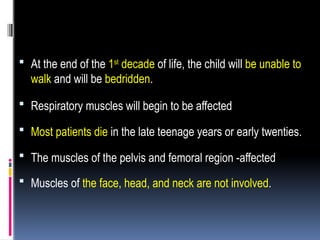
At theend of the 1st
decade of life, the child will be unable to
walk and will be bedridden.
Respiratory muscles will begin to be affected
Most patients die in the late teenage years or early twenties.
The muscles of the pelvis and femoral region -affected
Muscles of the face, head, and neck are not involved.
26.
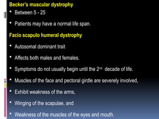
Becker’s muscular dystrophy
Between 5 - 25
Patients may have a normal life span.
Facio scapulo humeral dystrophy
Autosomal dominant trait
Affects both males and females.
Symptoms do not usually begin until the 2nd
decade of life.
Muscles of the face and pectoral girdle are severely involved,
Exhibit weakness of the arms,
Winging of the scapulae, and
Weakness of the muscles of the eyes and mouth.
27.
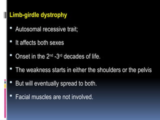
Limb-girdle dystrophy
Autosomalrecessive trait;
It affects both sexes
Onset in the 2nd
-3rd
decades of life.
The weakness starts in either the shoulders or the pelvis
But will eventually spread to both.
Facial muscles are not involved.
28.
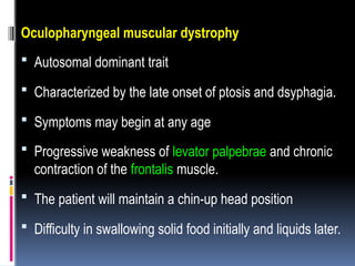
Oculopharyngeal muscular dystrophy
Autosomal dominant trait
Characterized by the late onset of ptosis and dsyphagia.
Symptoms may begin at any age
Progressive weakness of levator palpebrae and chronic
contraction of the frontalis muscle.
The patient will maintain a chin-up head position
Difficulty in swallowing solid food initially and liquids later.
29.
Myotonic dystrophy
Autosomaldominant trait
From birth to the age of 40 years,
Progressive muscular weakness, myotonia, cataracts, cardiac
abnormalities, hypogonadism, and frontal balding.
My occurs in the muscles of the head and neck and in the distal
extremities.
Unable to relax the muscles after contraction.
Observed in the forearm, thumb, and tongue.
Wasting of muscles and subsequent weakness
Prolapsed mitral valve and atrial flutter;
30.
Treatment
All formsof MD are incurable, and no satisfactory method
of retarding the muscle atrophy exists.
Corticosteroids have been shown to decrease the rate of
muscle loss, but only in the short term.
A physical therapy program will help to delay the
development of joint contractures
Orthopedic procedures may help to counteract deformities.
31.
Oral Health Considerations
Facioscapulo humeral
Myotonic forms of MD.
Difficulty in the ability to turn the head.
The muscles of facial expression and mastication are also
commonly affected.
The patient has difficulty in chewing or in pursing the lips.
Weakness of the facial muscles and enlargement of the
tongue due to fatty deposits.
32.
Oculopharyngeal form
Difficultyin swallowing.
Occlusal abnormalities
Lack of the proper muscle tension necessary to keep the teeth
properly aligned in the dental arch.
If the tongue is enlarged and the facial muscles are weak, the
teeth will be pushed out.
Macroglossia,
Anterior open bite,
TMJ dysfunction.
33.
Guillain-barré syndrome
(Acute idiopathicpolyneuropathy)
Acute symmetrical polyneuropathy, often occurring 1 to 3
weeks after an acute infection.
Often follows a nonspecific respiratory or GIT illness and
specific infections (CMV, EBV, and after immunization).
34.
Clinical Manifestations
Beginswith myalgia or paresthesias of the lower limbs,
followed by weakness
Involve abdominal, thoracic, and upper-limb muscles.
Respiration is compromised.
ANS - may induce changes in BP and pulse rate.
35.
Impaired swallowingor paresthesias of the mouth and
face
7th
cranial nerve is frequently involved, and bilateral
facial weakness is common.
Ptosis
Dysarthria, dysphagia and diplopia
36.
Treatment
Prednisone isineffective and may actually affect the
outcome adversely by prolonging recovery time
Plasmapheresis performed within the first few days of
illness.
37.
Epilepsy
Epilepsy isa condition characterized by abnormal,
recurrent, and excessive neuronal discharge precipitated
by many different disturbances within the CNS.
38.
Oral Health Considerations
Medicalhistory
What type of seizures the patient has,
How well the seizures are controlled,
The frequency and duration of seizures,
The potential triggers for seizures, and
What to expect if the patient has a seizure.
39.
Treatment planningmay be altered
Depending on the status of the seizure disorder.
Better to place a FPD rather than a removable appliance.
Gingival overgrowth. Phenytoin
Increase in the number of fibroblasts in the connective tissues.
Younger patients
Men and women are equally affected.
Does not correlation between dosage and the incidence of gingival
overgrowth.
Strong correlation between poor oral hygiene and the amount of
tissue
40.
Starts inthe interdental papillae and occurs only where teeth are
present.
The papillae enlarge buccally and lingually.
The enlarged areas are firm, pink, and covered with normal mucosa.
Treatment
Begins with prevention.
Careful oral hygiene can minimize the gingival enlargement.
Each patient should be referred to a dentist for oral hygiene
instruction and gingival curettage.
Gingivectomy.
Curettage
41.
Other side effects
Megaloblastic anemia,
Hirsutism, and
Lymphadenopathy.
Osteomalacia,
Thickening of the calvarium, and coarse facies
Patients with well-controlled epilepsy may be performed with
no change from normal treatment.
42.
Bell’s palsy
Unilateralparesis of the facial nerve.
Inflammatory reaction involving the facial nerve.
Herpes simplex virus 1
Bell’s palsy must be differentiated from other causes of facial
nerve palsy,
lyme disease,
Ramsay Hunt Syndrome
Acoustic neuromas.
43.
Clinical Manifestations
Beginswith slight pain around one ear, followed by an
abrupt paralysis of the muscles on that side of the face.
Eye on the affected side stays open,
The corner of the mouth drops,
Drooling.
Masseter weakness,
Food is retained in both the upper and lower buccal and
labial folds.
44.
The facialexpression changes remarkably,
The creases of the forehead are flattened.
Due to impaired blinking, corneal ulcerations from foreign
bodies can occur.
Involvement of the chorda tympani nerve leads to loss of
taste perception on the anterior two-thirds of the tongue.
Reduced salivary secretion
45.
Treatment
Systemic corticosteroidswithin the first few days after the onset
of paralysis,
Combining steroids with antiherpetic drugs
Protect the eye with lubricating drops or ointment
Facial plastic surgery
Anastomosis between the facial and hypoglossal nerves -
restore partial function.