- The document summarizes updates made in Version 2.2017 of the NCCN Guidelines for Thyroid Carcinoma from Version 1.2017.
- Key updates include revisions to diagnostic procedures, treatment guidelines, and surveillance recommendations for papillary carcinoma, follicular carcinoma, and Hürthle cell carcinoma.
- Molecular diagnostics were given a category 2A recommendation rather than category 2B for certain follicular lesions. Guidelines for radioiodine ablation and adjuvant therapy were also updated.


![Note: All recommendations are category 2A unless otherwise indicated.
Clinical Trials: NCCN believes that the best management of any cancer patient is in a clinical trial. Participation in clinical trials is especially encouraged.
Version 2.2017, 05/17/2017 © National Comprehensive Cancer Network, Inc. 2017, All rights reserved. The NCCN Guidelines®
and this illustration may not be reproduced in any form without the express written permission of NCCN®
.
NCCN Guidelines Index
Table of Contents
Discussion
NCCN Guidelines Version 2.2017 Updates
Thyroid Carcinoma
UPDATES
3 of 4
Papillary Carcinoma continued
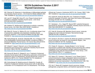
PAP-7
• Long-term surveillance statement was revised: Patients treated
with131I ablation, with a negative ultrasound, stimulated Tg 2
ng/mL (with negative antithyroglobulin antibodies), and negative
RAI imaging (if performed) may be followed by unstimulated
thyroglobulin annually and by periodic neck ultrasound. TSH-
stimulated testing, or other imaging (CT or MRI with contrast
or bone scan or chest x-ray) as clinically appropriate, may be
considered if clinical suggestion of recurrent disease. (Also for
FOLL-7 and HÜRT-7)
PAP-8
• “Locoregional recurrence” statement was revised: “Surgery
(preferred) if resectable and/or Radioiodine treatment, if radioiodine
imaging positive and/or EBRT/IMRT, if radioiodine imaging negative
and/or local therapies when available (ethanol ablation, RFA) and/
or EBRT/IMRT, if radioiodine imaging negative for select patients
not responsive to other therapies, or observation for low-volume
disease that is stable and distant from critical structures (Also for
FOLL-8 and HÜRT-8)
PAP-9
• 4th bullet for Iodine-refractory unresectable locoregional recurrent/
persistent disease or Iodine-refractory soft tissue metastases
(eg, lung, liver, muscle) excluding CNS metastases (see below)
was revised: Active surveillance may be is often appropriate in
asymptomatic patients with indolent disease assuming no brain
metastasis.(Also for FOLL-9 and HÜRT-9)
• A footnote was revised: While not FDA approved for treatment of
differentiated thyroid cancer, commercially available small-molecule
kinase inhibitors (such as axitinib, everolimus, pazopanib, sunitinib,
or vandetanib, vemurafenib (BRAF-positive), or cabozantinib [all are
category 2A]) can be considered if clinical trials are not available or
appropriate.
(Also for PAP-10, FOLL-9, FOLL-10, HÜRT-9 and HÜRT-10)
PAP-10
• A bullet was removed: Active surveillance may be appropriate in
asymptomatic patients with indolent disease. (See PAP-8) (Also for
FOLL-10 and HURT-10)
Follicular Carcinoma
FOLL-9
• 4th bullet for Iodine-refractory unresectable loco-regional recurrent/
persistent disease and Iodine-refractory soft tissue metastases (eg,
lung, liver, muscle) excluding CNS metastases (see below) was revised:
Active surveillance may be is often appropriate in asymptomatic patients
with indolent disease assuming no brain metastasis. (See FOLL-7)
FOLL-10
• For CNS Metastases a bullet was removed: Active surveillance may be
approriate in asymptomatic patients with indolent disease. (see FOLL-7)
(Also for HURT-10)
Medullary Carcinoma
MEDU-1
• Primary Treatment
�A bullet was revised: “Consider Adjuvant EBRT/IMRT is for gross
residual disease rarely recommended)” (Also for MEDU-3, MEDU-4)
MEDU-2
• Additional Workup
3rd bullet was revised: Screen for germline RET proto-oncogene
mutations
• Management
Statement revised: Germline RET positive mutation identified
Statement revised: Germline RET negative mutation not identified
MEDU-3
• Primary Treatment
4th bullet was revised: Consider Adjuvant EBRT/IMRT for gross
residual disease is rarely recommended (Also for MEDU-4)
Printed by Irfan Ashraf on 9/21/2017 12:01:47 PM. For personal use only. Not approved for distribution. Copyright © 2017 National Comprehensive Cancer Network, Inc., All Rights Reserved.](https://image.slidesharecdn.com/nccnguidelinesthyroid-230901141830-026f6616/85/NCCN-GUIDELINES-THYROID-pdf-6-320.jpg)


![NCCN Guidelines Index
Table of Contents
Discussion
Note: All recommendations are category 2A unless otherwise indicated.
Clinical Trials: NCCN believes that the best management of any cancer patient is in a clinical trial. Participation in clinical trials is especially encouraged.
Version 2.2017, 05/17/2017 © National Comprehensive Cancer Network, Inc. 2017, All rights reserved. The NCCN Guidelines®
and this illustration may not be reproduced in any form without the express written permission of NCCN®
.
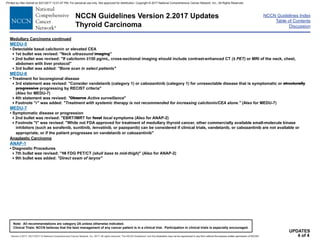
NCCN Guidelines Version 2.2017
Thyroid Carcinoma – Papillary Carcinoma
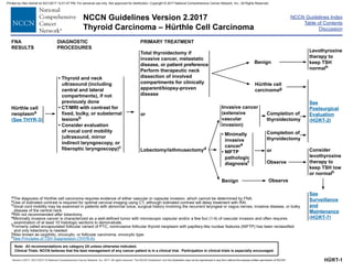
PAP-8
• Stimulated Tg 1–10 ng/mL
• Non-resectable tumors
• Non-radioiodine responsive
Suppress TSH with levothyroxineh
Locoregional
recurrence
Surgery (preferred) if resectabley
and/or
Radioiodine treatment,q
if radioiodine imaging positive
and/or
local therapies when available (ethanol ablation, radiofrequency ablation [RFA])
and/or
EBRT/IMRT, if radioiodine imaging negative for select patients not responsive to other therapies
or
observation for low-volume disease that is stable and distant from critical structures
Metastatic disease
See Treatment of Metastatic
Disease (PAP-9)
and/or
local therapies when available
RECURRENT DISEASE
hSee Principles of TSH Suppression (THYR-A).
qThe administered activity of RAI therapy should be adjusted for pediatric patients.
yPreoperative vocal cord assessment, if central neck recurrence.
Continue surveillance with unstimulated Tg,
ultrasound, and other imaging as clinically indicated
(see PAP-7)
• Stimulated Tg 10
ng/mL and rising
• Scans (including
PET) negative
Consider radioiodine therapy with 100–150 mCiq
and
post-treatment 131I imaging (category 3); additional RAI treatments
should be limited to patients who responded to previous RAI therapy
Printed by Irfan Ashraf on 9/21/2017 12:01:47 PM. For personal use only. Not approved for distribution. Copyright © 2017 National Comprehensive Cancer Network, Inc., All Rights Reserved.](https://image.slidesharecdn.com/nccnguidelinesthyroid-230901141830-026f6616/85/NCCN-GUIDELINES-THYROID-pdf-22-320.jpg)
![NCCN Guidelines Index
Table of Contents
Discussion
Note: All recommendations are category 2A unless otherwise indicated.
Clinical Trials: NCCN believes that the best management of any cancer patient is in a clinical trial. Participation in clinical trials is especially encouraged.
Version 2.2017, 05/17/2017 © National Comprehensive Cancer Network, Inc. 2017, All rights reserved. The NCCN Guidelines®
and this illustration may not be reproduced in any form without the express written permission of NCCN®
.
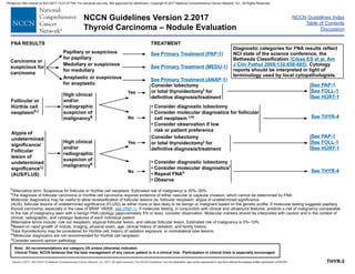
NCCN Guidelines Version 2.2017
Thyroid Carcinoma – Papillary Carcinoma
PAP-9
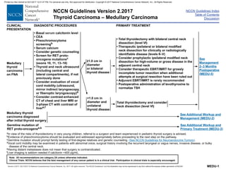
Structurally
persistent/recurrent
locoregional or
distant metastatic
disease not
amenable to RAI
therapy
• Continue to
suppress
TSH with
levothyroxineh
Iodine-refractory
soft tissue metastases
(eg, lung, liver, muscle)
excluding CNS
metastases (see below)
Iodine-
refractory
metastatic
bone
metastasesz
CNS metastases
TREATMENT OF METASTATIC DISEASE NOT AMENABLE TO RAI THERAPY
• For progressive and/or symptomatic disease, consider lenvatinib
(preferred) or sorafenib.aa
• While not FDA approved for the treatment of differentiated thyroid cancer,
other commercially available small molecular kinase inhibitors can be
considered for progressive and/or symptomatic disease if clinical trials or
other systemic therapies are not available or appropriate. bb,cc,dd
• Consider resection of distant metastases and/or EBRT/SBRT/IMRT/other
local therapiesee when available to metastatic lesions if progressive and/or
symptomatic.
• Active surveillance is often appropriate in asymptomatic patients with
indolent disease assuming no brain metastasis.bb (See PAP-7)
• Consider surgical palliation and/or EBRT/SBRT/other local therapiesee
when available if symptomatic, or asymptomatic in weight-bearing sites.
Embolization prior to surgical resection of bone metastases should be
considered to reduce the risk of hemorrhage.
• Consider embolization or other interventional procedures as alternatives to
surgical resection/EBRT/IMRT in select cases.
• Consider intravenous bisphosphonate or denosumab.z
• Active surveillance may be appropriate in asymptomatic patients with indolent
disease.bb (See PAP-7)
• For progressive and/or symptomatic disease, consider lenvatinib (preferred)
or sorafenib.bb While not FDA approved for the treatment of differentiated
thyroid cancer, other commercially available small molecular kinase inhibitors
can be considered for progressive and/or symptomatic disease if clinical
trials or other systemic therapies are not available or appropriate.bb,cc,dd
hSee Principles of TSH Suppression (THYR-A).
zDenosumab and intravenous bisphosphonates can be associated with severe hypocalcemia; patients with hypoparathyroidism and vitamin D deficiency are at increased risk.
aaThe decision of whether to use lenvatinib (preferred) or sorafenib should be individualized for each patient based on likelihood of response and comorbidities.
bbKinase inhibitor therapy may not be approriate for patients with stable or slowly progressive indolent disease. See Principles of Kinase Inhibitor Therapy (THYR-B).
ccWhile not FDA approved for treatment of differentiated thyroid cancer, commercially available small-molecule kinase inhibitors (such as axitinib, everolimus, pazopanib,
sunitinib, vandetanib, vemurafenib (BRAF-positive), or cabozantinib [all are category 2A]) can be considered if clinical trials are not available or appropriate.
ddCytotoxic chemotherapy has been shown to have minimal efficacy, although most studies were small and underpowered.
eeEthanol ablation, cryoablation, RFA, etc.
See (PAP-10)
Iodine-refractory
unresectable loco-
regional recurrent/
persistent disease
or
Printed by Irfan Ashraf on 9/21/2017 12:01:47 PM. For personal use only. Not approved for distribution. Copyright © 2017 National Comprehensive Cancer Network, Inc., All Rights Reserved.](https://image.slidesharecdn.com/nccnguidelinesthyroid-230901141830-026f6616/85/NCCN-GUIDELINES-THYROID-pdf-23-320.jpg)
![NCCN Guidelines Index
Table of Contents
Discussion
Note: All recommendations are category 2A unless otherwise indicated.
Clinical Trials: NCCN believes that the best management of any cancer patient is in a clinical trial. Participation in clinical trials is especially encouraged.
Version 2.2017, 05/17/2017 © National Comprehensive Cancer Network, Inc. 2017, All rights reserved. The NCCN Guidelines®
and this illustration may not be reproduced in any form without the express written permission of NCCN®
.
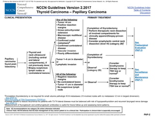
NCCN Guidelines Version 2.2017
Thyroid Carcinoma – Papillary Carcinoma
PAP-10
• For solitary CNS lesions, either neurosurgical resection or stereotactic radiosurgery is preferred.
and/or
• For multiple CNS lesions, consider resection and/or radiotherapy, including image-guided radiotherapy.
and/or
• For progressive and/or symptomatic disease, consider lenvatinib (preferred), or sorafenib.aa,ff
and/or
• While not FDA approved for the treatment of differentiated thyroid cancer, other commercially available
small molecular kinase inhibitors can be considered for progressive and/or symptomatic disease if
clinical trials or other systemic therapies are not available or appropriate.bb,cc,dd,ff
and/or
• Consider resection of distant metastases and/or EBRT/IMRT to metastatic lesions if progressive and/or
symptomatic.
TREATMENT OF METASTATIC DISEASE NOT AMENABLE TO RAI THERAPY
CNS metastases
aaThe decision of whether to use lenvatinib (preferred) or sorafenib should be individualized for each patient based on likelihood of response and comorbidities.
bbKinase inhibitor therapy may not be appropriate for patients with stable or slowly progressive indolent disease. See Principles of Kinase Inhibitor Therapy (THYR-B).
ccWhile not FDA approved for treatment of differentiated thyroid cancer, commercially available small-molecule kinase inhibitors (such as axitinib, everolimus, pazopanib,
sunitinib, vandetanib, vemurafenib (BRAF-positive), or cabozantinib [all are category 2A]) can be considered if clinical trials are not available or appropriate.
ddCytotoxic chemotherapy has been shown to have minimal efficacy, although most studies were small and underpowered.
ffAfter consultation with neurosurgery and radiation oncology, data on the efficacy of lenvatinib or sorafenib for patients with brain metastases have not
been established.
Printed by Irfan Ashraf on 9/21/2017 12:01:47 PM. For personal use only. Not approved for distribution. Copyright © 2017 National Comprehensive Cancer Network, Inc., All Rights Reserved.](https://image.slidesharecdn.com/nccnguidelinesthyroid-230901141830-026f6616/85/NCCN-GUIDELINES-THYROID-pdf-24-320.jpg)


![NCCN Guidelines Index
Table of Contents
Discussion
Note: All recommendations are category 2A unless otherwise indicated.
Clinical Trials: NCCN believes that the best management of any cancer patient is in a clinical trial. Participation in clinical trials is especially encouraged.
Version 2.2017, 05/17/2017 © National Comprehensive Cancer Network, Inc. 2017, All rights reserved. The NCCN Guidelines®
and this illustration may not be reproduced in any form without the express written permission of NCCN®
.
NCCN Guidelines Version 2.2017
Thyroid Carcinoma – Follicular Carcinoma
Structurally
persistent/recurrent
locoregional or
distant metastatic
disease not
amenable to RAI
therapy
• Continue to
suppress
TSH with
levothyroxineg
Iodine-refractory
unresectable loco-
regional recurrent/
persistent disease
or
Iodine-refractory
soft tissue metastases
(eg, lung, liver, muscle)
excluding CNS
metastases (see below)
Iodine-
refractory
metastatic
bone
metastasesx
CNS metastases
TREATMENT OF METASTATIC DISEASE NOT AMENABLE TO RAI THERAPY
• For progressive and/or symptomatic disease, consider lenvatinib
(preferred) or sorafenib.y
• While not FDA approved for the treatment of differentiated thyroid cancer,
other commercially available small molecular kinase inhibitors can be
considered for progressive and/or symptomatic disease if clinical trials or
other systemic therapies are not available or appropriate.z,aa,bb
• Consider resection of distant metastases and/or EBRT/SBRT/IMRT/other
local therapiescc when available to metastatic lesions if progressive and/or
symptomatic.
• Active surveillance is often appropriate in asymptomatic patients with
indolent disease assuming no brain metastasis.z (See FOLL-7)
• Consider surgical palliation and/or EBRT/SBRT/other local therapiescc
when available if symptomatic, or asymptomatic in weight-bearing sites.
Embolization prior to surgical resection of bone metastases should be
considered to reduce the risk of hemorrhage.
• Consider embolization or other interventional procedures as alternatives to
surgical resection/EBRT/IMRT in select cases.
• Consider intravenous bisphosphonate or denosumab.x
• Active surveillance may be appropriate in asymptomatic patients with indolent
disease.z (See FOLL-7)
• For progressive and/or symptomatic disease, consider lenvatinib (preferred) or
sorafenib.y While not FDA approved for the treatment of differentiated thyroid
cancer, other commercially available small molecular kinase inhibitors can
be considered for progressive and/or symptomatic disease if clinical trials or
other systemic therapies are not available or appropriate. z,aa,bb
gSee Principles of TSH Suppression (THYR-A).
xDenosumab and intravenous bisphosphonates can be associated with severe hypocalcemia; patients with hypoparathyroidism and vitamin D deficiency are at increased risk.
yThe decision of whether to use lenvatinib (preferred) or sorafenib should be individualized for each patient based on likelihood of response and comorbidities.
zKinase inhibitor therapy may not be appropriate for patients with stable or slowly progressive indolent disease.
See Principles of Kinase Inhibitor Therapy (THYR-B).
aaWhile not FDA approved for treatment of differentiated thyroid cancer, commercially available small-molecule kinase inhibitors (such as axitinib, everolimus, pazopanib,
sunitinib, vandetanib, vemurafenib (BRAF-positive), or cabozantinib [all are category 2A]) can be considered if clinical trials are not available or appropriate.
bbCytotoxic chemotherapy has been shown to have minimal efficacy, although most studies were small and underpowered.
ccEthanol ablation, cryoablation, RFA, etc.
See (FOLL-10)
FOLL-9
Printed by Irfan Ashraf on 9/21/2017 12:01:47 PM. For personal use only. Not approved for distribution. Copyright © 2017 National Comprehensive Cancer Network, Inc., All Rights Reserved.](https://image.slidesharecdn.com/nccnguidelinesthyroid-230901141830-026f6616/85/NCCN-GUIDELINES-THYROID-pdf-33-320.jpg)
![NCCN Guidelines Index
Table of Contents
Discussion
Note: All recommendations are category 2A unless otherwise indicated.
Clinical Trials: NCCN believes that the best management of any cancer patient is in a clinical trial. Participation in clinical trials is especially encouraged.
Version 2.2017, 05/17/2017 © National Comprehensive Cancer Network, Inc. 2017, All rights reserved. The NCCN Guidelines®
and this illustration may not be reproduced in any form without the express written permission of NCCN®
.
NCCN Guidelines Version 2.2017
Thyroid Carcinoma – Follicular Carcinoma
FOLL-10
• For solitary CNS lesions, either neurosurgical resection or stereotactic radiosurgery is preferred.
and/or
• For multiple CNS lesions, consider resection and/or radiotherapy, including image-guided radiotherapy.
and/or
• For progressive and/or symptomatic disease, consider lenvatinib (preferred), or sorafenib.y,dd
and/or
• While not FDA approved for the treatment of differentiated thyroid cancer, other commercially available
small molecular kinase inhibitors can be considered for progressive and/or symptomatic disease if
clinical trials or other systemic therapies are not available or appropriate.z,aa,bb,dd
and/or
• Consider resection of distant metastases and/or EBRT/IMRT to metastatic lesions if progressive and/or
symptomatic.
TREATMENT OF METASTATIC DISEASE NOT AMENABLE TO RAI THERAPY
CNS metastases
yThe decision of whether to use lenvatinib (preferred) or sorafenib should be individualized for each patient based on likelihood of response and comorbidities.
zKinase inhibitor therapy may not be appropriate for patients with stable or slowly progressive indolent disease. See Principles of Kinase Inhibitor Therapy (THYR-B).
aaWhile not FDA approved for treatment of differentiated thyroid cancer, commercially available small-molecule kinase inhibitors (such as axitinib, everolimus,
pazopanib, sunitinib, vandetanib, vemurafenib (BRAF-positive), or cabozantinib [all are category 2A]) can be considered if clinical trials are not available or
appropriate.
bbCytotoxic chemotherapy has been shown to have minimal efficacy, although most studies were small and underpowered.
ddAfter consultation with neurosurgery and radiation oncology; data on the efficacy of lenvatinib or sorafenib for patients with brain metastases have not been
established.
Printed by Irfan Ashraf on 9/21/2017 12:01:47 PM. For personal use only. Not approved for distribution. Copyright © 2017 National Comprehensive Cancer Network, Inc., All Rights Reserved.](https://image.slidesharecdn.com/nccnguidelinesthyroid-230901141830-026f6616/85/NCCN-GUIDELINES-THYROID-pdf-34-320.jpg)


![NCCN Guidelines Index
Table of Contents
Discussion
Note: All recommendations are category 2A unless otherwise indicated.
Clinical Trials: NCCN believes that the best management of any cancer patient is in a clinical trial. Participation in clinical trials is especially encouraged.
Version 2.2017, 05/17/2017 © National Comprehensive Cancer Network, Inc. 2017, All rights reserved. The NCCN Guidelines®
and this illustration may not be reproduced in any form without the express written permission of NCCN®
.
NCCN Guidelines Version 2.2017
Thyroid Carcinoma – Hürthle Cell Carcinoma
HÜRT-9
Structurally
persistent/recurrent
locoregional or
distant metastatic
disease not
amenable to RAI
therapy
• Continue to
suppress
TSH with
levothyroxineh
Iodine-refractory
unresectable loco-
regional recurrent/
persistent disease
or
Iodine-refractory
soft tissue metastases
(eg, lung, liver, muscle)
excluding CNS
metastases (see below)
Iodine-
refractory
metastatic
bone
metastasesy
CNS metastases
TREATMENT OF METASTATIC DISEASE NOT AMENABLE TO RAI THERAPY
• For progressive and/or symptomatic disease, consider lenvatinib (preferred),
or sorafenib.z
• While not FDA approved for the treatment of differentiated thyroid cancer, other
commercially available small molecular kinase inhibitors can be considered
for progressive and/or symptomatic disease if clinical trials or other systemic
therapies are not available or appropriateaa,bb,cc
• Consider resection of distant metastases and/or EBRT/SBRT/IMRT/other
local therapiesdd when available to metastatic lesions if progressive and/or
symptomatic.
• Active surveillance is often appropriate in asymptomatic patients with indolent
disease assuming no brain metastasis.aa (HÜRT-7)
• Consider surgical palliation and/or EBRT/SBRT/other local therapiesdd
when available if symptomatic, or asymptomatic in weight-bearing sites.
Embolization prior to surgical resection of bone metastases should be
considered to reduce the risk of hemorrhage.
• Consider embolization or other interventional procedures as alternatives to
surgical resection/EBRT/IMRT in select cases.
• Consider intravenous bisphosphonate or denosumab.y
• Active surveillance may be appropriate in asymptomatic patients with indolent
disease.aa (HÜRT-7)
• For progressive and/or symptomatic disease, consider lenvatinib (preferred)
or sorafenib.v While not FDA approved for the treatment of differentiated
thyroid cancer, other commercially available small molecular kinase inhibitors
can be considered for progressive and/or symptomatic disease if clinical trials
or other systemic therapies are not available or appropriate.aa,bb,cc
hSee Principles of TSH Suppression (THYR-A).
yDenosumab and intravenous bisphosphonates can be associated with severe hypocalcemia; patients with hypoparathyroidism and vitamin D deficiency are at
increased risk.
zThe decision of whether to use lenvatinib (preferred) or sorafenib should be individualized for each patient based on likelihood of response and comorbidities.
aaKinase inhibitor therapy may not be approriate for patients with stable or slowly progressive indolent disease. See Principles of Kinase Inhibitor Therapy (THYR-B).
bbWhile not FDA approved for treatment of differentiated thyroid cancer, commercially available small-molecule kinase inhibitors (such as axitinib, everolimus, pazopanib,
sunitinib, vandetanib, vemurafenib (BRAF-positive), or cabozantinib [all are category 2A]) can be considered if clinical trials are not available or appropriate.
ccCytotoxic chemotherapy has been shown to have minimal efficacy, although most studies were small and underpowered.
ddEthanol ablation, cryoablation, RFA, etc.
See (HÜRT-10)
Printed by Irfan Ashraf on 9/21/2017 12:01:47 PM. For personal use only. Not approved for distribution. Copyright © 2017 National Comprehensive Cancer Network, Inc., All Rights Reserved.](https://image.slidesharecdn.com/nccnguidelinesthyroid-230901141830-026f6616/85/NCCN-GUIDELINES-THYROID-pdf-43-320.jpg)
![NCCN Guidelines Index
Table of Contents
Discussion
Note: All recommendations are category 2A unless otherwise indicated.
Clinical Trials: NCCN believes that the best management of any cancer patient is in a clinical trial. Participation in clinical trials is especially encouraged.
Version 2.2017, 05/17/2017 © National Comprehensive Cancer Network, Inc. 2017, All rights reserved. The NCCN Guidelines®
and this illustration may not be reproduced in any form without the express written permission of NCCN®
.
NCCN Guidelines Version 2.2017
Thyroid Carcinoma – Hürthle Cell Carcinoma
HÜRT-10
• For solitary CNS lesions, either neurosurgical resection or stereotactic radiosurgery is preferred.
and/or
• For multiple CNS lesions, consider resection and/or radiotherapy, including image-guided radiotherapy.
and/or
• For progressive and/or symptomatic disease, consider lenvatinib (preferred) or sorafenib.z,ee
and/or
• While not FDA approved for the treatment of differentiated thyroid cancer, other commercially available
small molecular kinase inhibitors can be considered for progressive and/or symptomatic disease if clinical
trials or other systemic therapies are not available or appropriate. aa,bb,cc,ee
and/or
• Consider resection of distant metastases and/or EBRT/IMRT to metastatic lesions if progressive and/or
symptomatic.
TREATMENT OF METASTATIC DISEASE NOT AMENABLE TO RAI THERAPY
CNS metastases
zThe decision of whether to use lenvatinib (preferred) or sorafenib should be individualized for each patient based on likelihood of response and comorbidities.
aaKinase inhibitor therapy may not be appropriate for patients with stable or slowly progressive indolent disease. See Principles of Kinase Inhibitor Therapy (THYR-B).
bbWhile not FDA approved for treatment of differentiated thyroid cancer, commercially available small-molecule kinase inhibitors (such as axitinib, everolimus,
pazopanib, sunitinib, vandetanib, vemurafenib (BRAF-positive), or cabozantinib [all are category 2A]) can be considered if clinical trials are not available or
appropriate.
ccCytotoxic chemotherapy has been shown to have minimal efficacy, although most studies were small and underpowered.
eeAfter consultation with neurosurgery and radiation oncology; data on the efficacy of lenvatinib or sorafenib for patients with brain metastases have not been
established.
Printed by Irfan Ashraf on 9/21/2017 12:01:47 PM. For personal use only. Not approved for distribution. Copyright © 2017 National Comprehensive Cancer Network, Inc., All Rights Reserved.](https://image.slidesharecdn.com/nccnguidelinesthyroid-230901141830-026f6616/85/NCCN-GUIDELINES-THYROID-pdf-44-320.jpg)

![NCCN Guidelines Index
Table of Contents
Discussion
Note: All recommendations are category 2A unless otherwise indicated.
Clinical Trials: NCCN believes that the best management of any cancer patient is in a clinical trial. Participation in clinical trials is especially encouraged.
Version 2.2017, 05/17/2017 © National Comprehensive Cancer Network, Inc. 2017, All rights reserved. The NCCN Guidelines®
and this illustration may not be reproduced in any form without the express written permission of NCCN®
.
NCCN Guidelines Version 2.2017
Thyroid Carcinoma – Medullary Carcinoma
MEDU-3
CLINICAL
PRESENTATION
ADDITIONAL WORKUP PRIMARY TREATMENT
Germline
mutation of
RET
proto-
oncogenea,c
MEN 2B
(codon 918, 883, or
compound heterozygous
[V804M + E805K, Y806C,
or S904C] RET mutations)h
MEN 2A/Familial medullary
thyroid carcinoma
(codon 609, 611, 618, 620,
630, 634, 768, 790, 791, 804,
or 891 RET mutations)h
• Basal serum
calcitonin leveli
• CEA
• Pheochromocytoma
screeningb,j
• Central and lateral
neck compartments
ultrasound, if not
previously done
• Basal serum calcitonin leveli
• CEA
• Pheochromocytoma screeningb,j
• Serum calcium ± parathyroid hormone [PTH])
• Central and lateral neck compartments
ultrasound, if not previously done
• Total thyroidectomy during the first year of
life or at diagnosisa
• Therapeutic neck dissection as indicated;
consider prophylactic bilateral central neck
dissection (level VI)
• Consider more extensive node dissection
(levels II–V) if tumor(s) 0.5 cm in diameter
• Adjuvant EBRT/IMRT is rarely recommended
• Postoperative administration of
levothyroxine to normalize TSH
See
Management
2–3 Months
Postoperative
(MEDU-5)
See Primary
Treatment
(MEDU-4)
aIn view of the risks of thyroidectomy in very young children, referral to a surgeon and team experienced in pediatric thyroid surgery is advised.
bEvidence of pheochromocytoma should be evaluated and treated appropriately before proceeding to the next step on the pathway.
cGermline mutation should prompt family testing of first-degree relatives and genetic counseling. (See NCCN Guidelines for Neuroendocrine Tumors)
hThe timing of prophylactic thyroidectomy generally depends on the aggressiveness of the inherited RET mutation. Codon 634 mutations are considered highest risk with
MTC usually presenting at a younger age, whereas other RET mutations associated with MEN2A or FMTC are generally lower risk. Prophylactic thyroidectomy may be
delayed in patients with less high risk RET mutations that have later onset of MTC, provided the annual basal calcitonin measurement is normal, the annual ultrasound
is unremarkable, there is no history of aggressive MTC in the family, and the family is in agreement. (Brandi ML, Gagel RF, Angeli A, et al. Consensus: Guidelines for
diagnosis and therapy of MEN type 1 and type 2. J Clin Endocrinol Metab 2001;86(12):5658-5671 and American Thyroid Association Guidelines Task Force. Kloos RT,
Eng C, et al. Medullary thyroid cancer: management guidelines of the American Thyroid Association. Thyroid 2009;19:565-612.)
iNormal calcitonin ranges have not been established for very young children.
jScreening for pheochromocytoma (MEN 2A and 2B) and hyperparathyroidism (MEN 2A) should be performed annually. For some RET mutations (codons 768, 790, 804,
or 891), less frequent screening may be appropriate.
Printed by Irfan Ashraf on 9/21/2017 12:01:47 PM. For personal use only. Not approved for distribution. Copyright © 2017 National Comprehensive Cancer Network, Inc., All Rights Reserved.](https://image.slidesharecdn.com/nccnguidelinesthyroid-230901141830-026f6616/85/NCCN-GUIDELINES-THYROID-pdf-47-320.jpg)


![Version 2.2017, 05/17/17 © National Comprehensive Cancer Network, Inc. 2017, All rights reserved. The NCCN Guidelines® and this illustration may not be reproduced in any form without the express written permission of NCCN®. MS-5
NCCN Guidelines Index
Table of Contents
Discussion
NCCN Guidelines Version 2.2017
Thyroid Carcinoma
other short-lived 131Is were potent thyroid carcinogens in these
children, particularly those younger than 10 years of age when they
were exposed.39
Iodine deficiency increases the risk for
radiation-induced thyroid cancer.40
Although radiation-induced papillary
carcinoma tends to appear more aggressive histologically and to have
high recurrence rates, the prognosis for survival is similar to that of
spontaneously occurring tumors.41-43
Iodine deficiency is associated with
follicular carcinoma and anaplastic carcinomas.
Differentiated Thyroid Carcinoma
Clinical Presentation and Diagnosis
Differentiated (ie, papillary, follicular, Hürthle cell) thyroid carcinoma is
usually asymptomatic for long periods and commonly presents as a
solitary thyroid nodule. However, evaluating all nodules for malignancy
is difficult, because benign nodules are so prevalent and because
thyroid carcinoma is so uncommon.1,44,45
Moreover, both benign and
malignant thyroid nodules are usually asymptomatic, giving no clinical
clue to their diagnosis. About 50% of the malignant nodules are
discovered during a routine physical examination, by serendipity on
imaging studies, or during surgery for benign disease. The other 50%
are usually first noticed by the patient, usually as an asymptomatic
nodule.1,44
Regrettably, the typically indolent nature of differentiated
thyroid carcinoma often leads to long delays in diagnosis that may
substantially worsen the course of the disease.12
Initial Workup
For a patient with a thyroid nodule, the first step is to measure the
serum thyrotropin (thyroid-stimulating hormone [TSH]) level and to do
an ultrasound of the thyroid and neck; all nodules (even incidentalomas)
should have this assessment; there is no size cutoff.3,46-48
The TSH
level, ultrasound results, and clinical features are used to determine
whether is it necessary to do fine-needle aspiration (FNA) of the nodule
or whether there is a low risk of malignancy (see Nodule Evaluation in
the NCCN Guidelines for Thyroid Carcinoma).45,49
FNA, with or without ultrasound guidance, is the procedure of choice for
evaluating suspicious thyroid nodules.3,45,50
Data show that higher TSH
levels are associated with an increased risk for differentiated thyroid
carcinoma in patients with thyroid nodules, although TSH and
thyroglobulin (Tg) do not appear to be useful for screening for thyroid
cancer.51-54
FNA should be considered in patients with normal or
elevated TSH, certain ultrasound features, and clinical findings. FNA of
clinically significant or suspicious cervical lymph nodes should also be
considered if identified in the ultrasonographic evaluation of the thyroid
and neck. Ultrasound features that increase the threshold for FNA are
described in the NCCN algorithm (see Sonographic Features in Nodule
Evaluation in the NCCN Guidelines for Thyroid Carcinoma). RAI
imaging is recommended in patients with low TSH.
Sonographic (ultrasound) features can be used to predict either benign
or malignant thyroid nodules. Suspicious sonographic features include
hypoechoic, microcalcifications, infiltrative margins, and nodules that
are taller than they are wide in the transverse plane. Ultrasound
features associated with a low suspicion of malignancy include
isoechoic or hyperechoic solid nodules, mixed solid/cystic nodules, or
spongiform nodules without the suspicious features listed above.47,55-57
Standardized systems for assessing ultrasound features have been
created to improve consistency across centers.56,58
Other than the
presence of a pure cyst and nodule size, the inter-observer variability is
reported to be high, making comparisons between centers
challenging.57
Nonetheless, a constellation of findings—such as a
nodule with internal echogenicity consistent with microcalcifications,
irregular borders, and increased internal vascularity—conveys a higher
Printed by Irfan Ashraf on 9/21/2017 12:01:47 PM. For personal use only. Not approved for distribution. Copyright © 2017 National Comprehensive Cancer Network, Inc., All Rights Reserved.](https://image.slidesharecdn.com/nccnguidelinesthyroid-230901141830-026f6616/85/NCCN-GUIDELINES-THYROID-pdf-61-320.jpg)
![Version 2.2017, 05/17/17 © National Comprehensive Cancer Network, Inc. 2017, All rights reserved. The NCCN Guidelines® and this illustration may not be reproduced in any form without the express written permission of NCCN®. MS-6
NCCN Guidelines Index
Table of Contents
Discussion
NCCN Guidelines Version 2.2017
Thyroid Carcinoma
risk of malignancy. Because size is a comparatively reproducible
measure, its effect on likelihood of malignancy as an independent
variable has been assessed. Two articles suggest that size is a
relatively non-linear poor predictor of malignancy;47,59
however, it may
serve an important role in the setting of other concerning features.60
In the setting of a multinodular thyroid gland, selection of nodules for
FNA should be based on the pattern of radiographic features that
predict a higher likelihood of malignancy, such as the previous example,
or based on growth of a nodule over time. Similarly, choosing which
nodules are appropriate for active surveillance rather than FNA should
be based on the pattern of ultrasound features that predict benignity
(eg, spongiform appearance, a pure cyst, specific intranodular
appearances) or small size due to treatment considerations as
previously noted.55,56,61
At the time of thyroid ultrasound, a critical feature
that should be assessed is the presence or absence of concerning
lymphadenopathy in the central and lateral neck. The presence of a
node with concerning characteristics (eg, hypoechoic, rounded, absent
of fatty hilum, cystic or partially cystic, microcalcifications) should lead to
FNA of the node rather than, or in addition to, the most concerning
thyroid nodule.
Thyroid nodules smaller than 1 cm occur with such frequency in the
asymptomatic general population that they are often found by
serendipity when performing imaging studies for other head or neck
problems.17,62
Often termed “incidentalomas,” nodules smaller than 1 cm
are typically clinically insignificant lesions and usually do not require
FNA, unless there are suspicious findings (see Nodule Evaluation in the
NCCN Guidelines for Thyroid Carcinoma).4,14,47,63-67
In selected cases, it
may be reasonable to follow these nodules with serial ultrasounds. Data
indicate that older patients with intrathyroidal papillary microcarcinomas
may be good candidates for an active surveillance approach (rather
than immediate surgery) and usually show no evidence of clinically
significant disease progression over at least 5 to 10 years of follow-up.68
These observations cast doubt on the clinical benefit of diagnosing (and
treating) papillary microcarcinoma in these selected groups.69
Others
feel that surgery should be considered for select patients with papillary
carcinomas who are 45 years of age or older.70
The NCCN Panel uses recommendations from several organizations
(eg, American Thyroid Association [ATA], Society of Radiologists in
Ultrasound, NCI) and their expertise when formulating the NCCN
Guidelines for thyroid nodules (see Nodule Evaluation in the NCCN
Guidelines for Thyroid Carcinoma).3,49,71
The NCCN recommendations
describe which nodules require further assessment with FNA and which
can undergo active surveillance. In 2015, the ATA updated its
guidelines on the management of thyroid nodules and thyroid cancer; its
comprehensive guidelines also discuss ultrasound and FNA.72
In 2007,
the NCI had a conference on using FNA to manage thyroid nodules.
The NCI guidelines discuss which nodules should undergo FNA and
discuss the FNA results (ie, carcinoma, benign).45,49
The Society of
Radiologists in Ultrasound wrote a consensus statement in 2005 about
management of thyroid nodules identified at thyroid ultrasonography. Its
recommendations describe which nodules should undergo FNA based
on nodule size and ultrasound characteristics, and on clinical features
that might predict risk of morbidity from an undiagnosed malignancy.71
Suspicious criteria by ultrasound include increased central
hypervascularity, hypoechoic mass, microcalcifications, infiltrative
margins, and other features (see Sonographic Features in Nodule
Evaluation in the NCCN Guidelines for Thyroid Carcinoma).
Although more than 50% of all malignant nodules are asymptomatic, the
pretest probability of malignancy in a nodule increases considerably
when signs or symptoms are present (see Nodule Evaluation in the
Printed by Irfan Ashraf on 9/21/2017 12:01:47 PM. For personal use only. Not approved for distribution. Copyright © 2017 National Comprehensive Cancer Network, Inc., All Rights Reserved.](https://image.slidesharecdn.com/nccnguidelinesthyroid-230901141830-026f6616/85/NCCN-GUIDELINES-THYROID-pdf-62-320.jpg)
![Version 2.2017, 05/17/17 © National Comprehensive Cancer Network, Inc. 2017, All rights reserved. The NCCN Guidelines® and this illustration may not be reproduced in any form without the express written permission of NCCN®. MS-7
NCCN Guidelines Index
Table of Contents
Discussion
NCCN Guidelines Version 2.2017
Thyroid Carcinoma
NCCN Guidelines for Thyroid Carcinoma).73,74
For example, the
likelihood that a nodule is malignant increases about 7-fold if it is very
firm, fixed to adjacent structures, rapidly growing, associated with
enlarged regional lymph nodes, causes vocal cord paralysis, or
symptoms of invasion into neck structures are present.74,75
Family
history of thyroid cancer is also indicative of malignancy. If 2 or more of
these features are present, the likelihood of thyroid cancer is virtually
assured; however, this is a rare situation.75
A patient’s age and gender
also affect the probability of malignancy. Other factors that increase the
suspicion of malignancy include: 1) a history of head and neck
irradiation; 2) a history of diseases associated with thyroid carcinoma,
such as familial adenomatous polyposis (formerly called Gardner’s
syndrome), Carney complex, Cowden’s syndrome, and multiple
endocrine neoplasia (MEN) types 2A or 2B; 3) evidence of other thyroid
cancer–associated diseases or syndromes, such as
hyperparathyroidism, pheochromocytoma, marfanoid habitus, and
mucosal neuromas (suggestive of MEN2B), which make the presence
of medullary carcinoma more likely; or 4) the presence of suspicious
findings detected by imaging, such as focal FDG uptake on PET or
central hypervascularity, irregular border, and/or microcalcifications on
ultrasound.3,76
Some clinicians, especially in Europe,77
recommend obtaining serum
calcitonin levels from all patients with thyroid nodules to assess for
medullary carcinoma. However, this is controversial in the United
States, especially in the absence of confirmatory pentagastrin
stimulation testing and because it may not be cost effective. The ATA is
equivocal about measuring serum calcitonin to screen all patients with
thyroid nodules for medullary carcinoma.3
A study showed that
calcitonin screening may be cost effective in the United States.78
However, false-positive calcitonin readings that can result from minimal
calcitonin elevations have traditionally been ruled out with pentagastrin
testing, and pentagastrin is not available in the United States. Some
authors have suggested high-dose calcium infusion as an alternative to
pentagastrin stimulation testing in patients with minimal calcitonin
elevations.79
FNA Results
Cytologic examination of an FNA specimen is typically categorized as:
1) carcinoma (papillary, medullary, or anaplastic) or suspicious for
carcinoma; 2) follicular or Hürthle cell neoplasm; 3) atypia of
undetermined significance (AUS) or follicular lesion of undetermined
significance (FLUS); 4) thyroid lymphoma; 5) benign (ie, nodular goiter,
colloid goiter, hyperplastic/adenomatoid nodule, Hashimoto’s
thyroiditis); or 6) insufficient biopsy (nondiagnostic) (see Nodule
Evaluation in the NCCN Guidelines for Thyroid Carcinoma). These
diagnostic categories for FNA results reflect the NCI’s state of the
science conference held in 2007.45,49,80
Pathology and cytopathology
slides should be reviewed at the treating institution by a pathologist with
expertise in the diagnosis of thyroid disorders. Although FNA is a very
sensitive test—particularly for papillary carcinoma—false-negative
results are sometimes obtained; therefore, a reassuring FNA should not
override worrisome clinical or radiographic findings.81,82
Molecular diagnostic testing to detect individual mutations (eg, BRAF
V600E, RET/PTC, RAS, PAX8/PPAR [peroxisome proliferator-activated
receptors] gamma) or pattern recognition approaches using molecular
classifiers may be useful in the evaluation of FNA samples that are
indeterminate to assist in management decisions.83-91
The BRAF V600E
mutation occurs in about 45% of patients with papillary carcinoma and
is the most common mutation.92
Although controversial, data suggest
that BRAF V600E mutations may predict for increased recurrence of
Printed by Irfan Ashraf on 9/21/2017 12:01:47 PM. For personal use only. Not approved for distribution. Copyright © 2017 National Comprehensive Cancer Network, Inc., All Rights Reserved.](https://image.slidesharecdn.com/nccnguidelinesthyroid-230901141830-026f6616/85/NCCN-GUIDELINES-THYROID-pdf-63-320.jpg)


![Version 2.2017, 05/17/17 © National Comprehensive Cancer Network, Inc. 2017, All rights reserved. The NCCN Guidelines® and this illustration may not be reproduced in any form without the express written permission of NCCN®. MS-14
NCCN Guidelines Index
Table of Contents
Discussion
NCCN Guidelines Version 2.2017
Thyroid Carcinoma
Discussion) have been based on AJCC-TNM staging from earlier
editions, such as the 5th edition194
and not the 6th or 7th editions.9,193
Prognostic Scoring Strategies
Several staging and clinical prognostic scoring strategies use patient
age older than 40 years as a major feature to identify cancer mortality
risk from differentiated thyroid carcinoma.9,127,133,193,195
These strategies
include the EORTC, TNM 7th edition, AMES (Age, Metastases, Extent,
and Size), and AGES (Age, tumor Grade, Extent, and Size). All of these
strategies effectively distinguish between patients at low and high
risk.179
With incrementally worsening MACIS (Metastasis, Age,
Completeness of resection, Invasion, and Size) scores of less than 6, 6
to 6.99, 7 to 7.99, and 8+, however, the 20-year survival rates were
99%, 89%, 56%, and 24%, respectively.133
Unfortunately, a study that classified 269 patients with papillary
carcinoma according to 5 different prognostic paradigms found that
some patients in the lowest-risk group from each approach died of
cancer.136
This is particularly true of classification schemes that simply
categorize patients dichotomously as low or high risk.193,196
The AJCC
TNM staging approach (see Table 1 in the NCCN Guidelines for Thyroid
Carcinoma), which is perhaps the most widely used indicator of
prognosis, classifies tumors in all patients younger than 45 years as
stage I or stage II, even those with distant metastases. Although it
predicts cancer mortality reasonably well,197,198
TNM staging was not
established as a predictor of recurrence and therefore does not
accurately forecast the recurrences that often occur in patients who
developed thyroid carcinoma when they were young. Two studies have
shown the poor predictive value of most staging approaches for thyroid
carcinoma, including the TNM system.127,199
A three-tiered staging system—low, intermediate, high—that uses
clinicopathologic features to risk stratify with regard to the risk of
recurrence has been suggested and validated.3,200-203
This staging
system effectively risk stratifies patients with regard to the risk of
recurrence, risk of persistent disease after initial therapy, risk of having
persistent structural disease, likelihood of achieving remission in
response to initial therapy, and likelihood of being in remission at final
follow-up. In another approach, emphasis has been placed on
evaluation of response to therapy using a dynamic risk assessment
approach in which the initial risk estimates are modified during follow-up
as additional data are accumulated.204
This allows ongoing
re-assessment of risk and allows the management paradigm to be
better tailored to realistic estimates of risk that may change substantially
over time.
Surgical Management of Differentiated Thyroid Carcinoma
Ipsilateral Lobectomy Versus Total Thyroidectomy
The appropriate extent of thyroid resection—ipsilateral lobectomy
versus total thyroidectomy—is very controversial for lower-risk papillary
carcinoma, which is reflected in the NCCN category 2B
recommendations for these procedures (see Primary Treatment in the
NCCN Guidelines for Papillary [Thyroid] Carcinoma and Papillary
Thyroid Carcinoma in this Discussion). In most clinical settings,
decisions about the extent of thyroidectomy should be individualized
and done in consultation with the patient.205
Circumstances in which
lobectomy is not recommended are detailed in the NCCN Guidelines.
This debate reflects the limitations of prognostic scoring135
and the
morbidity often associated with total thyroidectomy performed outside of
major cancer centers. Patients treated at the Mayo Clinic Cancer Center
for low-risk PTCs (MACIS score ≤3.99) had no improvement in survival
rates after undergoing procedures more extensive than ipsilateral
Printed by Irfan Ashraf on 9/21/2017 12:01:47 PM. For personal use only. Not approved for distribution. Copyright © 2017 National Comprehensive Cancer Network, Inc., All Rights Reserved.](https://image.slidesharecdn.com/nccnguidelinesthyroid-230901141830-026f6616/85/NCCN-GUIDELINES-THYROID-pdf-70-320.jpg)
![Version 2.2017, 05/17/17 © National Comprehensive Cancer Network, Inc. 2017, All rights reserved. The NCCN Guidelines® and this illustration may not be reproduced in any form without the express written permission of NCCN®. MS-15
NCCN Guidelines Index
Table of Contents
Discussion
NCCN Guidelines Version 2.2017
Thyroid Carcinoma
lobectomy. Thus, the authors concluded that more aggressive surgery
was indicated only for those with higher MACIS scores.206
Cancer-specific mortality and recurrence rates after unilateral or
bilateral lobectomy were assessed in patients with papillary carcinoma
considered to be low risk by AMES criteria.207
No significant differences
were found in cancer-specific mortality or distant metastasis rates
between the 2 groups. However, the 20-year frequencies of local
recurrence and nodal metastasis after unilateral lobectomy were 14%
and 19%, respectively, which were significantly higher (P = .0001) than
the frequencies of 2% and 6% seen after bilateral thyroid lobe resection.
Hay et al concluded that bilateral thyroid resection is the preferable
initial surgical approach for patients with AMES low-risk papillary
carcinoma.207
Most NCCN Panel Members recommend total thyroidectomy for
patients with biopsy-proven papillary carcinoma who have large-volume
pathologic N1 metastases (5 involved nodes with metastases 2 mm
in largest dimension),3,34,208
because this procedure is associated with
improved disease-free survival.121,137,207,209
Some centers report that
patients treated by lobectomy alone have a 5% to 10% recurrence rate
in the opposite thyroid lobe.129,206
After lobectomy, these patients also
have an overall long-term recurrence rate of more than 30% (vs. 1%
after total thyroidectomy and 131I therapy)12
and the highest frequency
(11%) of subsequent pulmonary metastases.210
However, in properly
selected patients treated with lobectomy alone, recurrence rates may be
as low as 4%.41
Higher recurrence rates are also observed with cervical
lymph node metastases and multicentric tumors, providing some
additional justification for total thyroidectomy.12
However, some prominent thyroid cancer specialists (including some at
NCCN Member Institutions) oppose this view and advocate unilateral
lobectomy for most patients with papillary and follicular carcinoma
based on 1) the low mortality among most patients (ie, those patients
categorized as low risk by the AMES and other prognostic classification
schemes); and 2) the high complication rates reported with more
extensive thyroidectomy.134,195,211
The large thyroid remnant remaining
after unilateral lobectomy, however, may complicate long-term follow-up
with serum Tg determinations and whole-body 131I imaging. Panel
members recommend total lobectomy (without radioactive iodine RAI
ablation) for patients with papillary carcinoma who have small-volume
pathologic N1A metastases (5 involved nodes with no metastasis 2
mm, in largest dimension).212
NCCN Panel Members believe that total lobectomy alone is adequate
treatment for papillary microcarcinomas provided the patient has not
been exposed to radiation, has no other risk factors, and has a tumor
smaller than 1 cm that is unifocal and confined to the thyroid without
vascular invasion (see Primary Treatment in the NCCN Guidelines for
Papillary [Thyroid] Carcinoma).3,12,175,213-216
Total lobectomy alone is also
adequate treatment for NIFTP pathologies (see Tumor Variables
Affecting Prognosis, Histology) and minimally invasive follicular thyroid
carcinomas (see Primary Treatment in the NCCN Guidelines for
Follicular [Thyroid] Carcinoma). However, completion thyroidectomy is
recommended for any of the following: tumor more than 4 cm in
diameter, positive resection margins, gross extrathyroidal extension,
macroscopic multifocal disease, macroscopic nodal metastases,
confirmed contralateral disease, or vascular invasion.3
Note that “gross
extrathyroidal extension” refers to spread of the primary tumor outside
of the thyroid capsule with invasion into the surrounding structures such
as strap muscles, trachea, larynx, vasculature, esophagus, and/or
recurrent laryngeal nerve.149,217,218
Printed by Irfan Ashraf on 9/21/2017 12:01:47 PM. For personal use only. Not approved for distribution. Copyright © 2017 National Comprehensive Cancer Network, Inc., All Rights Reserved.](https://image.slidesharecdn.com/nccnguidelinesthyroid-230901141830-026f6616/85/NCCN-GUIDELINES-THYROID-pdf-71-320.jpg)

![Version 2.2017, 05/17/17 © National Comprehensive Cancer Network, Inc. 2017, All rights reserved. The NCCN Guidelines® and this illustration may not be reproduced in any form without the express written permission of NCCN®. MS-17
NCCN Guidelines Index
Table of Contents
Discussion
NCCN Guidelines Version 2.2017
Thyroid Carcinoma
is also recommended for select patients who are at greater risk for
recurrence with any of the following clinical indications such as primary
tumor 2 to 4 cm, high-risk histology, lymphatic invasion, cervical lymph
node metastases, macroscopic multifocality (one focus 1 cm), or
unstimulated postoperative serum Tg (5–10 ng/mL).3,232,233
However,
the NCCN Panel does not routinely recommend RAI for patients with all
of the following factors: 1) either unifocal or multifocal classic papillary
microcarcinomas (1 cm) confined to the thyroid; 2) no detectable anti-
Tg antibodies; and 3) postoperative unstimulated Tg less than 1 ng/mL.
Guidelines from the ATA list very similar indications for postoperative
RAI use and also provide specific guidance regarding the safe use of
RAI in the outpatient setting.3,234
Studies show decreased recurrence and disease-specific mortality for
populations at intermediate or higher risk when postoperative 131I
therapy is administered as part of the initial treatment.12,128,136,235-237
In a
study assessing outcomes in 1004 patients with differentiated thyroid
carcinoma, tumor recurrence was about 3-fold higher in patients either
treated with thyroid hormone alone or given no postoperative medical
therapy when compared with patients who underwent postoperative
thyroid remnant ablation with 131I (P .001). Moreover, fewer patients
developed distant metastases (P .002) after thyroid remnant 131I
ablation than after other forms of postoperative treatment. However, this
effect is observed only in patients with primary tumors 1.5 cm or more in
diameter.235
Another study of 21,870 intermediate risk patients with
differentiated thyroid cancer found that postoperative RAI improved OS
(P .001) and was associated with a 29% reduction in the risk of death
after adjustment for demographic and clinical factors (hazard risk, 0.71;
95% CI, .62–.82; P .001).237
Some studies have found that remnant
ablation had less of a therapeutic effect, perhaps because more
extensive locoregional surgery had been done.179
Previously, it was reported that postoperative RAI was associated with
decreased overall survival in patients with stage I thyroid cancer,
although the deaths seemed unrelated to thyroid cancer.238
Longer
follow-up suggests that overall survival is not decreased or increased in
these patients.239
However, a more recent study reported that the
incidence of secondary malignancies, such as leukemia and salivary
gland malignancies, has increased in patients with low-risk thyroid
cancer (ie, T1N0) who received RAI.240
Debate continues about ablating
the thyroid bed with 131I after total thyroidectomy.3,179,235,241
In patients
with papillary carcinoma who were at low risk for recurrence, thyroid
remnant ablation did not decrease recurrence rates.216,233,242
A long-term
study (n=1298) found that overall survival is not improved in patients
who receive RAI ablation.243
Reasons favoring remnant ablation include:
1) simplified patient follow-up, because elimination of thyroid bed uptake
prevents misinterpretation of it as disease; 2) elimination of normal
tissue as a source of Tg production, which facilitates identification of
patients who are free of disease and may simplify their care while
promoting early identification of those with residual cancer; and 3)
elimination of normal tissue, which may eliminate the nidus for
continued confounding anti-Tg antibody production. Conversely, others
argue that most recurrences can be easily detected with neck
ultrasound and that serum Tg levels are often quite low after a total
thyroidectomy. Therefore, in patients at low and intermediate risk, the
clinical benefit of routine remnant ablation as a requirement for optimal
follow-up remains uncertain.
Data suggest that lower doses of RAI are as effective as higher doses—
30 versus 100 mCi—for ablation in patients with low-risk thyroid cancer
(eg, T1b/T2 [1–4 cm], clinical N0 disease).32,33
The NCCN Guidelines
reflect a more cautious approach to using RAI ablation based on these
randomized trials.244
If RAI ablation is used, the NCCN Guidelines
Printed by Irfan Ashraf on 9/21/2017 12:01:47 PM. For personal use only. Not approved for distribution. Copyright © 2017 National Comprehensive Cancer Network, Inc., All Rights Reserved.](https://image.slidesharecdn.com/nccnguidelinesthyroid-230901141830-026f6616/85/NCCN-GUIDELINES-THYROID-pdf-73-320.jpg)


![Version 2.2017, 05/17/17 © National Comprehensive Cancer Network, Inc. 2017, All rights reserved. The NCCN Guidelines® and this illustration may not be reproduced in any form without the express written permission of NCCN®. MS-20
NCCN Guidelines Index
Table of Contents
Discussion
NCCN Guidelines Version 2.2017
Thyroid Carcinoma
while the patient continues thyroid hormone suppressive therapy and
avoids symptomatic hypothyroidism.269
Administration of rhTSH is well
tolerated; nausea (10.5%) and transient mild headache (7.3%) are its
main adverse effects.267
It is associated with significantly fewer
symptoms and dysphoric mood states than hypothyroidism induced by
thyroid hormone withdrawal.269
An international study was performed to assess the effects of 2 rhTSH
dosing schedules on whole-body 131I imaging and serum Tg levels
when compared with imaging and Tg levels obtained after thyroid
hormone withdrawal.267
Data showed that the combination of rhTSH–
stimulated whole-body imaging and serum Tg measurements detected
100% of metastatic carcinoma.267
In this study, 0.9 mg of rhTSH was
given intramuscularly every day for 2 days, followed by a minimum of 4
mCi of 131I on the third day. Whole-body imaging and Tg
measurements were performed on the fifth day. Whole-body 131I
images were acquired after 30 minutes of imaging or after obtaining
140,000 counts, whichever came first. A serum Tg of 2.0 ng/mL or
higher, obtained 72 hours after the last rhTSH injection, indicates that
thyroid tissue or thyroid carcinoma is present, regardless of the
whole-body imaging findings.267,270
Measuring Serum Tg and Anti-Tg Antibodies
Serum Tg measurement is the best means of detecting thyroid tissue,
including carcinoma. Tg can be measured when TSH has been
stimulated—either by thyroid hormone withdrawal or by rhTSH—
because in this setting, serum Tg has a lower false-negative rate than
whole-body 131I imaging.266-268,271
Serum Tg levels vary in response to
the increase in serum TSH after thyroid hormone withdrawal or rhTSH
stimulation. Serum Tg generally does not increase as much after rhTSH
administration as after withdrawal of thyroid hormone. The conditions
for rhTSH–stimulated, whole-body 131I imaging stipulate using 4-mCi
131I doses (based on the trial)267
and an imaging time of 30 minutes or
until 140,000 counts are obtained. Tg measurements may also be
obtained without stimulating TSH using ultrasensitive assays (ie,
second-generation Tg immunometric assays [TgIMAs]).272,273
It is useful
to measure serum Tg and anti-Tg antibody levels for follow-up and
assessing trend patterns.
The sensitivity and specificity of various Tg assays, however, vary
widely in different laboratories, even with the use of an international
standard (CRM 457).274,275
Thus, it is recommended that patients
undergo Tg monitoring via the same Tg assay performed in the same
laboratory. Ideally, serum is frozen and saved for future analyses if
needed, especially should a change in Tg assay be necessary. As the
sensitivity of commercially available Tg assays improves, the need for
stimulated Tg testing may become less important.
Anti-Tg antibodies should be measured in the same serum sample
taken for Tg assay, because these antibodies (which are found in ≤25%
of patients with thyroid carcinoma) invalidate serum Tg measurements
in most assays.272,275,276
These antibodies typically falsely lower the Tg
value in immunochemiluminometric assays (ICMAs) and
immunoradiometric assays (IRMAs), while raising the value in older
radioimmunoassays. Although the clinical importance of anti-Tg
antibodies is unclear, their persistence for more than 1 year after
thyroidectomy and RAI ablation probably indicates the presence of
residual thyroid tissue and possibly an increased risk of recurrence.276
In one study, 49% of patients had a recurrence if they had undetectable
serum Tg and serum anti-Tg antibody levels of 100 units/mL or more
when compared with only 3% of patients with undetectable serum Tg
and serum anti-Tg antibodies of less than 100 units/mL.277
In patients
with coexistent autoimmune thyroid disease at the time of surgery,
Printed by Irfan Ashraf on 9/21/2017 12:01:47 PM. For personal use only. Not approved for distribution. Copyright © 2017 National Comprehensive Cancer Network, Inc., All Rights Reserved.](https://image.slidesharecdn.com/nccnguidelinesthyroid-230901141830-026f6616/85/NCCN-GUIDELINES-THYROID-pdf-76-320.jpg)
![Version 2.2017, 05/17/17 © National Comprehensive Cancer Network, Inc. 2017, All rights reserved. The NCCN Guidelines® and this illustration may not be reproduced in any form without the express written permission of NCCN®. MS-21
NCCN Guidelines Index
Table of Contents
Discussion
NCCN Guidelines Version 2.2017
Thyroid Carcinoma
anti-Tg antibodies may persist far longer. In a study of 116 patients with
anti-Tg antibodies before thyroidectomy, antibodies remained
detectable for up to 20 years in some patients without detectable thyroid
tissue, and the median time to disappearance of antibodies was 3
years.278
Patients with persistently undetectable serum Tg and anti-Tg
antibody levels have longer disease-free survival when compared with
patients who have detectable levels.279
Treating Patients With Positive Tg and Negative Imaging
Post-treatment 131I imaging may indicate the location of metastases
when the serum Tg level is increased, but a tumor [or metastases]
cannot be found by physical examination or other localizing techniques
such as diagnostic 131I imaging, neck ultrasonography, CT, MRI, or
PET. Pulmonary metastases may be found only after administering
therapeutic doses of 131I and obtaining whole-body imaging within a
few days of treatment.280
In a study of 283 patients treated with 100 mCi
(3700 MBq) of 131I, 6.4% had lung and bone metastases detected after
treatment that had been suspected based on high serum Tg
concentrations alone but that had not been detected after 2-mCi (74
MBq) diagnostic imaging.281
Unfortunately, most patients who are diagnostic imaging–negative and
Tg-positive are not rendered disease free by 131I therapy; however, the
tumor burden may be diminished.282
Thus, most patients with residual or
recurrent disease confined to the neck undergo re-operation rather than
RAI therapy in the hopes of a cure. RAI therapy is more commonly
considered for those with distant metastases or inoperable local
disease. Patients not benefiting from this therapy can be considered for
clinical trials, especially those patients with progressive metastatic
disease. When a large tumor is not visible on diagnostic whole-body
imaging, its ability to concentrate 131I is very low; thus, the tumor will
not respond to 131I therapy.
Thyroid Hormone Suppression of TSH
The use of postoperative levothyroxine to decrease TSH levels is
considered optimal in treatment of patients with papillary, follicular, or
Hürthle cell carcinoma, because TSH is a trophic hormone that can
stimulate the growth of cells derived from thyroid follicular
epithelium.3,250,283,284
However, the optimal serum levels of TSH have not
been defined because of a lack of specific data; therefore, the NCCN
Panel recommends tailoring the degree of TSH suppression to the risk
of recurrence and death from thyroid cancer for each individual patient.
For patients with known residual carcinoma or those at high risk for
recurrence, the recommended TSH level is below 0.1 milliunits/L. For
patients at low risk and for those patients with an excellent response to
initial therapy who are in remission, the recommended TSH level is
either slightly below or slightly above the lower limit of the reference
range. The risk and benefit of TSH-suppressive therapy must be
balanced for each individual patient because of the potential toxicities
associated with TSH-suppressive doses of levothyroxine, including
cardiac tachyarrhythmias (especially in the elderly), bone
demineralization (particularly in post-menopausal women), and frank
symptoms of thyrotoxicosis.3,285
An adequate daily intake of calcium
(1200 mg/d) and vitamin D (1000 units/d) is recommended for patients
whose TSH levels are chronically suppressed. However, reports do not
suggest that bone mineral density is altered in patients receiving
levothyroxine.286,287
Decreased recurrence and cancer-specific mortality rates for
differentiated thyroid carcinoma have been reported for patients treated
with thyroid hormone suppressive therapy.12,235,238,284,288-290
The average
dosage needed to attain serum TSH levels in the euthyroid range is
higher in patients who have been treated for thyroid carcinoma (2.11
mcg/kg per day) than in those patients with spontaneously occurring
primary hypothyroidism (1.62 mcg/kg per day).290
Even higher doses are
Printed by Irfan Ashraf on 9/21/2017 12:01:47 PM. For personal use only. Not approved for distribution. Copyright © 2017 National Comprehensive Cancer Network, Inc., All Rights Reserved.](https://image.slidesharecdn.com/nccnguidelinesthyroid-230901141830-026f6616/85/NCCN-GUIDELINES-THYROID-pdf-77-320.jpg)

![Version 2.2017, 05/17/17 © National Comprehensive Cancer Network, Inc. 2017, All rights reserved. The NCCN Guidelines® and this illustration may not be reproduced in any form without the express written permission of NCCN®. MS-23
NCCN Guidelines Index
Table of Contents
Discussion
NCCN Guidelines Version 2.2017
Thyroid Carcinoma
objective responses.307
In a review of published series, 38% of patients
had a response (defined as a decrease in tumor mass) to
doxorubicin.308
Combination chemotherapy is not clearly superior to
doxorubicin therapy alone.129
Overall, traditional cytotoxic systemic
chemotherapy, such as doxorubicin, has minimal efficacy in patients
with metastatic differentiated thyroid disease.309
Novel treatments for
patients with metastatic differentiated thyroid carcinoma have been
evaluated.310-317
Agents include multitargeted kinase inhibitors, such as
lenvatinib,310,313,318-324
sorafenib,325-332
sunitinib,330,333
axitinib,334-336
everolimus,337
vandetanib,338
cabozantinib,311,339
and pazopanib;340
and
BRAF V600E mutation inhibitors, such as vemurafenib and
dabrafenib.341-344
Data suggest that anaplastic lymphoma kinase (ALK)
inhibitors may be effective in patients with papillary carcinoma who have
ALK gene fusion.345-348
Clinical trials suggest that kinase inhibitors have a clinical benefit
(partial response rates plus stable disease) in 50% to 60% of subjects,
usually for about 12 to 24 months.313,321,330,340,349-351
Lenvatinib and
sorafenib are recommended for the treatment of patients with RAI-
refractory differentiated thyroid cancer (see Papillary Thyroid Carcinoma
in this Discussion and the NCCN Guidelines for Papillary [Thyroid]
Carcinoma).320,325
Vandetanib and cabozantinib, oral kinase inhibitors,
are recommended for the treatment of medullary carcinoma in patients
with unresectable locally advanced or metastatic disease (see
Medullary Thyroid Carcinoma in this Discussion and the NCCN
Guidelines for Medullary [Thyroid] Carcinoma).352-355
Severe or fatal side
effects from kinase inhibitors include bleeding, hypertension, stroke,
and liver toxicity; however, most side effects can be managed and are
reversible with discontinuation of the drug.320,321,356-361
Dose modifications
of kinase inhibitors may be required. Pazopanib has been reported to
cause reversible hypopigmentation.362
Papillary Thyroid Carcinoma
Surgical Therapy
Imaging is performed before surgery to ascertain the extent of disease
and to aid in the surgical decision-making process. A cervical
ultrasound, including the thyroid and the central lateral compartments,
is the principal imaging modality that is recommended.363
In one report,
cervical ultrasound performed before primary surgery for newly
diagnosed thyroid cancer identified metastatic sites not appreciated on
physical examination in 20% of patients, and surgical strategy was
altered in 39% of patients.364
Surgeon-performed preoperative
ultrasound identified nonpalpable metastatic lymph nodes in 24% of
patients.365
In more than 700 patients with PTC, preoperative ultrasound
detected nonpalpable nodal metastases in 33% of subjects.366
Preoperative ultrasound findings altered the operation in more than
40% of cases. In another report,367
operative management was altered
in 23% of the total group due to findings on the preoperative ultrasound.
These studies indicate that preoperative ultrasound has a high
sensitivity for nodal disease and will detect nonpalpable nodal
metastases in 20% to 33% of patients, and ultrasound should alter the
index operation in a similar percentage of patients. In most cases,
lesions suspicious for locoregional recurrence, which are amenable to
needle biopsy, should be interrogated with FNA biopsy before surgery.
Tg washout assay may be a useful adjunct to FNA biopsy in these
cases. Cross-sectional imaging (CT or MRI) should be performed if the
thyroid lesion is fixed, bulky, or substernal. Iodinated contrast is
required for optimal cervical imaging with CT, although iodinated
contrast will delay treatment with RAI. Evaluation of vocal cord mobility
may be considered for patients with abnormal voice, a surgical history
involving the recurrent laryngeal or vagus nerves, invasive disease, or
bulky disease of the central neck. Vocal cord mobility may be evaluated
Printed by Irfan Ashraf on 9/21/2017 12:01:47 PM. For personal use only. Not approved for distribution. Copyright © 2017 National Comprehensive Cancer Network, Inc., All Rights Reserved.](https://image.slidesharecdn.com/nccnguidelinesthyroid-230901141830-026f6616/85/NCCN-GUIDELINES-THYROID-pdf-79-320.jpg)
![Version 2.2017, 05/17/17 © National Comprehensive Cancer Network, Inc. 2017, All rights reserved. The NCCN Guidelines® and this illustration may not be reproduced in any form without the express written permission of NCCN®. MS-24
NCCN Guidelines Index
Table of Contents
Discussion
NCCN Guidelines Version 2.2017
Thyroid Carcinoma
by ultrasound, mirror indirect laryngoscopy, or fiber-optic
laryngoscopy.368
The NCCN Panel agreed on the characteristics of patients at higher risk
who require total thyroidectomy and neck dissection as the primary
treatment (see Preoperative or Intraoperative Decision-Making Criteria
in the NCCN Guidelines for Papillary [Thyroid] Carcinoma).3,369,370
A total
thyroidectomy is recommended for patients with any one of the
following factors, including: known distant metastases, extrathyroidal
extension, tumor greater than 4 cm in diameter, cervical lymph node
metastases, or poorly differentiated histology. Total thyroidectomy may
be considered for patients with bilateral nodularity or a prior exposure to
radiation (category 2B for radiation exposure). Clinically positive and/or
biopsy-proven nodal metastases should be treated with a formal
compartmental resection. In the central neck, this is achieved through a
unilateral or bilateral level VI dissection. In the lateral compartment, a
formal modified radical neck dissection including levels II, III, IV, and Vb
should be performed.371
Extending the dissection field into levels I or Va
may be necessary when these levels are clinically involved. Based on
the results of a randomized controlled trial, the panel does not
recommend prophylactic central neck dissection if the cervical lymph
nodes are clinically negative. This trial of 181 patients with PTC
randomized patients to receive either total thyroidectomy alone or total
thyroidectomy plus central neck dissection and showed no difference in
outcomes between the two groups.372
Central neck dissection will be
required ipsilateral to a modified radical neck dissection done for
clinically involved lateral neck lymph nodes in most cases. Selective
dissection of individual nodal metastases (ie, cherry picking) is not
considered adequate surgery for nodal disease in a previously
undissected field.
The NCCN Panel did not uniformly agree about the preferred primary
surgery for patients with PTC who are assumed to be at lower risk of
cancer-specific mortality. As previously mentioned, the extent of thyroid
resection—ipsilateral lobectomy versus total thyroidectomy—is very
controversial for lower-risk PTC, which is reflected in the NCCN
category 2B recommendations for these procedures (see Ipsilateral
Lobectomy Versus Total Thyroidectomy in this Discussion). Lobectomy
plus isthmusectomy is recommended for patients who cannot (or refuse
to) take thyroid hormone replacement therapy for the remainder of their
lives.205
Note that some patients prefer to have total thyroidectomy to
avoid having a second surgery (ie, completion thyroidectomy). Other
patients prefer to have a lobectomy in an attempt to avoid thyroid
hormone replacement therapy.
A study of more than 5000 patients found that survival of patients after
partial thyroidectomy was similar to the survival after total thyroidectomy
for patients at low and high risk.373
An observational study (SEER
database) in more than 35,000 patients with PTC limited to the thyroid
gland suggests that survival is similar whether (or not) patients are
treated in the first year after diagnosis and whether they undergo
lobectomy or total thyroidectomy.374
However, most guidelines (eg,
NCCN, ATA) do not recommend active surveillance for patients with
PTC.3
Another study of 2784 patients with differentiated thyroid
carcinoma (86% with PTC) found that total thyroidectomy was
associated with increased survival in patients at high risk.238
A study in
52,173 patients found that total thyroidectomy reduces recurrence rates
and improves survival in patients with PTC of 1 cm or more when
compared with lobectomy.375
For patients at lower risk who undergo
lobectomy plus isthmusectomy, completion of thyroidectomy is
recommended for any one of the following risk factors: large tumor (4
cm), positive resection margins, gross extrathyroidal extension,
Printed by Irfan Ashraf on 9/21/2017 12:01:47 PM. For personal use only. Not approved for distribution. Copyright © 2017 National Comprehensive Cancer Network, Inc., All Rights Reserved.](https://image.slidesharecdn.com/nccnguidelinesthyroid-230901141830-026f6616/85/NCCN-GUIDELINES-THYROID-pdf-80-320.jpg)
![Version 2.2017, 05/17/17 © National Comprehensive Cancer Network, Inc. 2017, All rights reserved. The NCCN Guidelines® and this illustration may not be reproduced in any form without the express written permission of NCCN®. MS-25
NCCN Guidelines Index
Table of Contents
Discussion
NCCN Guidelines Version 2.2017
Thyroid Carcinoma
macroscopic multifocal disease, vascular invasion, or macroscopic
nodal metastases. While a retrospective study using the National
Cancer Database has shown that a sizable percentage of patients with
differentiated thyroid cancer receive RAI therapy following lobectomy376
,
the panel does not support this practice due to a lack of data showing
benefit. Therefore, RAI is not recommended following lobectomy for
differentiated thyroid cancer.
Incidentally discovered PTCs 1 to 4 cm in size may warrant a
completion thyroidectomy (category 2B) for lymphatic invasion (see
Primary Treatment in the NCCN Guidelines for Papillary [Thyroid]
Carcinoma); observation (category 2B) is another option for these
patients (ie, with measurement of Tg and anti-Tg antibodies).
Levothyroxine therapy can be considered for these patients to maintain
the TSH levels at low or normal (see Principles of TSH Suppression in
the NCCN Guidelines for Thyroid Carcinoma). Lobectomy is sufficient
for tumors resected with all of the following: negative resection margins,
no contralateral lesion, no suspicious lymph node(s), and small (1 cm)
PTCs found incidentally on the final pathology sections; these patients
are observed (ie, with measurement of Tg and anti-Tg antibodies).
Levothyroxine therapy to reduce serum TSH to low or low-normal
concentrations can be considered for these patients (see Principles of
TSH Suppression in the NCCN Guidelines for Thyroid Carcinoma).
Radioactive Iodine Therapy
Postoperative RAI administration is recommended when a number of
clinical factors predict a significant risk of recurrence, distant
metastases, or disease-specific mortality. Clinicopathologic factors can
be used to guide decisions about whether to use initial postoperative
RAI (see Clinicopathologic Factors in the NCCN Guidelines for Papillary
[Thyroid] Carcinoma). For example, RAI may be recommended when
the primary tumor is 1 to 4 cm, but the final decision depends on the
combination of individual clinical factors as outlined in the NCCN
Guidelines. Algorithms can assist in decision making about use of RAI
in different settings: 1) RAI is not typically indicated for patients
classified as having a low risk of recurrence/disease-specific mortality;
2) RAI is not recommended after lobectomy; 3) RAI may be considered
for patients without gross residual disease, but data are conflicting
regarding the benefit of RAI in this setting; and 4) RAI is often used for
patients with known or suspected distant metastatic disease at
presentation. However, some patients may have metastatic disease that
may not be amenable to RAI therapy, which is also known as iodine-
refractory disease (see Treatment of Metastatic Disease Not Amenable
to RAI Therapy in the NCCN Guidelines for Papillary [Thyroid]
Carcinoma).
Therapy with 131I is typically recommended for patients with: 1) gross
extrathyroidal extension; 2) primary tumor greater than 4 cm; or 3)
postoperative unstimulated Tg greater than 5 to 10 ng/mL. All patients
should be examined, and palpable neck disease should be surgically
resected before any RAI treatment. A negative pregnancy test is
required before the administration of RAI in women of child-bearing
potential. The administered activity of RAI therapy should be adjusted
for pediatric patients.262
RAI is not typically recommended for patients
with either unifocal or multifocal papillary microcarcinomas (2 cm)
confined to the thyroid, and clinical N0 and M0.244
The NCCN Panel
agrees that postoperative RAI administration after total thyroidectomy is
not needed for patients with classic PTC with small-volume N1a disease
if the postoperative unstimulated Tg levels are less than 1 ng/mL, 131I
imaging is negative, there are no concerning findings on ultrasound,
and anti-Tg antibody level is negative. RAI is selectively recommended
if any of the following are present: 1) primary tumor 2 to 4 cm; 2) poorly
Printed by Irfan Ashraf on 9/21/2017 12:01:47 PM. For personal use only. Not approved for distribution. Copyright © 2017 National Comprehensive Cancer Network, Inc., All Rights Reserved.](https://image.slidesharecdn.com/nccnguidelinesthyroid-230901141830-026f6616/85/NCCN-GUIDELINES-THYROID-pdf-81-320.jpg)
![Version 2.2017, 05/17/17 © National Comprehensive Cancer Network, Inc. 2017, All rights reserved. The NCCN Guidelines® and this illustration may not be reproduced in any form without the express written permission of NCCN®. MS-26
NCCN Guidelines Index
Table of Contents
Discussion
NCCN Guidelines Version 2.2017
Thyroid Carcinoma
differentiated histology, tall cell, columnar cell, and hobnail variants; 3)
lymphatic invasion; 4) cervical lymph node metastases; 5) macroscopic
multifocality (ie, one focus 1 cm); or 6) postoperative unstimulated Tg
less than 5 to 10 ng/mL. For patients with suspected or proven
RAI-responsive residual tumor, RAI treatment is recommended (100–
200 mCi) followed by post-treatment imaging; dosimetry can be
considered for distant metastases (see RAI Being Considered Based on
Clinicopathologic Features in the NCCN Guidelines for Papillary
[Thyroid] Carcinoma).3
For patients with unresectable gross residual disease in the neck
(suspected or proven) that is refractory to RAI, EBRT or IMRT can be
considered if disease is threatening vital structures (see Postsurgical
Evaluation in the NCCN Guidelines for Papillary [Thyroid]
Carcinoma).3,297,298,377-379
Surveillance and Maintenance
The recommendations for surveillance and maintenance are described
in the algorithm (see Surveillance and Maintenance in the NCCN
Guidelines for Papillary [Thyroid] Carcinoma).3
About 85% of patients
are considered to be low risk after surgery for papillary thyroid cancer.3
In patients who have had total (or near total) thyroidectomy and thyroid
remnant ablation, the ATA Guidelines define the absence of persistent
tumor (also known as no evidence of disease [NED]) as: 1) absence of
clinical evidence of tumor; 2) absence of imaging evidence of tumor;
and 3) undetectable Tg levels (during either TSH suppression or TSH
stimulation) and absence of anti-Tg antibodies.3
Patients treated with
131I ablation may be followed with unstimulated Tg annually and with
periodic neck ultrasound if they have negative ultrasounds, stimulated
Tg less than 2 ng/mL (with negative anti-Tg antibodies), and negative
RAI imaging (if performed). However, if they have a clinical suggestion
of recurrent disease, then TSH-stimulated testing (or other imaging)
may be considered. A subgroup of patients at low risk (eg,
micropapillary carcinomas entirely confined to the thyroid gland) may
only require periodic neck ultrasound follow-up (without stimulated Tg or
follow-up whole-body imaging) as long as their basal Tg remains low
(see Surveillance and Maintenance in the NCCN Guidelines for
Papillary [Thyroid] Carcinoma). Note that Tg should be measured using
the same laboratory and the same assay, because Tg levels vary widely
between laboratories.3
Patients with clinically significant residual
disease can typically be identified by the trend in Tg levels over time.3
RAI imaging (TSH-stimulated [during either TSH suppression or TSH
stimulation]) can be considered in patients at high risk for persistent or
recurrent disease, distant metastases, or disease-specific mortality;
patients with previous RAI-avid metastases; or patients with abnormal
Tg levels, stable or increasing anti-Tg antibodies, or abnormal
ultrasound results. In patients selected for monitoring with RAI imaging
it is recommended every 12 to 24 months until no clinically significant
response is seen to RAI treatment in patients with iodine-responsive
tumors and detectable Tg, distant metastases, or soft tissue invasion on
initial staging. Non-RAI imaging—such as ultrasound of the central and
lateral neck compartments, neck CT, chest CT, or FDG-PET/CT—may
be considered if RAI imaging is negative and stimulated Tg is greater
than 2 to 5 ng/mL. High-risk factors include incomplete tumor resection,
macroscopic tumor invasion, and distant metastases in patients at high
risk for persistent or recurrent disease, distant metastases, or disease-
specific mortality (see Consideration for Initial Postoperative RAI
Therapy in the NCCN Guidelines for Papillary [Thyroid] Carcinoma).3
Printed by Irfan Ashraf on 9/21/2017 12:01:47 PM. For personal use only. Not approved for distribution. Copyright © 2017 National Comprehensive Cancer Network, Inc., All Rights Reserved.](https://image.slidesharecdn.com/nccnguidelinesthyroid-230901141830-026f6616/85/NCCN-GUIDELINES-THYROID-pdf-82-320.jpg)
![Version 2.2017, 05/17/17 © National Comprehensive Cancer Network, Inc. 2017, All rights reserved. The NCCN Guidelines® and this illustration may not be reproduced in any form without the express written permission of NCCN®. MS-27
NCCN Guidelines Index
Table of Contents
Discussion
NCCN Guidelines Version 2.2017
Thyroid Carcinoma
Recurrent Disease
The NCCN Panel agrees that surgery is the preferred therapy for
locoregional recurrent disease if the tumor is resectable (see Recurrent
Disease in the NCCN Papillary [Thyroid] Carcinoma algorithm). Cervical
ultrasound, including the central and lateral compartments, is the
principal imaging modality when locoregional recurrence is suspected.3
Cross-sectional imaging with CT or MRI may also be valuable for
evaluation and surgical planning, especially when reliable
high-resolution diagnostic ultrasound is unavailable and/or there is
suspicion of invasion into the aerodigestive tract. In most cases, lesions
suspicious for locoregional recurrence, which are amenable to needle
biopsy, should be interrogated with FNA biopsy before surgery. Tg
washout assay may be a useful adjunct to FNA biopsy in these cases.
Clinically significant nodal recurrence in a previously undissected nodal
basin should be treated with a formal compartmental resection.3
In the
central neck, this is usually achieved through a unilateral level VI
dissection and, occasionally, a level VII dissection. In the lateral
compartment, a formal modified radical neck dissection—including
levels II, III, IV, and Vb—should be performed. Extending the dissection
field into levels I or Va may be necessary when these levels are
clinically involved. Selective dissection of individual nodal metastases
(cherry picking) is not considered adequate surgery for nodal disease in
a previously undissected field, and is not recommended in the NCCN
Guidelines for Thyroid Carcinoma. Clinically significant nodal recurrence
detected in a previously dissected nodal basin may be treated with a
more focused dissection of the region containing the metastatic
disease. For example, a level II recurrence detected in a patient who
underwent a modified radical neck dissection as part of the primary
treatment may only require selective dissection of level II. Likewise, a
central neck recurrence detected in a patient who underwent a central
neck dissection as part of the primary treatment may only require a
focused resection of the region of recurrence.
For unresectable locoregional recurrence, RAI treatment and EBRT or
IMRT are recommended if the 131I imaging is positive.296,372
Local
therapies, such as ethanol ablation or RFA, are also an option if
available. EBRT or IMRT alone is another option in the absence of 131I
uptake for select patients not responsive to other therapies. 298,380 300,382
300,382
When recurrent disease is suspected based on high
serum-stimulated Tg values (10 ng/mL) and negative imaging studies
(including PET scans), RAI therapy can be considered using an empiric
fixed dose of 100 to 150 mCi of 131I (see Recurrent Disease in the
NCCN Guidelines for Papillary [Thyroid] Carcinoma). The NCCN Panel
had a major disagreement about recommending (category 3) post-
treatment 131I imaging in this setting, because some do not feel that
these patients should have imaging. No study has shown a decrease in
morbidity or mortality in patients treated with 131I on the basis of
increased Tg measurements alone. In a long-term follow-up study, no
survival advantage was associated with empiric high-dose RAI in
patients with negative imaging.381
Further, potential long-term side
effects (ie, xerostomia, nasolacrimal duct stenosis, bone marrow and
gonadal compromise, the risk of hematologic and other malignancies)
may negate any benefit.382,383
Observation may be considered for
patients with low-volume disease that is stable and distant from critical
structures.
Metastatic Disease Not Amenable to RAI Therapy
For metastatic disease not amenable to RAI therapy, several
therapeutic approaches are recommended, depending on the site and
number of tumor foci (see Treatment of Metastatic Disease Not
Amenable to RAI Therapy in the NCCN Guidelines for Papillary
Printed by Irfan Ashraf on 9/21/2017 12:01:47 PM. For personal use only. Not approved for distribution. Copyright © 2017 National Comprehensive Cancer Network, Inc., All Rights Reserved.](https://image.slidesharecdn.com/nccnguidelinesthyroid-230901141830-026f6616/85/NCCN-GUIDELINES-THYROID-pdf-83-320.jpg)
![Version 2.2017, 05/17/17 © National Comprehensive Cancer Network, Inc. 2017, All rights reserved. The NCCN Guidelines® and this illustration may not be reproduced in any form without the express written permission of NCCN®. MS-28
NCCN Guidelines Index
Table of Contents
Discussion
NCCN Guidelines Version 2.2017
Thyroid Carcinoma
[Thyroid] Carcinoma).3,384
Patients should continue to receive
levothyroxine to suppress TSH levels. For skeletal metastases, consider
surgical palliation for symptomatic or asymptomatic tumors in
weight-bearing extremities; other therapeutic options are EBRT, SBRT,
or other local therapies.301,302,385-387
Intravenous bisphosphonate (eg,
pamidronate or zoledronic acid) or denosumab therapy may be
considered for bone metastases; data show that these agents prevent
skeletal-related events.388-390
Embolization (or other interventional
procedures) of metastases can also be considered either prior to
resection or as an alternative to resection.385,391
For solitary CNS lesions, either neurosurgical resection or stereotactic
radiosurgery is preferred (see the NCCN Guidelines for Central Nervous
System Cancers).303,304
For multiple CNS lesions, surgical resection
and/or EBRT can be considered (see Treatment of Metastatic Disease
Not Amenable to RAI Therapy in the NCCN Guidelines for Papillary
[Thyroid] Carcinoma).
For clinically progressive or symptomatic disease, recommended
options include: 1) lenvatinib (preferred) or sorafenib;320,325
2) clinical
trials for non-131I–responsive tumors; 3) consider other non-FDA
approved small molecule kinase inhibitors or systemic therapy if a
clinical trial is not available; or 4) consider resection of distant
metastases and/or EBRT or IMRT.392,393
The recommendations for
lenvatinib (preferred) or sorafenib are based on phase 3 randomized
trials.320,325
The NCCN Panel feels that lenvatinib is the preferred agent
in this setting based on a response rate of 65% for lenvatinib when
compared with 12% for sorafenib, although these agents have not been
directly compared.318,320,325
The decision to use lenvatinib or sorafenib
should be individualized for each patient based on likelihood of
response and comorbidities. The efficacy of lenvatinib or sorafenib for
patients with brain metastases has not been established; therefore,
consultation with neurosurgeons and radiation oncologists is
recommended. Kinase inhibitors have been used as second-line
therapy for thyroid cancer.321,322
Lenvatinib was compared with placebo in patients with metastatic
differentiated thyroid cancer that was refractory to RAI in a phase 3
randomized trial.320
Patients receiving lenvatinib had a progression-free
survival (PFS) of 18.3 months compared with 3.6 months for those
receiving placebo (hazard ratio [HR], 0.21; 99% CI, 0.14–0.31; P
.001). Six treatment-related deaths occurred in the lenvatinib group.
Another phase 3 randomized trial compared sorafenib with placebo in
patients with RAI-refractory metastatic differentiated thyroid cancer.325
Patients receiving sorafenib had a PFS of 10.8 months compared with
5.8 months for those receiving placebo (HR, 0.59; 95% CI, 0.45–0.76; P
.0001). One treatment-related death occurred in the sorafenib group.
Hand-foot syndrome is common with sorafenib and may require dose
adjustments.
Other commercially available small-molecule kinase inhibitors may also
be considered for progressive and/or symptomatic disease—including
axitinib, everolimus, pazopanib, sunitinib, vandetanib, cabozantinib, or
vemurafenib (for BRAF-positive disease)—although none have been
approved by the FDA for differentiated thyroid cancer (see Principles of
Kinase Inhibitor Therapy in Advanced Thyroid Carcinoma in the NCCN
Guidelines for Thyroid Carcinoma).392
Note that kinase inhibitor therapy
may not be appropriate for patients with stable or slowly progressive
indolent disease.320,325,357,394,395
Active surveillance is often appropriate for
asymptomatic patients with indolent disease and no brain
metastasis.321,357
Printed by Irfan Ashraf on 9/21/2017 12:01:47 PM. For personal use only. Not approved for distribution. Copyright © 2017 National Comprehensive Cancer Network, Inc., All Rights Reserved.](https://image.slidesharecdn.com/nccnguidelinesthyroid-230901141830-026f6616/85/NCCN-GUIDELINES-THYROID-pdf-84-320.jpg)
![Version 2.2017, 05/17/17 © National Comprehensive Cancer Network, Inc. 2017, All rights reserved. The NCCN Guidelines® and this illustration may not be reproduced in any form without the express written permission of NCCN®. MS-29
NCCN Guidelines Index
Table of Contents
Discussion
NCCN Guidelines Version 2.2017
Thyroid Carcinoma
Because chemotherapy is usually not effective for non-RAI avid tumors,
the NCCN Guidelines recommend clinical trials for treatment of these
tumors; small molecule kinase inhibitors (ie, axitinib, sunitinib,
pazopanib, vandetanib) or traditional cytotoxic systemic therapy can be
considered if a trial is not available.3,326,328,331,332,334,340,357,396,397
However,
kinase inhibitor therapy may be most appropriate for patients with
unresectable recurrent disease that is threatening vital structures or is
not responsive to EBRT or IMRT.398
Of interest, hypothyroidism has
been reported in some patients receiving sunitinib or sorafenib, but it
also seems to be associated with increased PFS.359,395
Follicular Thyroid Carcinoma
The diagnosis and treatment of papillary and follicular thyroid carcinoma
are similar; therefore, only the important differences in the management
of follicular carcinoma are highlighted. The diagnosis of follicular thyroid
carcinoma requires evidence of invasion through the capsule of the
nodule or the presence of vascular invasion.45,399
Unlike PTC, FNA is not
specific for follicular thyroid carcinoma and accounts for the main
differences in management of the 2 tumor types.74,81,114,400
The FNA
cytologic diagnosis of “[suspicious for] follicular neoplasm” will prove to
be a benign follicular adenoma in 80% of cases. However, 20% of
patients with follicular neoplasms on FNA are ultimately diagnosed with
follicular thyroid carcinoma when the final pathology is assessed.
Molecular diagnostic testing may be useful to determine the status of
follicular lesions or lesions of indeterminate significance (including
follicular neoplasms, AUS, or FLUS) as more or less likely to be
malignant based on the genetic profile. Further diagnostic and treatment
decisions for patients who present with follicular neoplasms are based
on their TSH levels (see Nodule Evaluation in the NCCN Guidelines for
Thyroid Carcinoma).
Because most patients with follicular neoplasms on FNA actually have
benign disease, total thyroidectomy is recommended only if invasive
cancer or metastatic disease is apparent at the time of surgery or if the
patient opts for total thyroidectomy to avoid a second procedure (ie,
completion thyroidectomy) if cancer is found at pathologic review.399,401
Otherwise, lobectomy plus isthmusectomy is advised as the initial
surgery. If invasive follicular thyroid carcinoma (extensive vascular
invasion) is found on the final histologic sections after lobectomy plus
isthmusectomy, prompt completion of thyroidectomy is recommended
(see Primary Treatment in the NCCN Guidelines for Follicular [Thyroid]
Carcinoma).
Completion thyroidectomy is also recommended for tumors that, on final
histologic sections after lobectomy plus isthmusectomy, are identified as
minimally invasive follicular thyroid carcinomas. Minimally invasive
cancer is characterized as a well-defined tumor with microscopic
capsular and/or few (1-4) foci of vascular invasion and often requires
examination of at least 10 histologic sections.402
Minimally invasive
cancers, as well as NIFTP tumors, may also be simply followed
carefully, because minimally invasive follicular carcinomas and NIFTP
usually have an excellent prognosis. However, deaths attributed to
minimally invasive follicular carcinoma do occasionally occur. For
patients who have a central neck recurrence, preoperative vocal cord
assessment should be considered (see Recurrent Disease in the NCCN
Guidelines for Follicular [Thyroid] Carcinoma).
The other features of management and follow-up for follicular thyroid
carcinoma are similar to those of PTC. Clinicopathologic factors can be
used to guide decisions about whether to administer initial postoperative
RAI (see Clinicopathologic Factors in the NCCN Guidelines for
Follicular [Thyroid] Carcinoma). For example, RAI may be
recommended when the primary tumor is 2 to 4 cm, but the final
Printed by Irfan Ashraf on 9/21/2017 12:01:47 PM. For personal use only. Not approved for distribution. Copyright © 2017 National Comprehensive Cancer Network, Inc., All Rights Reserved.](https://image.slidesharecdn.com/nccnguidelinesthyroid-230901141830-026f6616/85/NCCN-GUIDELINES-THYROID-pdf-85-320.jpg)
![Version 2.2017, 05/17/17 © National Comprehensive Cancer Network, Inc. 2017, All rights reserved. The NCCN Guidelines® and this illustration may not be reproduced in any form without the express written permission of NCCN®. MS-30
NCCN Guidelines Index
Table of Contents
Discussion
NCCN Guidelines Version 2.2017
Thyroid Carcinoma
decision depends on the combination of individual clinical factors as
outlined in the algorithm. The NCCN Guidelines provide algorithms to
assist in decision making about use of RAI in different settings: 1) RAI is
not typically indicated for patients classified as having a low risk of
recurrence/disease-specific mortality; 2) RAI may be considered for
patients without gross residual disease, but data are conflicting
regarding the benefit of RAI in this setting; and 3) RAI is often used for
patients with known or suspected distant metastatic disease (see
Clinicopathologic Factors in the NCCN Guidelines for Follicular
[Thyroid] Carcinoma).
RAI ablation may be used to destroy residual thyroid tissue for
suspected or proven thyroid bed uptake; alternatively, patients fitting
these criteria may be followed without RAI ablation. 131I ablation and
post-treatment imaging (with consideration of dosimetry for distant
metastasis) is recommended for suspected or proven 131I-avid
metastatic foci (see RAI Being Considered Based on Clinicopathologic
Features in the NCCN Guidelines for Follicular [Thyroid] Carcinoma).
The decision to perform diagnostic whole-body 131I imaging with
adequate TSH stimulation (thyroid withdrawal or rhTSH stimulation)
before 131I therapy is administered is a category 2B recommendation
for both follicular thyroid carcinoma and PTC because of the problem of
stunning (see section on Diagnostic Total Body Imaging and Thyroid
Stunning in this Discussion).
Hürthle Cell Carcinoma
This tumor (also known as oxyphilic cell carcinoma) is usually assumed
to be a variant of follicular thyroid carcinoma,9,166
although the prognosis
of Hürthle cell carcinoma is worse.171,399,401,403,404
Molecular diagnostic
testing is not currently recommended for Hürthle cell carcinomas due to
a lack of evidence showing benefit for molecular testing in this tumor
type. The Hürthle cell variant of PTC is rare and seems to have a
prognosis similar to follicular carcinoma.405
The management of Hürthle cell carcinoma is almost identical to
follicular thyroid carcinoma, except that 1) locoregional nodal
metastases may be more common, and therefore therapeutic lymph
node dissections of the affected compartment may be needed for
clinically apparent biopsy-proven disease; and 2) metastatic Hürthle cell
tumors are less likely to concentrate 131I (see Papillary Thyroid Cancer:
Surgical Therapy in this Discussion).406
Postoperative EBRT or IMRT
can be considered for: 1) unresectable primary Hürthle cell lesions that
do not concentrate 131I if disease is threatening vital structures; and 2)
unresectable locoregional recurrence (see Postsurgical Evaluation and
Recurrent Disease in the NCCN Guidelines for Hürthle Cell [Thyroid]
Carcinoma), similar to the management for follicular thyroid carcinoma.3
Clinicopathologic factors can be used to guide decisions about whether
to use initial postoperative RAI (see Clinicopathologic Factors in the
NCCN Guidelines for Hürthle Cell [Thyroid] Carcinoma). For example,
RAI may be recommended when the primary tumor is 2 to 4 cm, but the
final decision about whether to use RAI depends on the combination of
individual clinical factors as outlined in the algorithm. The NCCN
Guidelines provide algorithms to assist in decision making about use of
RAI in different settings: 1) RAI is not typically indicated for patients
classified as having a low risk of recurrence/disease-specific mortality;
2) RAI may be considered for patients without gross residual disease,
but data are conflicting regarding the benefit of RAI in this setting; and
3) RAI is often used for patients with known or suspected distant
metastatic disease (see Clinicopathologic Factors in the NCCN
Guidelines for Hürthle cell [Thyroid] Carcinoma).
Printed by Irfan Ashraf on 9/21/2017 12:01:47 PM. For personal use only. Not approved for distribution. Copyright © 2017 National Comprehensive Cancer Network, Inc., All Rights Reserved.](https://image.slidesharecdn.com/nccnguidelinesthyroid-230901141830-026f6616/85/NCCN-GUIDELINES-THYROID-pdf-86-320.jpg)
![Version 2.2017, 05/17/17 © National Comprehensive Cancer Network, Inc. 2017, All rights reserved. The NCCN Guidelines® and this illustration may not be reproduced in any form without the express written permission of NCCN®. MS-31
NCCN Guidelines Index
Table of Contents
Discussion
NCCN Guidelines Version 2.2017
Thyroid Carcinoma
RAI therapy has been reported to decrease the risk of locoregional
recurrence and is recommended for unresectable disease with positive
131I imaging. 131I therapy (100–150 mCi) may be considered after
thyroidectomy for patients with stimulated Tg levels of more than 10
ng/mL who have negative scans (including FDG-PET) (see Recurrent
Disease in the NCCN Guidelines for Hürthle Cell [Thyroid]
Carcinoma).171
Pretreatment diagnostic imaging (123I or low-dose 131I)
with adequate TSH stimulation (thyroid withdrawal or rhTSH stimulation)
may be considered based on pathology, postoperative Tg, and
intraoperative findings (see RAI Being Considered Based on
Clinicopathologic Features in the NCCN Guidelines for Hürthle Cell
[Thyroid] Carcinoma). However, some NCCN Panel Members did not
feel that diagnostic total body imaging should be recommended before
131I therapy is administered, because the thyroid remnant may interfere
with the scan, making this a category 2B recommendation.3
Medullary Thyroid Carcinoma
Medullary thyroid carcinoma (MTC) arises from the neuroendocrine
parafollicular C cells of the thyroid.407-410
Sporadic MTC accounts for
about 80% of all cases of the disease. The remaining cases consist of
inherited tumor syndromes, such as: 1) MEN type 2A (MEN 2A), which
is the most common type; and 2) MEN 2B.411,412
Familial MTC is now
viewed as a variant of MEN 2A.407,408,413
Sporadic disease typically
presents in the fifth or sixth decade of life. Inherited forms of the disease
tend to present at earlier ages.407,408
The 5-year relative survival for
stages I to III is about 93%, whereas 5-year survival for stage IV is
about 28%.9,10
Because the C cells are predominantly located in the
upper portion of each thyroid lobe, patients with sporadic disease
typically present with upper pole nodules. Metastatic cervical
adenopathy appears in about 50% of patients at initial presentation.
Symptoms of upper aerodigestive tract compression or invasion are
reported by up to 15% of patients with sporadic disease.414
Distant
metastases in the lungs or bones cause symptoms in 5% to 10% of
patients. Many patients with advanced MTC can have diarrhea,
Cushing’s syndrome, or facial flushing, because the tumor can secrete
calcitonin and sometimes other hormonally active peptides (ie,
adrenocorticotropic hormone [ACTH], calcitonin gene-related peptide
[CGRP]). Treatment with somatostatin analogs (eg, octreotide,
lanreotide) may be useful in patients with these symptoms.415
Patients
with unresectable or metastatic disease may have either slowly
progressive or rapidly progressive disease.
Nodule Evaluation and Diagnosis
Patients with MTC can be identified by using pathologic diagnosis or by
prospective genetic screening. Separate pathways are included in the
algorithm (see Clinical Presentation in the NCCN Guidelines for
Medullary [Thyroid] Carcinoma) depending on the method of
identification.
Sporadic MTC
Sporadic MTC is usually suspected after FNA of a solitary nodule (see
Nodule Evaluation in the NCCN Guidelines for Thyroid Carcinoma).
Reports suggest that about 3% of patients with nodular thyroid disease
will have an increased serum calcitonin level when measured by a
sensitive immunometric assay; 40% of these patients will have MTC at
thyroidectomy.416-418
However, routine measurement of the basal serum
calcitonin concentration is not recommended by the NCCN Panel for
evaluating a patient with nodular thyroid disease because of the
expense of screening all thyroid nodules and only finding a few cases of
MTC, the lack of confirmatory pentagastrin stimulation testing, and the
resulting need for thyroidectomy in some patients who actually have
benign thyroid disease.419,420
The ATA is equivocal about routine
calcitonin measurement.3
Printed by Irfan Ashraf on 9/21/2017 12:01:47 PM. For personal use only. Not approved for distribution. Copyright © 2017 National Comprehensive Cancer Network, Inc., All Rights Reserved.](https://image.slidesharecdn.com/nccnguidelinesthyroid-230901141830-026f6616/85/NCCN-GUIDELINES-THYROID-pdf-87-320.jpg)
![Version 2.2017, 05/17/17 © National Comprehensive Cancer Network, Inc. 2017, All rights reserved. The NCCN Guidelines® and this illustration may not be reproduced in any form without the express written permission of NCCN®. MS-32
NCCN Guidelines Index
Table of Contents
Discussion
NCCN Guidelines Version 2.2017
Thyroid Carcinoma
Inherited MTC
For patients in known kindreds with inherited MTC, prospective family
screening with testing for mutant RET genes can identify disease
carriers long before clinical symptoms or signs are noted.409,410
The
traditional approach of stimulating secretion of calcitonin by either
pentagastrin or calcium infusion to identify patients with MTC is no
longer recommended, because elevated calcitonin is not a specific or
adequately sensitive marker for MTC421
and because pentagastrin is no
longer available in the United States. When MEN 2A is suspected, the
NCCN Guidelines recommend measurement of calcium levels with (or
without) serum intact parathyroid hormone levels (see Additional
Workup in the NCCN Guidelines for Medullary [Thyroid] Carcinoma).
Compared with sporadic disease, the typical age of presentation for
familial disease is the third or fourth decade of life, without gender
preference. In patients with MEN 2A, signs or symptoms of
hyperparathyroidism or pheochromocytoma rarely present before those
of MTC, even in the absence of screening.
All familial forms of MTC and MEN 2 are inherited in an
autosomal-dominant fashion. Mutations in the RET proto-oncogene are
found in at least 95% of kindreds with MEN 2A and 88% of familial
MTC.409,410,422
The RET proto-oncogene codes for a cell
membrane-associated tyrosine kinase receptor for a glial, cell
line-derived neurotrophic factor. Mutations associated with MEN 2A and
familial MTC have been primarily identified in several codons of the
cysteine-rich extracellular domains of exons 10, 11, and 13; MEN 2B
and some familial MTC mutations are found within the intracellular
exons 14 to 16.407,408
Somatic mutations in exons 11, 13, and 16 have
also been found in at least 25% of sporadic MTC tumors—particularly
the codon 918 mutation that activates the tyrosine kinase function of the
receptor—and are associated with poorer prognosis of the patient.
About 6% of patients with clinically sporadic MTC carry a germline
mutation in RET, leading to identification of new kindreds with multiple
(previously undiagnosed) affected individuals.423,424
Genetic testing for
RET proto-oncogene mutations is recommended for all patients with
newly diagnosed clinically apparent sporadic MTC, and for screening
children and adults in known kindreds with inherited forms of MTC;425
genetic counseling should be considered. MTC can involve difficult
ethical decisions for clinicians if parents or guardians refuse screening
and/or treatment for children with possible MTC.426
The generally accepted preoperative workup includes measurement of
serum markers (basal serum calcitonin and serum carcinoembryonic
antigen [CEA]) and screening of patients with germline RET
proto-oncogene mutations for pheochromocytoma (MEN 2A and 2B)
and hyperparathyroidism (MEN 2A). Before surgery for MTC, it is
important to diagnose and address coexisting pheochromocytoma to
avoid hypertensive crisis during surgery (see
Pheochromocytoma/Paraganglioma in the NCCN Guidelines for
Neuroendocrine Tumors, available at NCCN.org). Pheochromocytoma
can be removed using laparoscopic adrenalectomy.407,408,427
Preoperative thyroid and neck ultrasound (including central and lateral
neck compartments) is recommended. Contrast-enhanced CT of chest
and liver MRI or 3-phase CT of liver can be considered, although distant
metastasis does not contraindicate surgery.407,408
Liver imaging is rarely
needed if the calcitonin is less than 400 pg/mL. Evaluation of vocal cord
mobility can also be considered for patients with abnormal voice,
surgical history involving the recurrent laryngeal or vagus nerves,
invasive disease, or bulky disease of the central neck.
Printed by Irfan Ashraf on 9/21/2017 12:01:47 PM. For personal use only. Not approved for distribution. Copyright © 2017 National Comprehensive Cancer Network, Inc., All Rights Reserved.](https://image.slidesharecdn.com/nccnguidelinesthyroid-230901141830-026f6616/85/NCCN-GUIDELINES-THYROID-pdf-88-320.jpg)

![Version 2.2017, 05/17/17 © National Comprehensive Cancer Network, Inc. 2017, All rights reserved. The NCCN Guidelines® and this illustration may not be reproduced in any form without the express written permission of NCCN®. MS-34
NCCN Guidelines Index
Table of Contents
Discussion
NCCN Guidelines Version 2.2017
Thyroid Carcinoma
Pheochromocytoma/Paraganglioma in the NCCN Guidelines for
Neuroendocrine Tumors, available at NCCN.org). Patients with
pheochromocytomas must be treated preoperatively with
alpha-adrenergic blockade (phenoxybenzamine) or with
alpha-methyltyrosine to avoid a hypertensive crisis during surgery.
Forced hydration and alpha-blockade are necessary to prevent
hypotension after the tumor is removed. After institution of
alpha-blockade and hydration, beta-adrenergic blockade may be
necessary to treat tachyarrhythmia.
Total thyroidectomy and bilateral central neck dissection (level VI) are
indicated in all patients with MTC whose tumor is 1 cm or larger or who
have bilateral thyroid disease; total thyroidectomy is recommended and
neck dissection can be considered for those whose tumor is less than 1
cm and for unilateral thyroid disease (see Primary Treatment in the
NCCN Guidelines for Medullary [Thyroid] Carcinoma).369,414
Given the
risks of thyroidectomy in very young children, referral to a surgeon and
team with experience in pediatric thyroid surgery is advised.
If a patient with inherited disease is diagnosed early enough, the
recommendation is to perform a prophylactic total thyroidectomy by age
5 years or when the mutation is identified (in older patients), especially
in patients with codon 609, 611, 618, 620, 630, or 634 RET
mutations.407,408,438
Note that C634 mutations are the most common
mutations.407,408
Total thyroidectomy is recommended in the first year of
life or at diagnosis for patients with MEN 2B who have codon 883 RET
mutations, 918 RET mutations, or compound heterozygous (V804M +
E805K, V804M + Y806C, or V804M + S904C) RET mutations (see
Clinical Presentation in the NCCN Guidelines for Medullary [Thyroid]
Carcinoma), because these RET mutations carry the highest risk for
MTC (ie, level D).407,408,439
However, for patients with codon 768, 790, 791, 804, and 891 RET (risk
level A) mutations, the lethality of MTC may be lower than with other
RET mutations.407,408,439,440
In patients with these less high-risk (ie,
lower-risk level A) RET mutations, annual basal calcitonin testing and
annual ultrasound are recommended; total thyroidectomy and central
node dissection may be deferred if these tests are normal, there is no
family history of aggressive MTC, and the family agrees to defer surgery
(see Additional Workup in the NCCN Guidelines for Medullary [Thyroid]
Carcinoma).407,408,441,442
Delaying thyroidectomy may also be appropriate
for children with lower-risk mutations (ie, level A) because of the late
onset of MTC development.407,408,440,441,443
A study found no evidence of
persistent or recurrent MTC 5 years or more after prophylactic total
thyroidectomy in young patients with RET mutations for MEN 2A; longer
follow-up is necessary to determine if these patients are cured.444
Variations in surgical strategy for MTC depend on the risk for
locoregional node metastases and on whether simultaneous parathyroid
resection for hyperparathyroidism is necessary.407,408
A bilateral central
neck dissection (level VI) can be considered for all patients with MEN
2B. For those patients with MEN 2A who undergo prophylactic
thyroidectomy, therapeutic ipsilateral or bilateral central neck dissection
(level VI) is recommended if patients have an increased calcitonin or
CEA test or if ultrasound shows a thyroid or nodal abnormality.
Similarly, more extensive lymph node dissection (levels II–V) is
considered for these patients with primary tumor(s) 1 cm or larger in
diameter (0.5 cm for patients with MEN 2B) or for patients with central
compartment lymph node metastases (see Primary Treatment in the
NCCN Guidelines for Medullary [Thyroid] Carcinoma).
With a concurrent diagnosis of hyperparathyroidism in MEN 2A or
familial MTC, the surgeon should leave or autotransplant the equivalent
mass of one normal parathyroid gland if multiglandular hyperplasia is
Printed by Irfan Ashraf on 9/21/2017 12:01:47 PM. For personal use only. Not approved for distribution. Copyright © 2017 National Comprehensive Cancer Network, Inc., All Rights Reserved.](https://image.slidesharecdn.com/nccnguidelinesthyroid-230901141830-026f6616/85/NCCN-GUIDELINES-THYROID-pdf-90-320.jpg)
![Version 2.2017, 05/17/17 © National Comprehensive Cancer Network, Inc. 2017, All rights reserved. The NCCN Guidelines® and this illustration may not be reproduced in any form without the express written permission of NCCN®. MS-35
NCCN Guidelines Index
Table of Contents
Discussion
NCCN Guidelines Version 2.2017
Thyroid Carcinoma
present. Cryopreservation of resected parathyroid tissue should be
considered to allow future implantation in the event of iatrogenic
hypoparathyroidism. Disfiguring radical node dissections do not improve
prognosis and are not indicated. In the presence of grossly invasive
disease, more extended procedures with resection of involved neck
structures may be appropriate. Function-preserving approaches are
preferred. In some patients, MTC is diagnosed after thyroid surgery. In
these patients, additional workup is recommended to ascertain whether
they have RET proto-oncogene mutations (eg, exons 10, 11, 13–16),
which will determine whether they need additional surgery (eg,
completion thyroidectomy and/or neck dissection); genetic counseling
should be considered (see Additional Workup in the NCCN Guidelines
for Medullary [Thyroid] Carcinoma).
Adjuvant RT
EBRT and IMRT have not been adequately studied as adjuvant therapy
in MTC.299,407,445
Slight improvements in local disease-free survival have
been reported after EBRT for selected patients, such as those with
extrathyroidal invasion or extensive locoregional node involvement.446
However, most centers do not have extensive experience with adjuvant
EBRT or IMRT for this disease. While therapeutic EBRT or IMRT may
be considered for grossly incomplete resection when additional
attempts at surgical resection have been ruled out, adjuvant EBRT or
IMRT is rarely recommended (see Primary Treatment in the NCCN
Guidelines for Medullary [Thyroid] Carcinoma).407,408
EBRT or IMRT can
also be given to palliate painful or progressing bone
metastases.301,302,387,407,408
Persistently Increased Calcitonin
Basal serum concentrations of calcitonin and CEA should be measured
2 or 3 months postoperatively. About 80% of patients with palpable
MTC and 50% of those with nonpalpable but macroscopic MTC who
undergo supposedly curative resection have serum calcitonin values
indicative of residual disease. Those patients with residual disease may
benefit from further evaluation to detect either residual resectable
disease in the neck or the presence of distant metastases. Patients with
detectable basal calcitonin or elevated CEA who have negative imaging
and who are asymptomatic may be followed (see Surveillance in the
NCCN Guidelines for Medullary [Thyroid] Carcinoma). Patients with a
basal serum calcitonin value greater than 1000 pg/mL—and with no
obvious MTC in the neck and upper mediastinum—probably have
distant metastases, most likely in the liver. However, occasionally
patients have relatively low serum CEA and calcitonin levels but have
extensive metastatic disease; initial postoperative imaging is therefore
reasonable despite the absence of very high serum markers.
The prognosis for patients with postoperative hypercalcitoninemia
depends primarily on the extent of disease at the time of initial surgery.
In a study of 31 patients (10 patients with apparently sporadic disease,
15 patients with MEN 2A, and 6 patients with MEN 2B), the 5- and
10-year survival rates were 90% and 86%, respectively.447
Two studies
have reported higher mortality rates for patients with high postoperative
serum calcitonin values, with more than 50% of patients having a
recurrence during a mean follow-up of 10 years.433,448
Routine
lymphadenectomy or excision of palpable tumor generally fails to
normalize the serum calcitonin concentrations in such patients;
therefore, some have focused on detection and eradication of
microscopic tumor deposits with a curative intent in patients without
distant metastases. Extensive dissection to remove all nodal and
perinodal tissue from the neck and upper mediastinum was first
reported to normalize the serum calcitonin levels in 4 of 11 patients at
least 2 years postoperatively.449
In subsequent larger studies, 20% to
Printed by Irfan Ashraf on 9/21/2017 12:01:47 PM. For personal use only. Not approved for distribution. Copyright © 2017 National Comprehensive Cancer Network, Inc., All Rights Reserved.](https://image.slidesharecdn.com/nccnguidelinesthyroid-230901141830-026f6616/85/NCCN-GUIDELINES-THYROID-pdf-91-320.jpg)
![Version 2.2017, 05/17/17 © National Comprehensive Cancer Network, Inc. 2017, All rights reserved. The NCCN Guidelines® and this illustration may not be reproduced in any form without the express written permission of NCCN®. MS-36
NCCN Guidelines Index
Table of Contents
Discussion
NCCN Guidelines Version 2.2017
Thyroid Carcinoma
40% of patients undergoing microdissection of the central and bilateral
neck compartments were biochemically cured, with minimal
perioperative morbidity.450,451
When repeat surgery is planned for
curative intent, preoperative assessment should include locoregional
imaging (ie, ultrasonography of the neck and upper mediastinum) and
attempts to exclude patients with distant metastases, which may include
contrast-enhanced CT or MRI of the neck, chest, and abdomen.451
Postoperative Management and Surveillance
Calcitonin is very useful for surveillance, because this hormone is only
produced in the parafollicular cells. Thus, measurements of serum
calcitonin and CEA levels are the cornerstone of postoperative
assessment for residual disease (see Surveillance in the NCCN
Guidelines for Medullary [Thyroid] Carcinoma). For patients with a
detectable basal calcitonin or elevated CEA level, neck ultrasound is
recommended. Patients with undetectable calcitonin levels and normal
CEA levels can subsequently be followed with annual measurements of
serum markers. Additional studies or more frequent testing can be done
for those with significantly rising calcitonin or CEA. Nonetheless, the
likelihood of significant residual disease is very low in patients with an
undetectable basal calcitonin level in a sensitive assay. If the patient
has MEN 2, annual screening for pheochromocytoma (MEN 2B or 2A)
and hyperparathyroidism (MEN 2A) should also be performed. For
some low-risk RET mutations (eg, codons 768, 790, 804, or 891), less
frequent screening may be appropriate.
Patients with detectable serum markers (ie, calcitonin levels ≥150
pg/mL) should have contrast-enhanced CT (±PET) or MRI of the neck,
chest, and abdomen with a liver protocol. Bone scan and MRI of axial
skeleton should be considered in select patients such as those with very
elevated calcitonin levels.407,408
The NCCN Panel recognizes that many
different imaging modalities may be used to examine for residual or
metastatic tumor, but there is insufficient evidence to recommend any
particular choice or combination of tests.407,408
For the asymptomatic patient with detectable markers in whom imaging
fails to identify foci of disease, the NCCN Panel recommends
conservative surveillance with repeat measurement of the serum
markers every 6 to 12 months. For patients who are asymptomatic with
abnormal markers and repeated negative imaging, continued
observation or consideration of cervical reoperation is recommended if
primary surgery was incomplete. For the patient with increasing serum
markers, more frequent imaging may be considered. Outside of clinical
trials, no therapeutic intervention is recommended on the basis of
abnormal markers alone.
Recurrent or Persistent Disease
Kinase inhibitors may be appropriate for select patients with recurrent or
persistent MTC that is not resectable (see Recurrent or Persistent
Disease in the NCCN Guidelines for Medullary [Thyroid] Carcinoma).
Although kinase inhibitors may be recommended for patients with MTC,
it is important to note that kinase inhibitors may not be appropriate for
patients with stable or slowly progressive indolent disease.321,452,453
Vandetanib and cabozantinib are oral receptor kinase inhibitors that
increase PFS in patients with metastatic MTC.353,355,454-456
Vandetanib is a multitargeted kinase inhibitor; it inhibits RET, vascular
endothelial growth factor receptor (VEGFR), and endothelial growth
factor receptor (EGFR).353
In a phase III randomized trial in patients with
unresectable, locally advanced, or metastatic MTC (n = 331),
vandetanib increased PFS when compared with placebo (HR, 0.46;
95% CI, 0.31–0.69; P .001); overall survival data are not yet
available.353
The FDA approved the use of vandetanib for patients with
Printed by Irfan Ashraf on 9/21/2017 12:01:47 PM. For personal use only. Not approved for distribution. Copyright © 2017 National Comprehensive Cancer Network, Inc., All Rights Reserved.](https://image.slidesharecdn.com/nccnguidelinesthyroid-230901141830-026f6616/85/NCCN-GUIDELINES-THYROID-pdf-92-320.jpg)
![Version 2.2017, 05/17/17 © National Comprehensive Cancer Network, Inc. 2017, All rights reserved. The NCCN Guidelines® and this illustration may not be reproduced in any form without the express written permission of NCCN®. MS-37
NCCN Guidelines Index
Table of Contents
Discussion
NCCN Guidelines Version 2.2017
Thyroid Carcinoma
locally advanced or metastatic MTC who are not eligible for surgery and
whose disease is causing symptoms or growing.352
However, access is
restricted through a vandetanib Risk Evaluation and Mitigation Strategy
(REMS) program because of potential cardiac toxicity.457
The NCCN
Panel recommends vandetanib (category 1) for patients with recurrent
or persistent MTC (see Recurrent or Persistent Disease in the NCCN
Guidelines for Medullary [Thyroid] Carcinoma).353
Cabozantinib is a multitargeted kinase inhibitor that inhibits RET,
VEGFR2, and MET. In a phase 3 randomized trial (EXAM) in patients
with locally advanced or metastatic MTC (n = 330), cabozantinib
increased median PFS when compared with placebo (11.2 vs. 4.0
months; HR, 0.28; 95% CI, 0.19–0.40; P .001); overall survival data
are not yet available.355
In 2012, the FDA approved the use of
cabozantinib for patients with progressive, metastatic MTC.354,355,458,459
The NCCN Panel recommends cabozantinib (category 1) based on the
phase III randomized trial and FDA approval (see Recurrent or
Persistent Disease in the NCCN Guidelines for Medullary [Thyroid]
Carcinoma). Rare adverse events with cabozantinib include severe
bleeding and gastrointestinal perforations or fistulas; severe
hemorrhage is a contraindication for cabozantinib.
When locoregional disease is identified in the absence of distant
metastases, surgical resection is recommended with (or without)
postoperative EBRT or IMRT. For unresectable locoregional disease
that is symptomatic or progressing by Response Evaluation Criteria in
Solid Tumors (RECIST) criteria,460
the following options can be
considered: 1) EBRT or IMRT; 2) vandetanib (category 1); or 3)
cabozantinib (category 1). Treatment can be considered for
symptomatic distant metastases (eg, those in bone); recommended
options include: 1) palliative resection, ablation (eg, radiofrequency,
embolization), or other regional treatment; 2) vandetanib (category 1);
or 3) cabozantinib (category 1) (see Recurrent or Persistent Disease in
the NCCN Guidelines for Medullary [Thyroid] Carcinoma). These
interventions may be considered for asymptomatic distant metastases
(especially for progressive disease), but active surveillance is
acceptable given the lack of data regarding alteration in outcome. The
NCCN Panel does not recommend treatment with systemic therapy for
increasing calcitonin or CEA alone.
In the setting of symptomatic disease or progression, the NCCN Panel
recommends the following: 1) vandetanib (category 1);353,456,461
2)
cabozantinib (category 1);355
3) clinical trial; or 4) consider other small
molecule kinase inhibitors (ie, sorafenib, sunitinib, lenvatinib,
pazopanib) if clinical trials, vandetanib, or cabozantinib are not available
or appropriate.333,462-466
If the patient progresses on vandetanib or
cabozantinib, systemic chemotherapy can be administered using
dacarbazine or combinations including dacarbazine.407,467-469
EBRT or
IMRT can be used for local symptoms. Intravenous bisphosphonate
therapy or denosumab can be considered for bone metastases.388-390
Best supportive care is also recommended.
Results from clinical trials have shown the effectiveness of novel
multitargeted therapies including sunitinib,333
sorafenib,394,463
lenvatinib,466
and pazopanib465
in MTC. Severe or fatal side effects from
kinase inhibitors include bleeding, hypertension, and liver toxicity;
however, many side effects can be managed.357,360,392,395
Because some
patients may have indolent and asymptomatic disease, potentially toxic
therapy may not be appropriate.357
Novel therapies and the management of aggressive MTC have been
reviewed.315,407,470-473
Of interest, calcitonin levels decreased dramatically
after vandetanib therapy, which did not directly correlate with changes
in tumor volume; thus, calcitonin may not be a reliable marker of tumor
Printed by Irfan Ashraf on 9/21/2017 12:01:47 PM. For personal use only. Not approved for distribution. Copyright © 2017 National Comprehensive Cancer Network, Inc., All Rights Reserved.](https://image.slidesharecdn.com/nccnguidelinesthyroid-230901141830-026f6616/85/NCCN-GUIDELINES-THYROID-pdf-93-320.jpg)

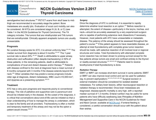
![Version 2.2017, 05/17/17 © National Comprehensive Cancer Network, Inc. 2017, All rights reserved. The NCCN Guidelines® and this illustration may not be reproduced in any form without the express written permission of NCCN®. MS-40
NCCN Guidelines Index
Table of Contents
Discussion
NCCN Guidelines Version 2.2017
Thyroid Carcinoma
Systemic Therapy
Treatment with single-drug chemotherapy is not very effective, although
some patients may show disease response or have stable disease.478,499
Hyperfractionated EBRT, combined with radiosensitizing doses of
doxorubicin, may increase the local response rate to about 80%, with
subsequent median survival of 1 year.500
Distant metastases then
become the leading cause of death.501
Similar improvement in local
disease control has been reported with a combination of
hyperfractionated RT and doxorubicin-based regimens, followed by
debulking surgery in responsive patients or other multimodality
approaches.499,502-504
IMRT may be useful to reduce toxicity.445,478,505-509
However, the addition of larger doses of other chemotherapeutic drugs
has not been associated with improved control of distant disease or with
improved survival.
Systemic therapy recommendations are described in the algorithm (see
Systemic Therapy for Anaplastic Thyroid Carcinoma in the NCCN
Guidelines for Anaplastic [Thyroid] Carcinoma).478,510
Recommended
regimens include paclitaxel and carboplatin combinations, docetaxel
and doxorubicin combinations, paclitaxel alone, or doxorubicin
alone.478,511
The dosage and frequency of administration of all the
recommended systemic therapy agents are provided in the algorithm.
Either concurrent chemoradiation or chemotherapy alone regimens may
be used depending on the clinical setting; however, chemoradiation is
generally more toxic. If using chemoradiation, the ATA Guidelines
recommend using weekly chemotherapy regimens.478
Chemotherapy
alone can be considered for patients with unresectable or metastatic
disease. Single-agent doxorubicin is the only agent that is approved by
the FDA for ATC.478
Single-agent paclitaxel may benefit some patients
with newly diagnosed ATC; increased survival has been reported in
patients with stage IVB disease.512-514
If weekly paclitaxel is used, the
ATA Guidelines recommend using paclitaxel at 60 to 90 mg/m2 IV
weekly and not the dose previously reported in the study by Ain et
al.478,514
Given the poor outcome with current standard therapy, all patients—
regardless of surgical resection—should be considered for clinical trials.
Previous clinical trials for ATC have tested therapies including
fosbretabulin (and its parent drug, combretastatin A4 phosphate
[CA4P], and crolibulin [EPC2407], which are vascular disrupting
agents), efatutazone (an oral PPAR gamma agonist), and novel
multitargeted therapies including bevacizumab with doxorubicin,
sorafenib, sunitinib, imatinib, and pazopanib.510,515-522
Outside of clinical
trials, targeted therapies are not currently recommended in the NCCN
Guidelines for patients with ATC, although some are recommended for
patients with papillary, follicular, Hürthle cell, or medullary carcinoma. A
trial in 80 patients (FACT) reported that the addition of fosbretabulin—to
a carboplatin/paclitaxel regimen—resulted in a nonsignificant increase
in median survival (5.2 vs. 4.0 months).510,523
Multimodality therapy is recommended in patients with locally
resectable disease (see Primary Treatment in the NCCN Guidelines for
Anaplastic [Thyroid] Carcinoma).478,505,510,524-528
Although optimal results
have been reported with hyperfractionated EBRT combined with
chemotherapy, the NCCN Panel acknowledged that considerable
toxicity is associated with such treatment and that prolonged remission
is uncommonly reported.529
Preliminary data suggest that ALK inhibitors
may be effective in a subset of patients with papillary thyroid cancer
who have ALK gene fusions; however, these ALK gene fusions are
rarely reported in patients with ATC.345-348
BRAF mutations have been
reported in patients with ATC.482,530-532
Printed by Irfan Ashraf on 9/21/2017 12:01:47 PM. For personal use only. Not approved for distribution. Copyright © 2017 National Comprehensive Cancer Network, Inc., All Rights Reserved.](https://image.slidesharecdn.com/nccnguidelinesthyroid-230901141830-026f6616/85/NCCN-GUIDELINES-THYROID-pdf-96-320.jpg)
![Version 2.2017, 05/17/17 © National Comprehensive Cancer Network, Inc. 2017, All rights reserved. The NCCN Guidelines® and this illustration may not be reproduced in any form without the express written permission of NCCN®. MS-41
NCCN Guidelines Index
Table of Contents
Discussion
NCCN Guidelines Version 2.2017
Thyroid Carcinoma
References
1. Mazzaferri EL. Thyroid carcinoma: Papillary and follicular. In:
Mazzaferri EL, Samaan N, eds. Endocrine Tumors. Cambridge:
Blackwell Scientific Publications 1993:278-333.
2. Hegedus L. Clinical practice. The thyroid nodule. N Engl J Med
2004;351:1764-1771. Available at:
http://www.ncbi.nlm.nih.gov/pubmed/15496625.
3. Cooper DS, Doherty GM, Haugen BR, et al. Revised American
Thyroid Association management guidelines for patients with thyroid
nodules and differentiated thyroid cancer. Thyroid 2009;19:1167-1214.
Available at: http://www.ncbi.nlm.nih.gov/pubmed/19860577.
4. Ezzat S, Sarti DA, Cain DR, Braunstein GD. Thyroid incidentalomas.
Prevalence by palpation and ultrasonography. Arch Intern Med
1994;154:1838-1840. Available at:
http://www.ncbi.nlm.nih.gov/pubmed/8053752.
5. Ron E, Lubin JH, Shore RE, et al. Thyroid cancer after exposure to
external radiation: a pooled analysis of seven studies. Radiat Res
1995;141:259-277. Available at:
http://www.ncbi.nlm.nih.gov/pubmed/7871153.
6. Schneider AB, Bekerman C, Leland J, et al. Thyroid nodules in the
follow-up of irradiated individuals: comparison of thyroid ultrasound with
scanning and palpation. J Clin Endocrinol Metab 1997;82:4020-4027.
Available at: http://www.ncbi.nlm.nih.gov/pubmed/9398706.
7. Howlader N, Noone A, Krapcho M, et al. SEER Cancer Statistics
Review, 1975-2014, based on November 2016 SEER data submission,
posted to the SEER web site, April 2017. Bethesda, MD: National
Cancer Institute; 2017. Available at:
https://seer.cancer.gov/csr/1975_2014/.
8. Siegel RL, Miller KD, Jemal A. Cancer Statistics, 2017. CA Cancer J
Clin 2017;67:7-30. Available at:
https://www.ncbi.nlm.nih.gov/pubmed/28055103.
9. Edge SB, Byrd DR, Compton CC, et al. AJCC Cancer Staging
Manual, 7th ed. New York: Springer; 2010:1-646.
10. Hundahl SA, Fleming ID, Fremgen AM, Menck HR. A National
Cancer Data Base report on 53,856 cases of thyroid carcinoma treated
in the U.S., 1985-1995 [see comments]. Cancer 1998;83:2638-2648.
Available at: http://www.ncbi.nlm.nih.gov/pubmed/9874472.
11. Jonklaas J, Nogueras-Gonzalez G, Munsell M, et al. The impact of
age and gender on papillary thyroid cancer survival. J Clin Endocrinol
Metab 2012;97:E878-887. Available at:
http://www.ncbi.nlm.nih.gov/pubmed/22496497.
12. Mazzaferri EL, Jhiang SM. Long-term impact of initial surgical and
medical therapy on papillary and follicular thyroid cancer. Am J Med
1994;97:418-428. Available at:
http://www.ncbi.nlm.nih.gov/pubmed/7977430.
13. Stroup AM, Harrell CJ, Herget KA. Long-term survival in young
women: hazards and competing risks after thyroid cancer. J Cancer
Epidemiol 2012;2012:641372. Available at:
http://www.ncbi.nlm.nih.gov/pubmed/23091489.
14. Ito Y, Higashiyama T, Takamura Y, et al. Long-term follow-up for
patients with papillary thyroid carcinoma treated as benign nodules.
Anticancer Res 2007;27:1039-1043. Available at:
http://www.ncbi.nlm.nih.gov/pubmed/17465240.
15. Li N, Du XL, Reitzel LR, et al. Impact of enhanced detection on the
increase in thyroid cancer incidence in the United States: review of
incidence trends by socioeconomic status within the surveillance,
epidemiology, and end results registry, 1980-2008. Thyroid
2013;23:103-110. Available at:
http://www.ncbi.nlm.nih.gov/pubmed/23043274.
16. Davies L, Welch HG. Current thyroid cancer trends in the United
States. JAMA Otolaryngol Head Neck Surg 2014;140:317-322.
Available at: http://www.ncbi.nlm.nih.gov/pubmed/24557566.
Printed by Irfan Ashraf on 9/21/2017 12:01:47 PM. For personal use only. Not approved for distribution. Copyright © 2017 National Comprehensive Cancer Network, Inc., All Rights Reserved.](https://image.slidesharecdn.com/nccnguidelinesthyroid-230901141830-026f6616/85/NCCN-GUIDELINES-THYROID-pdf-97-320.jpg)


![Version 2.2017, 05/17/17 © National Comprehensive Cancer Network, Inc. 2017, All rights reserved. The NCCN Guidelines® and this illustration may not be reproduced in any form without the express written permission of NCCN®. MS-45
NCCN Guidelines Index
Table of Contents
Discussion
NCCN Guidelines Version 2.2017
Thyroid Carcinoma
64. Neuhold N, Schultheis A, Hermann M, et al. Incidental papillary
microcarcinoma of the thyroid--further evidence of a very low malignant
potential: a retrospective clinicopathological study with up to 30 years of
follow-up. Ann Surg Oncol 2011;18:3430-3436. Available at:
http://www.ncbi.nlm.nih.gov/pubmed/21431405.
65. Takami H, Ito Y, Okamoto T, et al. Revisiting the guidelines issued
by the Japanese Society of Thyroid Surgeons and Japan Association of
Endocrine Surgeons: a gradual move towards consensus between
Japanese and western practice in the management of thyroid
carcinoma. World J Surg 2014;38:2002-2010. Available at:
http://www.ncbi.nlm.nih.gov/pubmed/24671301.
66. Ito Y, Uruno T, Nakano K, et al. An observation trial without surgical
treatment in patients with papillary microcarcinoma of the thyroid.
Thyroid 2003;13:381-387. Available at:
http://www.ncbi.nlm.nih.gov/pubmed/12804106.
67. Tan GH, Gharib H. Thyroid incidentalomas: management
approaches to nonpalpable nodules discovered incidentally on thyroid
imaging. Ann Intern Med 1997;126:226-231. Available at:
http://www.ncbi.nlm.nih.gov/pubmed/9027275.
68. Ito Y, Miyauchi A, Kihara M, et al. Patient age is significantly related
to the progression of papillary microcarcinoma of the thyroid under
observation. Thyroid 2014;24:27-34. Available at:
http://www.ncbi.nlm.nih.gov/pubmed/24001104.
69. Kim HJ, Kim NK, Choi JH, et al. Radioactive iodine ablation does
not prevent recurrences in patients with papillary thyroid
microcarcinoma. Clin Endocrinol (Oxf) 2013;78:614-620. Available at:
http://www.ncbi.nlm.nih.gov/pubmed/22957654.
70. Nilubol N, Kebebew E. Should small papillary thyroid cancer be
observed? A population-based study. Cancer 2015;121:1017-1024.
Available at: http://www.ncbi.nlm.nih.gov/pubmed/25425528.
71. Frates MC, Benson CB, Charboneau JW, et al. Management of
thyroid nodules detected at US: Society of Radiologists in Ultrasound
consensus conference statement. Radiology 2005;237:794-800.
Available at: http://www.ncbi.nlm.nih.gov/pubmed/16304103.
72. Haugen BR, Alexander EK, Bible KC, et al. 2015 American Thyroid
Association Management Guidelines for Adult Patients with Thyroid
Nodules and Differentiated Thyroid Cancer: The American Thyroid
Association Guidelines Task Force on Thyroid Nodules and
Differentiated Thyroid Cancer. Thyroid 2016;26:1-133. Available at:
https://www.ncbi.nlm.nih.gov/pubmed/26462967.
73. Bomeli SR, LeBeau SO, Ferris RL. Evaluation of a thyroid nodule.
Otolaryngol Clin North Am 2010;43:229-238, vii. Available at:
http://www.ncbi.nlm.nih.gov/pubmed/20510711.
74. Mazzaferri EL. Thyroid cancer in thyroid nodules: finding a needle in
the haystack. Am J Med 1992;93:359-362. Available at:
http://www.ncbi.nlm.nih.gov/pubmed/1415298.
75. Hamming JF, Goslings BM, van Steenis GJ, et al. The value of fine-
needle aspiration biopsy in patients with nodular thyroid disease divided
into groups of suspicion of malignant neoplasms on clinical grounds.
Arch Intern Med 1990;150:113-116. Available at:
http://www.ncbi.nlm.nih.gov/pubmed/2297281.
76. Chan BK, Desser TS, McDougall IR, et al. Common and uncommon
sonographic features of papillary thyroid carcinoma. J Ultrasound Med
2003;22:1083-1090. Available at:
http://www.ncbi.nlm.nih.gov/pubmed/14606565.
77. Henry JF, Denizot A, Puccini M, et al. [Early diagnosis of sporadic
medullary cancers of the thyroid: value of systematic assay of
calcitonin]. Presse Med 1996;25:1583-1588. Available at:
http://www.ncbi.nlm.nih.gov/pubmed/8952672.
78. Cheung K, Roman SA, Wang TS, et al. Calcitonin measurement in
the evaluation of thyroid nodules in the United States: a cost-
Printed by Irfan Ashraf on 9/21/2017 12:01:47 PM. For personal use only. Not approved for distribution. Copyright © 2017 National Comprehensive Cancer Network, Inc., All Rights Reserved.](https://image.slidesharecdn.com/nccnguidelinesthyroid-230901141830-026f6616/85/NCCN-GUIDELINES-THYROID-pdf-101-320.jpg)


![Version 2.2017, 05/17/17 © National Comprehensive Cancer Network, Inc. 2017, All rights reserved. The NCCN Guidelines® and this illustration may not be reproduced in any form without the express written permission of NCCN®. MS-52
NCCN Guidelines Index
Table of Contents
Discussion
NCCN Guidelines Version 2.2017
Thyroid Carcinoma
Clin Proc 1991;66:11-22. Available at:
http://www.ncbi.nlm.nih.gov/pubmed/1988751.
166. Hedinger CE. [Problems in the classification of thyroid tumors.
Their significance for prognosis and therapy]. Schweiz Med
Wochenschr 1993;123:1673-1681. Available at:
http://www.ncbi.nlm.nih.gov/pubmed/8211018.
167. Maxwell EL, Palme CE, Freeman J. Hurthle cell tumors: applying
molecular markers to define a new management algorithm. Arch
Otolaryngol Head Neck Surg 2006;132:54-58. Available at:
http://www.ncbi.nlm.nih.gov/pubmed/16415430.
168. Belchetz G, Cheung CC, Freeman J, et al. Hurthle cell tumors:
using molecular techniques to define a novel classification system. Arch
Otolaryngol Head Neck Surg 2002;128:237-240. Available at:
http://www.ncbi.nlm.nih.gov/pubmed/11886336.
169. Chen H, Nicol TL, Zeiger MA, et al. Hurthle cell neoplasms of the
thyroid: are there factors predictive of malignancy? Ann Surg
1998;227:542-546. Available at:
http://www.ncbi.nlm.nih.gov/pubmed/9563543.
170. Thompson NW, Dunn EL, Batsakis JG, Nishiyama RH. Hurthle cell
lesions of the thyroid gland. Surg Gynecol Obstet 1974;139:555-560.
Available at: http://www.ncbi.nlm.nih.gov/pubmed/4479589.
171. Lopez-Penabad L, Chiu AC, Hoff AO, et al. Prognostic factors in
patients with Hurthle cell neoplasms of the thyroid. Cancer
2003;97:1186-1194. Available at:
http://www.ncbi.nlm.nih.gov/pubmed/12599224.
172. Samaan NA, Schultz PN, Haynie TP, Ordonez NG. Pulmonary
metastasis of differentiated thyroid carcinoma: treatment results in 101
patients. J Clin Endocrinol Metab 1985;60:376-380. Available at:
http://www.ncbi.nlm.nih.gov/pubmed/3965495.
173. Ruegemer JJ, Hay ID, Bergstralh EJ, et al. Distant metastases in
differentiated thyroid carcinoma: a multivariate analysis of prognostic
variables. J Clin Endocrinol Metab 1988;67:501-508. Available at:
http://www.ncbi.nlm.nih.gov/pubmed/3410936.
174. Samaan NA, Schultz PN, Hickey RC, et al. The results of various
modalities of treatment of well differentiated thyroid carcinomas: a
retrospective review of 1599 patients. J Clin Endocrinol Metab
1992;75:714-720. Available at:
http://www.ncbi.nlm.nih.gov/pubmed/1517360.
175. Baudin E, Travagli JP, Ropers J, et al. Microcarcinoma of the
thyroid gland: the Gustave-Roussy Institute experience. Cancer
1998;83:553-559. Available at:
http://www.ncbi.nlm.nih.gov/pubmed/9690549.
176. Roti E, degli Uberti EC, Bondanelli M, Braverman LE. Thyroid
papillary microcarcinoma: a descriptive and meta-analysis study. Eur J
Endocrinol 2008;159:659-673. Available at:
http://www.ncbi.nlm.nih.gov/pubmed/18713843.
177. Mazzaferri EL. Management of low-risk differentiated thyroid
cancer. Endocr Pract 2007;13:498-512. Available at:
http://www.ncbi.nlm.nih.gov/pubmed/17872353.
178. Sugino K, Ito K, Jr., Ozaki O, et al. Papillary microcarcinoma of the
thyroid. J Endocrinol Invest 1998;21:445-448. Available at:
http://www.ncbi.nlm.nih.gov/pubmed/9766259.
179. Hay ID. Papillary thyroid carcinoma. Endocrinol Metab Clin North
Am 1990;19:545-576. Available at:
http://www.ncbi.nlm.nih.gov/pubmed/2261906.
180. Emerick GT, Duh QY, Siperstein AE, et al. Diagnosis, treatment,
and outcome of follicular thyroid carcinoma. Cancer 1993;72:3287-
3295. Available at: http://www.ncbi.nlm.nih.gov/pubmed/8080485.
Printed by Irfan Ashraf on 9/21/2017 12:01:47 PM. For personal use only. Not approved for distribution. Copyright © 2017 National Comprehensive Cancer Network, Inc., All Rights Reserved.](https://image.slidesharecdn.com/nccnguidelinesthyroid-230901141830-026f6616/85/NCCN-GUIDELINES-THYROID-pdf-108-320.jpg)


![Version 2.2017, 05/17/17 © National Comprehensive Cancer Network, Inc. 2017, All rights reserved. The NCCN Guidelines® and this illustration may not be reproduced in any form without the express written permission of NCCN®. MS-63
NCCN Guidelines Index
Table of Contents
Discussion
NCCN Guidelines Version 2.2017
Thyroid Carcinoma
advanced or metastatic radioactive iodine (RAI)-refractory, differentiated
thyroid cancer. BMC Cancer 2011;11:349. Available at:
http://www.ncbi.nlm.nih.gov/pubmed/21834960.
329. Hoftijzer H, Heemstra KA, Morreau H, et al. Beneficial effects of
sorafenib on tumor progression, but not on radioiodine uptake, in
patients with differentiated thyroid carcinoma. Eur J Endocrinol
2009;161:923-931. Available at:
http://www.ncbi.nlm.nih.gov/pubmed/19773371.
330. Cabanillas ME, Waguespack SG, Bronstein Y, et al. Treatment
with tyrosine kinase inhibitors for patients with differentiated thyroid
cancer: the M. D. Anderson experience. J Clin Endocrinol Metab
2010;95:2588-2595. Available at:
http://www.ncbi.nlm.nih.gov/pubmed/20392874.
331. Kloos RT, Ringel MD, Knopp MV, et al. Phase II trial of sorafenib in
metastatic thyroid cancer. J Clin Oncol 2009;27:1675-1684. Available
at: http://www.ncbi.nlm.nih.gov/pubmed/19255327.
332. Gupta-Abramson V, Troxel AB, Nellore A, et al. Phase II trial of
sorafenib in advanced thyroid cancer. J Clin Oncol 2008;26:4714-4719.
Available at: http://www.ncbi.nlm.nih.gov/pubmed/18541894.
333. Carr LL, Mankoff DA, Goulart BH, et al. Phase II study of daily
sunitinib in FDG-PET-positive, iodine-refractory differentiated thyroid
cancer and metastatic medullary carcinoma of the thyroid with
functional imaging correlation. Clin Cancer Res 2010;16:5260-5268.
Available at: http://www.ncbi.nlm.nih.gov/pubmed/20847059.
334. Locati LD, Licitra L, Agate L, et al. Treatment of advanced thyroid
cancer with axitinib: Phase 2 study with
pharmacokinetic/pharmacodynamic and quality-of-life assessments.
Cancer 2014;120:2694-2703. Available at:
http://www.ncbi.nlm.nih.gov/pubmed/24844950.
335. Cohen EE, Rosen LS, Vokes EE, et al. Axitinib is an active
treatment for all histologic subtypes of advanced thyroid cancer: results
from a phase II study. J Clin Oncol 2008;26:4708-4713. Available at:
http://www.ncbi.nlm.nih.gov/pubmed/18541897.
336. Cohen EE, Tortorici M, Kim S, et al. A Phase II trial of axitinib in
patients with various histologic subtypes of advanced thyroid cancer:
long-term outcomes and pharmacokinetic/pharmacodynamic analyses.
Cancer Chemother Pharmacol 2014;74:1261-1270. Available at:
https://www.ncbi.nlm.nih.gov/pubmed/25315258.
337. Lim SM, Chang H, Yoon MJ, et al. A multicenter, phase II trial of
everolimus in locally advanced or metastatic thyroid cancer of all
histologic subtypes. Ann Oncol 2013;24:3089-3094. Available at:
https://www.ncbi.nlm.nih.gov/pubmed/24050953.
338. Leboulleux S, Bastholt L, Krause T, et al. Vandetanib in locally
advanced or metastatic differentiated thyroid cancer: a randomised,
double-blind, phase 2 trial. Lancet Oncol 2012;13:897-905. Available at:
http://www.ncbi.nlm.nih.gov/pubmed/22898678.
339. Shah MH, De Souza J, Wirth L, et al. Cabozantinib in patients with
radioiodine-refractory differentiated thryoid cancer who progressed on
prior VEGFR-targeted therapy: results of NCI- and ITOG-sponsored
multicenter phase II clinical trial [abstract]. 15th International Thyroid
Congress 2015;Oral Abstract 73. Available at:
http://online.liebertpub.com/doi/pdfplus/10.1089/thy.2015.29004.abstrac
ts.
340. Bible KC, Suman VJ, Molina JR, et al. Efficacy of pazopanib in
progressive, radioiodine-refractory, metastatic differentiated thyroid
cancers: results of a phase 2 consortium study. Lancet Oncol
2010;11:962-972. Available at:
http://www.ncbi.nlm.nih.gov/pubmed/20851682.
341. Falchook GS, Millward M, Hong D, et al. BRAF inhibitor dabrafenib
in patients with metastatic BRAF-mutant thyroid cancer. Thyroid
2015;25:71-77. Available at:
http://www.ncbi.nlm.nih.gov/pubmed/25285888.
Printed by Irfan Ashraf on 9/21/2017 12:01:47 PM. For personal use only. Not approved for distribution. Copyright © 2017 National Comprehensive Cancer Network, Inc., All Rights Reserved.](https://image.slidesharecdn.com/nccnguidelinesthyroid-230901141830-026f6616/85/NCCN-GUIDELINES-THYROID-pdf-119-320.jpg)


![Version 2.2017, 05/17/17 © National Comprehensive Cancer Network, Inc. 2017, All rights reserved. The NCCN Guidelines® and this illustration may not be reproduced in any form without the express written permission of NCCN®. MS-67
NCCN Guidelines Index
Table of Contents
Discussion
NCCN Guidelines Version 2.2017
Thyroid Carcinoma
387. Lutz S, Berk L, Chang E, et al. Palliative radiotherapy for bone
metastases: an ASTRO evidence-based guideline. Int J Radiat Oncol
Biol Phys 2011;79:965-976. Available at:
http://www.ncbi.nlm.nih.gov/pubmed/21277118.
388. Henry DH, Costa L, Goldwasser F, et al. Randomized, double-
blind study of denosumab versus zoledronic acid in the treatment of
bone metastases in patients with advanced cancer (excluding breast
and prostate cancer) or multiple myeloma. J Clin Oncol 2011;29:1125-
1132. Available at: http://www.ncbi.nlm.nih.gov/pubmed/21343556.
389. Rosen LS, Gordon D, Tchekmedyian NS, et al. Long-term efficacy
and safety of zoledronic acid in the treatment of skeletal metastases in
patients with nonsmall cell lung carcinoma and other solid tumors: a
randomized, Phase III, double-blind, placebo-controlled trial. Cancer
2004;100:2613-2621. Available at:
http://www.ncbi.nlm.nih.gov/pubmed/15197804.
390. Vitale G, Fonderico F, Martignetti A, et al. Pamidronate improves
the quality of life and induces clinical remission of bone metastases in
patients with thyroid cancer. Br J Cancer 2001;84:1586-1590. Available
at: http://www.ncbi.nlm.nih.gov/pubmed/11401309.
391. Eustatia-Rutten CF, Romijn JA, Guijt MJ, et al. Outcome of
palliative embolization of bone metastases in differentiated thyroid
carcinoma. J Clin Endocrinol Metab 2003;88:3184-3189. Available at:
http://www.ncbi.nlm.nih.gov/pubmed/12843163.
392. Carhill AA, Cabanillas ME, Jimenez C, et al. The
noninvestigational use of tyrosine kinase inhibitors in thyroid cancer:
establishing a standard for patient safety and monitoring. J Clin
Endocrinol Metab 2013;98:31-42. Available at:
http://www.ncbi.nlm.nih.gov/pubmed/23185034.
393. Van Nostrand D, Atkins F, Yeganeh F, et al. Dosimetrically
determined doses of radioiodine for the treatment of metastatic thyroid
carcinoma. Thyroid 2002;12:121-134. Available at:
http://www.ncbi.nlm.nih.gov/pubmed/11916281.
394. Thomas L, Lai SY, Dong W, et al. Sorafenib in metastatic thyroid
cancer: a systematic review. Oncologist 2014;19:251-258. Available at:
http://www.ncbi.nlm.nih.gov/pubmed/24563075.
395. Fallahi P, Ferrari SM, Vita R, et al. Thyroid dysfunctions induced
by tyrosine kinase inhibitors. Expert Opin Drug Saf 2014;13:723-733.
Available at: http://www.ncbi.nlm.nih.gov/pubmed/24821006.
396. Crouzeix G, Michels JJ, Sevin E, et al. Unusual short-term
complete response to two regimens of cytotoxic chemotherapy in a
patient with poorly differentiated thyroid carcinoma. J Clin Endocrinol
Metab 2012;97:3046-3050. Available at:
http://www.ncbi.nlm.nih.gov/pubmed/22723320.
397. Cohen EE, Needles BM, Cullen KJ, et al. Phase 2 study of
sunitinib in refractory thyroid cancer [abstract]. J Clin Oncol
2008;26(Suppl 15):Abstract 6025. Available at:
http://meeting.ascopubs.org/cgi/content/abstract/26/15_suppl/6025.
398. Fagin JA, Tuttle RM, Pfister DG. Harvesting the low-hanging fruit:
kinase inhibitors for therapy of advanced medullary and nonmedullary
thyroid cancer. J Clin Endocrinol Metab 2010;95:2621-2624. Available
at: http://www.ncbi.nlm.nih.gov/pubmed/20525911.
399. McHenry CR, Sandoval BA. Management of follicular and Hurthle
cell neoplasms of the thyroid gland. Surg Oncol Clin N Am 1998;7:893-
910. Available at: http://www.ncbi.nlm.nih.gov/pubmed/9735140.
400. Ustun B, Chhieng D, Van Dyke A, et al. Risk stratification in
follicular neoplasm: a cytological assessment using the modified
Bethesda classification. Cancer Cytopathol 2014;122:536-545.
Available at: http://www.ncbi.nlm.nih.gov/pubmed/24753500.
401. Phitayakorn R, McHenry CR. Follicular and Hurthle cell carcinoma
of the thyroid gland. Surg Oncol Clin N Am 2006;15:603-623, ix-x.
Available at: http://www.ncbi.nlm.nih.gov/pubmed/16882500.
Printed by Irfan Ashraf on 9/21/2017 12:01:47 PM. For personal use only. Not approved for distribution. Copyright © 2017 National Comprehensive Cancer Network, Inc., All Rights Reserved.](https://image.slidesharecdn.com/nccnguidelinesthyroid-230901141830-026f6616/85/NCCN-GUIDELINES-THYROID-pdf-123-320.jpg)