The document discusses patellofemoral osteoarthritis (OA) and how it differs from tibiofemoral OA. It begins by describing how MRI has provided new insights into patellofemoral OA by enabling direct visualization of knee joint structures. While tibiofemoral and patellofemoral OA share some characteristics, they also have key differences. Specifically, patellofemoral OA presents more with activities involving the patella like squatting or stairs, while tibiofemoral OA pain is more from activities with axial loading. Treatment also differs between the compartments. MRI is now able to detect early cartilage changes associated with OA onset and progression in both compartments.
Studying relation between sitting position and knee osteoarthritiiosrjce
Osteoarthritis (OA) of the knee is the most common form of arthritis and leads to more activity
limitations (e.g., disability in walking and stair climbing) than any other disease, especially in the elderly. The
aim of this study was to clarify the relationship between the sitting position and knee osteoarthritis. The study
involved fat males of knee pain and clinical diagnosis of early knee osteoarthritis this research is applied and
the research method is "descriptive-correlative". In order to collecting data was used questionnaire tool. Also,
in order to analyzing data was used statistical method such as Pierson coefficient and Chi-squared test. Data is
analyzed from both descriptive and inferential statistics. Descriptive statistics and graphs on the table will
describe the characteristics of the study sample. The researcher to analyze the hypotheses used Chi-square
method. The statistical society is Osteoarthritis disease males.
Professor of Radiology and Medicine
Vice Chair, Academic Affairs
Assistant Dean of Diversity
Director, Quantitative Imaging Center (QIC)
Boston University School of Medicine, Boston, MA
This document discusses osteoarthritis of the knee joint. It begins with an introduction to knee joint anatomy and osteoarthritis. Common radiological signs of osteoarthritis including joint space narrowing and osteophyte formation are described. Methods for examining and measuring osteoarthritis via imaging and radiography are outlined. Results from measurements of 67 osteoarthritic knees in Myanmar are presented, finding greater tubercle spiking and ratios in females and increasing with age. The document concludes with references.
Presentation1, radiological imaging of diabetic foor and charcot joint.Abdellah Nazeer
This document discusses radiological imaging techniques for evaluating diabetic foot complications such as infection and Charcot foot. It provides examples of MRI, CT, ultrasound, bone scintigraphy, and PET/CT images showing osteomyelitis, soft tissue infections, and Charcot arthropathy in diabetic feet. MRI is highlighted as the most useful imaging method for diagnosing osteomyelitis, while PET/CT can help distinguish osteomyelitis from Charcot disease.
Osteoarthritis is defined by the presence of joint damage and is most commonly diagnosed through x-ray imaging. While many older individuals have radiographic evidence of osteoarthritis, only a portion experience joint pain and symptoms. The most frequently affected joints are the spine, fingers, knees and hips. Standard x-rays can reveal signs like reduced joint space, bone spurs and cysts. MRI may provide additional details on cartilage damage and other abnormalities but is generally not needed for routine osteoarthritis diagnosis and management.
Osteoid osteoma is among the commonest bone tumors, primarily affecting young subjects. Often localized in the diaphysis cortex of long bones, the disease has a well-described symptomatology and imagery of choice for diagnosis. When in a different location, the diagnosis is less evident. We describe a case herein of an intra-articular osteoid osteoma of the hip misdiagnosed as a femoro-acetabular impingement and treated by means of hip arthroscopy.
Osteoarthritis is a common joint disease that affects many sites in the body including the hands, knees, and hips. The incidence of osteoarthritis is increasing due to an aging population and rising obesity rates. There are many risk factors for osteoarthritis including age, female sex, genetics, obesity, joint injury, and abnormalities in joint shape. Local factors in the joint environment like muscle weakness, malalignment, and excessive or injurious joint loading can also increase the risk or progression of osteoarthritis. Accurately diagnosing and grading osteoarthritis involves both clinical assessment and radiographic evaluation using scales like the Kellgren-Lawrence grading system.
Structural Targets for Prevention of Post Traumatic OAOARSI
David Hunter MBBS, PhD, FRACP
Florance and Cope Chair of Rheumatology, Professor of Medicine
University of Sydney and Royal North Shore Hospital
Chair, Institute of Bone and Joint Research
Chair, Musculoskeletal, Sydney Medical Program
Consultant Rheumatologist, North Sydney Orthopedic and Sports Medicine
this presentation focus on a specific problem for patients with multiple hereditary exostosis who suffered from forearm deformity . It introduce a new technique to correct the deformity while preserving the epipyseal plate to maintain the growth of the bone.It avoids the complex surgery of distraction osteogenesis.
The anterior cruciate ligament (ACL) is a key ligament in the knee that prevents anterior tibial translation and rotational loads. It frequently tears during high-impact sports. The ACL inserts on the femur and tibia and is composed of two bundles that restrain movement differently based on knee flexion angle. While partial ACL tears may be treated nonsurgically, complete tears typically require surgical reconstruction using a graft to replace the torn ligament. Postoperative rehabilitation focuses initially on regaining range of motion and strength before gradually progressing to sport-specific activities.
This study retrospectively analyzed 310 patients treated for zygomatic bone and zygomatic arch fractures over a 10-year period. The majority of patients were young adult males injured in road traffic accidents. Patients were divided into three groups based on fracture location: zygomatic bone only, zygomatic arch only, or both. Most fractures were of the zygomatic bone alone. Treatment modalities included open reduction and internal fixation via miniplates and screws for 90.6% of patients and closed reduction for 9.4%. All patients achieved satisfactory postoperative results without complications, demonstrating the effectiveness of the treatment approaches used.
The document summarizes diagnostic radiology findings of musculoskeletal infectious diseases and joint disorders. It describes imaging features of acute and chronic osteomyelitis caused by bacteria and tuberculosis including bone destruction, periosteal reaction, and abscess formation. It also compares findings of pyogenic and tuberculous arthritis, noting tuberculosis involves bone margins and causes osteoporosis while pyogenic arthritis affects weight-bearing areas and leads to more bone destruction and sclerosis. Common joint disorders like osteoarthritis, ankylosis, and dislocations are outlined.
Presentation1 radiological film reading of wrist joint.Abdellah Nazeer
This document summarizes different types of wrist injuries seen on radiological films and MRI images. It describes various classifications of triangular fibrocartilage complex (TFCC) tears and discusses injuries to other ligaments including the scapholunate, lunotriquetral, and extrinsic ligaments. Common wrist abnormalities like scapholunate dissociation, SLAC wrist, and DISI/VISI deformities are also reviewed along with corresponding imaging findings. The document serves as a reference for interpreting radiological studies of the wrist joint and identifying associated ligament and cartilage injuries.
This document reviews various imaging modalities used to diagnose temporomandibular joint (TMJ) disorders. It discusses plain radiography, panoramic radiography, tomography, arthrography, computed tomography, cone beam computed tomography, magnetic resonance imaging, ultrasonography, and radionuclide imaging. For each modality, it describes its uses, advantages, and limitations in evaluating TMJ structures and pathologies. Magnetic resonance imaging is highlighted as the preferred method for assessing soft tissues like the disc position, while computed tomography is useful when bony involvement is suspected due to its ability to evaluate osseous changes with less radiation than CT. The correct imaging approach depends on the individual case and clinical findings.
This document summarizes a presentation on osteoarthritis (OA) phenotypes and risk factors. The presentation discusses evidence that OA may consist of distinct subtypes including generalized vs. joint-specific, secondary vs. primary, painful vs. non-painful, and malaligned vs. neutrally aligned joints. Identifying OA phenotypes is important for developing effective prevention and treatment strategies that may differ between subtypes.
A biomechanical approach for dynamic hip joint analysis 20pp 2011Victor Olivares
This document summarizes a study that aims to analyze hip joint mechanics during motion using subject-specific biomechanical modeling. The study presents methods to jointly model a subject's anatomy, kinematics, and dynamics through physically-based simulation of articular layers. Simulation results showed strong deformations and peak stresses in extreme hip postures, correlating with detected medical abnormalities. This suggests repetitive stresses within the joint could lead to early hip osteoarthritis.
Osteoartritis (OA) adalah salah satu jenis artritis yang paling sering dialami oleh sebagian orang. Penyakit ini merupakan penyakit sendi degeneratif yang mempengaruhi tulang rawan persendian. OA terjadi akibat rusaknya kartilago yang melindungi dan memberi bantalan bagi sendi.
This is my book review that came out on 24th Sunday 2017 in the EJOST(European Journal of Orthopaedic Surgery and Traumatology),which I have sent as an attachment.
K.Mohan Iyer(25/9/2017)
The Battle 2021 Castrocaro Terme (Italy). Achilles Insertional Tendinopathy a...Nicola Taddio
The aim of this presentation is to explain the background of Achilles Insertional Tendinopathy and Haglund's Triad, the rationale of conservative treatment and finally the therapeutic exercise evidence based approach.
Background: Management of large and massive rotator cuff tears remains controversial. Such tears are often irreparable, and results of treatment are unpredictable. This study documents the current
practice of orthopaedic surgeons in the British Elbow and Shoulder Society.
Methods: A questionnaire was prepared pertaining to the management of large and massive rotator cuff tears with minimal degenerative changes in three age groups: Patients of 50 years (young), 65 years (still active), aged 75 years (elderly) were considered. Various risk factors for failure of repair were
considered.
This document discusses imaging of the sacroiliac joint, including radiography and MRI findings. It begins with an overview of sacroiliac joint anatomy. It then describes common radiographic views and classifications of sacroiliac joint damage. The document focuses on using MRI to identify inflammatory lesions in the sacroiliac joints that can indicate early spondyloarthritis. It discusses how stopping NSAIDs before MRI may have little impact on findings. Repeating sacroiliac MRI months later generally does not reveal new cases, except possibly in HLA-B27 positive men. The document recommends sacroiliac MRI over spine MRI alone for axial spondyloarthritis workup given the low yield of isolated spinal findings without sac
Presentation1, radiological imaging of morel lavallee lesion.Abdellah Nazeer
Morel-Lavallée lesions are closed degloving injuries that result from shearing forces separating the skin and subcutaneous tissues from the underlying fascia during trauma. This creates a potential space that fills with blood, lymph, and fat. Morel-Lavallée lesions are most commonly seen near bony protuberances like the greater trochanter. MRI and ultrasound are useful for evaluating these lesions, which can vary in appearance from serous fluid collections to hematomas based on the acuity and inflammatory response. The Mellado-Bencardino classification system categorizes lesions based on their shape, signal characteristics, and enhancement pattern.
Foot orthoses for the treatment of patellofemoral painIsaac Knott
This document discusses foot orthoses for the treatment of patellofemoral joint pain. It summarizes that patellofemoral joint pain is common, affecting about 26% of the active population, and is caused by increased load through the joint from activities like prolonged walking or running. Conservative treatment including foot orthoses is usually prescribed to help correct foot posture and biomechanics to decrease pain. However, the evidence around the effectiveness of foot orthoses is conflicting, with higher quality studies finding little long-term impact on pain though a potential short-term benefit. More high-quality research is still needed.
This patient presents with knee pain. Imaging shows a lesion within the epiphysis of the knee. On MRI, the lesion has low signal on T1 and T2 weighted images with a low signal margin and no aggressive features. The most likely diagnosis is chondroblastoma, which is a rare benign epiphyseal tumor seen in children before growth plate closure that appears as a well-defined lytic lesion on radiographs and MRI.
Rheumatoid arthritis is a chronic inflammatory disease that commonly affects the small joints of the hands and feet. It results from an autoimmune response causing synovial inflammation and destruction of articular cartilage and bone. Early radiographic signs include soft tissue swelling, joint space widening, and juxta-articular osteopenia. Later findings consist of joint space narrowing, erosions, subluxations, and bony ankylosis. MRI is the best imaging modality for detecting early synovitis, bone marrow edema, and erosions. Characteristic sites of involvement include the second and third MCP and PIP joints bilaterally.
To investigate the geometric development of the
wrist in relation to the changes in its ossification pattern;
the study will help the treating surgeon to identify early deviations
from normal in children with musculoskeletal disorders
and provide a template for anatomic reduction after trauma
scenarios.
The document summarizes how Steel Belt Systems USA worked with the International Trade Center at College of DuPage to expand their export sales. The ITC provided market research, lead generation, marketing support, and guidance on trade show preparation and export grants. This comprehensive support allowed Steel Belt Systems to develop business in Brazil, Canada, Colombia, Mexico and beyond. As a result, they expect their Latin America exports to increase by 5-10% in 2016 through new purchase orders for machines and spare parts totaling millions of dollars. The company looks forward to continuing their work with the ITC to grow export sales further in coming years.
La combinación de correspondencia permite insertar datos de una lista de direcciones en un documento modelo para crear cartas personalizadas. Se abre Word y Excel, se crea un documento modelo en Word con espacios para los datos y una base de datos en Excel. Luego, en la pestaña de correspondencia de Word se inicia el asistente, se selecciona el documento modelo y la base de datos de Excel, y se insertan campos combinados en el modelo para los datos deseados. Esto genera una carta personalizada para cada registro en la base de datos.
O documento define o Estado como uma sociedade política formada por grupos de indivíduos unidos e organizados permanentemente para realizar um objetivo comum de bem público, dentro de um território determinado e com governo próprio. O Estado busca a ordem e defesa social através de meios que variam de acordo com a cultura, época e costumes, exercendo autoridade e poder sobre os governados.
SENTIDOS ATRIBUÍDOS POR PROFESSORES DA EDUCAÇÃO BÁSICA À APRESENTAÇÃO DA HIST...Everaldo Gomes
Objetivamos, com esse texto, apresentar dados de uma pesquisa de cunho qualitativo, que se fundamenta na teoria histórico-cultural. Tais dados indicam alguns sentidos que professores que ensinam Matemática na Educação Básica atribuem à História da Matemática sugerida em livros didáticos e em atividades de ensino. A investigação procura identificar e analisar, do ponto de vista de professores, inseridos em um contexto de formação, elementos que podem subsidiar a avaliação de livros didáticos e de atividades de ensino no que concerne ao papel pedagógico que a História da Matemática pode assumir no ensino, tais como: 1) a superação da visão da História da Matemática como fonte de motivação; 2) a superação da História do conteúdo com foco em personalidades; 3) a indicação da necessidade da História da Matemática nos diferentes níveis de ensino; 4) a superação da utilização da história de forma burocrática; 5) a indicação de que a História da Matemática faz diferença na sequência lógica do material didático; 6) a indicação de que a História da Matemática pode auxiliar o professor na organização do ensino; 7) a indicação de que a História pode ser fonte para a percepção do movimento do pensamento no surgimento e desenvolvimento do conceito, no que diz respeito à organização do ensino.
El documento resume las principales etapas históricas de la prehistoria y la historia de España desde el Paleolítico hasta la Edad Media, incluyendo la llegada de diferentes pueblos como fenicios, griegos y cartagineses, la conquista musulmana y el avance posterior de los reinos cristianos hasta la caída del último reino musulmán de Granada en 1492.
Serre Financial provides specialized consulting services that save clients money through opportunities within the Income Tax Act. They offer customized plans to meet individual client needs. With over a decade of experience, Serre Financial helps clients increase tax savings, cash flow, retirement benefits, and business exit strategies through working with their existing team of advisors.
Erin Foust is seeking a career in fundraising and development. She has over 5 years of experience in fundraising, event planning, and communications work for nonprofit organizations. Her education includes a B.A. in Journalism and Mass Communication from the University of Iowa with an emphasis in Philanthropy and Fundraising.
Shakeela is inviting applications for the roles of Organizing Committee Vice President for AIESEC's July Recruitment event. The roles include Vice President of Delegate Servicing, Marketing, Operations, and Agenda. Applicants will be interviewed on May 11th and selected candidates will be announced on May 12th. The timeline provides the application deadline, selection process dates, and first meeting date for the new Organizing Committee Vice Presidents. Shakeela looks forward to receiving applications and provides contacts for any inquiries about the roles.
A demographic profile_of_nondestructive_inspection_and_testing_(ndi-ndt)_pers...Mueed Liaqat
This document provides a demographic profile and preliminary analysis of nondestructive inspection and testing (NDI/NDT) personnel from nine aircraft maintenance facilities across three major US air carriers. It finds that the workforce is predominantly male (99%), with a median age of 45, and includes job classifications of visual inspectors (52%), visual-NDI/NDT personnel (36%), and NDI/NDT specialists (12%). Eddy current inspection was performed most frequently, while radiographic inspection was performed least. The analysis suggests that vision screening programs should give special consideration to the visual capabilities and ophthalmic conditions of males over 40 years old.
Building a Marine Renewables Industry in the United States: The Need for A "...Carolyn Elefant
Emergence of a robust marine renewables energy industry has been stymied in part by a regulatory process better suited for large, well funded entities. This paper presents my first phase of work on a Third Wave model of regulation for marine renewables, as well as other future renewable technologies that may be developed
Tiffany Wan- Honors Project PresentationTiffany Wan
This document summarizes Tiffany Wan's honors thesis presentation on women working in the public relations industry in Hong Kong. The presentation explored reasons for the high proportion of women in PR through an online survey and interviews. Key findings included that communications majors attract more female than male students, stereotypical views of women as better communicators, and Hong Kong's gender ratio imbalance with more women may influence the industry composition. The presentation concluded there are implications for diversity and incorporating different perspectives.
European citizenship can be achieved through recognizing our shared rights and responsibilities as citizens of Europe. Romanian students created a friendship boat named "European Citizens" to promote this idea of a shared identity between countries like Slovakia, Austria, Czech Republic, and Romania. Being active citizens who embrace our common European ideals makes us stronger together than if we were to stand alone.
This document provides addition problems and solutions for counting on by 1, 2, 3, and 4. It gives examples of adding single digit numbers together through counting on, with the goal of teaching how to use this method to solve addition problems up to 10.
A Large Intra-Articular Ossicle in the Knee Joint-A Rare Occurrence_Crimson P...CrimsonPublishersAICS
This document presents a case report of a rare occurrence of a large intra-articular ossicle in the knee joint of a 14-year-old male. Magnetic resonance imaging found a large ossicle impinging on the anterior cruciate ligament, causing lifting of the ligament anteriorly and laterally. Intra-articular ossicles in the knee are uncommon but can grow to a significant size, as seen in this case, producing mass effect and clinically mimicking an ACL injury. The etiology of intra-articular ossicles is unclear but may be congenital, traumatic, or degenerative in origin.
Background: The third most common musculoskeletal symptom in orthopaedic clinical practice is a sore shoulder, which can cause significant morbidity. It has been reported that 7–27% of the general population has it, and 36–66% of overhead arm athletes have it. Pathophysiology includes functional, degenerative, and mechanical factors. Most shoulder pain is subacromial pain syndrome (SAPS), often known as ‘shoulder impingement syndrome’. Impingement hypothesis: shoulder joint structures mechanically clash. SAPS accounts for 36–48% of shoulder discomfort. Methods: This observational study was conducted in the Department of Orthopaedics, MKCG Medical College and Hospital, Berhampur, among Eastern Indian outpatients. The study included adult patients (ages 18–75) of both sexes who presented to MKCG Medical College and Hospital's OPD with shoulder pain from December 2020 to November 2022 and were diagnosed with Shoulder Impingement Syndrome (SIS). Thorough histories and clinical exams were done. The Department of Radiology, MKCG Medical College and Hospital, Berhampur, performed conventional shoulder MRIs on the selected participants. Results: Most cases and controls were Type-II (43.3%), followed by Type-I (28.3% and 30%, 29.2% of the total group). The study's least common acromial shape was type-IV, seen in 5% of cases and 10% of controls (7.5% of the sample). Fisher's exact test showed no significant connection between subacromial impingement and acromial shape (p=0.65). With a p-value of 0.045, cases had a significantly greater acromial width (8.12±2.16 mm) than controls (7.51±0.81 mm). Conclusion: Sub-acromial impingement was unrelated to acromion morphology. There was no correlation between acromial morphology and rotator cuff injuries.
Key-words: Shoulder Impingement Syndrome, Acromion Morphology, MRI
This document summarizes a study that assessed patients' pain levels before and after undergoing ultrasound-guided knee arthrocentesis and steroid injection. A sample of 23 patients rated their knee pain on a numerical scale before the procedure and 2 weeks after. Results found a statistically significant decrease in mean pain scores, demonstrating a positive patient outcome. The implications are that portable ultrasound should be considered for other clinical settings like remote primary care where referrals are not possible, as ultrasound provides advantages over physical exam alone for assessing and treating knee effusions.
A SYSTEMATIC REVIEW STUDY TO DETERMINE THE CAUSATIVE FACTORS AND THE REHABILI...paperpublications3
This document summarizes a systematic review study that examined the causative factors and rehabilitation approaches for lateral ankle sprains. The review studied the pathophysiology, predisposing risk factors, and current evidence on therapeutic modalities and exercises used to treat ankle sprains. The review found that immobilization after ankle sprains facilitates ligament healing and rehabilitation. Graded joint mobilization, proprioceptive training, and balance training should also be included as adjunct treatments.
A SYSTEMATIC REVIEW STUDY TO DETERMINE THE CAUSATIVE FACTORS AND THE REHABILI...paperpublications3
Abstract:Ankle sprain is one of the most common musculoskeletal sports injury encountered. In sports injuries throughout the countries studied, the ankle was the second most common injured body site after the knee. The purpose of the review study is to study the pathophysiology, predisposing factors, and the current evidence regarding therapeutic modalities and exercises used in the treatment of ankle sprain. There is a high incidence rate of approximately 75% of lateral ankle sprain; it also possesses a high incidence rate of re-injury. Recent researches have proved that immobilization post ankle sprain facilitates ligament healing and enhances the rehabilitative protocol. In addition to that the other treatment protocols are to be implemented as an adjunct for instance graded joint mobilization, proprioceptive training and balance training. Altering current rehabilitative protocol to enhance the joint range of motion and to maintain the soft tissue integrity with stringent immobilization, and including graded joint mobilizations and balance training may be the first step to decreasing the incidence of short and long term ankle joint dysfunction.
Keyword:Ankle joint, Sprain, Ligament, Immobilization, Proprioceptive, Pathophysiology, Sports, Athletic, Degeneration.
Introduction: Spontaneous Osteonecrosis of the Knee (SONK) is a devastating and debilitating disease that mainly affl icts the
elderly. A conservative approach may forgo the need for surgical intervention. This case report describes an orthopaedic patient diagnosed with SONK. After fi ve months of conservative treatment, the patient was able to walk without pain and remained clinically stable for seven years.
Case Presentation: A 49-year-old male patient diagnosed with SONK of the left knee elected to undergo conservative management in lieu of surgical intervention. Seven years later the patient’s symptoms and MRI demonstrate a dramatic improvement.
Rheumatoid arthritis and osteoarthritisSonal Saran
Rheumatoid arthritis is a chronic inflammatory disease that predominantly affects the joints, especially small joints of the hands and feet. It is more common in women and involves symmetrical polyarticular inflammation of joints. The pathophysiology involves both cellular and humoral immune mechanisms leading to synovial membrane proliferation and cartilage/bone erosion. Osteoarthritis is the most common type of arthritis and is characterized by degeneration of joint cartilage and underlying bone, commonly affecting weight bearing joints like the hips and knees. Risk factors include age, obesity, trauma and genetics. Symptoms include joint pain, stiffness and decreased range of motion.
This document provides a supplement and revision to the 2001 Clinical Practice Guideline on the diagnosis and treatment of heel pain published in the Journal of Foot & Ankle Surgery. It outlines a pathway for evaluating and managing plantar heel pain, which is the most common type of heel pain seen in clinical practice. A thorough history and physical exam can usually determine the cause is mechanical in nature and rule out other potential etiologies. Conservative treatment is recommended first and includes padding, strapping, orthotics, medications, stretching, and corticosteroid injections. For patients who do not improve with conservative care, further treatment options are outlined in a three-tier ladder approach including night splints, additional orthotics, immobilization, injections
Fractures that occur against the background of osteoporosis represent a global medical and social problem. In elderly people, 90% of hip fractures, as international studies have shown, occur against the background of osteoporosis. According to WHO, it is the fractures of the proximal femur that put osteoporosis on the 4th place among all causes of disability and mortality.
This document summarizes osteoporosis, including its definition, prevalence, risk factors, pathogenesis, diagnosis, and treatment options. It discusses how osteoporosis is a widespread condition characterized by compromised bone strength and increased fracture risk. Diagnostic tools like DXA scans and emerging methods like multi-detector CT are used to assess bone mineral density and structure. Treatment involves lifestyle changes as well as pharmacotherapy like bisphosphonates, calcitonin, PTH, and emerging drugs. Overall the document provides a comprehensive overview of osteoporosis from causes and diagnosis to current and novel treatment approaches.
This case report describes a 56-year-old man who presented with an 8-month history of painful swelling in his right thumb. Imaging showed extensive destruction of the proximal phalanx bone and soft tissue swelling. Biopsy revealed granulomatous inflammation and acid-fast bacilli, leading to a diagnosis of tuberculous dactylitis. The patient underwent surgical debridement and 6 months of anti-tuberculosis treatment. At a 7-year follow-up, the thumb was shortened but he had good hand function with no significant disability.
The document discusses osteoarthritis of the knee, including:
1. Risk factors for osteoarthritis like age, gender, genetics, obesity, and joint injuries.
2. Clinical features like pain, stiffness, swelling, crepitus, and deformity.
3. Diagnostic tools like x-rays, MRI, CT, and arthroscopy that can assess cartilage damage and bone changes.
4. Treatment approaches including medications, physical therapy, weight loss, bracing, injections, and surgeries like arthroscopy, osteotomies, knee replacements, and arthrodesis.
Ankle and foot fractures are common injuries that require immobilization or surgery followed by physical therapy. Physiotherapists play an important role in rehabilitation by addressing range of motion, strength, and functional mobility deficits caused by fractures and associated soft tissue damage. Treatment involves casting or surgery to properly align bone fragments, followed by progressive weight bearing and exercises under physiotherapy guidance to restore function and prevent long-term issues like osteoarthritis. Outcome measures evaluate factors like pain, activity level, and quality of life to assess recovery.
This study compared muscle activation and knee mechanics during gait in patients with non-traumatic knee osteoarthritis (OA), post-traumatic knee OA caused by an anterior cruciate ligament injury, and healthy adults. The post-traumatic OA group had lower gastrocnemius muscle activation compared to healthy adults. The non-traumatic OA group had higher activation of the quadriceps and hamstring muscles compared to the post-traumatic OA group. The non-traumatic OA group also had lower knee extension and medial rotation moments during gait compared to the post-traumatic and healthy groups. The results indicate differences in muscle function and knee biomechanics between non-traumatic and
2. 2
REVIEW – Wijayaratne, Teichtahl, Wluka, Hanna & Cicuttini
Future Rheumatol. (2007) 2(2) future science groupfuture science group
Signs & symptoms
The most common symptom of knee OA is pain
that is aggravated by activity [4]. Nevertheless,
compartmental pain tends to be task-specific.
For instance, patellofemoral pain is more com-
mon among activities that increase retropatellar
load, such as squatting, rising from the seated
position and stair climbing [10]. By contrast, pain
in tibiofemoral OA tends to be more common
among activities that increase axial joint loads,
such as long-distance walking [11].
On clinical examination, the location of ten-
derness may help diagnose compartmental knee
OA. Patellofemoral tenderness, using the grind
test, is a reliable sign of patellofemoral OA [12].
Tenderness of the undersurface of the patella,
most commonly the lateral facet, is also said to
suggest patellofemoral involvement [4,13]. Ten-
derness over the medial or lateral joint lines has
been identified as a reliable sign of tibiofemoral
OA when examined by rheumatologists [4,12].
Other clinical signs in knee OA may include
bone swelling, joint effusions, crepitus, restricted
range of movements and muscle atrophy, but are
not distinguished between the patellofemoral
and tibiofemoral compartments [14].
Radiographic assessment of
patellofemoral & tibiofemoral OA
Radiographic examination of the arthritic joint
serves three purposes: to establish the diagnosis
and severity of OA; to monitor progression and
possible therapeutic responses; and to look for
complications of the disorder or the treatment
[15]. The most common features of radiographic
OA are joint-space narrowing, the presence of
osteophytes and subchondral sclerosis [16]. For
both tibiofemoral and patellofemoral OA, radio-
logical joint-space width (JSW), which is consi-
dered a surrogate measure of articular cartilage, is
the current gold standard for assessing the
natural history of radiographic OA [17].
The choice of views to identify radiographic
patellofemoral OA has evolved over the last few
decades. Previously, radiographic imaging of
knee OA was restricted to the tibiofemoral joint,
mainly owing to easy accessibility of antero-
posterior radiographs [18]. After patellofemoral
OA was recognized as a major source of pain and
disability, skyline and lateral radiographic views
were used to examine the patellofemoral com-
partment [18]. For the purposes of epidemio-
logical studies, atlas’s, such as the Osteoarthritis
Research Society International Atlas [16], are used
to define radiographic disease in each joint by
grading the severity of individual radiographic
characteristics of disease. In addition to defining
disease, these can be used to examine for patella
alta (high riding patella) and baja (low riding
patella), each of which has been associated with
patellar pathologies that cause pain [19,20]. How-
ever, little work has been done to standardize
patellofemoral views in epidemiological studies,
and many issues have been raised regarding the
reliability and validity of radiographic examina-
tion of the patellofemoral compartment [18].
Optimization of radiological assessment of
the patellofemoral compartment
Assessment of the severity of OA in the patello-
femoral compartment by lateral or skyline views
is potentially problematic. The lateral view is
often not a true lateral image and is further com-
plicated if patella tilt or subluxation are present
[21]. The presence of patella subluxation impedes
interpretation of the JSW and, thus, limits the
ability to accurately qualify, and subsequently
quantify, the presence of joint-space narrowing
both cross-sectionally and longitudinally [22].
Similarly, differences in knee flexion may affect
radiographic joint-space narrowing in the skyline
view, reducing validity of the measure [23].
Indeed, these methodological issues may have
contributed to inconsistent findings among
studies examining risk factors for the onset and
progression of patellofemoral OA [23]. In turn,
this may account for the limited data regarding
the relationship between risk factors and the
natural history of patellofemoral OA.
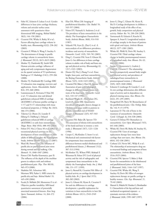
Figure 1. Sagittal T1-weighted
fat-saturated 3D MRI images showing a
normal patella cartilage (Grade 0).
A
uthorProof
3. 3
Patellofemoral osteoarthritis: new insights into a neglected disease – REVIEW
future science groupfuture science group www.futuremedicine.com
Management of tibiofemoral &
patellofemoral OA
Whilst the use of analgesia and self-management
strategies are similar for the involvement of both
compartments, there are differences in physical
therapies employed in the management of tibio-
femoral and patellofemoral OA.
Physiotherapy tends to be the mainstay of
conservative treatment for both patellofemoral
and tibiofemoral OA. Although the aims of
treatment in both conditions are identical (i.e.,
maintaining or improving joint range of move-
ment and muscle strength to enable independent
function), different strategies to reduce pain and
improve function are used to address the extent
of involvement of the different compartments.
For example, patellofemoral pathology and pain
often benefits from reducing laterally directed
translation of the patella [24]. This can be
achieved via strengthening medial muscles, such
as the vastus medialis, while reducing tension in
lateral supports, such as the iliotibial band. Tap-
ing the patella with a medially directed force may
also be beneficial [24–26]. Such techniques are not
standard for tibiofemoral pathology, which often
responds to exercise in a reduced weight-bearing
environment, such as hydrotherapy.
Investigation of the use of orthotic footwear to
correct malalignment as a treatment strategy in
knee OA has only been examined in tibiofemoral
disease [4]. However, the results of these studies
have been inconsistent [4].
With respect to surgical management, joint-
replacement surgery is the mainstay of therapy
for tibiofemoral disease. However, there may also
be a role for osteotomy in the presence of signifi-
cant malalignment or partial joint replacement
surgery. Although total knee-joint replacement
may also be used successfully to treat patello-
femoral OA in the absence of tibiofemoral OA,
some surgeons believe that this sacrifices too
much healthy tissue [14,27]. Other less traumatic
approaches include a lateral retinacular release,
which aims to reduce the tendency for lateral dis-
placement [28]. Although this should theoreti-
cally correct some of the forces contributing to
disease progression, there is limited published
long-term follow-up of this procedure [28]. In an
older procedure, the Maquet procedure, the tib-
ial tuberosity is transferred anteriorly to reduce
the loading on the patellofemoral joint [29]. Pub-
lished results in pure populations of subjects
with patellofemoral osteoarthritis are small case
series only, with significant loss to follow-up [14].
Anteromedial transfer of the tibial tuberosity, a
modification of the Maquet procedure, is more
common in the USA [14]. Since this procedure
moves the patellofemoral contact area medially,
it would be expected to be most effective where
disease is isolated to the lateral facet [30]. Patello-
femoral replacement may play a role, providing
the disease is truly isolated to the patellofemoral
compartment, or may be attributed to malalign-
ment, trauma or trochlear dysplasia [14]. The
combined assessment of pre- and post-operative
patients with imaging and biomechanical studies
will enable these therapies to be further refined
and assessed.
Recent developments in the assessment
of knee-joint OA
MRI
It has been recognized that a major limitation in
understanding the pathogenesis of knee OA is the
indirect manner in which the articular cartilage is
examined when using radiography. Previous
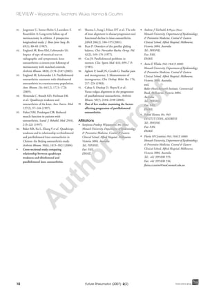
Table 1. Investigation, diagnosis and treatment of patellofemoral and tibiofemoral osteoarthritis.
Patellofemoral osteoarthritis Tibiofemoral osteoarthritis
Symptoms Pain with activities that increase retropatellar load, such
as squatting and stair climbing
Pain with activities such as long-distance walking that
increase axial joint loads
Signs Patellofemoral tenderness
Joint effusions
Muscle atrophy
Crepitus
Tenderness over lateral and medial lines
Joint effusions
Muscle atrophy
Crepitus
Radiography Skyline or lateral radiographic views Anteroposterior radiographs
Treatment:
– Conservative
– Medical
– Surgical
Physiotherapy
Analgesia, NSAIDs (analgesic ladder)
Total knee replacement
Physiotherapy
Analgesia, NSAIDs (analegesic ladder)
Joint replacement rare, efforts to reduce force on the
patellofemoral joint
NSAID: Nonsteroidal anti-inflammatory drug.
A
uthorProof
4. 4
REVIEW – Wijayaratne, Teichtahl, Wluka, Hanna & Cicuttini
Future Rheumatol. (2007) 2(2) future science groupfuture science group
studies have defined OA based on radiographic
changes, although it has been shown that when
the first changes of radiological OA are detected,
an average of 13% of the cartilage has already
been lost [31]. Therefore, radiographic assessment
of the knee joint is insensitive to potential early
degenerative change. Indeed, radiographic
change at the patellofemoral joint, using either
lateral or skyline views, correlates poorly with the
change in the amount of cartilage present [32].
Use of MRI has expanded the ability to
directly assess the knee joint in its entirety. Its use
is established in the clinical management of joint
disease. By measuring structural change, it has
recently begun to be developed as a tool for stud-
ying disease pathogenesis. For instance, MRI
allows the direct visualization of all structures,
including articular cartilage, within the knee
joint [33]. Recent studies examining the suitabil-
ity of MRI for assessing the features of OA have
demonstrated accurate assessment of cartilage
thickness, demonstrated internal cartilage
changes and signal abnormalities in subchondral
bone, and have also shown the morphological
changes occurring at cartilage surfaces [33,34].
Moreover, MRI is more sensitive than radiogra-
phy for the detection of soft-tissue changes in the
joint [35]. With MRI, it is possible to directly vis-
ualize the soft tissues in the joint, and to detect
change over time [36]. Thus, structural change in
the joint may be quantified and studied non-
invasively, in both healthy and arthritic subjects,
to examine risk factors for both the onset and
progression of disease more sensitively than has
previously been possible.
Use of MRI & cartilage defects
Progressive articular cartilage loss has been one
of the major hallmarks of OA [15]. The earliest
detectable changes in cartilage are irregularities
of the articular cartilage surface, observed on
MRI as cartilage defects. Defects are independ-
ent predictors of cartilage loss [37]. Ding and col-
leagues evaluated 325 healthy adult subjects at
baseline and 2 years later. They found that the
prevalence of patella cartilage defects was 1.9%
and that after an average of 2.3 years the severity
of patella cartilage defects increased significantly
[38]. This study also found that an increase in car-
tilage defect scores was associated with cartilage
loss in all compartments in both men and
women [38]. The severity of cartilage defects also
predicts the need for joint replacement in people
with knee OA, regardless of the amount or thick-
ness of cartilage present [39].
Use of MRI & cartilage volume
Knee-cartilage volume measurements derived
from MRI have been assessed at both the tibio-
femoral and patellofemoral compartments, have
been shown to have high reproducibility and are
a valid indicator of the radiographic grade of
both patellofemoral and tibiofemoral OA [40–42].
Additionally, loss of tibial cartilage, as assessed by
MRI, correlates with worsening symptoms [43]
and predicts the risk for knee replacement [44].
Using MRI, it is possible to measure the change
in cartilage volume over short periods of time,
both at the tibiofemoral and patellofemoral
compartments in healthy [7,45] and arthritic sub-
jects [36,46]. The assessment of cartilage volume
in both tibiofemoral and patellofemoral com-
partments by MRI has enabled investigators to
examine risk factors for cartilage loss and carti-
lage deterioration in all compartments of the
knee [7,36].
Use of MRI & cartilage quality
The potential of MRI to image joint structure
has not been fully exploited. New sequences and
techniques are being developed to be used as
markers of disease severity. Although neither of
the following two examples have been assessed in
longitudinal studies, they show early promise.
For example, the transverse relaxation time con-
stant (T2) of articular cartilage has been pro-
posed as a biomomarker for OA [47,48]. These
maps may identify localized degeneration of
articular cartilage [47,49]. Another technique, the
delayed Gadolinium Enhanced MRI of Cartilage
(dGEMRIC), shows promise in identifying areas
of abnormal cartilage signal based on different
levels of glycosaminoglycan, which are thought
to mirror cartilage health [49,50]. These and other
new techniques have still to be evaluated over
time, before they can be used as research tools.
Use of MRI & anatomical definition
The importance of biomechanical factors in the
pathogenesis of OA has become better appreci-
ated [51,52]. Consequently, there is increasing
interest in how the geometric characteristics of
the joint relate to the symptoms and develop-
ment of disease [20,53–55]. In the past, many unidi-
mensional measures were made from radiographs
to approximate joint shape, and these correlated
with clinical presentation. With increasing
understanding of biomechanical factors and
forces acting on each individual joint, there have
been many attempts to measure more biome-
chanically important and relevant components of
A
uthorProof
5. 5
Patellofemoral osteoarthritis: new insights into a neglected disease – REVIEW
future science groupfuture science group www.futuremedicine.com
the joint from the images obtained using com-
puted tomography, and more recently MRI
[20,52,54]. These may be used to assess patellar
tracking in various degrees of knee flexion [56].
Although these methods show promise, they have
not yet elucidated the causes of patellofemoral
pain [14]. Longitudinal use of these techniques
may be useful in identifying factors associated
with pathogenesis of OA.
Risk factors – similarities & differences
between patellofemoral &
tibiofemoral OA
Age & gender
Studies have consistently demonstrated that the
prevalence of OA increases with age regardless of
what definition of OA (clinical or radiographic)
is used [5,57]. Moreover, it has been consistently
demonstrated that women are more likely to have
knee OA than men, as confirmed by a recent
meta-analysis [58]. This may be related to a gender
difference in the amount of cartilage present:
men have substantially higher knee cartilage than
women [33]. Nevertheless, when cartilage volume
or JSW is adjusted for, there is still a female
disparity among the elderly with knee OA [59].
McAlindon and colleagues found that in
women with symptomatic knee OA, isolated
patellofemoral OA was more common than
medial tibiofemoral OA and tended to increase
with age [5]. For men in the same study the oppo-
site was observed; medial-tibiofemoral compart-
ment OA was the more common than
patellofemoral OA and the frequency tended to
increase with age. In the Beijing study the preva-
lence of both radiographic patellofemoral and
tibiofemoral OA was higher in women than in
men (25.9% in men vs 35.7% in women for
patellofemoral OA; and 21.9% in men vs 41.8%
in women for tibiofemoral OA) [60]. These data
indicate that although knee OA may be more
common in women, the pattern of compartmen-
tal involvement may have a gender disparity.
Obesity & body mass index
Obesity is a major risk factor for both patello-
femoral and tibiofemoral OA [5,61]. People with a
larger body mass index (BMI) are at an increased
risk for tibiofemoral OA, with an estimated 40%
increase in risk with each 10-lb weight gain [62].
In a population-based twin study of women aged
48–70 years, obesity increased the risk of develop-
ing OA at both the tibiofemoral and patellofemo-
ral compartments, with a 9–13% increased risk
for OA per kg weight gain [63].
The obesity–OA relationship may vary between
the different compartments of the knee joint: MRI
may be helpful in clarifying this relationship. For
example, it was demonstrated that although obesity
is a strong risk factor for medial tibiofemoral OA, it
did not affect the risk of patellofemoral OA [64]. In
addition, MRI studies have found that whereas
change in tibial cartilage volume was affected by
BMI [36], the association between patella-cartilage
volume and BMI was nonsignificant [7]. However,
these data were obtained from a group of healthy
men and may not be generalizable to women, or in
the presence of established OA.
Physical activity
The issue of whether physical activity, independ-
ent of joint injury, is detrimental to joints is
unclear. Sporting activities that excessively load
joints may increase the risk of OA, whereas, light
and moderate activities do not appear to increase
this risk [65]. Cross-sectional and longitudinal data
in children suggest that cartilage growth responds
to stimulation [66]. Children who exercised more
had higher tibial-cartilage volumes than their sed-
entary counterparts, although it is unclear
whether the same phenomenon occurs in mature
adults [67,68]. However, adult cartilage appears to
require loading for health. A study of subjects fol-
lowing recent paraplegia showed increased tibial,
femoral and patella cartilage loss over 12 months
(9–13%) [69]. Healthy adults lose approximately
2% of their knee articular cartilage per year [70].
Studies also suggest that frequently high levels
of physical activity increases the prevalence of
patellofemoral OA [71]. However, only small stud-
ies have assessed the relationship between physical
activity and patella-cartilage change in healthy
subjects [7]. It is likely that different types of exer-
cise affect the individual knee compartments dif-
ferently. A study that compared seven weightlifters
with seven sprinters and 14 untrained subjects
reported that patella cartilage deformation dem-
onstrated a dose-dependent response, where more
intense loading led to greater cartilage deforma-
tion [72]. A potential explanation may be that
when the knee flexes to 15 degrees at initial con-
tact during walking, the patellofemoral joint reac-
tion force is reportedly 50% of the total body
weight, while at 60 degrees knee flexion, the retro-
patellar force may have increased to 3.3-times the
total body weight [73]. Hence, people who take
part in weightlifting or other load-bearing exer-
cises that require deep knee flexion may impart
excessive loads across their articular patella carti-
lage, which may predate degenerative change.
A
uthorProof
6. 6
REVIEW – Wijayaratne, Teichtahl, Wluka, Hanna & Cicuttini
Future Rheumatol. (2007) 2(2) future science groupfuture science group
Estrogen replacement therapy
Estrogen deficiency, as a result of the onset of
menopause, has demonstrated to be associated
with rapidly progressive OA [74]. Estrogen
replacement therapy (ERT) is gaining increasing
support from observational studies, suggesting a
protective effect against tibiofemoral OA [75].
Supportive of this, MRI studies have shown
long-term ERT to be positively associated with
tibial cartilage volume [76] and decreased preva-
lence of knee OA-related subchondral bone
lesions [77]. However, there are limited data
examining the effect of ERT on patella cartilage
and patellofemoral OA. A radiographic study by
Cicuttini and colleagues found that premeno-
pausal status was protective for patellofemoral
OA but not associated with tibiofemoral OA [78].
When the effect of long-term ERT use on patel-
lar cartilage volume in postmenopausal women
was examined, no effect was seen [79].
Joint injury & meniscectomy
It is well established that major joint injury is a
common cause of OA, especially at the knee [80].
Isolated patellofemoral OA is common following
patella injury [18]. Previous knee injury increased
the risk of all forms of knee OA (odds ratio:
2–5.5) [9].
Meniscectomy has been recognized as a strong
risk factor for tibiofemoral OA [81]. Studies have
shown that there is a sixfold increase of develop-
ing tibiofemoral OA following total meniscec-
tomy compared with unoperated controls [82].
Nevertheless, the effects of meniscectomy are not
confined to the tibiofemoral compartment
alone; increased patellofemoral OA was also
demonstrated in a meniscectomy population,
after adjusting for age, gender and BMI [83].
Quadriceps weakness
Lower extremity muscle weakness may play an
important role in knee OA. Cross-sectional stud-
ies have shown that individuals with sympto-
matic knee OA have weaker quadriceps
compared with healthy subjects [84,85]. The
majority of studies investigating muscle weakness
in knee OA have defined the disease as limited to
the tibiofemoral compartment. These studies
demonstrated a strong correlation between quad-
riceps weakness and women with tibiofemoral
OA [84]. The only study to examine the relation-
ship between quadriceps weakness and patel-
lofemoral and tibiofemoral OA showed that
quadricep weakness was associated with patel-
lofemoral, tibiofemoral and combined patel-
lofemoral and tibiofemoral OA in both men and
women [86].These findings suggest that muscle
weakness may affect knee OA in all compart-
ments. Further longitudinal work is required to
determine the relationship between muscle weak-
ness and compartmental knee OA.
Varus–valgus alignment
There is mounting evidence to suggest that the
mechanical effects of alignment on load distribu-
tion are significantly higher in patients with
genu varum or valgum deformities. In a longitu-
dinal study of knee OA, baseline varus alignment
increased the risk for the progression of radio-
graphic medial-tibiofemoral OA, whereas valgus
alignment increased the risk of lateral tibiofemo-
ral OA progression [87].
At the patellofemoral compartment, increased
varus angulation reduces the Q-angle, which, in
turn, increases medial-patellofemoral forces; by
contrast, increased valgus angulation increases
the Q-angle, thus increasing the lateral patel-
lofemoral forces [88]. Moreover, given that
women tend to have slightly larger Q angles than
men, secondary to the relatively wider female
pelvis as well as greater femoral anteversion and
genu valgum [89,90], biomechanical factors, such
as the Q angle, may contribute toward the
female disparity of knee OA. Findings from a
longitudinal study examining the effect of align-
ment on patellofemoral OA demonstrated pro-
gression of medial patellofemoral OA in people
with genu varum, and progression of lateral
patellofemoral OA in those with genu valgum
[91]. From these findings it appears that
varus–valgus alignment is associated with the
progression of both patellofemoral and tibiofem-
oral OA in a compartment-specific manner.
Conclusion
Knee OA is a major cause of chronic pain and
disability among the elderly [15]. Pain associated
with knee OA frequently emanates from the
patellofemoral joint, which has been linked to
greater disability and a reduced quality of life,
compared with tibiofemoral OA [4]. The preva-
lence of OA increases with age for both patel-
lofemoral and tibiofemoral compartments,
particularly in women [5]. Modifiable risk fac-
tors, such as obesity and quadricep weakness,
have been associated with the progression of
both tibiofemoral and patellofemoral OA.
Tibiofemoral and patellofemoral compart-
ments have independent anatomical structure
and function. Therefore, it is important to
A
uthorProof
7. 7
Patellofemoral osteoarthritis: new insights into a neglected disease – REVIEW
future science groupfuture science group www.futuremedicine.com
consider patellofemoral OA as a separate entity
from tibiofemoral disease and to consider risk
factors for disease in the context of compartmen-
tal OA rather than global knee OA. Recent
developments in MRI of the knee have meant
that, for the first time, it is possible to examine
the patellofemoral joint noninvasively prior to,
and in the presence of, OA. Therefore, the advent
of MRI as a valid, reliable and sensitive assess-
ment tool for the patellofemoral joint structure
offers new opportunities to help better under-
stand patellofemoral pathologies, including OA.
Future perspectives
The advent of MRI has provided a noninvasive,
reliable, valid and sensitive tool for the assess-
ment of knee joint structures, such as cartilage
volume, in both healthy and arthritic states. Fea-
tures such as cartilage defects found on MRI have
been used as predictors to examine the change in
knee cartilage in both normal and osteoarthritic
knee joints. MRI allows noninvasive visualization
of all structures within the knee joint and has
subsequently enabled accurate assessment of car-
tilage thickness, internal cartilage changes, evalu-
ation of the subchondral bone for evidence of
signal abnormalities and also shows the morpho-
logical changes occurring at cartilage surfaces.
With MRI, it is possible to examine knee struc-
ture and change in both people with OA and in
the prediseased state over short periods of time,
which has the potential to optimize preventative
and therapeutic strategies for OA in both the
patellofemoral and tibiofemoral compartments.
Executive summary
Why is patellofemoral osteoarthritis significant?
• Osteoarthritis (OA) of the knee affects approximately one in three people over the age of 65 years.
• The knee has three compartments: the lateral, medial tibiofemoral and patellofemoral.
• Pain associated with knee OA often originates from the patellofemoral joint, but little is known about patellofemoral OA.
• Patellofemoral pain has been linked to significant disability and reduced knee-related quality of life.
• Different factors are likely to affect the risk of patellofemoral and tibiofemoral OA.
Radiographical assessment
• Skyline and lateral radiographic views are used for radiographic assessment of the patellofemoral compartment.
• These views are not reliable, which may have led to inconsistent findings regarding factors affecting progression of
patellofemoral OA.
Magnetic resonance imaging
• Magnetic resonance imaging (MRI) allows for direct noninvasive visualization of all structures within the knee joint and is sensitive
to change (e.g., demonstrates small amounts of cartilage loss).
• Therefore, MRI can assess disease progression at both the patellofemoral and tibiofemoral compartments in a valid, reliable and
sensitive manner.
• MRI is being more frequently used in epidemiological studies of the tibiofemoral joint. However, MRI also offers new
opportunities to help better understand patellofemoral pathologies, including OA.
Risk factors for patellofemoral & tibiofemoral OA
• Prevalence of both tibiofemoral and patellofemoral OA increase with age, and women are at a higher risk for the disease than men.
• Risk factors such as obesity, varus–valgus alignment, quadricep weakness, joint injury and meniscectomy appear to affect the risk
of both patellofemoral and tibiofemoral OA.
• From a limited number of studies, estrogen replacement therapy appears to be more important in the risk of tibiofemoral than
patellofemoral OA.
• It is not well established whether physical activity affects the risk for either tibiofemoral or patellofemoral OA. Nevertheless, different
activities, such as squatting, are believed to be more important in mediating patellofemoral pathology than tibiofemoral disease.
Bibliography
Papers of special note have been highlighted as
either of interest (•) or of considerable interest (••)
to readers.
1. Hohe J, Ateshian G, Reiser M, Englmeier KH,
Eckstein F: Surface size, curvature analysis,
and assessment of knee joint incongruity
with MRI in vivo. Magn. Reson. Med. 47(3),
554–561 (2002).
2. van der Esch M, Steultjens M, Wieringa H,
Dinant H, Dekker J: Structural joint
changes, malalignment, and laxity in
osteoarthritis of the knee. Scand. J.
Rheumatol. 34(4), 298–231 (2005).
3. Cerejo R, Dunlop DD, Cahue S et al.: The
influence of alignment on risk of knee
osteoarthritis progression according to
baseline stage of disease. Arthritis Rheum.
46(10), 2632–2636 (2002).
4. Felson DT: Clinical practice. Osteoarthritis
of the knee. N. Engl. J. Med. 354(8),
841–848 (2006).
5. McAlindon TE, Snow S, Cooper C,
Dieppe PA: Radiographic patterns of
osteoarthritis of the knee joint in the
community: the importance of the
patellofemoral joint. Ann. Rheum. Dis.
51(7), 844–849 (1992).
• Describes the prevalence of tibiofemoral
and patellofemoral radiographic
osteoarthritis and correlates this with
symptoms, demonstrating the magnitude
of the problem.
A
uthorProof
8. 8
REVIEW – Wijayaratne, Teichtahl, Wluka, Hanna & Cicuttini
Future Rheumatol. (2007) 2(2) future science groupfuture science group
6. Elahi S, Cahue S, Felson DT, Engelman L,
Sharma L: The association between varus-
valgus alignment and patellofemoral
osteoarthritis. Arthritis Rheum. 43(8),
1874–1880 (2000).
• Relates biomechanical factors to
patellofemoral arthritis.
7. Hanna F, Wluka AE, Ebeling PR et al.:
Determinants of change in patella cartilage
volume in healthy subjects. J. Rheumatol.
33(8), 1658–1661 (2006).
8. Lawrence RC, Helmick CG, Arnett FC
et al.: Estimates of the prevalence of arthritis
and selected musculoskeletal disorders in the
United States. Arthritis Rheum. 41(5),
778–799 (1998).
9. McAlindon T, Zhang Y, Hannan M et al.:
Are risk factors for patellofemoral and
tibiofemoral knee osteoarthritis different?
J. Rheumatol. 23(2), 332–337 (1996).
10. Cartier P, Sanoullier JL, Khefacha A: Long
term results with the first patellofemroal
prosthesis. Clin. Orthop. Rel. Res. 436,
47–54 (2005).
11. Altman R, Asch E, Bloch D et al.:
Development of criteria for the classification
and reporting of osteoarthritis.
Classification of osteoarthritis of the knee.
Diagnostic and Therapeutic Criteria
Committee of the American Rheumatism
Association. Arthritis Rheum. 29(8),
1039–1049 (1986).
12. Cibere J, Bellamy N, Thorne A et al.:
Reliability of the knee examination in
osteoarthritis: effect of standardization.
Arthritis Rheum. 50(2), 458–468 (2004).
• Demonstrates reproducibility of the
clinical examination in osteoarthritis.
13. Grelsamer RP: Patellar malalignment.
J. Bone Joint Surg. Am. 82, 1639–1650
(2000).
14. Grelsamer RP, Stein DA: Patellofemoral
arthritis. J. Bone Joint Surg. Am. 88(8),
1849–1860 (2006).
15. Hochberg M, Silman AJ, Smolen JS,
Weinblatt ME, Weisman MH:
Rheumatology (3rd Edition) Vol. 2, Hochberg
M (Ed.). Vol. 2. Edinburgh, Mosby, 2190
(2003).
16. Altman RD, Hochberg M, Murphy WA,
Wolfe F, Lequesne M: Atlas of individual
radiographic features in osteoarthritis.
Osteoarthr. Cartil. 3(Suppl. A), 3–70 (1995).
17. Altman R, Brandt K, Hochberg M et al.:
Design and conduct of clinical trials in
patients with osteoarthritis:
recommendations from a task force of the
Osteoarthritis Research Society. Results
from a workshop. Osteoarthr. Cartil. 4(4),
217–243 (1996).
18. Felson DT, Radin EL: What causes knee
osteoarthrosis: are different compartments
susceptible to different risk factors?
J. Rheumatol. 21(2), 181–183 (1994).
19. Ahlback S, Mattson S: Patella alta and
gonarthrtosis. Acta. Radiol. Diagn. 19(4),
578–584 (1978).
20. Biedert RM, Albrecht S: The
patellotrochlear index: a new index for
assessing patellar height. Knee Surg. Sports
Traumatol. Arthrosc. 14, 707–712 (2006).
21. Jones AC, Ledingham J, McAlindon T et al.:
Radiographic assessment of patellofemoral
osteoarthritis. Ann. Rheum. Dis. 52(9),
655–658 (1993).
22. Cicuttini FM, Baker J, Hart DJ,
Spector TD: Choosing the best method for
radiological assessment of patellofemoral
osteoarthritis. Ann. Rheum. Dis. 55(2),
134–136 (1996).
23. Lanyon P, Jones A, Doherty M: Assessing
progression of patellofemoral osteoarthritis:
a comparison between two radiographic
methods. Ann. Rheum. Dis. 55(12),
875–879 (1996).
24. McConnell J: Management of
patellofemoral problems. Manual Ther. 1(2),
60–66 (1996).
25. Crossley K, Bennell K, Green S, Cowan S,
McConnell J: Physical therapy for
patellofemoral pain: a randomized, double-
blinded, placebo-controlled trial. Am. J.
Sports Med. 30(6), 857–865 (2002).
26. Puniello MS: Iliotibial band tightness and
medial patella glide in patients with
patellofemoral dysfunction. J. Orthop. Sports
Phys. Ther. 17(3), 144–148 (1993).
27. Laskin RS , van Steijn M: Total knee
replacement for patients with patellofemoral
arthritis. Clin. Orthop. 367, 89–95
(1999).
28. Aderinto J, Cobb AG: Lateral release for
patellofemoral arthritis. Arthroscopy 18(4),
399–403 (2002).
29. Lewallen DG, Riegger CL: Effects of
retinacular release and tibial tubercle
elevation in patellofemoral degenerative
joint disease. J. Orthop. Res. 8(6), 856–862
(1990).
30. Pidoriano AJ, Weinstein RN, Buuck DA,
Fulkerson JP: Correlation of patellar
articular lesions with results from
anteromedial tibial tubercle transfer. Am. J.
Sports Med. 25(4), 533–537 (1997).
31. Jones G, Ding C, Scott F, Glisson M,
Cicuttini F: Early radiographic osteoarthritis
is associated with substantial changes in
cartilage volume and tibial bone surface area
in both males and females. Osteoarthr.
Cartil. 12(2), 169–174 (2004).
32. Cicuttini FM, Wluka AE, Hankin J,
Stuckley S: Comparison of patella cartilage
volume and radiography in the assessment
of longitudinal joint change at the
patellofemoral joint. J. Rheumatol. 31(7),
1369–1372 (2004).
33. Cicuttini F, Forbes A, Morris K et al.:
Gender differences in knee cartilage volume
as measured by magnetic resonance imaging.
Osteoarthr. Cartil. 7(3), 265–271 (1999).
34. Andriacchi TP, Lang PL, Alexander EJ,
Hurwitz DE: Methods for evaluating the
progression of osteoarthritis. J. Rehabil. Res.
Dev. 37(2), 163–170 (2000).
35. Chan WP, Lang P, Stevens MP et al.:
Osteoarthritis of the knee: comparison of
radiography CT, and MR imaging to assess
extent and severity. Am. J. Roentgenol.
157(4), 799–806 (1991).
36. Wluka AE, Stuckey S, Snaddon J,
Cicuttini FM: The determinants of change
in tibial cartilage volume in osteoarthritic
knees. Arthritis Rheum. 46(8), 2065–2072
(2002).
37. Cicuttini FM, Ding C, Wluka AE et al.:
Association of cartilage defects with loss of
knee cartilage in healthy middle-age adults:
a prospective study. Arthritis Rheum. 52(7),
2033–2039 (2005).
• Demonstrates the clinical relevance of
cartilage defects to loss of cartilage in
healthy subjects.
38. Ding C, Cicuttini F, Scott F, Boon C, Jones G:
Association of prevalent and incident knee
cartilage defects with loss of tibial and patellar
cartilage: a longitudinal study. Arthritis Rheum.
52(12), 3918–3927 (2005).
39. Wluka AE, Ding C, Jones G, Cicuttini FM:
The clinical correlates of articular cartilage
defects in symptomatic knee osteoarthritis: a
prospective study. Rheumatology 44(10),
1311–1316 (2005).
40. Eckstein F, Westhoff J, Sittek H et al.:
In vivo reproducibility of three-dimensional
cartilage volume and thickness
measurements with MR imaging. Am. J.
Roentgenol. 170(3), 593–597 (1998).
41. Dupuy DE, Spillane RM, Rosol MS et al.:
Quantification of articular cartilage in the
knee with three-dimensional MR imaging.
Acad. Radiol. 3, 919–924 (1996).
42. Sittek H, Eckstein F, Gavazzeni A et al.:
Assessment of normal patellar cartilage
volume and thickness using MRI: an analysis
of currently available pulse sequences. Skeletal
Radiol. 25(1), 55–62 (1996).
43. Hudelmaier M, Glaser C, Hohe J et al.:
Age-related changes in the morphology and
deformational behavior of knee joint cartilage.
Arthritis Rheum. 44(11), 2556–2561 (2001).
A
uthorProof
9. 9
Patellofemoral osteoarthritis: new insights into a neglected disease – REVIEW
future science groupfuture science group www.futuremedicine.com
44. Faber SC, Eckstein F, Lukasz S et al.: Gender
differences in knee joint cartilage thickness,
volume and articular surface areas:
assessment with quantitative three-
dimensional MR imaging. Skeletal Radiol.
30(3), 144–150 (2001).
45. Cicuttini FM, Wluka A, Bailey M et al.:
Factors affecting knee cartilage volume in
healthy men. Rheumatology 42(2), 258–262
(2003).
46. Cicuttini F, Wluka A, Wang Y, Stuckey S:
The determinants of change in patella
cartilage volume in osteoarthritic knees.
J. Rheumatol. 29(12), 2615–2619 (2002).
47. Mosher TJ, Dardzinski BJ, Smith MB:
Human articular cartilage: influence of
aging and early symptomatic degeneration
on the spatial variation of T2–preliminary
findings at 3 T. Radiology 214(1), 259–266
(2000).
48. Mosher TJ, Dardzinski BJ: Cartilage MRI
T2 relaxation time mapping: overview and
applications. Semin. Musculoskelet. Radiol.
8(4), 355–368 (2004).
49. Lammentausta E, Kiviranta P, Nissi MJ
et al.: T2 relaxation time and delayed
gadolinium-enhanced MRI of cartilage
(dGEMRIC) of human patellar cartilage at
1.5 T and 9.4 T: relationships with tissue
mechanical properties. J. Orthop. Res. 24(3),
366–374 (2006).
50. Tiderius CJ, Olsson LE, Leander P,
Ekberg O, Dahlberg L: Delayed
gadolinium-enhanced MRI of cartilage
(dGEMRIC) in early knee osteoarthritis.
Magn. Reson. Med. 49(3), 488–492 (2003).
51. Felson DT, Niu J, Clancy M et al.: Effect of
recreational physical activities on the
development of knee osteoarthritis in older
adults of different weights: the Framingham
Study. Arthritis Rheum. 57(1), 6–12 (2007).
52. Ward SR, Powers CM: The influence of
patella alta on patellofemoral joint stress
during normal and fast walking. Clin.
Biomech. 19, 1040–1047 (2004).
53. Powers CM: Patellar Kinematics, part II:
The influence of the depth of the trochlear
groove in subjects with and without
patellofemoral pain. Phys. Ther. 80(10),
965–973 (2000).
54. Shabshin NE, Schweitzer ME,
Morrison WB, Parker L: MRI criteria for
patella alta and baja. Skeletal Radiol. 33,
445–450 (2004).
55. Escala JS, Mellado JM, Montserrat O et al.:
Objective patellar instability: MR-based
quantitative assessment of potentially
associated anatomical features. Knee Surg.
Sports Traumatol. Arthrosc. 14, 264–272
(2006).
56. Elias DA, White LM: Imaging of
patellofemoral disorders. Clin. Radiol. 59,
543–557 (2004).
57. Felson DT, Naimark A, Anderson J et al.:
The prevalence of knee osteoarthritis in the
elderly. The Framingham Osteoarthritis
Study. Arthritis Rheum. 30(8), 914–918
(1987).
58. Srikanth VK, Fryer JL, Zhai G et al.: A
meta-analysis of sex differences prevalence,
incidence and severity of osteoarthritis.
Osteoarthr. Cartil. 13(9), 769–781 (2005).
59. Ding C, Cicuttini FM, Scott F, Glisson M,
Jones G: Sex differences in knee cartilage
volume in adults: role of body and bone size,
age and physical activity. Rheumatology 42,
1317–1323. (2003).
60. Hunter DJ, Niu J, Zhang Y et al.: Knee
height, knee pain, and knee osteoarthritis:
the Beijing Osteoarthritis Study. Arthritis
Rheum. 52(5), 1418–1423 (2005).
61. Cicuttini FM, Baker J, Hart DJ, Spector TD:
Association of pain with radiological
changes in different compartments and
views of the knee joint. Osteoarthr. Cartil.
4(2), 143–147 (1996).
62. Buckland-Wright JC, MacFarlane DG,
Lynch JA, Jasani MK: Quantitative
microfocal radiography detects changes in
OA knee joint space width in patients in
placebo controlled trial of NSAID
therapy. J. Rheumatol. 22(5), 937–943
(1995).
63. Cicuttini FM, Baker JR, Spector TD:
The association of obesity with osteoarthritis
of the hand and knee in women: a twin
study. J. Rheumatol. 23(7), 1221–1226
(1996).
64. Cooper C, McAlindon T, Snow S et al.:
Mechanical and constitutional risk factors
for symptomatic knee osteoarthritis:
differences between medial tibiofemoral and
patellofemoral disease. J. Rheumatol. 21(2),
307–313 (1994).
65. McAlindon TE, Wilson PWF, Aliabadi P,
Weissman B, Felson DT: Level of physical
activity and the risk of radiographic and
symptomatic knee osteoarthritis in the
elderly: the Framingham Study. Am. J. Med.
106, 151–157. (1999).
66. Jones G, Bennell K, Cicuttini FM: Effect of
physical activity on cartilage development in
healthy kids. Br. J. Sports Med. 37(5),
382–383 (2003).
67. Jones G, Glisson M, Hynes K, Cicuttini F:
Sex and site differences in cartilage
development: a possible explanation for
variations in knee osteoarthritis in later life.
Arthritis Rheum. 43(11), 2543–2549
(2000).
68. Jones G, Ding C, Glisson M, Hynes K,
Ma D: Cartilage development in children: a
longitudinal study of the effect of sex,
growth, body composition and physical
activity. Pediatr. Res. 54, 230–236 (2003).
69. Vanwanseele B, Eckstein F, Knecht H,
Spaepen A, Stussi E: Longitudinal analysis
of cartilage atrophy in the knees of patients
with spinal cord injury. Arthritis Rheum.
48(12), 3377–3381 (2003).
70. Wluka AE, Wolfe R, Davis SR, Stuckey S,
Cicuttini FM: Tibial cartilage volume
change in healthy postmenopausal women:
a longitudinal study. Ann. Rheum. Dis. 63,
444–449 (2004).
71. Szoeke C, Dennerstein L, Guthrie J,
Clark M, Cicuttini F: The relationship
between prospectively assessed body weight
and physical activity and prevalence of
radiological knee osteoarthritis in
postmenopausal women. J. Rheumatol.
33(9), 1835–1840 (2006).
72. Eckstein F, Lemberger B, Gratzke C et al.:
In vivo cartilage deformation after different
types of activity and its dependence on
physical training status. Ann. Rheum. Dis.
64(2), 291–295 (2005).
73. Hungerford DS, Barry M: Biomechanics of
the patellofemoral joint. Clin. Orthop. Relat.
Res. 144, 9–15 (1979).
74. Lajeunesse D: The role of bone in the
treatment of osteoarthritis. Osteoarthr.
Cartil. 12(Suppl. A), S34–S38 (2004).
75. Garnero P, Delmas PD: Biomarkers in
osteoarthritis. Curr. Opin. Rheumatol. 15(5),
641–646 (2003).
76. Wluka AE, Davis SR, Bailey M, Stuckey SL,
Cicuttini FM: Users of oestrogen
replacement therapy have more knee
cartilage than non-users. Ann. Rheum. Dis.
60(4), 332–336 (2001).
77. Carbone LD, Nevitt MC, Wildy K et al.:
The relationship of antiresorptive drug use
to structural findings and symptoms of knee
osteoarthritis. Arthritis Rheum. 50(11),
3516–3525 (2004).
78. Cicuttini FM, Spector T, Baker J: Risk
factors for osteoarthritis in the tibiofemoral
and patellofemoral joints of the knee.
J. Rheumatol. 24(6), 1164–1167 (1997).
79. Cicuttini FM, Wluka AE, Wang Y,
Stuckey SL,Davis SR: Effect of estrogen
replacement therapy on patella cartilage in
healthy women. Clin. Exp. Rheumatol. 21,
79–82 (2003).
80. Maetzel A, Makela M, Hawker G, Bombardier
C: Osteoarthritis of the hip and knee and
mechanical occupational exposure – a
systematic overview of the evidence.
J. Rheumatol. 24(8), 1599–1607 (1997).
A
uthorProof
10. 10
REVIEW – Wijayaratne, Teichtahl, Wluka, Hanna & Cicuttini
Future Rheumatol. (2007) 2(2) future science groupfuture science group
81. Jorgensen U, Sonne-Holm S, Lauridsen F,
Rosenklint A: Long-term follow-up of
meniscectomy in athletes. A prospective
longitudinal study. J. Bone Joint Surg. Br.
69(1), 80–83 (1987).
82. Englund M, Roos EM, Lohmander LS:
Impact of type of meniscal tear on
radiographic and symptomatic knee
osteoarthritis: a sixteen-year followup of
menisectomy with matched controls.
Arthritis Rheum. 48(8), 2178–2187 (2003).
83. Englund M, Lohmander LS: Patellofemoral
osteoarthritis coexistent with tibiofemoral
osteoarthritis in a meniscectomy population.
Ann. Rheum. Dis. 64(12), 1721–1726
(2005).
84. Slemenda C, Brandt KD, Heilman DK
et al.: Quadriceps weakness and
osteoarthritis of the knee. Ann. Intern. Med.
127(2), 97–104 (1997).
85. Fisher NM, Pendergast DR: Reduced
muscle function in patients with
osteoarthritis. Scand. J. Rehabil. Med. 29(4),
213–221 (1997).
86. Baker KR, Xu L, Zhang Y et al.: Quadriceps
weakness and its relationship to tibiofemoral
and patellofemoral knee osteoarthritis in
Chinese: the Beijing osteoarthritis study.
Arthritis Rheum. 50(6), 1815–1821 (2004).
• Cross-sectional study comparing
relationship between quadriceps
weakness and tibiofemoral and
patellofemoral knee osteoarthritis.
87. Sharma L, Song J, Felson DT et al.: The role
of knee alignment in disease progression and
functional decline in knee osteoarthritis.
JAMA 286(2), 188–195 (2001).
88. Ficat P: Disorders of the patellar gliding
balance. Chir. Narzadow Ruchu. Ortop. Pol.
42(2), 169–176 (1977).
89. Cox JS: Patellofemoral problems in
runners. Clin. Sports Med. 4(4), 699–715
(1985).
90. Aglietti P, Insall JN, Cerulli G: Patellar pain
and incongruence. I: Measurements of
incongruence. Clin. Orthop. Relat. Res. 176,
217–224 (1983).
91. Cahue S, Dunlop D, Hayes K et al.:
Varus–valgus alignment in the progression
of patellofemoral osteoarthritis. Arthritis
Rheum. 50(7), 2184–2190 (2004).
•• One of few studies examining the factors
affecting progression of patellofemoral
osteoarthritis.
Affiliations
• Sanjeewa Pradeep Wijayaratne, BSc (Hons)
Monash University, Department of Epidemiology
& Preventive Medicine, Central & Eastern
Clinical School, Alfred Hospital, Melbourne,
Victoria 3004, Australia
Tel.: PHONE;
Fax: FAX;
EMAIL
• Andrew J Teichtahl, B.Physio (Hons)
Monash University, Department of Epidemiology
& Preventive Medicine, Central & Eastern
Clinical School, Alfred Hospital, Melbourne,
Victoria 3004, Australia
Tel.: PHONE;
Fax: FAX;
EMAIL
• Anita E Wluka, PhD, FRACP, MBBS
Monash University, Department of Epidemiology
& Preventive Medicine, Central & Eastern
Clinical School, Alfred Hospital, Melbourne,
Victoria 3004, Australia,
and,
Baker Heart Research Institute, Commercial
Road, Melbourne, Victoria 3004,
Australia
Tel.: PHONE;
Fax: FAX;
EMAIL
• Fahad Hanna, BSc, PhD
INSTITUTION, ADDRESS
Tel.: PHONE;
Fax: FAX;
EMAIL
• Flavia M Cicuttini, PhD, FRACP, MBBS
Monash University, Department of Epidemiology
& Preventive Medicine, Central & Eastern
Clinical School, Alfred Hospital, Melbourne,
Victoria 3004, Australia
Tel.: +61 399 030 555;
Fax: +61 399 030 556;
flavia.cicuttini@med.monash.edu.au
A
uthorProof