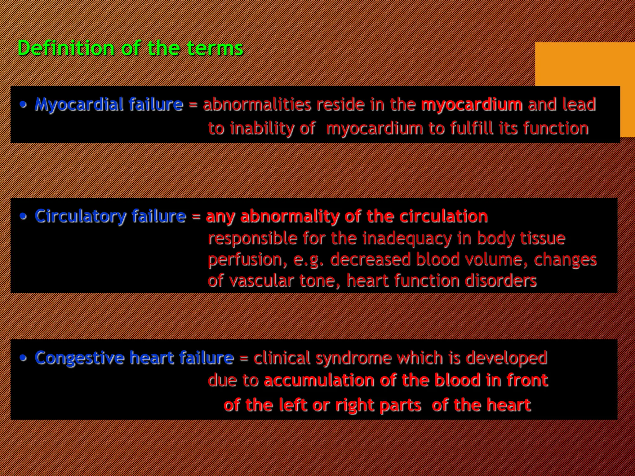
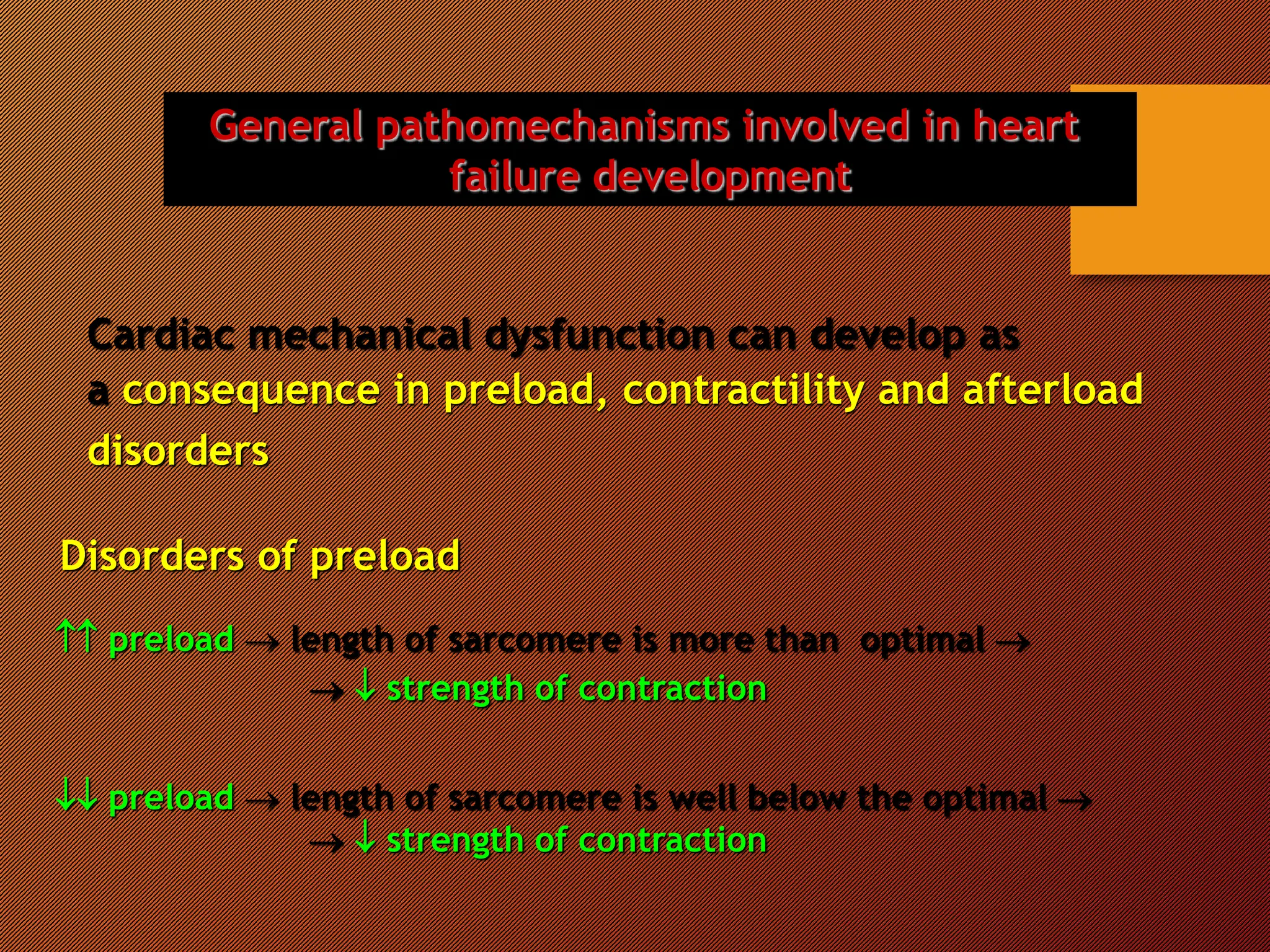
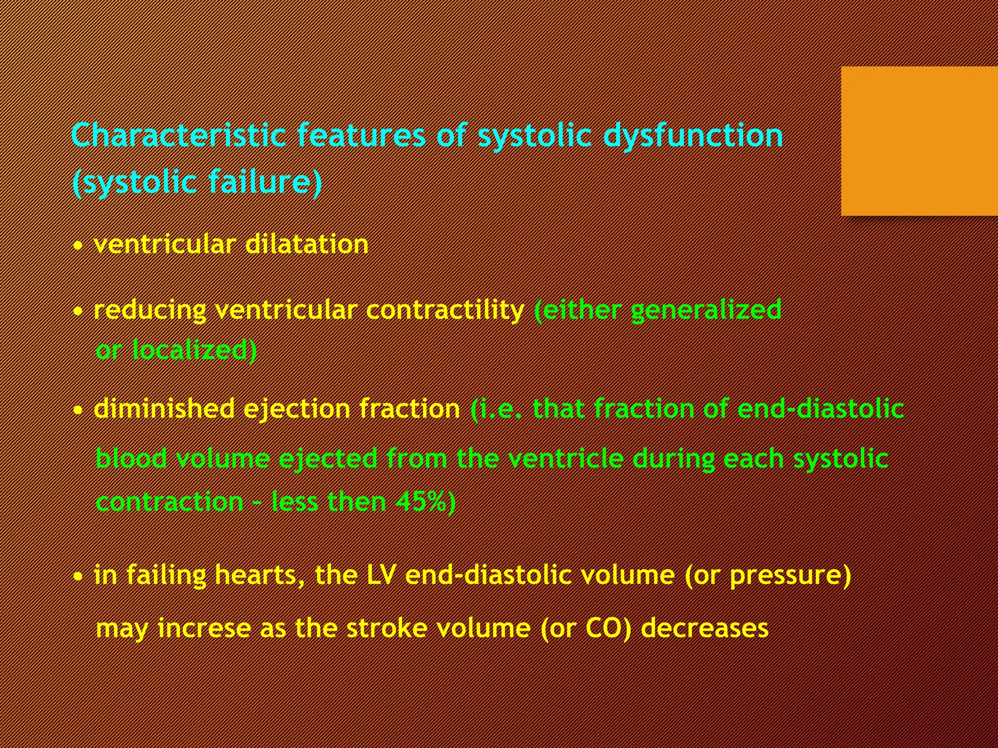
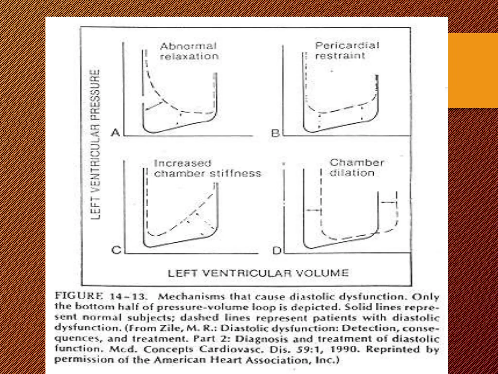
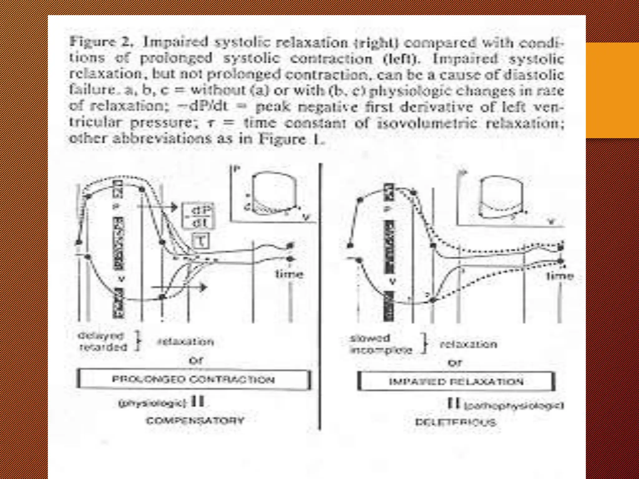
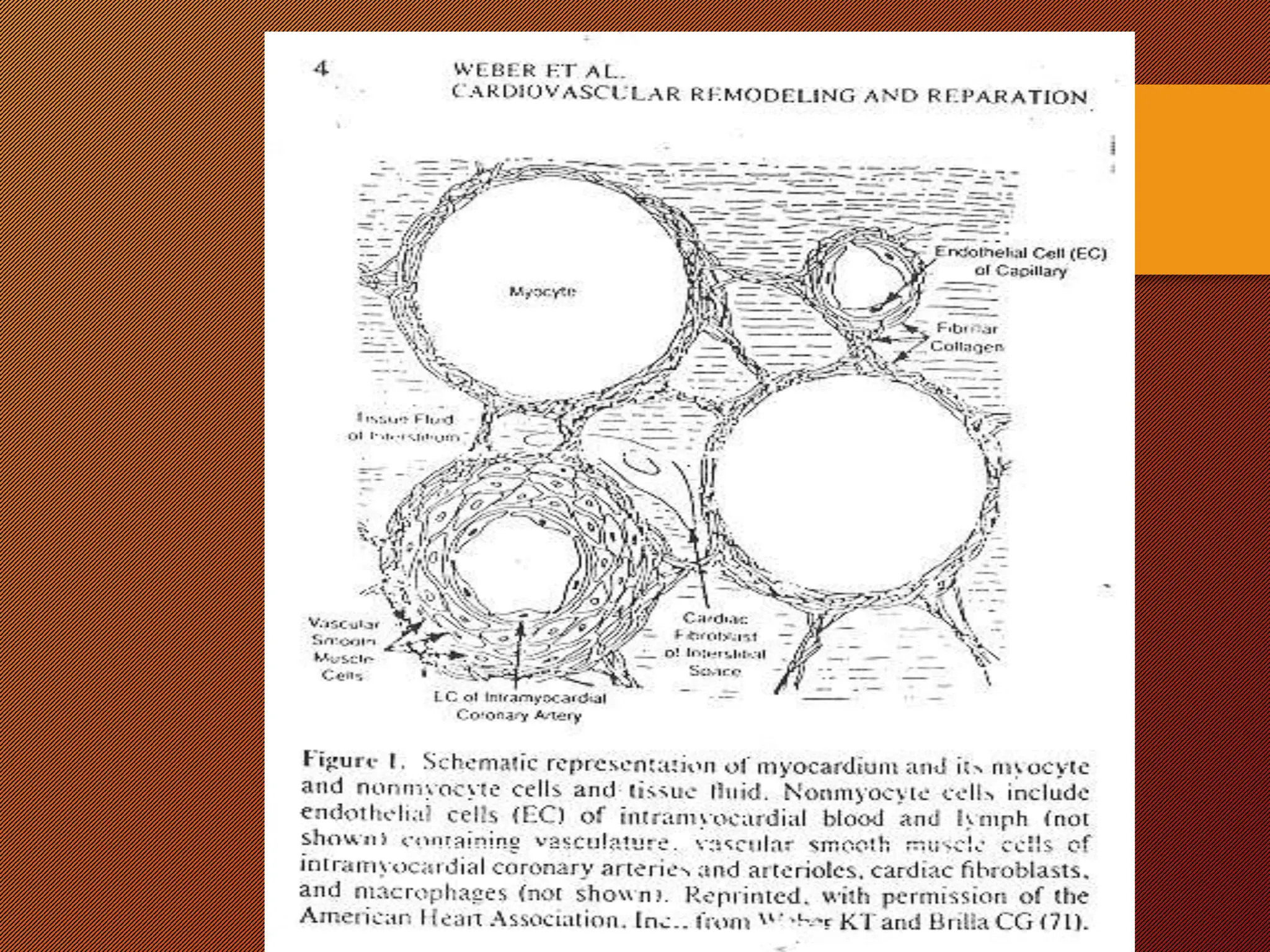
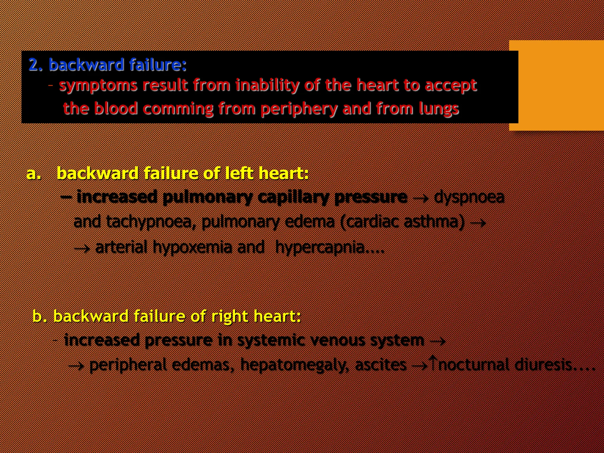
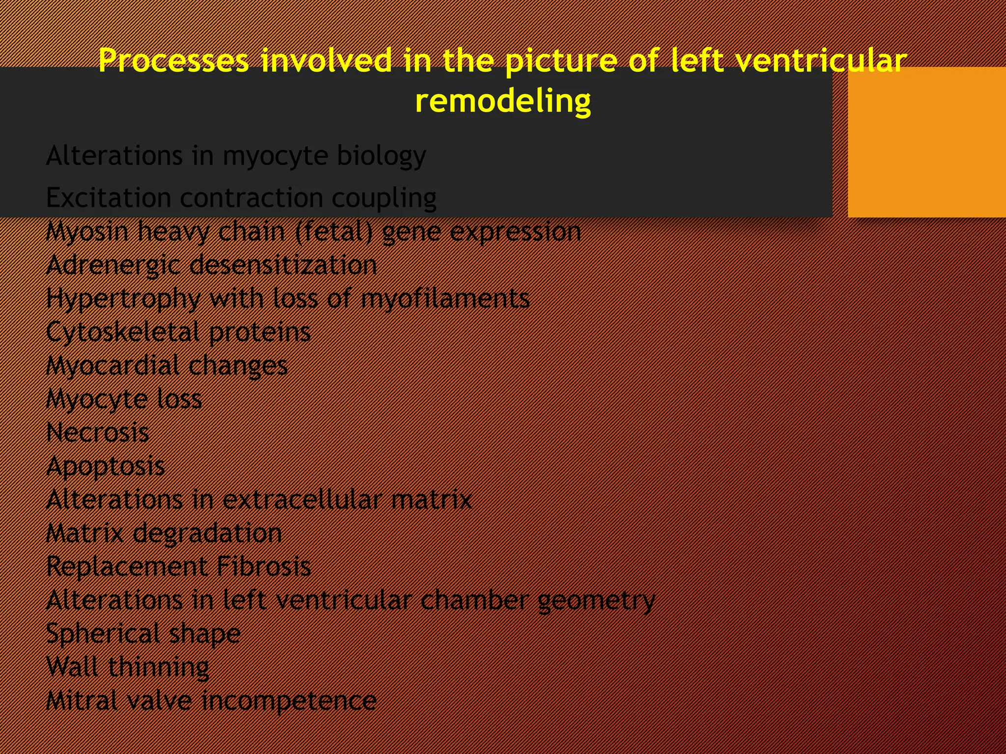
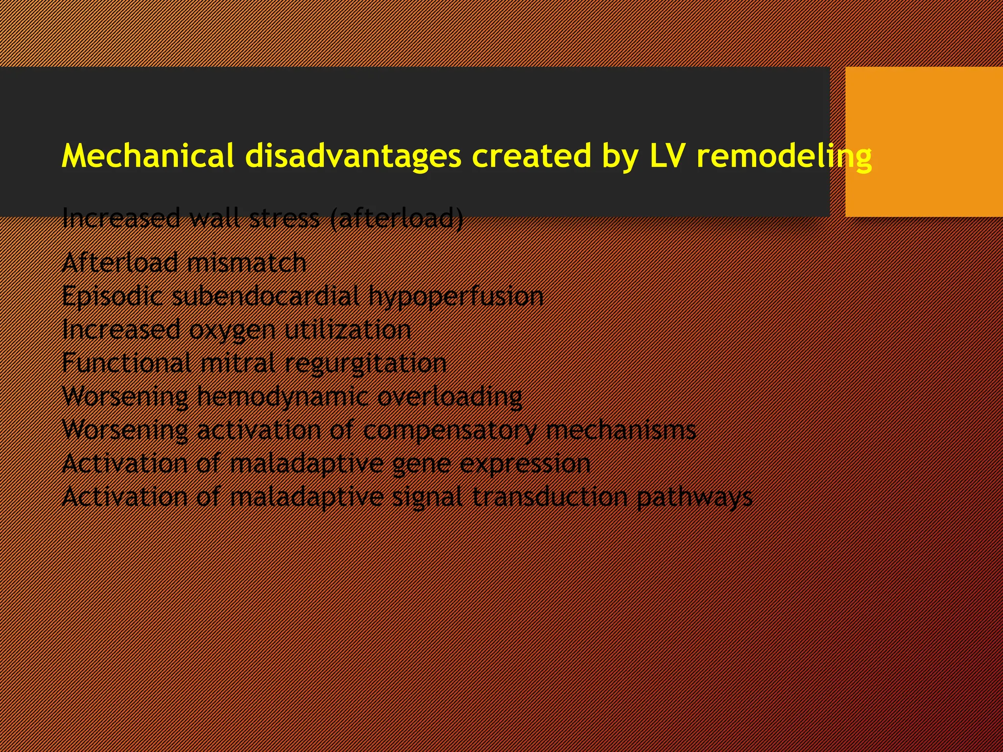
PATHOPHYSIOLOGY
OF HEART FAILURE
Myocardial contractility
Contractility of myocardium
Changes in ability of myocardium to develop force
by contraction that occurs independently on changes
in myocardial fibre length
Mechanisms involved in changes of contractility
•  amount of created cross-bridges in the sarcomere
by  of Ca ++i
- catecholamines  Ca++i  contractility
- inotropic drugs  Ca++i  contractility
 contractility  shifting the entire ventricular function
curve upward and to the left
 contractility  shifting the entire ventricular function
curve (hypoxia, acidosis) downward and
to the right
The pressure – volume loop
• It is the relation between ventricular volume and pressure
• This loop provides a convenient framework for understanding
the response of individual left ventricular contractions
to alterations in preload, afterload, and contractility
• It is composed of 4 phases:
- filling of the ventricle
- isovolumic contraction of ventricle
- isotonic contraction of ventricle (ejection of blood)
- isovolumic relaxation of ventricle
Pressure – volume loops recorded under different
conditions
It is expressed as tension which must be developed in the wall of
ventricles during systole to open the semilunar valves and eject
blood to aorta/pulmunary artery
Laplace law:
intraventricular pressure x radius of ventricle
wall tension = --------------------------------------------------------
2 x ventricular wall thickness
 afterload: due to - elevation of arterial resistance
-  ventricular size
-  intrathoracic pressure (loss of myocard)
 afterload: due to -  arterial resistance
- myocardial hypertrophy
-  ventricular size
Afterload
Heart failure
Definition
It is the pathophysiological process in which
the heart as a pump is unable to meet
the metabolic requirements of the tissue for
oxygen and substrates despite the venous
return to heart is either normal or increased
Definition of the terms
• Myocardial failure = abnormalities reside in the myocardium and lead
to inability of myocardium to fulfill its function
• Circulatory failure = any abnormality of the circulation
responsible for the inadequacy in body tissue
perfusion, e.g. decreased blood volume, changes
of vascular tone, heart function disorders
• Congestive heart failure = clinical syndrome which is developed
due to accumulation of the blood in front
of the left or right parts of the heart
General pathomechanisms involved in heart
failure development
Cardiac mechanical dysfunction can develop as
a consequence in preload, contractility and afterload
disorders
Disorders of preload
 preload  length of sarcomere is more than optimal 
  strength of contraction
 preload  length of sarcomere is well below the optimal 
  strength of contraction
Important: failing ventricle requires higher end-diastolic volume
to achieve the same CO that normal ventricle
achieves with lower ventricular volumes
Disorders of contractility
In the most forms of heart failure the contractility of myocardium
is decreased (ischemia, hypoxia, acidosis, inflammation, toxins,
metabolic disorders... )
Disorders of afterload due to:
• fluid retention in the body  increased blood volume
•  arterial resistance
• valvular heart diseases ( stenosis )
Characteristic features of systolic dysfunction
(systolic failure)
• ventricular dilatation
• reducing ventricular contractility (either generalized
or localized)
• diminished ejection fraction (i.e. that fraction of end-diastolic
blood volume ejected from the ventricle during each systolic
contraction – less then 45%)
• in failing hearts, the LV end-diastolic volume (or pressure)
may increse as the stroke volume (or CO) decreases
Characteristic features of diastolic dysfunctions
(diastolic failure)
• ventricular cavity size is normal or smaller than normal
• myocardial contractility is normal or hyperdynamic
• ejection fraction is normal (>50%) or supranormal
• ventricle is usually hypertrophied
• ventricle is filling slowly in early diastole (during the period
of passive filling)
 end-diastolic ventricular pressure is increased
Causes of heart pump failure
A. MECHANICAL ABNORMALITIES
1. Increased pressure load
– central (aortic stenosis, aortic coarctation...)
– peripheral (systemic hypertension)
2. Increased volume load
– valvular regurgitation
– hypervolemia
3. Obstruction to ventricular filling
– valvular stenosis
– pericardial restriction
B. MYOCARDIAL DAMAGE
1. Primary
a) cardiomyopathy
b) myocarditis
c) toxicity (e.g. alcohol)
d) metabolic abnormalities (e.g. hyperthyreoidism)
2. Secondary
a) oxygen deprivation (e.g. coronary heart disease)
b) inflammation (e.g. due to increased metabolic demands)
c) chronic obstructive lung disease
C. ALTERED CARDIAC RHYTHM
1. ventricular flutter and fibrilation
2. extreme tachycardias
3. extreme bradycardias
Pathomechanisms involved in heart failure
A. Pathomechanisms involved in myocardial failure
1. Damage of cardiomyocytes   contractility,
 compliance
Consequences:
 defect in ATP production and utilisation
 changes in contractile proteins
 uncoupling of excitation – contraction process
  number of cardiomyocytes
 impairment relaxation of cardiomyocytes with decrease
compliance of myocardium
 impaired of sympato-adrenal system (SAS)   number of
1-adrenergic receptors on the surface of cardiomycytes
In normal conditions, the ryanoid channel in SR is stabilized, but in heart
failure abnormal calcium leak is induced. In heart failure, channel gating is
hypersensitized to calcium: at a lower concentration of calcium, the channel
is more activated.
2. Changes of neurohumoral control of the heart
function
• Physiology: • SNS   contractility
 HR
 activity of physiologic pacemakers
Mechanism:   sympathetic activity  cAMP 
Ca ++i  contractility
  sympathetic activity  influence
of parasympathetic system on the heart
• Pathophysiology: normal neurohumoral control is
changed and creation of pathologic
neurohumoral mechanisms are present
Endothelin
Nitric oxyde
Bradykinin
Pro-proliferative effects Anti-proliferative effects
Chronic heart failure (CHF) is characterized by an imbalance of
neurohumoral adaptive mechanisms with a net results of excessive
vasoconstriction and salt and water retention
Catecholamines : - concentration in blood :
- norepinephrin – 2-3x higher at the rest than in healthy subjects
- circulating norepinephrin is increased much more
during equal load in patients suffering from CHF than
in healthy subject
-  number of beta 1 – adrenergic receptors 
 sensitivity of cardiomyocytes to catecholamines 
  contractility
System rennin – angiotensin – aldosteron
heart failure  CO  kidney perfusion  stim. of RAA system
Important:
Catecholamines and system RAA = compensatory mechanisms
 heart function and arterial BP
The role of angiotensin II in development of heart
failure
 vasoconstriction (mainly in resistant vesels)
 retention of Na  blood volume
  releasing of arginin – vasopresin peptide (AVP –antidiuretic
hormon) from neurohypophysis
  sensitivity of vessel wall to norepinephrine
 mitogenic effect on smooth muscles in vessels and on
cardiomyocytes in the heart  hypertrophy
 constriction of vas efferens (in glomerulus)
  sensation of thirst
  secretion of aldosteron from adrenal gland
 mesangial conctraction  glomerular filtration rate
 facilitation of norepinephrine release from sympathetic nerve
endings
 mitogenic effect on fibrocytes in vessel wall and in
myocardium
Pathogenesis of heart failure
Douglas L. Mann, 2004
Index event – primary
cause of heart damage
Secondary damage –
remodeling
Adrenergic, RAA,
cytokine systems are
involved in the remodeling
Pathophysiology of diastolic heart failure
 systolic heart failure = failure of ejecting function of the heart
 diastolic heart failure = failure of filling the ventricles,
 resistance to filling of ventricles
But, which of the cardiac cycle is real diastole ?
Diastolic failure is a widely recognized clinical entity
Definition of diastolic heart failure
It is pathophysiological process characterized by symptoms and signs of
congestive heart failure, which is caused by increased filling resistance
of ventricles and increased intraventricular diastolic pressure
Primary diastolic heart failure
- no signs and symptoms of systolic dysfunction is present
- ! up to 40% of patients suffering from heart failure!
Secondary diastolic heart failure
- diastolic dysfunction is the consequence of primary
systolic dysfunction
Main causes and pathomechanisms of diastolic
heart failure
1. structural disorders passive chamber stiffness
a) intramyocardial
– e.g. myocardial fibrosis, amyloidosis, hypertrophy,
myocardial ischemia...
b) extramyocardial – e.g. constrictive pericarditis
2. functional disorders   relaxation of chambers e. g.
myocardial ischemia, advanced hypertrophy of ventricles, failing
myocardium, asynchrony in heart ventricle functions
Causes and mechanism participating on impaired
ventricular relaxation
a) physiological changes in chamber relaxation due to:
– prolonged ventricular contraction
Relaxation of ventricles is not impaired !
b) pathological changes in chamber relaxation due to:
Impaired relaxation process
 delayed relaxation (retarded)
 incomplete (slowed) relaxation
 Consequences of impaired ventricular relaxation
- filling of ventricles is more dependent on diastasis
and on the systole of atrias than in healthy subjects
Symptoms and signs:
 exercise intolerance = early sign of diastolic failure
  coronary blood flow during diastole
 Causes and mechanisms involved in development
of ventricular stiffness
 ventricular compliance = passive property of ventricle
Source of compliance: cardiomyocytes and other types of cells in
the heart tissue to stretching
 Ventricular compliance is caused by structural abnormalities
localized in myocardium and in extramyocardial tissue
a) Intramyocardial causes : myocardial fibrosis, hypertrophy of
ventricular wall, restrictive cardiomyopathy
b. Extramyocardial causes : constrictive pericarditis
The role of myocardial remodelling in genesis of
heart failure
 adaptive remodelling of the heart
 pathologic remodelling of the heart
Main causes and mechanisms involved in
pathological remodelation of the heart
1.Increased amount and size of myocytes = hypertrophy
Due to: -  volume and/or pressure load
(excentric, concentric hypertrophy)
- hormonal stimulation of cardiomyocytes by
norepinephrine, angiotenzine II, endothelin...
2. Increased % of non-myocytic cells in myocardium
and their influence on structure and function of heart
a. endothelial cells – endothelins : mitogenic ability 
 stimulation growth of smooth muscle cells of vessels, fibroblasts
b. fibroblasts -  production of kolagens
Symptoms and signs of heart failure
1. forward failure:
symptoms result from inability of the heart to pump enough
blood to the periphery (from left heart), or to the lungs (from
the right heart)
a) forward failure of left heart:- muscle weakness, fatigue,
dyspepsia, oliguria....
 general mechanism: tissue hypoperfusion
b) forward failure of right heart: - hypoperfusion of the
lungs  disorders of gas
exchange
- decreased blood supply
to the left heart
2. backward failure:
– symptoms result from inability of the heart to accept
the blood comming from periphery and from lungs
a. backward failure of left heart:
– increased pulmonary capillary pressure  dyspnoea
and tachypnoea, pulmonary edema (cardiac asthma) 
 arterial hypoxemia and hypercapnia....
b. backward failure of right heart:
– increased pressure in systemic venous system 
 peripheral edemas, hepatomegaly, ascites nocturnal diuresis....
Processes involved in the picture of left ventricular
remodeling
Alterations in myocyte biology
Excitation contraction coupling
Myosin heavy chain (fetal) gene expression
Adrenergic desensitization
Hypertrophy with loss of myofilaments
Cytoskeletal proteins
Myocardial changes
Myocyte loss
Necrosis
Apoptosis
Alterations in extracellular matrix
Matrix degradation
Replacement Fibrosis
Alterations in left ventricular chamber geometry
Spherical shape
Wall thinning
Mitral valve incompetence
Mechanical disadvantages created by LV remodeling
Increased wall stress (afterload)
Afterload mismatch
Episodic subendocardial hypoperfusion
Increased oxygen utilization
Functional mitral regurgitation
Worsening hemodynamic overloading
Worsening activation of compensatory mechanisms
Activation of maladaptive gene expression
Activation of maladaptive signal transduction pathways
Characteristics of pathological and physiological cardiac hypertrophy
Pathologic cardiac hypertrophy Physiologic cardiac hypertrophy
Stimuli Pressure load in a disease setting Regular physical activity or chronic exercise
training
(e.g. hypertension, aortic coarction) Volume load (e.g. running, walking, swimming)
or volume load (e.g. valvular disease) Pressure load (e.g. strength training: weight lifting
Cardiomyopathy (familial, viral, toxic,
metabolic)
Cardiac
morphology Increased myocyte volume Increased myocyte volume
Formation of new sarcomeres Formation of new sarcomeres
Interstitial fibrosis
Myocyte necrosis and apoptosis
Fetal gene
expression Usually upregulated* Relatively normal*
Cardiac
function Depressed over time Normal or enhanced
Completely
reversible Not usually Usually
Association with
heart failure
and increased Yes No
mortality

Heart failure.ppt

  • 1.
  • 2.
    Myocardial contractility Contractility ofmyocardium Changes in ability of myocardium to develop force by contraction that occurs independently on changes in myocardial fibre length Mechanisms involved in changes of contractility •  amount of created cross-bridges in the sarcomere by  of Ca ++i - catecholamines  Ca++i  contractility - inotropic drugs  Ca++i  contractility  contractility  shifting the entire ventricular function curve upward and to the left  contractility  shifting the entire ventricular function curve (hypoxia, acidosis) downward and to the right
  • 3.
    The pressure –volume loop • It is the relation between ventricular volume and pressure • This loop provides a convenient framework for understanding the response of individual left ventricular contractions to alterations in preload, afterload, and contractility • It is composed of 4 phases: - filling of the ventricle - isovolumic contraction of ventricle - isotonic contraction of ventricle (ejection of blood) - isovolumic relaxation of ventricle
  • 4.
    Pressure – volumeloops recorded under different conditions
  • 5.
    It is expressedas tension which must be developed in the wall of ventricles during systole to open the semilunar valves and eject blood to aorta/pulmunary artery Laplace law: intraventricular pressure x radius of ventricle wall tension = -------------------------------------------------------- 2 x ventricular wall thickness  afterload: due to - elevation of arterial resistance -  ventricular size -  intrathoracic pressure (loss of myocard)  afterload: due to -  arterial resistance - myocardial hypertrophy -  ventricular size Afterload
  • 6.
    Heart failure Definition It isthe pathophysiological process in which the heart as a pump is unable to meet the metabolic requirements of the tissue for oxygen and substrates despite the venous return to heart is either normal or increased
  • 7.
    Definition of theterms • Myocardial failure = abnormalities reside in the myocardium and lead to inability of myocardium to fulfill its function • Circulatory failure = any abnormality of the circulation responsible for the inadequacy in body tissue perfusion, e.g. decreased blood volume, changes of vascular tone, heart function disorders • Congestive heart failure = clinical syndrome which is developed due to accumulation of the blood in front of the left or right parts of the heart
  • 8.
    General pathomechanisms involvedin heart failure development Cardiac mechanical dysfunction can develop as a consequence in preload, contractility and afterload disorders Disorders of preload  preload  length of sarcomere is more than optimal    strength of contraction  preload  length of sarcomere is well below the optimal    strength of contraction
  • 10.
    Important: failing ventriclerequires higher end-diastolic volume to achieve the same CO that normal ventricle achieves with lower ventricular volumes Disorders of contractility In the most forms of heart failure the contractility of myocardium is decreased (ischemia, hypoxia, acidosis, inflammation, toxins, metabolic disorders... ) Disorders of afterload due to: • fluid retention in the body  increased blood volume •  arterial resistance • valvular heart diseases ( stenosis )
  • 12.
    Characteristic features ofsystolic dysfunction (systolic failure) • ventricular dilatation • reducing ventricular contractility (either generalized or localized) • diminished ejection fraction (i.e. that fraction of end-diastolic blood volume ejected from the ventricle during each systolic contraction – less then 45%) • in failing hearts, the LV end-diastolic volume (or pressure) may increse as the stroke volume (or CO) decreases
  • 13.
    Characteristic features ofdiastolic dysfunctions (diastolic failure) • ventricular cavity size is normal or smaller than normal • myocardial contractility is normal or hyperdynamic • ejection fraction is normal (>50%) or supranormal • ventricle is usually hypertrophied • ventricle is filling slowly in early diastole (during the period of passive filling)  end-diastolic ventricular pressure is increased
  • 14.
    Causes of heartpump failure A. MECHANICAL ABNORMALITIES 1. Increased pressure load – central (aortic stenosis, aortic coarctation...) – peripheral (systemic hypertension) 2. Increased volume load – valvular regurgitation – hypervolemia 3. Obstruction to ventricular filling – valvular stenosis – pericardial restriction
  • 15.
    B. MYOCARDIAL DAMAGE 1.Primary a) cardiomyopathy b) myocarditis c) toxicity (e.g. alcohol) d) metabolic abnormalities (e.g. hyperthyreoidism) 2. Secondary a) oxygen deprivation (e.g. coronary heart disease) b) inflammation (e.g. due to increased metabolic demands) c) chronic obstructive lung disease
  • 16.
    C. ALTERED CARDIACRHYTHM 1. ventricular flutter and fibrilation 2. extreme tachycardias 3. extreme bradycardias
  • 17.
    Pathomechanisms involved inheart failure A. Pathomechanisms involved in myocardial failure 1. Damage of cardiomyocytes   contractility,  compliance Consequences:  defect in ATP production and utilisation  changes in contractile proteins  uncoupling of excitation – contraction process   number of cardiomyocytes  impairment relaxation of cardiomyocytes with decrease compliance of myocardium  impaired of sympato-adrenal system (SAS)   number of 1-adrenergic receptors on the surface of cardiomycytes
  • 19.
    In normal conditions,the ryanoid channel in SR is stabilized, but in heart failure abnormal calcium leak is induced. In heart failure, channel gating is hypersensitized to calcium: at a lower concentration of calcium, the channel is more activated.
  • 20.
    2. Changes ofneurohumoral control of the heart function • Physiology: • SNS   contractility  HR  activity of physiologic pacemakers Mechanism:   sympathetic activity  cAMP  Ca ++i  contractility   sympathetic activity  influence of parasympathetic system on the heart • Pathophysiology: normal neurohumoral control is changed and creation of pathologic neurohumoral mechanisms are present
  • 21.
  • 22.
    Chronic heart failure(CHF) is characterized by an imbalance of neurohumoral adaptive mechanisms with a net results of excessive vasoconstriction and salt and water retention Catecholamines : - concentration in blood : - norepinephrin – 2-3x higher at the rest than in healthy subjects - circulating norepinephrin is increased much more during equal load in patients suffering from CHF than in healthy subject -  number of beta 1 – adrenergic receptors   sensitivity of cardiomyocytes to catecholamines    contractility System rennin – angiotensin – aldosteron heart failure  CO  kidney perfusion  stim. of RAA system
  • 23.
    Important: Catecholamines and systemRAA = compensatory mechanisms  heart function and arterial BP The role of angiotensin II in development of heart failure  vasoconstriction (mainly in resistant vesels)  retention of Na  blood volume   releasing of arginin – vasopresin peptide (AVP –antidiuretic hormon) from neurohypophysis
  • 24.
      sensitivityof vessel wall to norepinephrine  mitogenic effect on smooth muscles in vessels and on cardiomyocytes in the heart  hypertrophy  constriction of vas efferens (in glomerulus)   sensation of thirst   secretion of aldosteron from adrenal gland  mesangial conctraction  glomerular filtration rate  facilitation of norepinephrine release from sympathetic nerve endings  mitogenic effect on fibrocytes in vessel wall and in myocardium
  • 26.
    Pathogenesis of heartfailure Douglas L. Mann, 2004 Index event – primary cause of heart damage Secondary damage – remodeling Adrenergic, RAA, cytokine systems are involved in the remodeling
  • 27.
    Pathophysiology of diastolicheart failure  systolic heart failure = failure of ejecting function of the heart  diastolic heart failure = failure of filling the ventricles,  resistance to filling of ventricles But, which of the cardiac cycle is real diastole ? Diastolic failure is a widely recognized clinical entity
  • 29.
    Definition of diastolicheart failure It is pathophysiological process characterized by symptoms and signs of congestive heart failure, which is caused by increased filling resistance of ventricles and increased intraventricular diastolic pressure Primary diastolic heart failure - no signs and symptoms of systolic dysfunction is present - ! up to 40% of patients suffering from heart failure! Secondary diastolic heart failure - diastolic dysfunction is the consequence of primary systolic dysfunction
  • 30.
    Main causes andpathomechanisms of diastolic heart failure 1. structural disorders passive chamber stiffness a) intramyocardial – e.g. myocardial fibrosis, amyloidosis, hypertrophy, myocardial ischemia... b) extramyocardial – e.g. constrictive pericarditis 2. functional disorders   relaxation of chambers e. g. myocardial ischemia, advanced hypertrophy of ventricles, failing myocardium, asynchrony in heart ventricle functions
  • 31.
    Causes and mechanismparticipating on impaired ventricular relaxation a) physiological changes in chamber relaxation due to: – prolonged ventricular contraction Relaxation of ventricles is not impaired ! b) pathological changes in chamber relaxation due to: Impaired relaxation process  delayed relaxation (retarded)  incomplete (slowed) relaxation
  • 34.
     Consequences ofimpaired ventricular relaxation - filling of ventricles is more dependent on diastasis and on the systole of atrias than in healthy subjects Symptoms and signs:  exercise intolerance = early sign of diastolic failure   coronary blood flow during diastole  Causes and mechanisms involved in development of ventricular stiffness  ventricular compliance = passive property of ventricle Source of compliance: cardiomyocytes and other types of cells in the heart tissue to stretching
  • 35.
     Ventricular complianceis caused by structural abnormalities localized in myocardium and in extramyocardial tissue a) Intramyocardial causes : myocardial fibrosis, hypertrophy of ventricular wall, restrictive cardiomyopathy b. Extramyocardial causes : constrictive pericarditis The role of myocardial remodelling in genesis of heart failure  adaptive remodelling of the heart  pathologic remodelling of the heart
  • 36.
    Main causes andmechanisms involved in pathological remodelation of the heart 1.Increased amount and size of myocytes = hypertrophy Due to: -  volume and/or pressure load (excentric, concentric hypertrophy) - hormonal stimulation of cardiomyocytes by norepinephrine, angiotenzine II, endothelin... 2. Increased % of non-myocytic cells in myocardium and their influence on structure and function of heart a. endothelial cells – endothelins : mitogenic ability   stimulation growth of smooth muscle cells of vessels, fibroblasts b. fibroblasts -  production of kolagens
  • 38.
    Symptoms and signsof heart failure 1. forward failure: symptoms result from inability of the heart to pump enough blood to the periphery (from left heart), or to the lungs (from the right heart) a) forward failure of left heart:- muscle weakness, fatigue, dyspepsia, oliguria....  general mechanism: tissue hypoperfusion b) forward failure of right heart: - hypoperfusion of the lungs  disorders of gas exchange - decreased blood supply to the left heart
  • 39.
    2. backward failure: –symptoms result from inability of the heart to accept the blood comming from periphery and from lungs a. backward failure of left heart: – increased pulmonary capillary pressure  dyspnoea and tachypnoea, pulmonary edema (cardiac asthma)   arterial hypoxemia and hypercapnia.... b. backward failure of right heart: – increased pressure in systemic venous system   peripheral edemas, hepatomegaly, ascites nocturnal diuresis....
  • 40.
    Processes involved inthe picture of left ventricular remodeling Alterations in myocyte biology Excitation contraction coupling Myosin heavy chain (fetal) gene expression Adrenergic desensitization Hypertrophy with loss of myofilaments Cytoskeletal proteins Myocardial changes Myocyte loss Necrosis Apoptosis Alterations in extracellular matrix Matrix degradation Replacement Fibrosis Alterations in left ventricular chamber geometry Spherical shape Wall thinning Mitral valve incompetence
  • 41.
    Mechanical disadvantages createdby LV remodeling Increased wall stress (afterload) Afterload mismatch Episodic subendocardial hypoperfusion Increased oxygen utilization Functional mitral regurgitation Worsening hemodynamic overloading Worsening activation of compensatory mechanisms Activation of maladaptive gene expression Activation of maladaptive signal transduction pathways
  • 42.
    Characteristics of pathologicaland physiological cardiac hypertrophy Pathologic cardiac hypertrophy Physiologic cardiac hypertrophy Stimuli Pressure load in a disease setting Regular physical activity or chronic exercise training (e.g. hypertension, aortic coarction) Volume load (e.g. running, walking, swimming) or volume load (e.g. valvular disease) Pressure load (e.g. strength training: weight lifting Cardiomyopathy (familial, viral, toxic, metabolic) Cardiac morphology Increased myocyte volume Increased myocyte volume Formation of new sarcomeres Formation of new sarcomeres Interstitial fibrosis Myocyte necrosis and apoptosis Fetal gene expression Usually upregulated* Relatively normal* Cardiac function Depressed over time Normal or enhanced Completely reversible Not usually Usually Association with heart failure and increased Yes No mortality