Cardiac Output Measurements
By Yerukneh Solomon
07/18/17 Prepared by Yerukneh Solomon 1
07/18/17 Prepared by Yerukneh Solomon 2
Outlines
➢General consideration of cardiac output measurement
• Historical perspective cardiac output measurement
• Importance of cardiac output measurement
• Features of an ideal Cardiac Output monitor
• Factors affecting selection of cardiac output monitoring devices
➢Methods of measuring cardiac output
• Types of cardiac output measurement
➢cardiac reserve
• Mechanisms of cardiac reserve:
➢Cardiac index
Adolf Eugen Fick ( 1829 – 1901)
•1855- Introduced a law
of diffusion called Fick’s
law of diffusion
•1870- Was the first one
to develop a technique
for measuring cardiac
output
07/18/17 Prepared by Yerukneh Solomon 3
07/18/17 Prepared by Yerukneh Solomon 4
Cardiac output
•Is volume of blood pumped into the aorta each minute
by the left vetricle .
•It is the determinant of global oxygen transport to the
body
•It reflects the efficiency of cardiovascular system
•There no absolute value for cardiac output
measurement
07/18/17 Prepared by Yerukneh Solomon 5
Cardiac Output Measurement
•AIM : hemodynamic monitoring and support in the
critically ill so as to optimize oxygen delivery to the
tissues
•Oxygen delivery is determined by Cardiac Output
and amount of oxygen carried in the blood
•Allows us to assess the blood flow to the tissues,
and provides information on how to best support
a failing circulation
07/18/17 Prepared by Yerukneh Solomon 6
Why should we measure?
1.The recognition that in many critically ill patients, low
cardiac output leads to significant morbidity and mortality
2.The clinical assessment of cardiac output is unreliable/
inaccurate
07/18/17 Prepared by Yerukneh Solomon 7
When should we monitor?
•High risk critically ill surgical patients in whom
large fluid shifts are expected along with bleeding
and hemodynamic instability
•An important component of goal directed therapy
(GDT), i.e., when a monitor is used in conjunction
with administration of fluids and vasopressors to
achieve set therapeutic endpoints thereby
improving patient care and outcome
Features of an ideal Cardiac Output monitor
•Safe and accurate
•Quick and easy to use both in terms of set-up and
interpretation of information
•Operator independent i.e. the skill of the operator doesn’t
affect the information collected
•Provide continuous measurement
•Rel0
i7
a
/18
b
/17
le during various p
Pre
h
par
y
ed b
s
y
i
Ye
o
ruk
ln
o
eh S
g
olo
im
c
on
al states 8
07/18/17 Prepared by Yerukneh Solomon 9
Methods of measuring cardiac output
Non-Invasive
Invasive
• Pulmonary Artery Catheter I.
✓(Dye, Thermodilution)
II.
✓Fick Principle
III.
TEE / Esophageal Doppler
Endotracheal Cardiac
Output Monitor (ECOM)
Trans Thoracic
Echocardiography (TTE)
IV. impedance Cardiography
(ICG)
Factors affecting selection of cardiac
output monitoring devices
07/18/17 Prepared by Yerukneh Solomon 10
07/18/17 Prepared by Yerukneh Solomon 11
Methods of measuring cardiac output
Simple method:
• CO(L/min) = HR(beats /min) x SV(L/beat.)
• SV: volume ejected during each beat , depends on venous
return, can be equal to venous return.
• CO (at rest) = 5-6 L/min.
• HR=72 beats/min , SV=0.07L/min (70ml) .
• CO increases when metabolic need (eg. Exercise), Therefore,
HR or SV or both can increase.
Methods of measuring cardiac output
Fick Principle: “Gold standard”
•Fick Principle relies on the total uptake of a substances
by peripheral tissue is equal to the product of blood
flow to the peripheral tissue and arterial – venous
con
07
c
/1e
8/17
ntration difference
Prepo
ared
fbyt
Ye
h
ruke
neh S
s
olu
omo
b
n stances 12
13
Prepared by Yerukneh Solomon
07/18/17
❖Mixed venous blood is usually obtained
•Through a catheter inserted into:
✓The brachial vein of the forearm,
✓Through the subclavian vein,
✓Down to the right atrium,
✓The right ventricle or pulmonary artery
❖Systemic arterial blood can then be obtained from
any
07/
s
18/
y
17
stemic artery in th
Pre
e
pare
b
d by
o
Ye
d
rukn
y
eh
.Solomon 14
07/18/17 Prepared by Yerukneh Solomon 15
•Fick cardiac outputs are infrequently used because
difficulties in collecting and analyzing exhaled gas
concentration in critically ill patients because may
not have normal VO2 value.
07/18/17 Prepared by Yerukneh Solomon 16
❑Indicator dilution method
•Based on how fast the flowing blood can dilute the
substances introduced into the circulation
•A known amount of a substance, such as a dye, is injected
into a large systemic vein or, preferably, into the right
atrium.
•The concentration of the dye is recorded as the dye passes
through one of the peripheral arteries, giving a curve
Stewart-Hamilton Equation
07/18/17 Prepared by Yerukneh Solomon 17
Area under the curve is inversely proportion to the
rate of blood flow.
07/18/17 Prepared by Yerukneh Solomon 18
07/18/17 Prepared by Yerukneh Solomon 19
❑is Thermodilution Method, in which the indicator
used is cold saline.
•Affected by the phase of respiration and should be
measured at the same point of respiratory cycle
•Variations in the speed of cold water injection can
result in altered measurement
07/18/17 Prepared by Yerukneh Solomon 20
Thermodilution: A popular indicator
dilution technique
•A cold solution of D/W 5% or normal saline
(temperature 0°C) is injected into the right atrium from
a proximal catheter port
•This solution causes a decrease in blood temperature
in right heart and flows to the pulmonary artery where
the temperature is measured by a thermistor placed in
the pulmonary artery catheter
07/18/17 Prepared by Yerukneh Solomon 21
07/18/17 Prepared by Yerukneh Solomon 22
•The thermistor records the change in blood
temperature with time and sends this information
to an electronic instrument that records and
displays a temperature-time curve /
Thermodilution curve
07/18/17 Prepared by Yerukneh Solomon 23
07/18/17 Prepared by Yerukneh Solomon 24
• The Cardiac Output can be derived from the Modified
Stewart-Hamilton conservation of heat equation
• CO= V1( Tb- T1) K1 K2
ξ ΔTb (t) dt
Where,
V1= injectate volume in ml
Tb= temperature of pulmonary artery blood
T1= injectate temperature °C
K1
= density factor
K2= computation constant taking in account the catheter
dead-space and heat exchange in transit ; both
computation constant
Denominator: change in temp and change in time:
corresponds to the area under thermodilution curve
07/18/17 Prepared by Yerukneh Solomon 25
•The degree of change is inversely
proportional to cardiac output
•Temperature change is minimal if there is a
high blood flow but Temperature change high
if blood flow is low
07/18/17 Prepared by Yerukneh Solomon 26
Non Invasive Methods
07/18/17 Prepared by Yerukneh Solomon 27
Endotracheal Cardiac Output Monitor
(ECOM)
• ECOM (Con-Med, Irvine, Calif, United States) measures CO
using impedance plethysmography
• Is based on the principle of bio-impedance
• Current is passed through electrodes attached to endotracheal
tube shaft and cuff
• Current is passed from electrode on the shaft of endotracheal
tube and change in impedance secondary to aortic blood flow is
detected by electrode on the cuff
07/18/17 Prepared by Yerukneh Solomon 28
•An algorithm calculates SV based on impedance
changes and CO can be calculated
•Impedance is affected by aortic blood flow
•Limitations:
•Electro cautery affects its accuracy
•Coronary blood flow is not calculated
•Is still adequately not validated in humans
•Is costly and has not become very popular
07/18/17 Prepared by Yerukneh Solomon 29
Trans Esophageal Echocardiography (TEE) /
Doppler
•A widely used monitor in perioperative setting
•Is an important tool for the assessment of
cardiac structures, filling status and cardiac
contractility
•Aortic pathology can also be detected by TEE
•Doppler technique is used to measure CO by
Simpson’s rule measuring SV multiplied by HR
07/18/17 Prepared by Yerukneh Solomon 30
•Measurement can be done at the level of pulmonary
artery, mitral or aortic valve
•TEE can quantify Cardiac Output more precisely by
measuring both the velocity and the cross-sectional
area of blood flow at appropriate locations in the heart
or great vessels
i.e. Flow = CSA X Velocity
▪ SV= flow X ET ( Systolic Ejection time)
▪ CO=SV X HR
07/18/17 Prepared by Yerukneh Solomon 31
07/18/17 Prepared by Yerukneh Solomon 32
07/18/17 Prepared by Yerukneh Solomon 33
Advantages
• Minimally invasive
• Minimal interference
form bone, lungs and
soft tissue (as seen with
transthoracic route)
• Quickly inserted and
analyzed
• Little training required
• Very few complications
Disadvantages
• May require sedation
• User dependent
• Probe may detect other
vessels e.g. intracardiac,
intrapulmonary
• Contraindicated in
Esophageal Surgeries
• Depends on accurate
probe positioning
07/18/17 Prepared by Yerukneh Solomon 34
Trans Thoracic Echocardiography (TTE)
•Echocardiography is cardiac ultrasound
•Can be used to estimate Cardiac Output by direct
visualization of the contracting heart in real time
•Gaining acceptance as one of the safest and
most widely used cardiac output monitors in the
critically ill
07/18/17 Prepared by Yerukneh Solomon 35
•Be used to assess cardiac output (intermittently)
•The USCOMTMdevice targets the pulmonary and
aortic valves accessed via the parasternal and
suprasternal windows in order to assess cardiac
output completely non-invasively
07/18/17 Prepared by Yerukneh Solomon 36
Advantages
•Non-invasive
•Quick
•Allows for
measurement of the
ventricles,
visualization of the
valves, estimation of
ejection fraction
Disadvantages
▪ User dependent
▪ Possible
interference from
bone, lung and
soft tissues
07/18/17 Prepared by Yerukneh Solomon 37
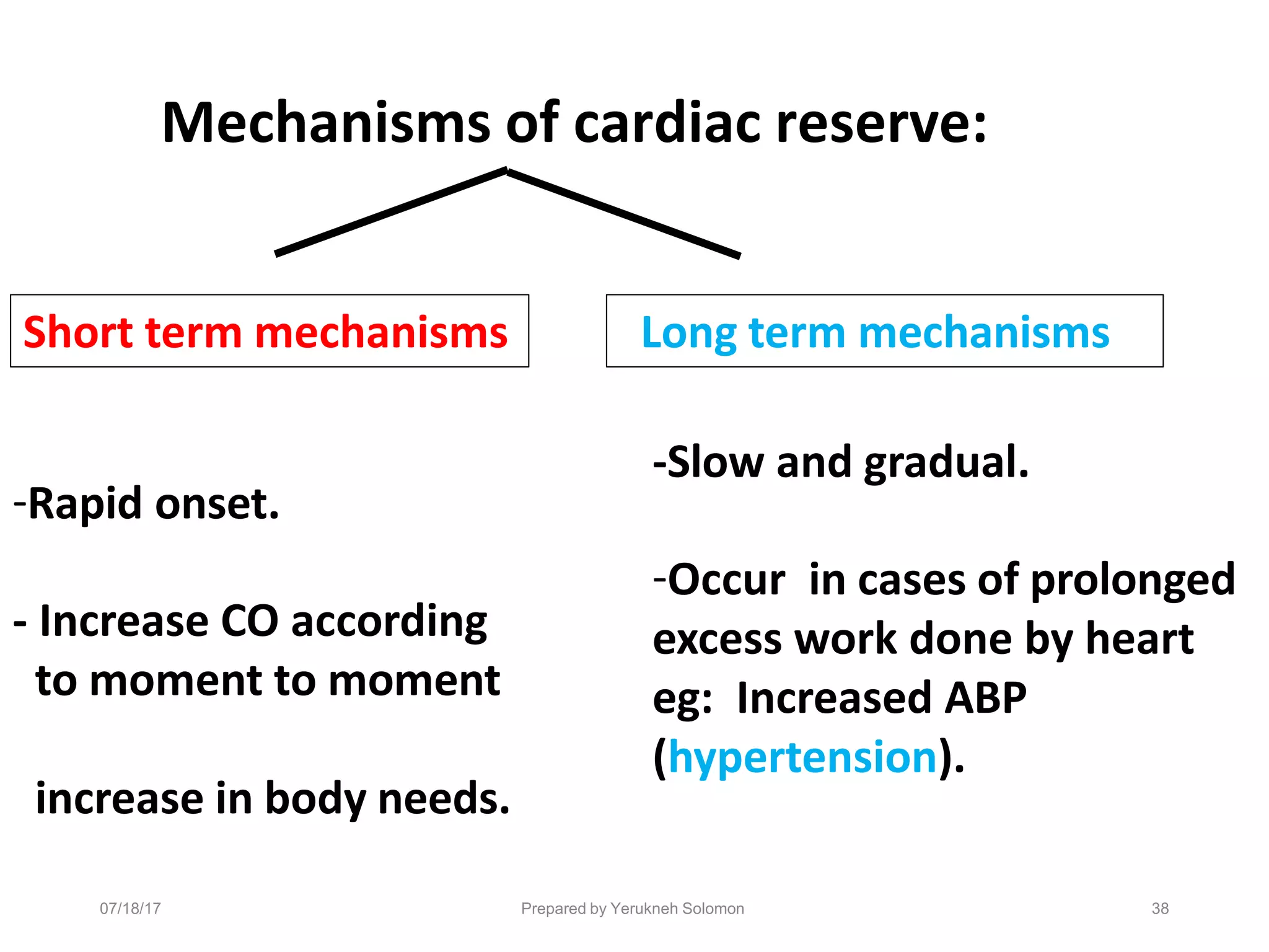
Cardiac reserve
•Cardiac reserve refers to the heart's ability to adjust to
the demands placed upon it.
❑Cardiac reserve =
[cardiac output during stress - cardiac output at rest.]
•In the normal young adult the resting cardiac output is
about 5-6 L/min and the cardiac reserve is nearly 30
L/min.
Short term mechanisms Long term mechanisms
Mechanisms of cardiac reserve:
-Rapid onset.
- Increase CO according
to moment to moment
increase in body needs.
07/18/17 Prepared by Yerukneh Solomon 38
-Slow and gradual.
-Occur in cases of prolonged
excess work done by heart
eg: Increased ABP
(hypertension).
Prepared by Yerukneh Solomon
Short term mechanisms:
1. Heart rate (HR) reserve:
The possibility of the increase of heart rate up to 2 – 3 times.
•Heart rate reserve is limited because the increase in heart rate above
180 beats/min causes decrease in the CO.
As this marked increase in the heart rate will be associated with
marked decrease in diastolic period causing:
Decrease in ventricular filling Decrease in coronary blood flow
Decrease in SV and CO Decrease in myocardial contraction
Decrease in CO
39
07/18/17
2. Stroke volume reserve:
•Increase in SV causes increase of CO.
•This mechanism is mediated by either:
Increase of the EDV
(EDV reserve)
Decrease of the ESV
(ESV reserve)
40
Prepared by Yerukneh Solomon
07/18/17
2.1. EDV reserve:
• Increase of the venous return (as in Ms exercise) increases
EDV ➔ stretch of ventricular muscle fibers
✓Increases force of contraction of cardiac muscle
and increases SV and CO.(Starling law.)
❖This mechanism is limited as the marked increase in EDV
causes overstretch of ventricular muscle fibers decreases
forc
07
e
/18/1
o
7
f contraction of car
P
d
repa
ir
a
ed
c
by Y
m
erukn
u
eh
s
So
c
lom
le
on
. This will decrease 41
Prepared by Yerukneh Solomon
❑ 2.2 ESV reserve:
• Increased sympathetic stimulation to the heart increases
the force of cardiac muscle contraction and decreases the
ESV. This will increase SV and CO.
• This mechanism is also limited as the marked decrease in
ESV causes myocardial injury (athletic injury).
42
07/18/17
Long term mechanisms:
1. Cardiac muscle hypertrophy:
•It is the increase in the size of the individual cardiac muscle
fiber (increase its protein content). This increases the force
of contraction of cardiac muscle increase SV and CO.
•Marked hypertrophy of the myocardium is not
associated with parallel increase in coronary blood flow.
•This cause myocardial ischemia with subsequent
decrease in the force of contraction of cardiac muscle.
This will decrease the SV and CO.
Prepared by Yerukneh Solmon 43
07/18/17
2. Dilatation of cardiac chambers:
• This mechanism occurs when the blood accumulated
inside the cardiac chambers as in case of heart failure.
• This dilatation of the cardiac chambers causes stretch of
the cardiac muscle fibers Increases its force of
contraction according to Starling law.
• Marked increase in EDV causes overstretch of ventricular
muscle fibers and decreases force of contraction of cardiac
muscle.
•Thi0
s
7/18
w
/17
ill decrease the SVP
a
rep
n
are
d
d by
t
Ye
h
ruk
e
neh
C
Sol
O
omo
.
n 44
07/18/17 Prepared by Yerukneh Solomon 45
•In severe heart failure the cardiac reserve can be
markedly diminished or totally abolished
•The cardiac reserve is affected by a number of
different factors like:
▪ The percentage of fibrosis,
▪ The degree of apoptosis or necrosis and
▪ Presence of auto-antibodies against the β1-
adrenoceptor in the heart.
Cardiac Index
•Both of these people’s
hearts pump 3 liters of
blood per minute.
•Who feels better?
•The smaller one!
•Why ?
•Less body surface area.
07/18/17 Prepared by Yerukneh Solomon 46
07/18/17 Prepared by Yerukneh Solomon 47
Cardiac Index
• Is the cardiac output per square meter of body surface area
• Cardiac output is frequently stated in terms of the cardiac
index
• Average human being who weighs 70 kilograms has a body
surface area of about 1.7 square meters, which means that the
normal average cardiac index for adults is about 3 L/min/m2 of
body surface area
Variation oP
frep
C
are
a
d by
rY
d
eru
ikn
a
eh
c
Solo
Im
n
on
dex with age
07/18/17 48
07/18/17 Prepared by Yerukneh Solomon 49
THANK YOU!!!
07/18/17 Prepared by Yerukneh Solomon 50
• Ganong’s Review of Medical Physiology - 23rdEd
• Medical physiology : a cellular and molecular approach /
[edited by] Walter F. Boron, Emile L. Boulpaep. 2nded.
• Guyton and hall textbook of medical physiology, 13th
edition
• http://heart.bmj.com/content/79/3/289#BIBL
• Chew MS, Aneman A. Haemodynamic monitoring using
arterial waveform analysis. Curr Opin Crit Care.
2013;19:234-41

myassignment-170718074752 (1).pptx

  • 1.
    Cardiac Output Measurements ByYerukneh Solomon 07/18/17 Prepared by Yerukneh Solomon 1
  • 2.
    07/18/17 Prepared byYerukneh Solomon 2 Outlines ➢General consideration of cardiac output measurement • Historical perspective cardiac output measurement • Importance of cardiac output measurement • Features of an ideal Cardiac Output monitor • Factors affecting selection of cardiac output monitoring devices ➢Methods of measuring cardiac output • Types of cardiac output measurement ➢cardiac reserve • Mechanisms of cardiac reserve: ➢Cardiac index
  • 3.
    Adolf Eugen Fick( 1829 – 1901) •1855- Introduced a law of diffusion called Fick’s law of diffusion •1870- Was the first one to develop a technique for measuring cardiac output 07/18/17 Prepared by Yerukneh Solomon 3
  • 4.
    07/18/17 Prepared byYerukneh Solomon 4 Cardiac output •Is volume of blood pumped into the aorta each minute by the left vetricle . •It is the determinant of global oxygen transport to the body •It reflects the efficiency of cardiovascular system •There no absolute value for cardiac output measurement
  • 5.
    07/18/17 Prepared byYerukneh Solomon 5 Cardiac Output Measurement •AIM : hemodynamic monitoring and support in the critically ill so as to optimize oxygen delivery to the tissues •Oxygen delivery is determined by Cardiac Output and amount of oxygen carried in the blood •Allows us to assess the blood flow to the tissues, and provides information on how to best support a failing circulation
  • 6.
    07/18/17 Prepared byYerukneh Solomon 6 Why should we measure? 1.The recognition that in many critically ill patients, low cardiac output leads to significant morbidity and mortality 2.The clinical assessment of cardiac output is unreliable/ inaccurate
  • 7.
    07/18/17 Prepared byYerukneh Solomon 7 When should we monitor? •High risk critically ill surgical patients in whom large fluid shifts are expected along with bleeding and hemodynamic instability •An important component of goal directed therapy (GDT), i.e., when a monitor is used in conjunction with administration of fluids and vasopressors to achieve set therapeutic endpoints thereby improving patient care and outcome
  • 8.
    Features of anideal Cardiac Output monitor •Safe and accurate •Quick and easy to use both in terms of set-up and interpretation of information •Operator independent i.e. the skill of the operator doesn’t affect the information collected •Provide continuous measurement •Rel0 i7 a /18 b /17 le during various p Pre h par y ed b s y i Ye o ruk ln o eh S g olo im c on al states 8
  • 9.
    07/18/17 Prepared byYerukneh Solomon 9 Methods of measuring cardiac output Non-Invasive Invasive • Pulmonary Artery Catheter I. ✓(Dye, Thermodilution) II. ✓Fick Principle III. TEE / Esophageal Doppler Endotracheal Cardiac Output Monitor (ECOM) Trans Thoracic Echocardiography (TTE) IV. impedance Cardiography (ICG)
  • 10.
    Factors affecting selectionof cardiac output monitoring devices 07/18/17 Prepared by Yerukneh Solomon 10
  • 11.
    07/18/17 Prepared byYerukneh Solomon 11 Methods of measuring cardiac output Simple method: • CO(L/min) = HR(beats /min) x SV(L/beat.) • SV: volume ejected during each beat , depends on venous return, can be equal to venous return. • CO (at rest) = 5-6 L/min. • HR=72 beats/min , SV=0.07L/min (70ml) . • CO increases when metabolic need (eg. Exercise), Therefore, HR or SV or both can increase.
  • 12.
    Methods of measuringcardiac output Fick Principle: “Gold standard” •Fick Principle relies on the total uptake of a substances by peripheral tissue is equal to the product of blood flow to the peripheral tissue and arterial – venous con 07 c /1e 8/17 ntration difference Prepo ared fbyt Ye h ruke neh S s olu omo b n stances 12
  • 13.
    13 Prepared by YeruknehSolomon 07/18/17
  • 14.
    ❖Mixed venous bloodis usually obtained •Through a catheter inserted into: ✓The brachial vein of the forearm, ✓Through the subclavian vein, ✓Down to the right atrium, ✓The right ventricle or pulmonary artery ❖Systemic arterial blood can then be obtained from any 07/ s 18/ y 17 stemic artery in th Pre e pare b d by o Ye d rukn y eh .Solomon 14
  • 15.
    07/18/17 Prepared byYerukneh Solomon 15 •Fick cardiac outputs are infrequently used because difficulties in collecting and analyzing exhaled gas concentration in critically ill patients because may not have normal VO2 value.
  • 16.
    07/18/17 Prepared byYerukneh Solomon 16 ❑Indicator dilution method •Based on how fast the flowing blood can dilute the substances introduced into the circulation •A known amount of a substance, such as a dye, is injected into a large systemic vein or, preferably, into the right atrium. •The concentration of the dye is recorded as the dye passes through one of the peripheral arteries, giving a curve
  • 17.
  • 18.
    Area under thecurve is inversely proportion to the rate of blood flow. 07/18/17 Prepared by Yerukneh Solomon 18
  • 19.
    07/18/17 Prepared byYerukneh Solomon 19 ❑is Thermodilution Method, in which the indicator used is cold saline. •Affected by the phase of respiration and should be measured at the same point of respiratory cycle •Variations in the speed of cold water injection can result in altered measurement
  • 20.
    07/18/17 Prepared byYerukneh Solomon 20 Thermodilution: A popular indicator dilution technique •A cold solution of D/W 5% or normal saline (temperature 0°C) is injected into the right atrium from a proximal catheter port •This solution causes a decrease in blood temperature in right heart and flows to the pulmonary artery where the temperature is measured by a thermistor placed in the pulmonary artery catheter
  • 21.
    07/18/17 Prepared byYerukneh Solomon 21
  • 22.
    07/18/17 Prepared byYerukneh Solomon 22 •The thermistor records the change in blood temperature with time and sends this information to an electronic instrument that records and displays a temperature-time curve / Thermodilution curve
  • 23.
    07/18/17 Prepared byYerukneh Solomon 23
  • 24.
    07/18/17 Prepared byYerukneh Solomon 24 • The Cardiac Output can be derived from the Modified Stewart-Hamilton conservation of heat equation • CO= V1( Tb- T1) K1 K2 ξ ΔTb (t) dt Where, V1= injectate volume in ml Tb= temperature of pulmonary artery blood T1= injectate temperature °C K1 = density factor K2= computation constant taking in account the catheter dead-space and heat exchange in transit ; both computation constant Denominator: change in temp and change in time: corresponds to the area under thermodilution curve
  • 25.
    07/18/17 Prepared byYerukneh Solomon 25 •The degree of change is inversely proportional to cardiac output •Temperature change is minimal if there is a high blood flow but Temperature change high if blood flow is low
  • 26.
    07/18/17 Prepared byYerukneh Solomon 26 Non Invasive Methods
  • 27.
    07/18/17 Prepared byYerukneh Solomon 27 Endotracheal Cardiac Output Monitor (ECOM) • ECOM (Con-Med, Irvine, Calif, United States) measures CO using impedance plethysmography • Is based on the principle of bio-impedance • Current is passed through electrodes attached to endotracheal tube shaft and cuff • Current is passed from electrode on the shaft of endotracheal tube and change in impedance secondary to aortic blood flow is detected by electrode on the cuff
  • 28.
    07/18/17 Prepared byYerukneh Solomon 28 •An algorithm calculates SV based on impedance changes and CO can be calculated •Impedance is affected by aortic blood flow •Limitations: •Electro cautery affects its accuracy •Coronary blood flow is not calculated •Is still adequately not validated in humans •Is costly and has not become very popular
  • 29.
    07/18/17 Prepared byYerukneh Solomon 29 Trans Esophageal Echocardiography (TEE) / Doppler •A widely used monitor in perioperative setting •Is an important tool for the assessment of cardiac structures, filling status and cardiac contractility •Aortic pathology can also be detected by TEE •Doppler technique is used to measure CO by Simpson’s rule measuring SV multiplied by HR
  • 30.
    07/18/17 Prepared byYerukneh Solomon 30 •Measurement can be done at the level of pulmonary artery, mitral or aortic valve •TEE can quantify Cardiac Output more precisely by measuring both the velocity and the cross-sectional area of blood flow at appropriate locations in the heart or great vessels i.e. Flow = CSA X Velocity ▪ SV= flow X ET ( Systolic Ejection time) ▪ CO=SV X HR
  • 31.
    07/18/17 Prepared byYerukneh Solomon 31
  • 32.
    07/18/17 Prepared byYerukneh Solomon 32
  • 33.
    07/18/17 Prepared byYerukneh Solomon 33 Advantages • Minimally invasive • Minimal interference form bone, lungs and soft tissue (as seen with transthoracic route) • Quickly inserted and analyzed • Little training required • Very few complications Disadvantages • May require sedation • User dependent • Probe may detect other vessels e.g. intracardiac, intrapulmonary • Contraindicated in Esophageal Surgeries • Depends on accurate probe positioning
  • 34.
    07/18/17 Prepared byYerukneh Solomon 34 Trans Thoracic Echocardiography (TTE) •Echocardiography is cardiac ultrasound •Can be used to estimate Cardiac Output by direct visualization of the contracting heart in real time •Gaining acceptance as one of the safest and most widely used cardiac output monitors in the critically ill
  • 35.
    07/18/17 Prepared byYerukneh Solomon 35 •Be used to assess cardiac output (intermittently) •The USCOMTMdevice targets the pulmonary and aortic valves accessed via the parasternal and suprasternal windows in order to assess cardiac output completely non-invasively
  • 36.
    07/18/17 Prepared byYerukneh Solomon 36 Advantages •Non-invasive •Quick •Allows for measurement of the ventricles, visualization of the valves, estimation of ejection fraction Disadvantages ▪ User dependent ▪ Possible interference from bone, lung and soft tissues
  • 37.
    07/18/17 Prepared byYerukneh Solomon 37 Cardiac reserve •Cardiac reserve refers to the heart's ability to adjust to the demands placed upon it. ❑Cardiac reserve = [cardiac output during stress - cardiac output at rest.] •In the normal young adult the resting cardiac output is about 5-6 L/min and the cardiac reserve is nearly 30 L/min.
  • 38.
    Short term mechanismsLong term mechanisms Mechanisms of cardiac reserve: -Rapid onset. - Increase CO according to moment to moment increase in body needs. 07/18/17 Prepared by Yerukneh Solomon 38 -Slow and gradual. -Occur in cases of prolonged excess work done by heart eg: Increased ABP (hypertension).
  • 39.
    Prepared by YeruknehSolomon Short term mechanisms: 1. Heart rate (HR) reserve: The possibility of the increase of heart rate up to 2 – 3 times. •Heart rate reserve is limited because the increase in heart rate above 180 beats/min causes decrease in the CO. As this marked increase in the heart rate will be associated with marked decrease in diastolic period causing: Decrease in ventricular filling Decrease in coronary blood flow Decrease in SV and CO Decrease in myocardial contraction Decrease in CO 39 07/18/17
  • 40.
    2. Stroke volumereserve: •Increase in SV causes increase of CO. •This mechanism is mediated by either: Increase of the EDV (EDV reserve) Decrease of the ESV (ESV reserve) 40 Prepared by Yerukneh Solomon 07/18/17
  • 41.
    2.1. EDV reserve: •Increase of the venous return (as in Ms exercise) increases EDV ➔ stretch of ventricular muscle fibers ✓Increases force of contraction of cardiac muscle and increases SV and CO.(Starling law.) ❖This mechanism is limited as the marked increase in EDV causes overstretch of ventricular muscle fibers decreases forc 07 e /18/1 o 7 f contraction of car P d repa ir a ed c by Y m erukn u eh s So c lom le on . This will decrease 41
  • 42.
    Prepared by YeruknehSolomon ❑ 2.2 ESV reserve: • Increased sympathetic stimulation to the heart increases the force of cardiac muscle contraction and decreases the ESV. This will increase SV and CO. • This mechanism is also limited as the marked decrease in ESV causes myocardial injury (athletic injury). 42 07/18/17
  • 43.
    Long term mechanisms: 1.Cardiac muscle hypertrophy: •It is the increase in the size of the individual cardiac muscle fiber (increase its protein content). This increases the force of contraction of cardiac muscle increase SV and CO. •Marked hypertrophy of the myocardium is not associated with parallel increase in coronary blood flow. •This cause myocardial ischemia with subsequent decrease in the force of contraction of cardiac muscle. This will decrease the SV and CO. Prepared by Yerukneh Solmon 43 07/18/17
  • 44.
    2. Dilatation ofcardiac chambers: • This mechanism occurs when the blood accumulated inside the cardiac chambers as in case of heart failure. • This dilatation of the cardiac chambers causes stretch of the cardiac muscle fibers Increases its force of contraction according to Starling law. • Marked increase in EDV causes overstretch of ventricular muscle fibers and decreases force of contraction of cardiac muscle. •Thi0 s 7/18 w /17 ill decrease the SVP a rep n are d d by t Ye h ruk e neh C Sol O omo . n 44
  • 45.
    07/18/17 Prepared byYerukneh Solomon 45 •In severe heart failure the cardiac reserve can be markedly diminished or totally abolished •The cardiac reserve is affected by a number of different factors like: ▪ The percentage of fibrosis, ▪ The degree of apoptosis or necrosis and ▪ Presence of auto-antibodies against the β1- adrenoceptor in the heart.
  • 46.
    Cardiac Index •Both ofthese people’s hearts pump 3 liters of blood per minute. •Who feels better? •The smaller one! •Why ? •Less body surface area. 07/18/17 Prepared by Yerukneh Solomon 46
  • 47.
    07/18/17 Prepared byYerukneh Solomon 47 Cardiac Index • Is the cardiac output per square meter of body surface area • Cardiac output is frequently stated in terms of the cardiac index • Average human being who weighs 70 kilograms has a body surface area of about 1.7 square meters, which means that the normal average cardiac index for adults is about 3 L/min/m2 of body surface area
  • 48.
  • 49.
    07/18/17 Prepared byYerukneh Solomon 49 THANK YOU!!!
  • 50.
    07/18/17 Prepared byYerukneh Solomon 50 • Ganong’s Review of Medical Physiology - 23rdEd • Medical physiology : a cellular and molecular approach / [edited by] Walter F. Boron, Emile L. Boulpaep. 2nded. • Guyton and hall textbook of medical physiology, 13th edition • http://heart.bmj.com/content/79/3/289#BIBL • Chew MS, Aneman A. Haemodynamic monitoring using arterial waveform analysis. Curr Opin Crit Care. 2013;19:234-41