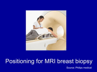
Vacuum assisted breast biopsy systems use image-guided breast biopsy techniques to precisely locate breast lesions and safely obtain tissue samples. MRI is a highly accurate imaging method that can detect breast abnormalities early, making it useful for evaluating high-risk women. The ATEC system is an example of a vacuum assisted biopsy device that quickly collects core biopsy samples under MRI guidance in a closed system.