
Recommended
More Related Content
What's hot
What's hot (20)
Similar to Humeral shaft fractures
Similar to Humeral shaft fractures (20)
More from Supun Dhanasekara
Recently uploaded
Recently uploaded (20)
Humeral shaft fractures
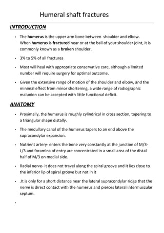
- 1. Humeral shaft fractures INTRODUCTION • The humerus is the upper arm bone between shoulder and elbow. When humerus is fractured near or at the ball of your shoulder joint, it is commonly known as a broken shoulder. • 3% to 5% of all fractures • Most will heal with appropriate conservative care, although a limited number will require surgery for optimal outcome. • Given the extensive range of motion of the shoulder and elbow, and the minimal effect from minor shortening, a wide range of radiographic malunion can be accepted with little functional deficit. ANATOMY • Proximally, the humerus is roughly cylindrical in cross section, tapering to a triangular shape distally. • The medullary canal of the humerus tapers to an end above the supracondylar expansion. • Nutrient artery- enters the bone very constantly at the junction of M/3- L/3 and foramina of entry are concentrated in a small area of the distal half of M/3 on medial side. • Radial nerve- it does not travel along the spiral groove and it lies close to the inferior lip of spiral groove but not in it • .It is only for a short distance near the lateral supracondylar ridge that the nerve is direct contact with the humerus and pierces lateral intermuscular septum. •
- 2. MECHANISM OF INJURY • Direct trauma is the most common especially MVA. • Indirect trauma such as fall on an outstretched hand. • Fracture pattern depends on stress applied; • Compressive- proximal or distal humerus • Bending- transverse fracture of the shaft • Torsional- spiral fracture of the shaft • Torsion and bending- oblique fracture usually associated with a butterfly fragment CLINICAL FEATURES • Pain. • Deformity. • Bruising. • Crepitus. • Abnormal mobility • Swelling. • Any neurovascular injury CLASSIFICATION
- 3. • Closed • Open • Location- Proximal, Middle, Distal • Fracture pattern:- Tranverse, Spiral, Oblique,Comminuted segmental • Soft tissue status– Tscherene & Gotzen Gustilo & Anderson AO CLASSIFICATION OF THE HUMERUS FRACTURE SHAFT INVESTIGATION • Skin integrity . • Examine the shoulder and elbow joints and the forearm, hand, and clavicle for associated trauma. • Check the function of the median, ulnar, and, particularly, the radial nerves. • Assess for the presence of the radial pulse. • Radiographs • CT scan
- 4. • MRI scan • Nerve conduction studies • AP and lateral views of the humerus, • including the joints below and above the injury. • Computed Tomographic (CT) scans of associated intra-articular injuries proximally or distally. • MRI for pathological. TREATMENT • Non operative • Operative NON OPERATIVE • INDICATIONS Undisplaced closed simple fractures. Displaced closed fractures with less than 20 anterior angulation, 30 varus/ valgus angulation. Spiral fractures. Short oblique fractures. • CONSERVATIVE TREATMENT; • >90% of humeral shaft fractures heal with nonsurgical management • 20degrees of anterior angulation, 30 degrees of varus angulation and up to 3 cm of shortening are acceptable. • Most treatment begins with application of a coaptation splint or a hanging arm cast followed by placement of a fracture brace.
- 5. • Splinting: • Fractures are splinted with a hanging splint, which is from the axilla, under the elbow, postioned to the top of the shoulder . • The U splint. • The splinted extremity is supported by a sling. • Immobilization by fracture bracing is continued for at least 2 months or until clinical and radiographic evidence of fracture healing is observed. OPERATIVE INDICATIONS • Fractures in which reduction is unable to be achieved or maintained. • Fractures with nerve injuries after reduction maneuvers. • Open fractures. • Intra articular extension injury. • Neurovascular injury. • Impending pathologic fractures. • Segmental fractures. • Multiple extremity fractures. relative operative indications • Polytrauma • Bilateral humeral fractures • Morbid obesity • Segmental fractures • Need to use crutches
- 6. METHODS OF SURGICAL MANAGEMENT • Plating • Nailing • External fixation Plate Osteosynthesis • The best functional results: use of plates and screws • Direct fracture reduction • Stable fixation of the humeral shaft • No violation of the rotator cuff • Visualization of radial nerve • Results: • Union rates averaged 96% with significant complications ranging from 3% to 13% • Motion restrictions at the elbow Flexible nails • Ender nails • Antegrade or retrograde • Rotational control • Migration problems
- 7. Antegrade locked IM nails • Pathological and osteopenic fractures • Good rotational/length control • Good healing rates • Often allows weight bearing • Insertion often damages rotator cuff tendons • Inrtamedullary canal narrows distally • Neurovascular injury at interlocking sites Retrograde IM nailing • Interlocked nails may also be inserted through distal site • Care to avoid fracture at entrance site External fixation • Open fractures with extensive soft-tissue injuries • Severe contamination
- 8. OPERATIVE ANTERO LATERAL APPROACH Proximally, the plane lies between the deltoid laterally (axillary nerve) and the pectoralis major medially(medial and lateral pectoral nerves). Distally, the plane lies between the medial fibers of the brachialis (musculocutaneous nerve) medially and the lateral fibers of the brachialis (radial nerve) laterally POSTERIOR APPROACH Position of the patient for the approach to the upper arm in either the (A) lateral or (B) prone position. • Incision • Tip of olecranon distally to postero lateral corner of acromion proximally.Incise the deep fascia of the arm in line with the skin
- 9. incision. Identify the gap between the lateral and long heads of the triceps muscle. COMPLICATIONS OF OPERATIVE MANAGEMENT • Injury to the radial nerve. • Nonunion rates are higher when fractures are treated with intramedullary nailing. • Malunion. • Shoulder pain -when fractures are treated with nails and with plates . • Elbow or shoulder stiffness. Thank you
- 11. Supun Maduranga Dhanasekara Ganeshan Shalika Navini TSMU Group No:- 04 Sri Lanka