
Recommended
More Related Content
Similar to MONTEGGIA FRACTURES CLASSIFICATIONS.pptx
Similar to MONTEGGIA FRACTURES CLASSIFICATIONS.pptx (20)
Recently uploaded
Recently uploaded (20)
MONTEGGIA FRACTURES CLASSIFICATIONS.pptx
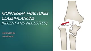
- 1. MONTEGGIA FRACTURES CLASSIFICATIONS (RECENT AND NEGLECTED) PRESENTED BY: DR AGGOUN
- 2. PLAN INTRODUTION CLINICAL PRESENTATION MECHANISM DIAGNOSIS CLASSIFICATIONS NEGLECTED MONTEGGIA FRACTURE CONCLUSION REFERENCES
- 3. INTRODUCTION A Monteggia fracture is defined as a proximal 1/3 ulna fracture with an associated radial head dislocation. Diagnosis is made with forearm and elbow radiographs to check for congruency of the radiocapitellar joint in the setting of an ulna fracture
- 4. CLINICAL PRESENTATION pain and swelling at elbow join dislocation at radiocapitellar joint should evaluate skin integrity elbow dislocation neurovascular exam radial deviation of hand with wrist extension weakness of thumb extension weakness of MCP extension most likely nerve injury
- 5. MECHANISM Typically, Monteggia fracture- dislocations occur as the result of a fall onto an outstretched hand DIAGNOSIS Clinic + X ray (AP and lateral views )
- 6. BADO CLASSIFICATION • The most commonly used classification system for Monteggia fractures • Four types • based on : the direction of the radial head dislocation the presence or absence of associated fractures.
- 7. BADO CLASSIFICATION Type I: Most common type (around 60% of cases) Anterior dislocation of the radial head Fracture of the proximal or middle third of the ulna, typically with an angulation anteriorly (towards the front) most common in children and young adults
- 8. BADO CLASSIFICATION Type II: Less common (around 15% of cases) Posterior dislocation of the radial head Fracture of the proximal or middle third of the ulna, typically with an angulation posteriorly (towards the back) 70 to 80% of adult Monteggia fractures
- 9. Jupiter Classification of Type II Monteggia Fracture-Dislocations
- 10. BADO CLASSIFICATION Type III: Less common (around 20% of cases) Lateral or anterolateral dislocation of the radial head Fracture of the proximal or middle third of the ulna, typically with an angulation laterally (towards the outside)
- 11. BADO CLASSIFICATION Type IV: Least common (around 5% of cases) Fracture of both the ulna and radius, often with comminution (fragmentation) Dislocation of the radial head in any direction
- 12. BADO CLASSIFICATION Limitations of the bado classification It does not consider the involevement of the proximal radioulnar joint(PRUJ) which can significantly impact prognosis and treatment It does not account the severity of the fracture or the degree of angulation and displacement
- 13. AO CLASSIFICATION the AO/OTA system offers a more comprehensive approach by incorporating: Fracture morphology Mechanism of injury Associated injuries (PRUJ) It doesn't specifically categorize Monteggia fractures as a separate entity individual fractures in the ulna and radius according to their respective anatomical locations.
- 14. TRILLAT CLASSIFICATION The Trillat classification focuses on associated injuries and complexity of the fracture pattern PROGNOSIS VALUE THREE groups
- 15. TRILLAT CLASSIFICATION Group I: all diaphyseal ulnar fractures, whatever the direction of dislocation of the radial head (anterior, posterior or lateral). Group II: all metaphyseal-epiphyseal ulnar fractures, whatever the direction of dislocation of the radial head. Group III: all ulnar fractures in group I or group II which are associated with damage to the radius or humerus (complete fracture of the radial head, diaphysis or wrist).
- 16. TRILLAT CLASSIFICATION IN PEDIATRIC POPULATION : type I : -Diaphyseal FR of the ulna - Ant dislocation of the radial head, producing the typical MONTEGGIA FR type II : High metaphyseal FR, most often greenwood External dislocation of the radial head
- 17. LETTS CLASSIFICATION(PEDIATRIC) based both on direction of radial head dislocation and the type of ulnar fracture
- 18. NEGLECTED MONTEGGIA FRACTURE Defined as the fracture of the proximal ulna associated with radial head dislocation (RHD) without undergoing any treatment for 4 weeks or more following injury Clinical examination palpable mass a decrease in elbow flexion and forearm pronation and supination valgus elbow deformity instability of the elbow joint and late (tardy) ulnar palsy SURGICAL TREATEMENT
- 19. NEGLECTED MONTEGGIA FRACTURE The classifications of moneteggia fractures can be applied to the neglected fractures also The most commonely used is the bado classification Type I: Anterior angulation of the ulnar fracture with an anterior dislocation of the radial head Type II: Posterior angulation of the ulnar fracture with a posterior dislocation of the radial head Type III: Angulation of the ulnar fracture in any direction with an anterolateral dislocation of the radial head. Type IV: Fracture of both the ulna and radius, with a dislocation of the radial head in any direction.
- 20. NEGLECTED MONTEGGIA FRACTURE Neglected Monteggia fracture: (a) anterior dislocation of the radial head with heterotopic ossification visible on a lateral view
- 21. NEGLECTED MONTEGGIA FRACTURE (a) Radial head dislocation (dotted line) and ulnar angulation (heavy line) left unnoticed on initial X-rays; (b) chronic radial head dislocation and consolidation of ulnar fracture after cast removal at 4 weeks
- 22. CONCLUSION Monteggia fractures are a comlex type of elbow injury that can be classified based on the direction of the radial head dislocation and the presence of any associated fractures. The most common classification system is the bado classification , which divides montegia fractures into four types The treatement of monteggia fractures depends on the type of fracture ,the severity of the injury , and the patiens age and health The best option for a neglected Monteggia injury is prevention
- 23. REFERENCES https://www.rch.org.au/clinicalguide/guideline_index/fractures/monteggia_ fracturedislocations_emergency_department_setting/ https://journals.lww.com/md- journal/fulltext/2021/03120/two_stage_strategy_for_neglected_monteggia. 108.aspx [1]. Goyal T, Arora SS, Banerjee S, et al. Neglected Monteggia fracture dislocations in children: a systematic review. J Pediatr Orthop B 2015;24:191–9 [3]. Chin K, Kozin SH, Herman M, et al. Pediatric Monteggia fracture- dislocations: avoiding problems and managing complications. Instr Course Lect 2016;65:399–407.
Editor's Notes
- Symptoms pain and swelling at elbow joint Physical exam inspection may or may not be obvious dislocation at radiocapitellar joint should evaluate skin integrity ROM & instability may be loss of ROM at elbow due to dislocation neurovascular exam PIN neuropathy radial deviation of hand with wrist extension weakness of thumb extension weakness of MCP extension most likely nerve injury
- The most commonly used classification system for Monteggia fractures is the Bado classification, which is based on : the direction of the radial head dislocation and the presence or absence of associated fractures. 04 types
- Anterior dislocation of the radial head with an angulated fracture of the ulna in the anterior direction. This is the most common type of Monteggia fracture, accounting for about 60% of all cases.
- Posterior dislocation of the radial head with fracture of the ulna shaft (diaphysis) or metaphysis
- The Jupiter classification is a refinement of the Bado classification system for Type II Monteggia fracture-dislocations Type IIA Coronoid level Type IIB Metaphyseal-diaphyseal junction Type IIC Distal to coronoid Type IID Fracture extending to distal half of ulna
- Fracture of the ulnar metaphysis (distal to coronoid process) with lateral dislocation of the radial head
- the AO/OTA classification system is another important way to categorize Monteggia fractures. While the Bado classification focuses primarily on the direction of the radial head dislocation, the AO/OTA system offers a more comprehensive approach by incorporating: Fracture morphology: This refers to the shape and pattern of the bone fragments in the ulna and radius. Mechanism of injury: This considers how the fracture occurred, such as a fall on an outstretched hand (FOOSH) or a direct blow. Associated injuries: This includes any other bone or soft tissue damage that may be present.(PROXIMAL RU JOINT) However, it's important to understand that the AO/OTA system doesn't specifically categorize Monteggia fractures as a separate entity. Instead, it classifies them based on the individual fractures in the ulna and radius according to their respective anatomical locations.
- IT HAS A PROGNOSIS VALUE
- Gravity increase from groupe I to groupe III I ;LESIONS LOCATED IN THE FORARM II;ELBOW FOR ADULTS BUT IN PEDIATRIC POPULATION
- Letts et al.81 have described an alternate classification schedule for pediatric Monteggia fracture-dislocations based both on direction of radial head dislocation and the type of ulnar fracture A: Anterior dislocation of the radial head with plastic deformation of the ulna. B: Anterior dislocation of the radial head with greenstick fracture of the ulna. C: Anterior dislocation of the radial head with complete fracture of the ulna. D: Posterior dislocation of the radial head with fracture of the ulnar metaphysis. E: Lateral dislocation of the radial head and metaphyseal greenstick fracture of the ulna.
- Permanent dislocation of the radial head, which is considered chronic after four weeks [7], can lead to several further complications such as palpable mass, a decrease in elbow flexion and forearm pronation and supination, valgus elbow deformity, instability of the elbow joint and late (tardy) ulnar palsy THE TREAMENT MUST BE SURGICAL TO REDUCE THE DISLOCATION OF RADIAL HEAD